CARDIOVASCULAR PHYSIOLOGY: THE HEARTfaculty.mtsac.edu/mpresch/36_lecture_files_unit_3/18 The Heart...
31
CARDIOVASCULAR PHYSIOLOGY: THE HEART Physiology Unit 3
Transcript of CARDIOVASCULAR PHYSIOLOGY: THE HEARTfaculty.mtsac.edu/mpresch/36_lecture_files_unit_3/18 The Heart...
CARDIOVASCULARPHYSIOLOGY:THEHEART
PhysiologyUnit3
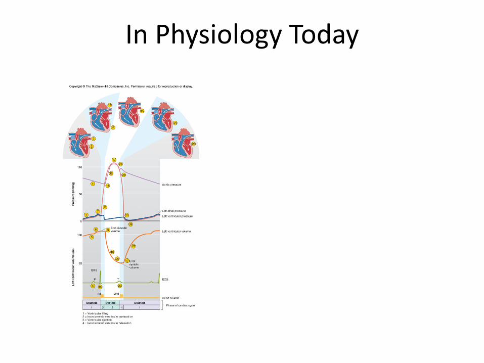
InPhysiologyToday
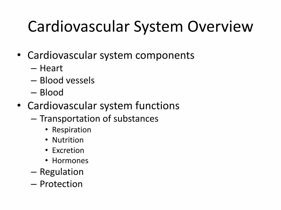
CardiovascularSystemOverview• Cardiovascularsystemcomponents
– Heart– Bloodvessels– Blood
• Cardiovascularsystemfunctions– Transportationofsubstances
• Respiration• Nutrition• Excretion• Hormones
– Regulation– Protection

CardiacMuscle• Characteristics
– Somecellsintheatriasecreteapeptidehormonecalledatrialnatriureticfactor(ANF)
• Causesnatriuresis• Vasodilation
• Conductingsystem– 1%ofcells– Initiatesheartbeatandspreadstheimpulsethroughouttheheart
• Innervation• Bloodsupply
– Coronarycirculation
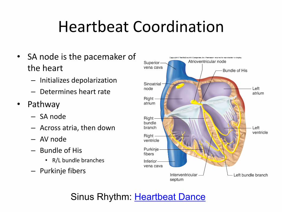
HeartbeatCoordination• SAnodeisthepacemakerof
theheart– Initializesdepolarization– Determinesheartrate
• Pathway– SAnode– Acrossatria,thendown– AVnode– BundleofHis
• R/Lbundlebranches
– Purkinjefibers
Sinus Rhythm: Heartbeat Dance
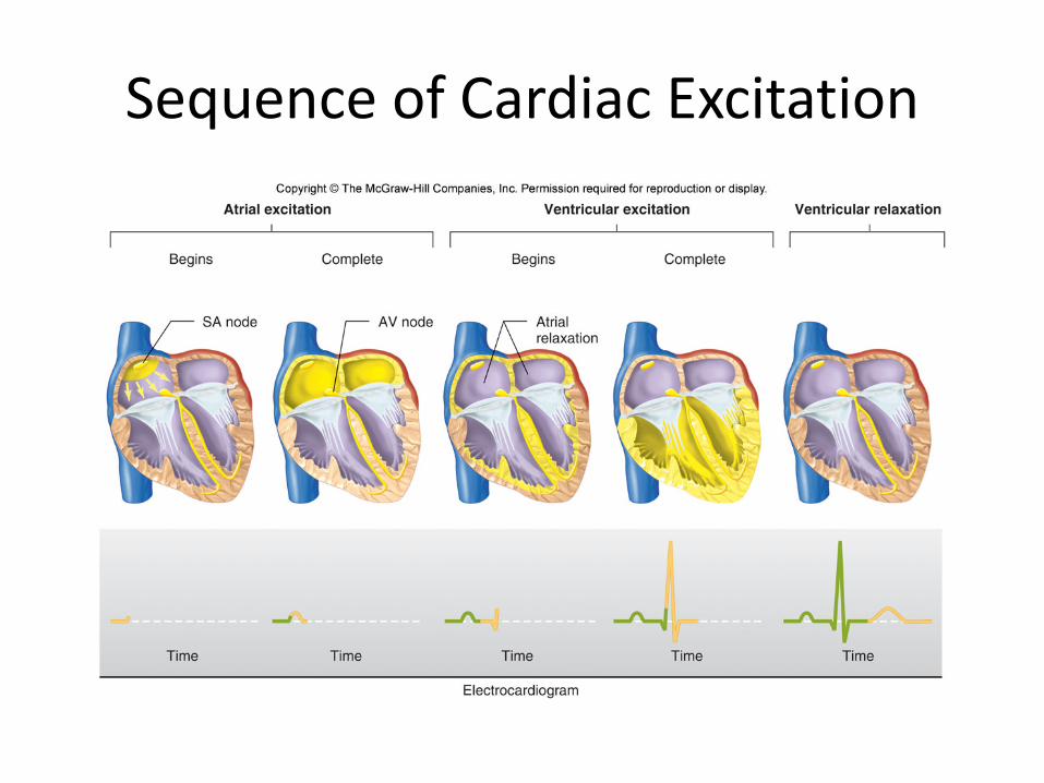
SequenceofCardiacExcitation
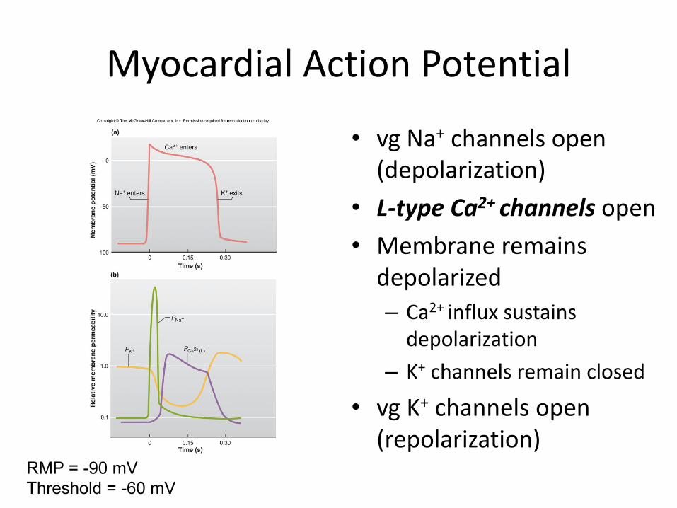
MyocardialActionPotential
• vgNa+ channelsopen(depolarization)
• L-typeCa2+channelsopen• Membraneremainsdepolarized– Ca2+influxsustainsdepolarization
– K+ channelsremainclosed
• vgK+ channelsopen(repolarization)
RMP = -90 mVThreshold = -60 mV
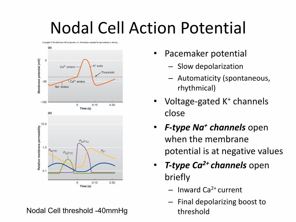
NodalCellActionPotential• Pacemakerpotential
– Slowdepolarization– Automaticity(spontaneous,
rhythmical)
• Voltage-gatedK+ channelsclose
• F-typeNa+ channelsopenwhenthemembranepotentialisatnegativevalues
• T-typeCa2+channelsopenbriefly– InwardCa2+current– Finaldepolarizingboostto
thresholdNodal Cell threshold -40mmHg
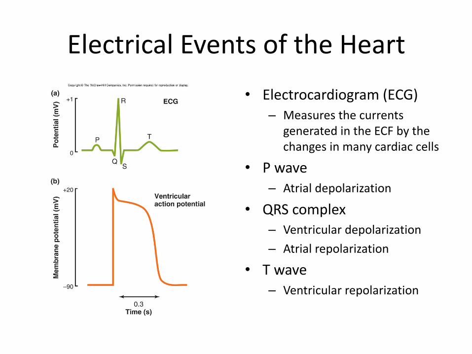
ElectricalEventsoftheHeart• Electrocardiogram(ECG)
– MeasuresthecurrentsgeneratedintheECFbythechangesinmanycardiaccells
• Pwave– Atrialdepolarization
• QRScomplex– Ventriculardepolarization– Atrialrepolarization
• Twave– Ventricularrepolarization
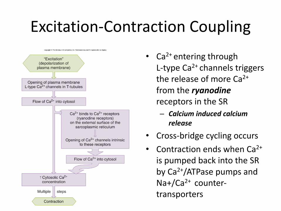
Excitation-ContractionCoupling• Ca2+enteringthrough
L-typeCa2+channelstriggersthereleaseofmoreCa2+fromtheryanodinereceptorsintheSR– Calciuminducedcalcium
release
• Cross-bridgecyclingoccurs• ContractionendswhenCa2+
ispumpedbackintotheSRbyCa2+/ATPasepumpsandNa+/Ca2+ counter-transporters
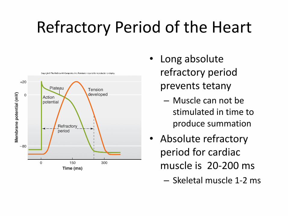
RefractoryPeriodoftheHeart
• Longabsoluterefractoryperiodpreventstetany– Musclecannotbestimulatedintimetoproducesummation
• Absoluterefractoryperiodforcardiacmuscleis20-200ms– Skeletalmuscle1-2ms

MechanicalEventsoftheHeart• Cardiaccycle
– Pressureandvolumechangesthatoccurduringthecardiaccycle– Averageheartrate72bpm– Eachcardiaccyclelasts0.8s
• 0.3sinsystole• O.5sindiastole
• 2alternatingphases– Systole
• Ventricularcontractionandbloodejection– Diastole
• Ventricularrelaxationandbloodfilling
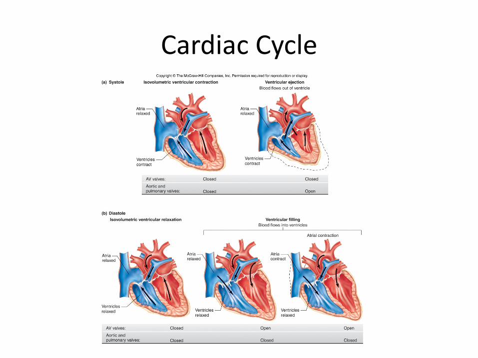
CardiacCycle
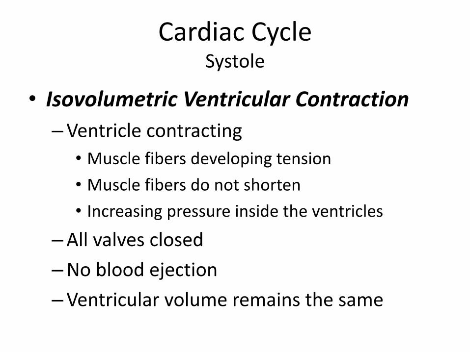
CardiacCycleSystole
• IsovolumetricVentricularContraction–Ventriclecontracting
• Musclefibersdevelopingtension• Musclefibersdonotshorten• Increasingpressureinsidetheventricles
–Allvalvesclosed–Nobloodejection–Ventricularvolumeremainsthesame
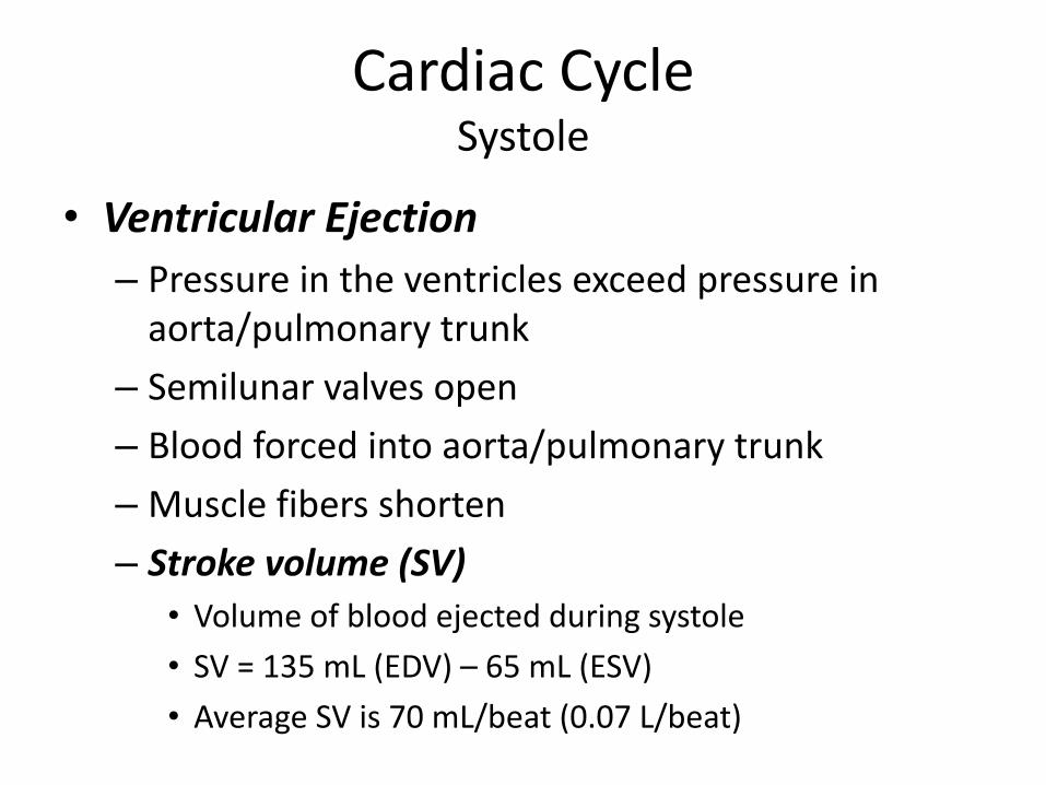
CardiacCycleSystole
• VentricularEjection– Pressureintheventriclesexceedpressureinaorta/pulmonarytrunk
– Semilunarvalvesopen– Bloodforcedintoaorta/pulmonarytrunk– Musclefibersshorten– Strokevolume(SV)
• Volumeofbloodejectedduringsystole• SV=135mL(EDV)– 65mL(ESV)• AverageSVis70mL/beat(0.07L/beat)
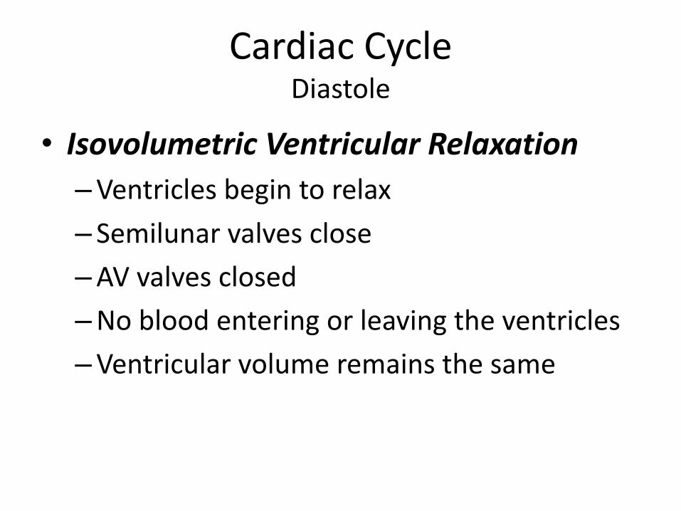
CardiacCycleDiastole
• IsovolumetricVentricularRelaxation–Ventriclesbegintorelax– Semilunarvalvesclose–AVvalvesclosed–Nobloodenteringorleavingtheventricles–Ventricularvolumeremainsthesame
CardiacCycleDiastole
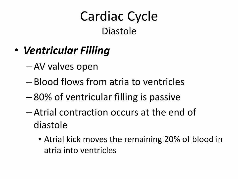
• VentricularFilling–AVvalvesopen–Bloodflowsfromatriatoventricles–80%ofventricularfillingispassive–Atrialcontractionoccursattheendofdiastole• Atrialkickmovestheremaining20%ofbloodinatriaintoventricles
CardiacCycleVolumes
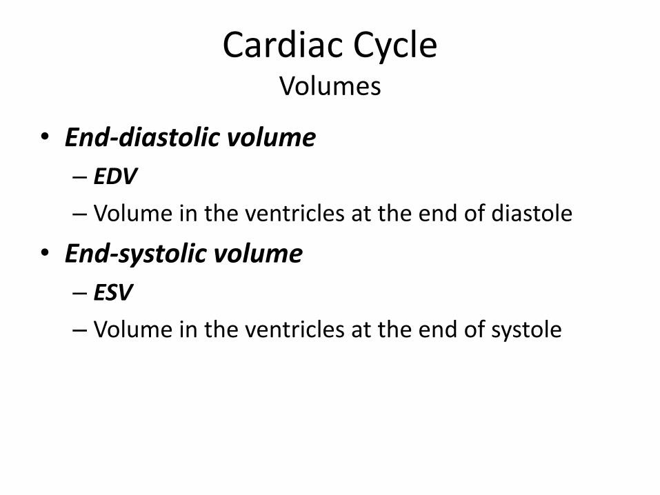
• End-diastolicvolume– EDV– Volumeintheventriclesattheendofdiastole
• End-systolicvolume– ESV– Volumeintheventriclesattheendofsystole
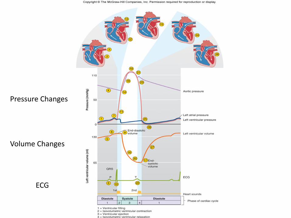
PressureChanges
VolumeChanges
ECG
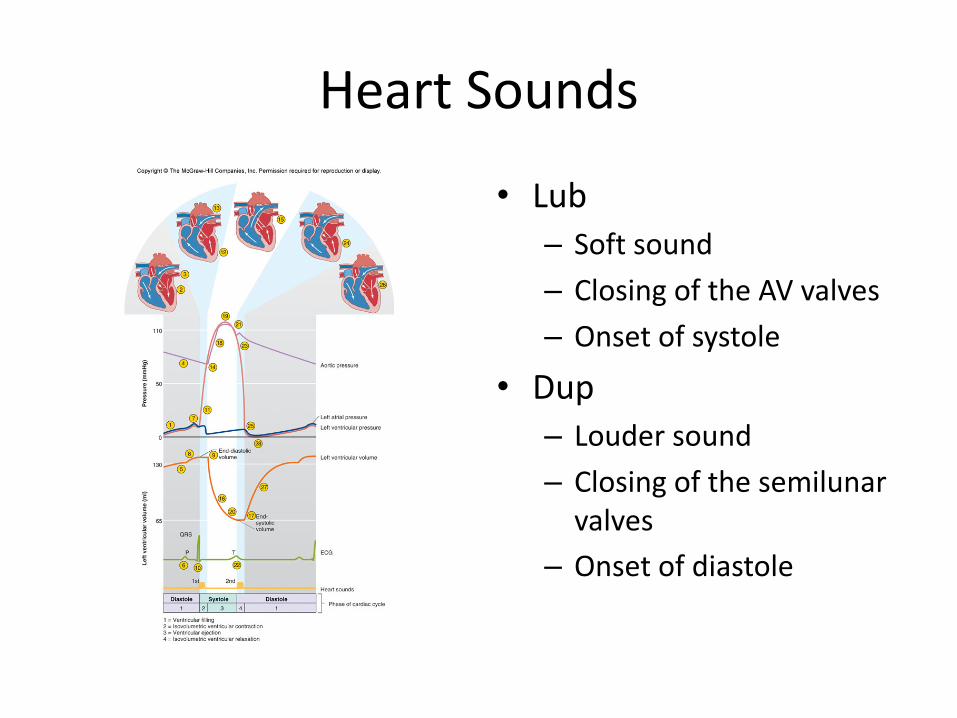
HeartSounds
• Lub– Softsound– ClosingoftheAVvalves– Onsetofsystole
• Dup– Loudersound– Closingofthesemilunarvalves
– Onsetofdiastole
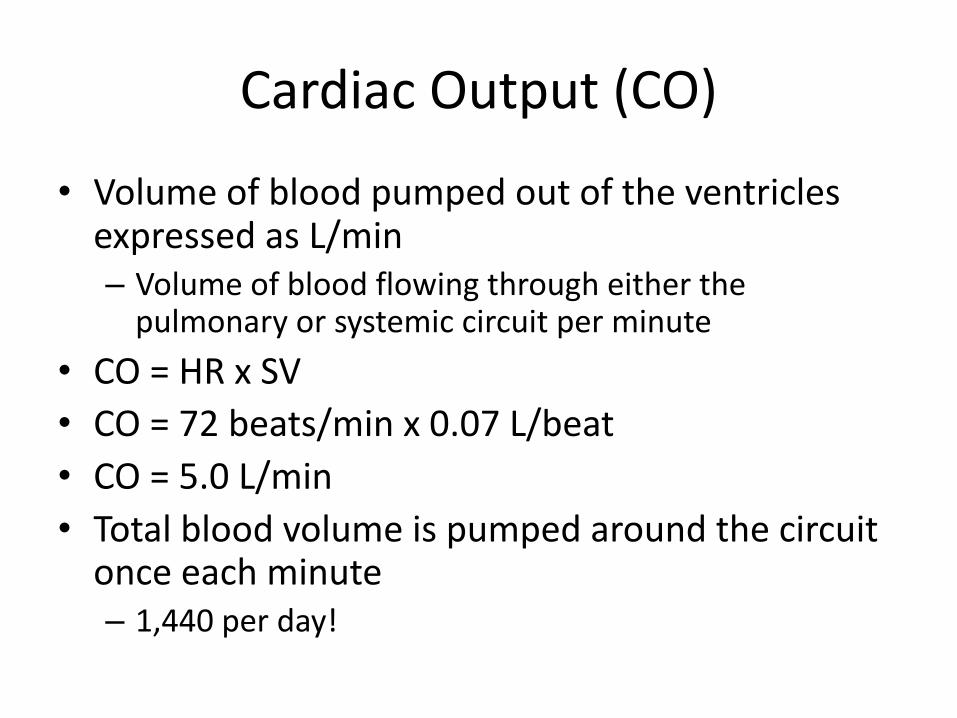
CardiacOutput(CO)
• VolumeofbloodpumpedoutoftheventriclesexpressedasL/min– Volumeofbloodflowingthrougheitherthepulmonaryorsystemiccircuitperminute
• CO=HRxSV• CO=72beats/minx0.07L/beat• CO=5.0L/min• Totalbloodvolumeispumpedaroundthecircuitonceeachminute– 1,440perday!
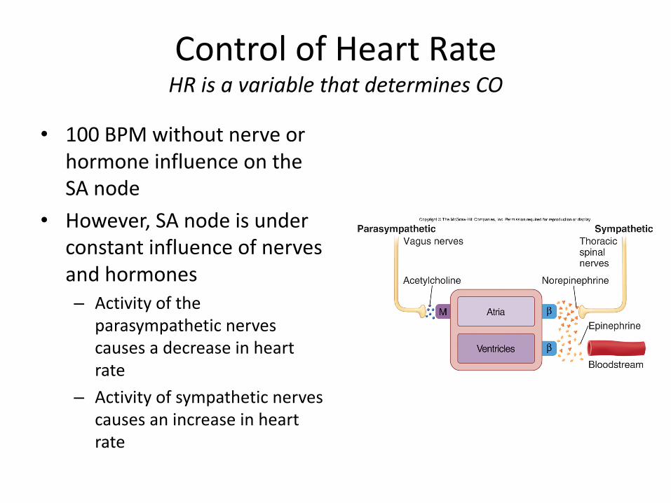
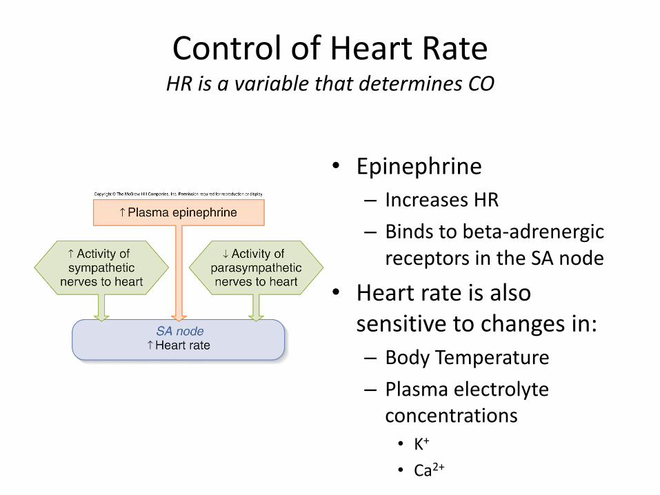
ControlofHeartRateHRisavariablethatdeterminesCO
• 100BPMwithoutnerveorhormoneinfluenceontheSAnode
• However,SAnodeisunderconstantinfluenceofnervesandhormones– Activityofthe
parasympatheticnervescausesadecreaseinheartrate
– Activityofsympatheticnervescausesanincreaseinheartrate
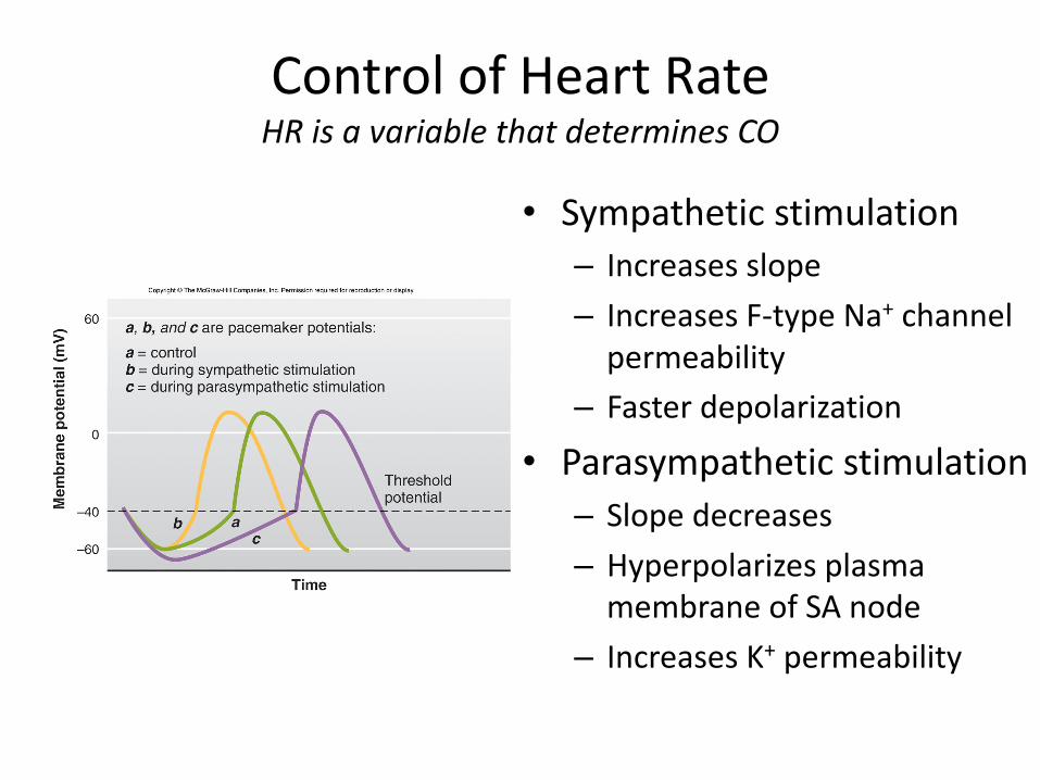
ControlofHeartRateHRisavariablethatdeterminesCO
• Sympatheticstimulation– Increasesslope– IncreasesF-typeNa+ channelpermeability
– Fasterdepolarization
• Parasympatheticstimulation– Slopedecreases– HyperpolarizesplasmamembraneofSAnode
– IncreasesK+ permeability
ControlofHeartRateHRisavariablethatdeterminesCO
• Epinephrine– IncreasesHR– Bindstobeta-adrenergicreceptorsintheSAnode
• Heartrateisalsosensitivetochangesin:– BodyTemperature– Plasmaelectrolyteconcentrations
• K+
• Ca2+
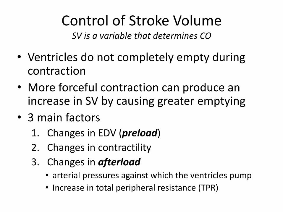
ControlofStrokeVolumeSVisavariablethatdeterminesCO
• Ventriclesdonotcompletelyemptyduringcontraction
• MoreforcefulcontractioncanproduceanincreaseinSV bycausinggreateremptying
• 3mainfactors1. ChangesinEDV (preload)2. Changesincontractility3. Changesinafterload
• arterialpressuresagainstwhichtheventriclespump• Increaseintotalperipheralresistance(TPR)

Starling’sLawoftheHeartRelationshipbetweenEDVandSV
• Ventriclescontractmoreforcefullyduringsystolewhenithasbeenfilledtoagreaterdegreeduringdiastole
• SVincreasesasEDVincreases
• SVisDependentofEDV• Increaseinvenousreturn
forcesanincreaseinCObyincreasingEDVwhichincreasesSV
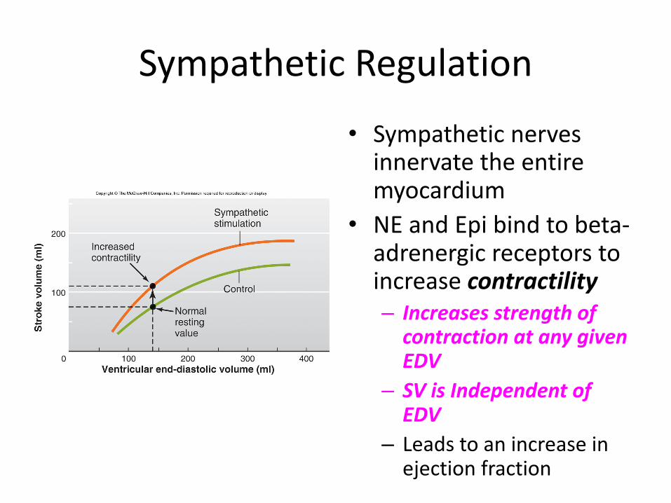
SympatheticRegulation
• Sympatheticnervesinnervatetheentiremyocardium
• NEandEpibindtobeta-adrenergicreceptorstoincreasecontractility– IncreasesstrengthofcontractionatanygivenEDV
– SVisIndependentofEDV
– Leadstoanincreaseinejectionfraction
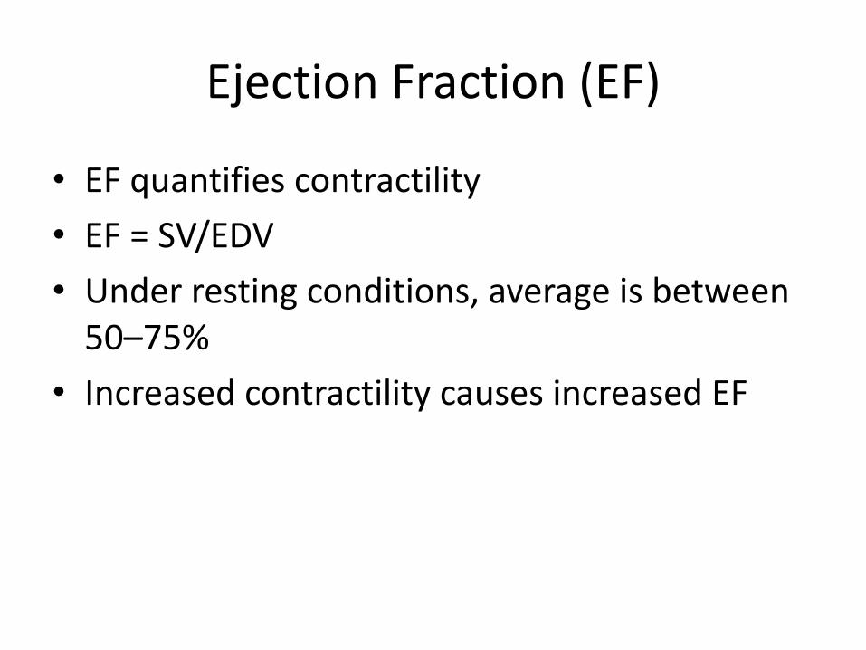
EjectionFraction(EF)
• EFquantifiescontractility• EF=SV/EDV• Underrestingconditions,averageisbetween50–75%
• IncreasedcontractilitycausesincreasedEF
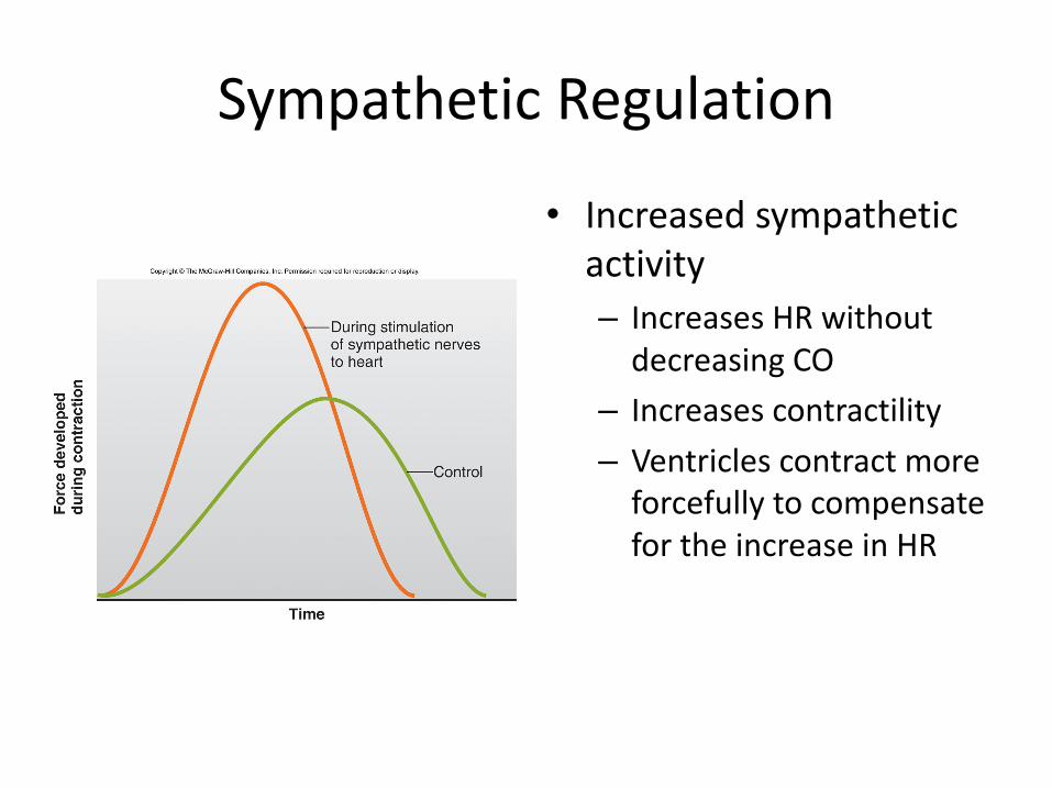
SympatheticRegulation
• Increasedsympatheticactivity– IncreasesHRwithoutdecreasingCO
– Increasescontractility– VentriclescontractmoreforcefullytocompensatefortheincreaseinHR
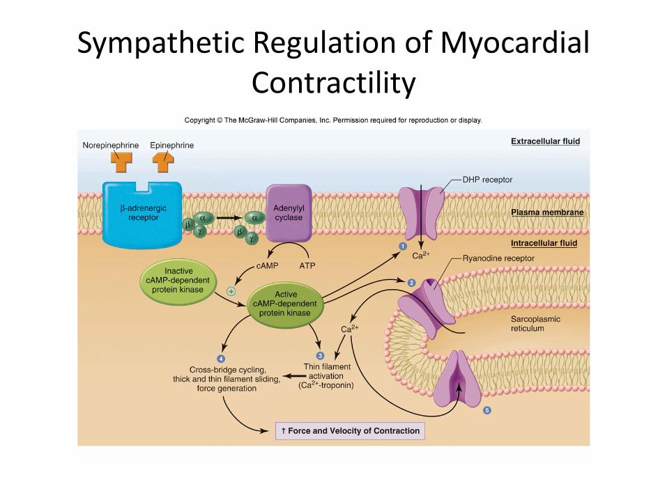
SympatheticRegulationofMyocardialContractility
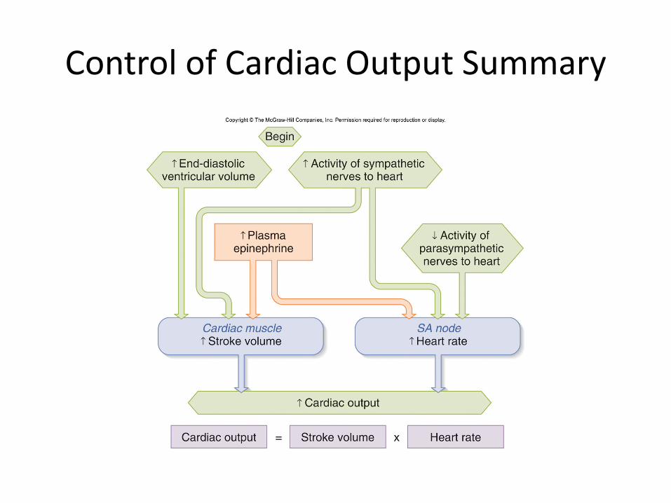
ControlofCardiacOutputSummary


![Raw264.7 Cells Secrete Fibroblast Growth Stimulating Activity … · healing, macrophages secrete growth factors [16] [17]. In this paper, we show that Raw264.7 cells secrete cyto-kines](https://static.fdocuments.us/doc/165x107/6064900f81fe4b40bf056aaa/raw2647-cells-secrete-fibroblast-growth-stimulating-activity-healing-macrophages.jpg)