'IAION PG A A- D -- 2 A~a..A24 46 o eg e o .ti If ent ... · A A- D -- 2 ~en- eg e o .ti normation...
98
. 'IAION PG Form Approved "rATIO PAGE0MB No, 0704-0788 o ~ie ao hur ec response. Ancivbdrn the tirne fior eviewinginstructions, searhin. e,,tnq daia sources, eg e o .ti If normation Send cm ent egadigti burden "s~mate or anv ither ai-k of this A D -- A- 2 ~en- to rivashinq ' 6 ,ton Headoarters Services. Directorate for-information Operations and Reports, 1215 jefferson A~a..A24 46 of Managvement an Budget. Paperwork Reduction Project (0704-0188), Washington,.V 20503. II 1111 111111ii 11111I1111 ol1 111 _________________________ ________________________ 4. TITLE AND SUBTITLE Analysis of the Extent of Completion of 5. FUNDING NUMBERS Skin Assessments and Documentation of Risk for skin Break- down in the Trauma Patient Who Experiences hypovolemic Shock Reciuiring Massive Transfusion Resuscitation and 6. AUTHOR(S) Surgical Intervention Maryanne Kolesar, Major 7. PERFORMING ORGANIZATION NAME(S) AND ADDRESS(ES) B. PERFORMING ORGANIZATION REPORT NUMBER AFIT Student Atten-ding: University of Washington AFIT/CI/C1A-91-085 9. SPONSORING/ MONITORING AGENCY NAME(S) AND ADDRESS(E~ (' 10. SPONSORING/ MONITORING I. ~. ~ AGENCY REPORT NUMBER AFIT/CI 6K 7 LE% GTE Wright-Patterson AFB OH 45433-6583 K rE~19~J 11. SUPPLEMENTARY NOTES 12a. DISTRIBUTION /AVAILABILITY STATEMENT 12b. DISTRIBUTION CODE Approved for Public Release IAW 190-1 Distributed Unlimited ERNEST A. HAYGOOD, Captain, USAF Executive Officer 13. ABSTRACT (Maximum 200 words) 14. SUBJECT TERMS 15. NUMBER OF PAGES 87 16. PRICE CODE 17. SECURITY CLASSIFICATION 18. SECURITY CLASSIFICATION 19. SECURITY CLASSIFICATION 20. LIMITATION OF ABSTRACT NSN 7540-01-280-5500 Standard Form 298 (Rev. 2-89)
Transcript of 'IAION PG A A- D -- 2 A~a..A24 46 o eg e o .ti If ent ... · A A- D -- 2 ~en- eg e o .ti normation...

. 'IAION PG Form Approved"rATIO PAGE0MB No, 0704-0788
o ~ie ao hur ec response. Ancivbdrn the tirne fior eviewinginstructions, searhin. e,,tnq daia sources,eg e o .ti If normation Send cm ent egadigti burden "s~mate or anv ither ai-k of thisA D -- A- 2 ~en- to rivashinq ' 6 ,ton Headoarters Services. Directorate for-information Operations and Reports, 1215 jeffersonA~a..A24 46 of Managvement an Budget. Paperwork Reduction Project (0704-0188), Washington,.V 20503.
II 1111 111111ii 11111I1111 ol1 111 _________________________ ________________________
4. TITLE AND SUBTITLE Analysis of the Extent of Completion of 5. FUNDING NUMBERS
Skin Assessments and Documentation of Risk for skin Break-down in the Trauma Patient Who Experiences hypovolemicShock Reciuiring Massive Transfusion Resuscitation and6. AUTHOR(S) Surgical Intervention
Maryanne Kolesar, Major
7. PERFORMING ORGANIZATION NAME(S) AND ADDRESS(ES) B. PERFORMING ORGANIZATIONREPORT NUMBER
AFIT Student Atten-ding: University of Washington AFIT/CI/C1A-91-085
9. SPONSORING/ MONITORING AGENCY NAME(S) AND ADDRESS(E~ (' 10. SPONSORING/ MONITORINGI. ~. ~ AGENCY REPORT NUMBER
AFIT/CI 6K 7 LE% GTEWright-Patterson AFB OH 45433-6583 K rE~19~J
11. SUPPLEMENTARY NOTES
12a. DISTRIBUTION /AVAILABILITY STATEMENT 12b. DISTRIBUTION CODE
Approved for Public Release IAW 190-1Distributed UnlimitedERNEST A. HAYGOOD, Captain, USAFExecutive Officer
13. ABSTRACT (Maximum 200 words)
14. SUBJECT TERMS 15. NUMBER OF PAGES87
16. PRICE CODE
17. SECURITY CLASSIFICATION 18. SECURITY CLASSIFICATION 19. SECURITY CLASSIFICATION 20. LIMITATION OF ABSTRACT
NSN 7540-01-280-5500 Standard Form 298 (Rev. 2-89)

University of Washington
Abstract
Analysis of the Extent of Completion of Skin Assessments andDocumentation of Risk for Skin Breakdown in the Trauma
Patient Who Experiences Hypovolemic Shock Requiring MassiveTransfusion Resuscitation and Surgical Intervention
by Maryanne KolesarMaj, USAF NC
Chai enson- -of--te-Su vi sory Committee:Associate Professor Janet Marvin
K i School of Nursing
This retrospective random chart audit spanning the
years 1985 through 1989 was conducted in a sample of 105
charts of trauma patients. Patients included in the study
had to be 18 years or old, r who survived the event longer
than 24 hours after admission to the ICU. Patients who had
a pressure sise prior to admission were excluded from the
study.//
|"The purpose of the study was to determine the number
of charts with documentation of skin assessment and if
deemed appropriate, the nursing diagnosis actual or
potential for impaired skin integrity, Ninety-seven (92.4%)
patients were deemed at risk for pressure sore development
according to the Braden Scale for Predicting Pressure Sore
Risk. Thirty-one charts had documentation of a skin
assessment. No documentation of the nursing diagnosis
actual or potential for impaired skin integrity was found.
91-18008 91 12i3 2521111 I~i~ iII 11111 1111 11111111

A second purpose was to determine the frequency with
which cues listed on the Braden Scale for Predicting
Pressure Sore Risk were documented in the nursing
assessment. The most frequently documented cues for sensory
perception were receiving sedatives (50.5%) and comatose
(26.7%). Exposure of skin to moisture was found most
frequently under the cues skin dry and intact (20.0%).
Activity was documented under the cues bedrest (93.3%) and
out of bed in chair (6.7%). Mobility had documentation of
the cues receiving sedatives (50.5%), and comatose (26.7%)
most frequently. Friction and shear was documented most
frequently under the cue able to move/change position by
self (53.3%). Nutrition was documented under the cues
nothing by mouth (98.1%) and liquid diet (1.9%) most
frequently.
A third purpose, to determine if there was any
association with additional data and documentation of skin
assessment, yielded significant results for length of stay
in the emergency department only (p=.02).
This study points out gaps in nursing documentation
that may or may not be reflective of practice. Further
research is warranted to determine potential problems with
documentation of nursing practice related to prevention of
pressure injury to skin.

Analysis of the Extent of Completion of Skin Assessments andDocumentation of Risk for Skin Breakdown in the Trauma
Patient Who Experiences Hypovol:emic Shock Requiring MassiveTransfusion Resuscitation and Surgical Intervention
by
MARYANNE KOLESARMaj, USAF NC
A thesis submitted in partial fulfillmentof the requirements for the degree of
Master of Nursing
University of Washingt n
Approved (C i edno u6,isory Committee)
Program Authorizedto Offer egree
Date re Z /
I AD ,isiC T or
OOP' It ~C .B~~jcopy'! U't. utead IIN P C E Ju s t t fi at ! ____
G
• D1tribotion/
i.Ava!_' tnd/orjDist P0a

In presenting this thesis in partial fulfillment of therequirements for a Master's degree at the University ofWashington, I agree that the library shall make its copiesfreely available for inspection. I further agree thatextensive copying of this thesis is allowable only forscholarly purposes, consistent with "fair use' as prescribedin the U. S. Copyright Law. Any other reproduction for anypurposes or by any means shall be allowed without mypermission.
Signature
Date

TABLE OF CONTENTS
Page
List of Figures ...................................... vi
List of Tables ...................................... vii
Chapter I: Problem Statement ........................ 1
Chapter II: Conceptual Framework ..................... 4
Alterations in Blood Circulation DuringHypovolemic Shock ................................. 4
Cellular Metabolism During Hypovolemic Shock ...... 5
Cellular Metabolism of Nonvital Tissues ....... 6
Skin and Connective Tissue ................ 6
Gastrointestinal Tract .................... 7
Skeletal Muscle ........................... 7
Cell Death Related to Hypoxia ................. 7
Reperfusion Injury ............................ 8
Factors Affecting Skin Breakdown .................. 8
Pressure, Friction, and Shear ................. 8
Immobilization and Other Factors ............. 10
Assessment Tool for Identification of atRisk Patients ................................ 13
Prevention of Skin Breakdown by Skin Assessment..15
Purpose .............................................. 19
Chapter III: Methodology ............................. 20
Design ........................................... 20
Sample ........................................... 21

Population ................................... 21
Inclusion Criteria ........................... 21
Exclusion Criteria ........................... 22
Selection .................................... 22
Measurement ...................................... 22
Documentation of Skin Assessment ............. 23
Validity and Reliability ................. 24
Nursing Diagnosis Actual or Potential forImpaired Skin Integrity .................. ... 24
Validity and Reliability ................. 25
Cues for Predicting Pressure Sore Risk ....... 27
Sensory Perception ....................... 27
Moisture ................................. 28
Activity .................................. 28
Mobility ................................. 28
Friction and Shear ....................... 28
Nutrition ................................ 29
Additional Data ............................. 29
Acuity Indices ........................... 29
Demographic Data ......................... 30
Transportation and Mobility Status ....... 31
Physiologic Factors ...................... 31
Methods of Procedure ............................. 31
Protection of Human Subjects ..................... 32
iii

Methods of Analysis ...............................32
Summary.. ......................................... 34
Chapter IV: Results .................................. 35
Sample ........................................... 35
Pilot Study for Inter-rater Reliability of theData Collection Tool ............................. 36
Additional Data .............................. 36
Demographic -Data ......................... 36
Physiologic Factors ...................... 37
Acuity Indices ............................ 37
Transportation and Mobility Factors ...... 38
Documentation of Skin Assessment ............. 38
Documentation of the Nursing DiagnosisPotential for or Actual Impaired SkinIntegrity ..................................... 41
Documentation of Cues for Related to theBraden Scale for Predicting Pressure SoreRisk ......................................... 41
Study Results .................................... 44
Additional Data .............................. 44
Demographic Data ......................... 44
Physiologic Factors ...................... 46
Acuity Indices ........................... 47
Transportation and Mobility Factors ...... 47
Skin Assessment .................................. 50
Nursing Diagnosis Actual or Potential forImpaired Skin Integrity .......................... 52
iv

Cues for Predicting Pressure Sore Risk ........... 54
Association of Additional Data WithDocumentation of Skin Assessment ................. 56
Association of Documentation of the NursingDiagnosis Actual or Potential Impaired SkinIntegrity with Skin Assessment ................... 61
Summary .......................................... 65
Chapter V: Discussion ................................ 68
Discussion of Findings ...................... ..... 68
Limitations of the Study ......................... 72
Recommendations .................................. 73
Conclusions ...................................... 74
References ........................................... 76
Appendix A: Braden Scale for Predicting PressureSore Risk ................................ 80
Appendix B: Schematic Model ........................... 81
Appendix C: Data Collection Tool ..................... 82
Appendix D: Letter Granting Premission to Usethe Braden S,.ale for PredictingPressure Sore Risk ....................... 85
Appendix E: Letter Granting Permission to UtilizeMedical Facility for Data Collection ..... 86
V
LIST OF FIGURES
Number Page
1. Chi Square Analysis of Additional Data with SkinAssessment ............................................. 62
2. Chi Square Analysis of Skin Assessment withthe Nursing Diagnosis Potential for Impaired SkinIntegrity, and Actual Skin Breakdown With theNursing Diagnosis Impaired Skin Integrity ............. 64
vi

LIST OF TABLES
Number I Page
1. Percentage of Inter-rater Agreement forDocumentation of Additional Data ...................... 39
2. Percentage of Inter-rater Agreement forDocumentation of Cues Related to Skin Assessmentand Body Surface Area Inspected, Actual SkinBreakdown, Time Skin Assessed, and Nursing DiagnosisPotential for or Actual Impaired Skin Integrity ....... 42
3. Percentage of Inter-rater Agreement forDocumentation of Cues from the Braden Scale forPredicting Pressure Sore Risk ......................... 45
4. Results of Additional Data ............................ 51
5. Frequency of Documentation of Descriptors of SkinAssessment ............................................ 53
6. Frequency of Documentation Related to Cues Fromthe Braden Scale for Predicting Pressure Sore Risk .... 57
vii

CHAPTER I
Problem Statement
The National Pressure Ulcer Advisory Panel (1989) found
that well over one million patients in acute and chronic
care facilities develop skin breakdown each year. One study
of a Surgical Intensive Care Unit (SICU) reported 21 of 63
patients as arriving and leaving the SICU with skin
breakdown (Robnett, 1986). Cost of decubiti care in acute
care settings ranges from $2,000 to $30,000 per case
(National Pressure Ulcer Advisory Panel, 1989). Treatment
costs of between $715 to $43,460 per patient were reported
in four case studies of patients who developed pressure
ulcers while in the operating room (Vermillion, 1990).
Length of stay for these patients increased by 11 to 64 days
for treatment of pressure ulcers alone (Vermillion, 1990).
Skin breakdown can occur if delivery of oxygen and
nutrients to the cell are interrupted (Iverson-Carpenter,
1989). Pressure on skin over boney prominences has been
indicated as the primary factor which interrupts delivery of
oxygen and nutrients to skin tissue. Recent studies
(Bennett & Lee, 1988; Mawson, Biundo, Neville, Linares,
Winchester & Lopez, 1988) report the amount of pressure
required to cause injury to skin as related to the patient's
systolic blood pressure combined with the forces of friction

2
and vertical sheer. These findings suggest that patients in
shock who are immobilized could experience tissue injury
when exposed to lower external skin pressures combined with
friction and sheer as compared to immobilized patients with
relatively normal systolic blood pressures. The trauma
patient who experiences hypovolemic shock is an example of a
critically ill patient who could be at high risk for skin
breakdown.
The Joint Commission for the Accreditation of
Healthcare Organizations (JCAHO) requires nurses to complete
and document physical assessments on all patients (1990).
The physical assessment, to include skin assessment, should
result in documentation of nursing diagnoses and treatment
plans in the patient chart to guide nursing practice and
promote continuity of care.
Accreditation agencies (Pieper, Mikols, Mance & Adams,
1990) consider the incidence of pressure ulcers a reflection
of the quality of patient care. Research has been done to
explain what causes pressure ulcers; however, little is
known about the extent to which nursing personnel are
completing and documenting skin assessments and identifying
patients at risk for pressure ulcers. This is particularly
important as skin breakdown is a costly problem with respect
to length of stay. And it is preventable through systematic

3
identification of risk factors and with treatment aimed at
prevention.
The goals of this study are to determine if nursing
personnel are documenting, within the first 24 hours of
admission to the intensive care unit (ICU), skin assessments
of trauma patients who experience hypovolemic shock
requiring massive transfusion resuscitation and surgical
intervention. And when deemed appropriate, identifying
those patients that have actual skin breakdown or are at
risk for potential skin breakdown.
The findings of this study could be used to identify
gaps in nursing chart documentation reflective of a lack of
systematic assessment of risk for skin breakdown. High cost
and increased length of stay associated with skin breakdown
could be avoided through early identification and
preventative treatment.
The information obtained from this study could be
used to develop a flow sheet which facilitates documentation
of skin assessment and serves as a standard of care with
which to orient nurses. A list of indicators related to
skin breakdown and prevention could be developed as a tool
to monitor quality of care.

CHAPTER I-I
Conceptual Framework
This chapter introduces the concepts of alterations in
blood circulation during hypovolemic shock, cellular
metabolism during shock, factors affecting skin breakdown,
and prevention of skin breakdown by skin assessment.
Alterations in BdQ Circulation During Hynovolemic Shock
Hypovolemic shock occurs as whole blood and its
components are lost from the intravascular space through
hemorrhage. Baroreceptors and chemoreceptors located
throughout the body sense changes in intravascular pressure
and oxygen tension of blood and chemically mediate responses
aimed at maintaining homeostasis by continued circulation of
nutrients and oxygen to cells and removal of waste products
from cells (Berne & Levy, 1990). But as more and more
circulating blood volume is lost through hemorrhage, the
body is unable to maintain a homeostatic balance. The shock
state ensues and blood is shifted from nonvital tissues
which are the: skin and connective tissue, gastrointestinal
tract, and skeletal muscle to vital tissues which are
essential for survival such as: kidney, lung, liver, heart,
and brain (Schumer, 1987). The nonvital tissues must
convert to anaerobic metabolism as the supply of nutrients

5
and oxygen diminishes in hypovolemic shock states.
Cellular MetabolilsmDj HyDovolemic Shock
Under fed conditions, ingested carbohydrates, fats, and
proteins are utilized as sources of nutrients for cellular
metabolism. These nutrients are broken down during
digestion into smaller components which are absorbed into
the blood stream and transported along with oxygen and
chemical mediators of metabolism to cells. In starvation or
fasting states, stored food sources are mobilized,
processed, and delivered as nutrients to cells. In the
presence of oxygen these nutritional components are
aerobically metabolized for production of adenosine
triphosphate (ATP), the body's major energy source.
Metabolism can occur anaerobically for brief periods;
however, it is less efficient for producing ATP (Genuth,
1990).
Adenosine triphosphate is the energy source for several
essential physiologic functions. It is used for muscular
work and cell movement otherwise known as mechanical work.
Synthetic reactions which are necessary for tissue
regeneration, storage of nutrients, and production of
chemicals necessary for regulation of bodily functions
require ATP. Transport of substances across cell membranes
essential for electrical, chemical, and mechanical signal

6
generation and conduction could not occur without it.
Detoxification degradation proce.3es such as urea formation,
oxidation, reduction, and conjugation need ATP in order to
occur. And ATP allows for heat production for maintenance
of body temperature (Genuth, 1990).
Cellular Metabolism of Nonvital Tissues
Skin pnd connective tissue. Fourteen percent of the
total body cell mass is made up of skin and connective
tissue (Schumer, 1987). Skin functions as a barrier
protecting the body from invasion by environmental hazards
and from loss of vital fluids. If anaerobic metabolism
continues and ATP generation for maintenance of cellular
function diminishes, cell death will occur (Schumer, 1987).
When cells die, they release digestive enzymes which
instigate an inflammatory response. Connective tissue serves
as the medium for inflammatory activity. The inflammatory
response serves to remove dead cells and toxins, provide
immunologic support against infection, and supply nutrients
for tissue repair (Beisel, 1987). However, this process
requires an adequate delivery of the cellular components
necessary for the inflammatory process, oxygen, and
nutrients. Because the blood supply to skin and connective
tissue is decreased during shock, immune defenses will
diminish and wound healing will be impaired, predisposing

7
the patient to skin breakdown and invasion by
microorganisms.
Gastrointestinal tract. The gastrointestinal tract,
normally responsible for nutrient intake, absorption, and
solid waste exchange is limited in the ability to perform
these functions due to reduced blood flow during hypovolemic
shock. Adenosine triphosphate production falls resulting in
insufficient energy for absorption of nutrients across the
intestinal membrane. A starvation or fasting state occurs
and stored nutrients are mobilized as sources of energy
(SChumer, 1987).
Skeletal muscle. Skeletal muscle serves as a protein
reservoir during severe shock when the body is unable to
obtain nutrients via the gastrointestinal tract. Muscle
tissue is broken down releasing amino acids and ketones into
circulation. However, during the shock state, altered
circulation and hypoxia interfere with the metabolism of
these stored nutrients (Genuth, 1990).
Cell Death Related to Hypoxia
Anaerobic metabolism or glycolysis cannot be maintained
for prolonged periods of time such as may occur in
hypovolemic shock (Genuth, 1990). It is thought that cells
die as energy levels are depleted and the sodium-potassium-
calcium pumps fail. Sodium and calcium move into the cell

8
taking water with them. Potassium moves out of the cell.
Cell swelling occurs, the membrane ruptures, and the cell
dies (Schumer, 1987).
Reperfusion Injury
Hypovolemic (hemorrhagic) shock pati'ents are treated
with infusion of blood in an attempt to restore volume and
circulation of oxygen to the tissues. In severe hypovolemic
(hemorrhagic) shock, patients may require massive blood
transfusions. Massive transfusion is defined as infusion of
greater that eight units or 4,000 milliliters (ml) of blood
in a four hour period (G. Jurkovich, personal communication,
May 5, 1990).
Reperfusion injuries can occur as flow of oxygenated
blood returns to ischemic tissues. It has been postulated
that reperfused ischemic cells do not metabolize oxygen
appropriately, resulting in the formation of oxygen free
radicals. These highly reactive types cause further cell
injury (Carden, Smith; Zimmerman, Korthuis & Granger, 1989).
Consequently, the hypovolemic shock patient can sustain skin
tissue injury from hypoxia related to the shock state and
from reperfusion of ischelmic tissue.
Factors Affecting Skin Breakdown
Pressure, Friction, and Shar
Pressure on skin over boney prominences has been

9
identified as the major factor contributing to tissue injury
because it interrupts tho supply of oxygen and nutrients to
cells (Iverson--Carpent#-. '49). Several researchers
referenced a study by nd z i1930) who found an amount of
pressure equal to or greater than 35 millimeters of mercury
(mm Hg) for a period of eo. to two hours (Iverson-Carpenter,
1989; Linares, Mawson, Suarez & Liundo, 1987; Patterson &
Fisher, 1980) as sufficient to cause tissue injury.
However, Landis (1930) derived this pressure by cann~llation
of an arteriole in a human fingernail bed in which the
pressure was measured during the bleeding process. This
finding does not take into account a reactive pressure that
could occur when external pressure is applied to an intact
vessel or conversely, diminished blood flow to a bleeding
vessel. In fact, recent studies (Bennett & Lee, 1988;
Mawson et al, 1988) indicate that pressure sufficient to
cause tissue injury is a function of the patient's systolic
blood pressure and exposure of skin to the forces of
friction and sheer. Friction occurs when skin surface is
rubbed against a rough surface. Sheer occurs when
superficial skin layers remain stationary while deeper
layers slide causing stretching and torsion of blood vessels
which results in disruption of the flow of b~ood to skin
cells (Braden & Bergstrom, 1989). For example, shear occurs

10
when a patient slides down in bed and the outer layers of
skin adhere to the sheets.
Immobilization and Other Factors
Studies of the prevalence of pressure sores in the
hypovolemic shock patient undergoing surgical intervention
could not be for-,d. However, studies concerning spinal cord
injury patients, many of which are traua patients who have
surgical intervention, were found in the literature.
In a retrospective chart audit, Linares et al. (1987)
found a significant difference related to immobilization
during the immediate post injury period between spinal cord
injury patients who developed pressure ulcers (ages 20-63
years, mean age 31.2 years) and those that did not (ages 17-
61 years, mean age 28 years). Of 32 patients, the 16 who
developed pressure sores had longer transit time to the
hospital (p=0.003 1), a longer wait to be x-rayed (p=0.0004),
took longer actually having the x-ray done (p=0.0029 ), and
had the longest delay before being admitted to the hospital
unit (p<O.O0001).
In a prospective study of 39 consecutively admitted
patients with spinal cord injury, Mawson et al. (1988) found
that most patients (combined age range reported of '6.5-59
years, mean age 29.5 years) who were going to develop
pressure ulcers related to the immediate post injury period

11
did so four to five days after the injury. They found in 20
patients who developed pressure ulcers as compared to 1-1 who
did not, significant differences reported respectively for:
length of time they were immobilized prior to admission to
the ward ( mean hours 20 +/- 12 hours SD versus mean hours
11 +/- 8 hours SD, p=0.04), time immobilized on a spinal
board (mean hours 13 +/- 5 SD hours versus mean hours 6 +/-
5 hours SD, p=0.01), and systolic blood pressure on
admission to the e-Mergency room (mean mm Hg 99 +/- 22.8 mm
Hg SD versus mean mm 1Ig 120 +/- 23.7 mm Hg SD, p=0.03). No
significant differences were associated with arterial blood
gases, biochemical data and anthropometrics. Although this
sample was small and included onlh wales, the data support
findings from the previously reported study.
The physiologically unstable hypovolemic shock patient
requiring massive transfusion and surgical intervention is
at increased risk for pressure sore development because they
will probably be immobilized for several hours while in
transport via the ambulance, in the emergency room, in
transport for diagnostic tests, and in the operating room,
which could contribute to pressure injury of skin.
Allman, Laprade, Noel, Walker, Moores, Dear & Smith,
(1986) conducted a cross sectional survey of 634 patients to
determine which ones either had pressure ulcers or were

12
deemed at risk for pressure ulcers because they were
immobilized in bed or a chair for at least one week. Thirty
patients mean age 67 +/- 13 years SD who had pressure ulcers
were compared to 78 patients mean age 61 +/- 18 years SD who
were deemed at risk. Logistical regression analysis
revealed significant associations between hypoalbuminemia
(p=O.01), fecal incontinence (p=0.03), and fractures
(p=0.04) with development of pressure sores. Albumin levels
are an indication of nutritional status. The association of
pressure ulcers with fractures could be a reflection of the
degree of immobility. The researchers in this study did not
indicate the location of the fractures. Fecal incontinence
has been described as an alkaline irritant to the acid
mantle of skin (Alterescu & Alterescu, 1988). This sample
size was fairly large and included a diverse group of
patients which makes it more generalizable; however, the age
of this population may have contributed to the findings. As
people age, fibroblast activity and collagen synthesis
decrease thereby slowing tissue growth necessary for
replacement of injured cells (Preston, 1987).
Several other factors have been associated with skin
breakdown; however, research findings to substantiate
significant associations with pressure sore formation could
not be found. These factors include: obesity, urinary

13
incontinence, excessive perspiration, medications,
hydration, and chronic illness. Prospective studies with
larger samples need to be conducted in more diverse
populations to determine significance of these factors in
relation to pressure ulcer formation. Gender and race have
not been found to be associated with pressure sore formation
(Gosnell, 1987), although, some authors report difficulty in
assessing black skin for erythema. Host authors agree that
pressure sores result from systemic causes (intrinsic) as
well as extrinsic and pressure factors.
Assessment Tool for Identification of at Risk Patients
The Braden Scale for Predicting Pressure Sore Risk
(Braden & Bergstrom, 1989) is a tool based upon significant
research findings indicative of risk for pressure sore
development (Appendix A). A conceptual schema was developed
to score factors related to pressure and tissue tolerance.
The concepts of sensory perception, moisture, activity,
mobility, and nutrition are scored individually from one to
four. Friction and shear, a sixth concept, is scored from
one to three. Total scores can range from six to 23. A
score of 16 or less has been shown to be a valid indicator
of risk for pressure sore development (Braden & Bergstrom,
1989).
Braden (1989) operationalized measurement of pressure
14
by the patient's mobility status (ability to move by their
own volition), activity level (degree of physical activity
permitted such as bed, chair, ambulatory), and sensory
perception (ability to sense pain or pressure discomfort in
skin). Most people normally shift their body weight
frequently in response to pain or discomfort from pressure
on skin over boney prominences (Siegreen, 1987). If a
patient were unable to move about on their own, get out of
bed, or perceive discomfort due to altered mental status
from use of analgesics, exposure to anesthesia, or
paralysis, they would be at increased risk for pressure
injury to skin.
T:ssue tolerance includes both extrinsic and intrinsic
factors. Extrinsic factors include moisture, friction and
shear. Intrinsic factors include nutrition, age, arteriolar
pressure, and possibly interstitial fluid flow, emotional
stress, smoking and skin temperature (Braden & Bergstrom,
1989). These findings are operationalized in reference to
exposure of skin to moisture (from excessive perspiration,
fecal and urinary incontinence), nutritional intake (if they
are allowed food or not and. how much they consume), and
friction and shear (the patient's ability to assist with
positioning which may be altered by sedation, paralysis, or
paralyzing medications). Not all intrinsic factors are

15
measured by the Braden Scale.
The Acute Physiologic and Chronic Health Evaluation
(APACHE II) severity of disease classification system
(Knaus, Draper, Wagner & Zimmerman, 1985) evaluates the
criticality of patients based on 12 physiologic variables,
age, and chronic health problems. Because many of the
variables include measurement of oxygenation, tissue
perfusion, and chronic health risk, the patient's APACHE II
scores will be collected in this study as demographic data
to describe this patient population. Scores range from 0-
71. The higher the score, the poorer the prognosis.
Physiologic variables include: body temperature, mean
arterial pressure, heart rate, respiratory rate,
oxygenation, arterial ph, serum sodium, serum potassium,
serum creatinine, hematocrit, white blood cell count and
Glascow Coma Score.
Prevention 21 f inBreLkown y Skin Assesment
Nursing is the "diagnosis and treatment of human
responses to actual or potential health problems" (American
Nurses' Association [ANA], 1980, p.9). In order to prevent
skin breakdown, a skin assessment must be completed and
documented (Gosnell, 1989). A nursing diagnosis and written
treatment plan provides an outline of interventions to be
followed by all who care for the patient.

16
The nursing diagnosis "Impaired Skin Integrity"
(Carroll-Johnson, 1989, p.5 3 2) is used to describe
"disruption of skin surface, destruction of skin layers, or
invasion of body structures". Environmental factors that
contribute to this nursing diagnosis include "hyper or
hypothermia, chemical substances, mechanical factors
(shearing forces, pressure, restraint), radiation, physical
immobilization, and humidity (Carroll-Johnson, 1989, p.532).
Internal factors contributing to this nursing diagnosis
include: "medication, altered nutritional state (obesity,
emaciation), altered metabol-ic state, altered circulation,
altered sensation, altered pigmentation, skeletal
prominence, developmental factors such as stress level and
degree to which the individual takes care of their own skin,
immunologic deficit such as occurs during fever and
infection, and alterations in turgor (change in elasticity)"
(Carroll-Johnson, 1989, p. 532). The nursing diagnosis
"Potential for Impaired Skin Integrity " describes a
situation where the skin remains intact, but is predisposed
to these environmental and internal factors (Carroll-
Johnson, 1989, p. 532).
Pressure ulcers occur 60-90% of the time over boney
prominences (Gosnell, 1987). The most common sites of
pressure ulcers are the: "heel, malleolus, greater

17
trochanter, ischial tuberosity, and sacrum" (Iverson-
Carpenter, 1989, p. 276).
Erythema is an early sign of pressure injury to skin
(Gosnell, 1987). Erythema results when pressure is relieved
and the area floods with a supply of blood. Documentation
of a skin assessment related to pressure ulcers would
include the words: "tissue swelling, induration, erythema,
rashes, lesions, pressure sores, ulcerations, and disruption
of skin" (Iverson-Carpenter, 1989, p. 275).
Peiper, Mikols, Mance, and Adams (1990) conducted a
retrospective study of 167 records of patients who were
placed in special air flow beds for treatment of pressure
ulcers to determine which characteristics were used most
often to describe pressure sores. Of the 167 records 11 had
no documentation concerning pressure ulcers. Location was
documented 74,2% of the time. The descriptors of "color,
exudate, odor, size in measurement, size in descriptor
words, stage, healing, and nharacteristics of surrounding
tissue" were documented in the nurse's notes less than 40%
of the time (Peiper et al, 1990, p.33). This was the only
study found which examined nursing documentation concerning
pressure ulcers. Because pressure sores can be prevented by
skin assessment with documentation of treatment plans to
guide care givers, more studies need to be conducted to

18
determine if nurses are completing and documenting skin
assessments and identifying patients at risk for skin
breakdown.
Van Ness (1989) conducted a survey of an "acute care
medical center" (p.61) to determine the prevalence of
hospital acquired pressure sores. Although no statistics
were given, this researcher found the ICU to have the
highest prevalence. Critically ill patients are at high
risk for pressure ulcers-due to the "disease state,
physiologic and environmental variables and physical forces"
to which they are exposed (Mechanic & Perkins, 1988, p.217).
Because the trauma patient who has experienced
hypovolemic shock requiring massive transfusion
resuscitation and surgical intervention is at risk for skin
injury from hypoxia related to decreased tissue perfusion
and reperfusion, pressure injury, from lower external
pressures due to hypotension, nutritional needs that exceed
intake, stress from surgery and the trauma, decreased
mobility, friction, and shear; nursing has a responsibility
for completing skin assessments and determining risk for
skin breakdown in these patients. Studies have been done to
explain what contributes to skin breakdown; however, little
research has been done to determine if nurses are assessing
patients for skin breakdown and documenting actual impaired

skin integrity or potential for impaired skin integrity in
patients deemed at risk. A schematic model of this study is
located in Appendix B.
Purpose
The purpose of this study is to determine, within the
first 24 hours of admission to the ICU, in a sample of 150
charts of trauma patients who experienced hypovolemic shock
requi-ring massive transfusion of greater than eight units or
4,000 ml of blood in a four hour period (G. Jurkovich,
personal communication, May 5, 1990) and surgical
intervention: the number of charts with documentation of
skin assessments and if deemed appropriate, the nursing
diagnosis actual or potentiai for impaired skin integrity.
A second purpose is to determine the frequency with which
cues listed on the Braden Scale for Predicting Pressure Sore
Risk are documented in the nursing assessment. A Braden
Score will be calculated for each patient based on
documentation within the first 24 hours of admission to the
ICU. Additional data to include acuity indices, demographic
data, transportation and mobility status, and physiologic
factors will be collected to describe this sample and to
determine if there is any association between these
variables and the presence of documentation of a skin
assessment.

CHAPTER III
Methodology
This chapter describes the research design, sample,
measurement, methods of-procedure, protection of human
subjects, and methods of analysis.
Design
This retrospective chart review described the extent to
which nursing personnel were (within 24 hours of the
patient's admission to ICU) documenting skin assessments,
diagnosing patients with impaired skin integrity or
potential for impaired skin integrity when deemed
appropriate as evidenced by a Braden Score of 16 or less,
and the frequency with which cues from the Braden Scale for
Predicting Pressure Sore Risk were used in documentation of
patient assessments. Demographic data were collected and
related to documentation.
The descriptive design was chosen because this study
attempted to describe the prevalence (Fugate Woods,
Catanzaro, 1988) with which nursing personnel were
documenting skin assessments and impaired skin integrity or
identifying those patients at risk for impaired skin
integrity when they met predictive criteria defined as a
score of 16 or less on the Braden Scale for Predicting
Pressure Sore Risk. Data were also collected to determine
21
the frequency with which cues listed on the Braden Scale for
Predicting Pressure Sore Risk were used in documentation
regarding patient assessment within the first 24 hours of
admission to the ICU.
Sample
Population
The study sample was drawn from charts of trauma
patients who experienced hypovolemic shock requiring massive
transfusion resuscitation with greater than 4,000 mls or
eight units of blood acutely over a four hour period (G.
Jurkovich, personal communication, May 5, 1990) and surgical
intervention. A list of approximately 1,008 charts of
patients who experienced traumatic injury requiring surgical
intervention, and who survived the event for a period of
greater than three days was used to obtain a sample of 105
medical records used for data analysis. The medical records
of patients from a metropolitan trauma center were audited-
retrospectively from the years 1985 through 1989.
Inclusion Criteria
Patients 18 years or older who had experienced any type
of traumatic injury, to include spinal cord injury,
resulting in hypovolemic shock requiring massive transfusion
resuscitation and surgical intervention were included in
this study.

22
Exclusion Criteria
Patients who did not survive the trauma event longer
than 24 hours after admission to ICU were excluded from the
study. Patients who had pressure sores prior to the trauma
event were also excluded from th-is study.
Selection
Charts were selected from a computer generated
alphabetized list of traumatized patients who were admitted
through the emergency department, transferred directly to
the operating room, and survived the event for a period of
greater than three days. A random sample of charts that met
inclusion criteria was selected for data analysis by using a
table of random numbers. Beginning with the 1989 list and
working backward consecutively through the 1985 list, all
even numbered charts were selected and screened to determine
if they met the criteria of massive transfusion- and age.
Measurement
The variabl-es measured in this- study were:
documentation of skin assessment, the nursing diagnosis of
actual or potential for impaired skin integrity, cues for
predicting pressure sore risk as defined by the Braden Scale
for Predicting Pressure Sore Risk, and additional data to
include acuity indices, demographic data, transportation and
mobility status, and physiologic factors.

23
Documentation of Skin Assess!Tent
Consideration that a skin assessment was completed was
reflected in the chart by the fcl lowing terms: no evidence
of skin breakdown, skin dry and intact, or mention
of skin characteristics such as tissue swelling, induration,
disruption of skin, lesions, rashes, erythema, pressure sore
or ulceration in reference to a specific body surface prone
to pressure such as the heel, mall eolus, greater trochanter,
ischial tuberosity, sacrum, or coccyx (Iverson-Carpenter,
1989). The specif.ic adjectives listed on the data
collection tool (Appendix C)- were used to describe the skin
and the location of pressure injury as outlined above. The
researcher circled the adjectives found in documentation on
the-data collection tool. A listing for no skin assessment
and no body surface area recorded were also Listed on the
data gathering tool. An answer which allowed for recording
of other adjectives used to describe the skin or the
location was also among the selections found on the data
gathering tool.
Skin assessments were not limited to the wound area(s)
caused by the trauma or surgery. Pressure areas resulting
from lying in one position for lengthy periods of time had
to be assessed and reflected in documentation in the chart
within the first 24 hours of admission to the ICU. If none

24
of these criteria were met, the medical record received the
rating that no assessment to determine possible pressure
injury to skin was accomplished.
Validity and Reliability.
Face validity of these criteria is supported in the
literature as referenced by Iverson-Carpenter (1989). A
panel of experts also confirmed validity of these criteria.
Inter-rater reliability will be discussed under the pilot
study findings.
Nursing Diagnosis Actual or Potential for Impaired Skin
Integrity
Potential for impaired skin integrity was determined
by rating documentation of the concepts related to sensory
perception, moisture, activity, mobility, nutrition, and
friction and shear as identified on the Braden Scale for
Predicting Pressure Sore Risk (Bergstrom & Braden, 1989).
If no documentation existed to substantiate a rating for a
specific concept on the Braden Scale for Predicting Pressure
Sore Risk, that concept received the highest rating which
signifies less risk than the lowest rating which signifies
greatest risk for pressure sore development. If the patient
was deemed at risk by a score of 16 or less according to the
ratings on the Braden Scale for Predicting Pressure Sore
Risk, documentation of the nursing diagnosis potential for

25
impaired skin integrity was sought. Documentation of the
nursing diagnosis actual impaired skin integrity was sought
if the characteristics associated with pressure injury under
skin assessment were documented as present on the patient.
Documentation of the nursing diagnosis actual or potential
impairment of skin integrity was answered by a "yes". "no"
response on the data collection tool.
Validity and Rfeliability
Validity of the cues on the Braden Scale for Predicting
Pressure Sore Risk was established by a convenience sample
of 12 nurses who worked on two rehabilitation units located
within an 800 bed teaching hospital. Validity was
established by Fehring's method. This method-yields a
correlation coeffTicient. A high correlation coefficient
does not neces 'rily indicate:a causal relationship. The
results were: mobility 0.97, activity 0.91, and sensory
percepcion 0.87. These ratings indicate these cues as
critical risk factors with a >0.75 clinial validity rating
(Copeland-Fields & Hoshiko, 1989). Moisture was rated 0.64,
friction .77, and shear as 0.66. All are considered risk
factors with a >0.50 clinical validity rating (Copeland-
Fields & Hoshiko, 1989). Validity of findings was also
determined for a population of 60 adult patients in an
intensive care unit (Bergstrom, Demuth & Braden, 1987). In

26
this study titled A Clinical Trial of the Braden Scate for
Predicting Pressure Sore Risk, the mean score predictive of
pressure sore development was 13.8 +/- 3 SD versus 16.9 +/-
3 SD, significant at p<0.001.
Bergstrom, Braden, Laguzza, and Holman (1987) reported
an interrater reliability score of r=0.99, p<0.O01 for
registered nurses using the Braden Sca'le for Predicting
Pressure Sore Risk in a rehabil-itation center. These
authors also report an interrater reliability score ranging
from r=0.83, p(0.001 to r=0.87, p<0.001 when used by
licensed practical nurses (LPN's) and nurses' aides (NA's)
during two different work shifts in a geriatric care
facility. Bergstrom, Demuth, and Braden (1987) tested the
Braden Scale for Pressure Sore Risk ina population of 60
patients in an adult intensive care unit. Interrater
reliability was found to be r=0.89.
A clinical nurse specialist at a local trauma center
(C. Goodrich, personal communication, July 24, 1991)
recently completed data collection related to risk for skin
breakdown utilizing the Braden Scale for Predicting Pressure
Sore Risk. No test to determine inter-rater reliability of
the Braden Scale for Predicting Pressure Sore Risk in the
intensive care units at that facility was conducted.
This researcher acknowledges that reliability and
27
validity for use of the Braden Scale for Predicting Pressure
Sore Risk in a retrospective study has not been established.
Permission to use the Braden Scale for Predicting Pressure
Sore Risk was obtained from the author (Appendix D).
Cues for Predicting Pressure Sore Risk
Data was collected to determine which cues from the
Braden Scale for Predicting Pressure Sore Risk were
documented when assessing the patient. The idea was to
determine if these cues were evaluated and documented even
though they may not have led to a nursing diagnosis of
potential or actual impaired skin integrity. The cues are
sensory perception, moisture, activity, mobil-i-ty, nutrition,
and friction and shear. The appropriate answer was circled
on the data collection tool under each category.
Sensory erception. Sensory perception was determined
by the ability of the patient to perceive pain associated
with pressure on skin over boney prominences. The level of
consciousness to include awake and alert, or coma, spinal
cord injury with paralysis, use of sedatives, use of
paralyzing agents, or exposure to anesthesia, was considered
when evaluating this characteristic. The appropriate
answers were circled on the data collection tool. If no
documentation existed to evaluate this cue the answer no
comment was circled.

28
Moisture. The cue of moisture was determined by the
exposure of skin to urinary or fecal incontinence, excessive
perspiration, diaphoresis, or documentation that the skin
was dry and intact. If no documentation existed to evaluate
this cue according to these words, the answer no comment was
circled.
Activity. Activity level was determined by what level
of physical activity was ordered for the patient within the
first 24 hours of admission to the ICU. The choices from
which to select included bedrest, up in chair, ambulate with
assistance, up ad lib, or no comment.
Mobility. Mobility was determined by the patient's
ability to move and change position by their own volition.
Because the use of sedatives, anesthesia, paralyzing agents
or actual paralysis affected this characteristic, these were
some of the choices listed under this cue. In addition to
these cues the choice of moving about in bed, restless, and
thrashing, awake and alert, and comatose were also listed.
If documentation did not exist to evaluate this cue the
answer no comment was circled.
Friction and shear. Friction and shear are determined
by the exposure of skin to rough surfaces on which it slides
such that surface tissue is displaced from underlying
tissues. If the patient was unable to move by their own

29
volition they were considered to be exposed to friction and
sheer. The choice of answers included unable to move or
change position by self, awake and alert moving about by
self, or no comment.
Nutrition. The diet ordered for the patient within the
first 24 hours of admission to the ICU Was used to determine
nutritional intake. The choices included regular diet, soft
diet, liquid diet, nothing by mouth (NPO), total parenteral
nutrition (TPN), or nasogastric (N/G) feeding. The
appropriate answer was circled on the data collection tool.
Additional Data
Additional data to include acuity indices, demographic
data, transportation and mobility status, and physiologic
factors was collected to describe the sample population.
These data were compared with documentation of skin
assessment.
Acuity indices. An APACHE II score was calculated on
each patient based on data recorded at the time of admission
to the ICU. APACHE II scores were gathered and compared
with evidence of documentation of skin assessment.
Validation of the APACHE scores is reported in reference to
prediction of mortality rates. Knaus et al (1985) report
chi-square of 5.28, p=0.02 for death rates of 1.9% at the 0
to 4 point rating as compared to 3.9% death rate at the 5 to

30
9 point rating. Comparison of a 73% death rate at 30 to 34
points to 84% death rate at 35 or greater points resulted in
a chi-square of 7.5, p=0.01. Inter-rater reliability of
this tool in the research setting for this study was
determined and will be discussed under inter-rater
reliability of the data collection tool for this study.
Demographic data. (a) The admitting diagnosis was
recorded on the data collection tool. (b) The choices of
yes or no were used to record the presence of spinal cord
injury. (c) Skin color determination was made by what race
was listed on the admissions form. The choices of black or
nonblack were circled on the data collection tool. (d) Age
was obtained from the admission form and recorded in years.
(e) During the pilot study it was discovered that height was
not recorded in the medical record of any of these patients.
This entry was deleted. (f) The admission weight was
obtained from the chart and recorded in pounds. (g) The
actual time the skin assessment was completed within the
first 24 hours of admission was obtained by noting the time
of entry in the medical record. This time was compared with
actual admission time to the ICU as noted in the nursing
notes. The time the skin assessment was completed after
admission to ICU was recorded as minutes after admission to
ICU on the data coileti on tool.

31
Transportation and mobility status. (a) Length of stay
in the ED was reported in minutes as obtained from the
emergency room record. (b) Length of stay in the OR was
reported in minutes as obtained from the anesthesia record.
(c) Length of time in transport to diagnostic tests was
obtained from the patient chart. This time was subtracted
from the total time spent in the ED if the patient was
transported for diagnostic tests while still admitted to the
ED. (d) Length of time immobilized on the back board was
obtained from the medical record and recorded in minutes.
Physiologic factors. (a) The white blood cell count
was obtained from the laboratory report and recorded as
measured on admission to the ED. (b) The oxygen saturation
level was not available so the partial pressure of arterial
oxygen was obtained from arterial blood gas analysis as
recorded on admission to the ED.
Methods oL Procedure
Permission was obtained from the University of
Washington Human Subjects committee before proceeding with
this study. This proposal was presented to the nursing
research committee at the hospital used for data collection.
A letter granting permission to conduct the study was
received from the director of the nursing education
department at that facility (Appendix E). The people in

32
charge of the trauma registry were contacted and told the
purpose of the study. Their help was solicited in order to
identify patients meeting inclusion criteria. Data
collection continued until a sample of 105 charts had been
achieved.
Protection of Human Subjects
This researcher and the colleague who assisted with
establishing inter-rater reliability took an oath of
confidentiality prior to data collection. Thus it was not
necessary to inform the patients in the study population
that an audit of their medical records for data collection
purposes was conducted. Medical records review continued
retrospectively until a sample of 105 charts that met
inclusion and exclusion criteria had been obtained. This
researcher personally collected all data. Each patient was
coded for identification. Results of this study are
reported in group form only.
Methods of Analysis
Data were reported by descriptive statistics to include
the range, mean, and standard deviation. Three exceptions
were that of skin color, spinal cord injury, and Braden
Score of 16 or less. Actual numbers and percentages per
category of black or nonblack skin were reported. Spinal
cord injury and a Braden Score of 16 or less were reported

33
as a percentage of the total sample by a "yes" "no"
response.
Documentation of skin assessment sias reported as a
percentage. Analysis of data concerning identification of
the patient as at risk for development of skin breakdown was
reported as follows. Documentation in the medical record
was categorized under the concepts found in the Braden Scale
for Predicting Pressure Sore Risk. If the patient was
deemed at risk for skin breakdown as evidenced by a Braden
Score of 16 or less, documentation of the nursing diagnosis
potential for impaired skin integrity was sought in the
chart. Documentation of potential for impaired skin
integrity was reported as a percentage of the total. The
number of patients reported as having actual impaired skin
integrity was reported as a percentage of the total. The
frequency with which characteristics related to the six
concepts from the Braden Scale for Predicting Sore Risk were
identified in the chart were reported in a table. Chi
square analysis was used by constructing a contingency table
comparing skin assessment (yes, no) with documentation of
the nursing diagnosis altered skin integrity (yes, no). A
contingency table comparing skin assessment (yes, no) with
documentation of the nursing diagnosis potential for
impaired skin integrity (yes, no) was also plotted.

34
Additional data were compared with presence or absence of a
documented skin assessment using chi square analysis to
determine if there was a pattern associated with
documentation of skin assessment.
Summary
The aims of this study were to determine, within the
first 24 hours of admission to the ICU, the extent to which
nursing personnel were documenting skin assessments as
related to pressure injury, and identifying and documenting
risk for skin breakdown related to the nursing diagnosis
potential for or actual impaired skin integrity. Data
concerning the frequency with which the cues listed on the
Braden Scale for Predicting Pressure Sore Risk were
documented in reference to physical assessments were also
gathered. Collection of additional data to include acuity
indices, demographic data, transportation and mobility
status, and physiologic factors were used to describe this
population. This study was designed as a descriptive
retrospective random audit of the charts of 105
physiologically unstable patients who suffered hypovolemic
shock necessitating massive transfusion and surgical
intervention.

CHAPTER IV
Results
This chapter describes findings related to nursing
documentation-of skin assessment, diagnosis of patients with
impaired skin integrity or potential for impaired skin
integrity, and the frequency with which cues from the Braden
Scale for Predicting Pressure Sore Risk are documented in
the patient chart. Demographic data collected to describe
the study population of 105 charts of trauma patients who
experienced hypovolemic shock requiring massive transfusion
with greater than 4,000 mls or eight units of blood acutely
over a four hour period (G. Jurkovich, personal
communication, May 5, 1990) and surgical intervention will
also be reported.
Samnle
Three hundred eighty-nine charts of the randomized
computer generated list of 1,008 charts of traumatized
patients who were admitted through the emergency department
(ED), transported directly to the operating room (OR), and
survived the event for a period of greater than three days
were reviewed to determine if they met the inclusion
criteria of age and massive transfusion resuscitation. The
charts were also reviewed to determine that the patient did
not have documentation of a pressure sore prior to the

36
trauma event. One hundred five charts spanning the years
1985 through 1989 were randomly selected for the study. All
patients were admitted to intensive care units post
operatively.
Pilot Study for Inter-rater Re-Liability o the D
Collection Tool
Inter-rater reliability was established by a pilot
study in which a nurse colleague and this investigator
recorded data from a sample of the same ten charts.
Percentage of inter-rater agreement was calculated by
dividing the number of agreements with the number of
agreements and disagreements (Woods, 1988). Agreement of
inter-rater reliability ranged from 40% to 100%. Even
though the second rater was briefed on where to locate
evidence of documentation, there were some discrepancies.
Additional Data
Demographic data. There was 100% agreement with the
admitting diagnosis and a Braden Score of less than or equal
to 16, or greater than or equal to 17. Evaluation of spinal
cord injury had 90% agreement as did age of the patient and
skin color. Evaluation of spinal cord injury was found most
often in medical documentation. The discrepancy for age and
skin color occurred because of a variation between the
admission form and the admission nursing assessment

37
completed in the ED. There was 50% agreement on time of
admission to the ICU. Of the five charts that differed, the
longest time span of discrepancy was 16 minutes with the
shortest time span being 10 minutes. This may have occurred
because admission time was taken from either the timed
nursing assessment note or documentation of the admission
vital signs. Sometimes these two times varied by a few
minutes. A standard was established whereby entry time of
the admission vital signs was used as admission time to the
ICU.
Physiologic factors. Levels of partial pressure of
arterial oxygen and the white blood cell count had 100%
agreement. These entries were clearly timed on the
laboratory report.
Acuity indices. There was only 40% agreement for the
APACHE II score. Even though there was such a low
percentage of agreement, the scores varied by only 2 points
at the most. Kruse, Thell-Baharozian, and Carlson (1988)
conducted a study utilizing APACHE II scores to predict
mortality of patients in a medical intensive care unit.
They reported inter-rater agreement of 60% when allowed a 5%
range in score among second year residents and fellows. In
comparison, this study would have 100% inter-rater agreement
utilizing this percentage agreement calculation.

38
Transportation and mobility factors. Length of time on
the backboard had 90% agreement as did length of stay in the
OR. Length of stay for diagnostic tests had 80% agreement.
Length of stay in the ED had 60% agreement. Because length
of stay for diagnostic tests was subtracted from length of
time in the ED these two variables showed less agreement.
Time of admission to the ED was often recorded differently
on the nursing assessment and on the vital signs portion of
the form. A standard was established whereby entry time of
the vital signs was used as the admission time of the
patient to tha ED. It was easy to determine when a patient
went for diagnostic testing, however, it was difficult to
determine from documentation as to when the patient returned
to the ED. There was no way to correct for this problem.
Total time from admission to the ED until admission to the
ICU had 50% agreement. Disagreement spanned seven to 16
minutes only and was probably due to disagreement between ED
admission times and ICU admission times. Standardization of
where these variables would be obtained in documentation
increased rater reliability. Table I displays percentage of
inter-rater agreement for documentation of additional data.
Documentation of Skin Assessment
Skin assessment was evaluated by evidence of
documentation of cues related to this variable and a

39
Table 1
Percentage of Inter-rater Agreement for Documentation ofAdditional Data
Admitting Diagnosis 100%Braden Score of _ 16 or >17 100%Spinal Cord Injury 90%Age 90%Skin Color 90%Time of Adm to ICU 50%Pa02 100%WBC Count 100%APACI II Score 40%Length of Time on Backboard 90%LOS in OR 90%LOS for Diagnostic Tests 80%LOS in ED 60%Total Time From Adm to ED to Adm to ICU 50%

40
specific body surface area prone to pressure injury. The
following cues had 100% agreement: tissue swelling,
induration, erythema, pressure sore, lesion, rash,
disruption of skin, and ulceration. The cues skin dry and
intact and other had 90% agreement. No evidence of skin
breakdown had 80% agreement. And no skin assessment had 60%
agreement. There was disagreement between the raters
concerning documentation of skin assessment in four records.
More careful evaluation of the record corrected this
discrepancy.
Body surface area inspected had 100% agreement for the
cues heel, malleolus, greater trochanter, sacrum, and
coccyx. There was 90% agreement for the cues ischial
tuberosity, no area recorded, and other area prone to
pressure. These discrepancies occurred because of the
disagreement between documentation of skin assessment.
Actual skin breakdown had 70% agreement. This was due
to the disagreement concerning documentation of a skin
assessment. Time skin assessment completed after admission
to the ICU had 60% agreement. Again, this was because there
were four charts where the raters disagreed as to
documentation of skin assessment. More careful evaluation
of documentation was instituted.

41
Documentation of the Nursino Diagnosis Potential fo or
Actual Impaired Skin Integrity
There was 100% agreement that neither the nursing
diagnosis potential for or actual impaired skin integrity
was documented in the patient chart within the first 24
hours of admission to the ICU. Percentage of inter-rater
agreement of documentation of cues related to skin
assessment, body surface area inspected, actual skin
breakdown, time skin assessed, and the nursing diagnosis
potential for or actual impaired skin integrity are
summarized in Table 2.
Documentation of Cues Related to the Braden Scale For
Predicting Pressure Sore Risk
The cues listed under sensory perception had the
following percentage agreement: paralysis due to spinal cord
injury, comatose, and no comment 100%, receiving sedatives
and under influence of anesthesia 90%, and awake/alert and
receiving paralyzing agents 70%. The cues of awake and
alert and receiving sedatives were confusing in the
documentation. Often times the neurological exam completed
upon admission to the ICU listed the patient as awake and
alert and moving all extremities, yet they were receiving
sedatives. Response to the sedatives was rarely recorded in
nursing documentation.

42
Table 2
Percentage of Inter-rater Agreement for Documentation ofCues Related to Skin Assessment and Body Surface AreaInspected, Actual Skin Breakdown, Time Skin Assessed, andNursing Diagnosis Potential for or Actual Impaired SkinIntegrity
Skin AssessmentTissue Swelling 100%Induration 100%Erythema 100%Pressure Sore 100%Lesion 100%Rash 100%Disruption of Skin 100%Ulceration 100%Skin Dry and Intact 90%Other 90%No Evidence of Skin Breakdown 80%No Skin Assessment 60%
Body Surface InspectedHeel 100%Malleolus 100%Greater Trochanter 100%Sacrum 100%Coccyx 100%Ischial Tuberosity 90%No Area Recorded 90%Other Area Prone to Pressure 90%
Actual Skin Breakdown 70%
Time Skin Assessed After Adm to ICU 60%
Nursing Diagnosis Potential for Impaired SkinIntegrity 100%
Nursing Diagnosis Actual Impaired SkinIntegrity 100%

43
Cues related to exposure of skin to moisture had the
following percentage agreement: diaphoretic, incontinent of
urine and feces, and excessive perspiration 100%, no comment
60%, and skin dry 50%. The cue of skin dry was generally
documented in the cardiovascular assessment. The rating of
this cue also affected the rating of the cue no comment.
More careful evaluation of documentation was instituted.
Activity level had 100% agreement of all the cues. The
cues were bedrest, out of bed in chair, ambulate with
assistance, ambulate ad lib, and no comment.
Cues related to mobility had 100% agreement for actual
paralysis, no comment, and comatose. Moving about in bed,
restless, and thrashing, awake and alert, receiving
sedatives, receiving paralyzing agents, and under the
influence of anesthesia had 90% agreement.
The cues related to friction and shear of awake and
alert/moving about by self and no comm,'nt had 100%
agreement. Unable to move/change position by self had 90%
agreement.
All cues related to nutrition had 100% agreement.
These were regular diet, liquid diet, soft diet, nothing by
mouth, total parenteral nutrition, and nasogastric feeding.
Findings of percentage agreement related to documentation of
cues from the Braden Scale for Predicting Pressure Sore Risk

44
are summarized in Table 3.
Study Results
Additional Data
DemograDhic data. The admitting diagnoses were
generally multiple trauma due to motor vehicle accident,
self inflicted trauma, accidental injury, or injury related
to violent acts such as gun shot wounds or stab wounds.
The study population ranged in age f'rom 18 years to 89
years. The mean age was 36.83 years with +/- 16.79 years
standard deviation (SD).
The sample population consisted of 7.6% (8) black
skinned patients. Non black skinned patients, which
included any race other than blacks, were 92.4% (97) of the
sample population.
Twenty-three or 21.9% of the patients sustained a
spinal cord injury. The other 78.1% (82) of the patients
did not have spinal cord injury.
Ninety-seven or 92.4% of the sample population had a
Braden Score of 16 or less. A score of 16 or less indicates
that the patient is at risk for skin breakdown. Eight or
7.6% of the sample population had a Braden Score of 17 or
greater.

45
Table 3
Percentage of Inter-rater Agreement for Documentation ofCues from The Braden Scale for Predicting Pressure Sore Risk
n = 105 More Than One Answer Could Be Selected Per Chart
Sensory PerceptionParalysis Due to Spinal Cord Injury 100%Comatose 100%No Comment 100%Receiving Sedatives 90%Under Influence of Anesthesia 90%Awake/alert 70%Receiving Paralyzing Agents 70%
Exposure of Skin to MoistureDiaphoretic 100%Incontinent of Urine and Feces 100%Excessive Perspiration 100%No Comment 60%Skin Dry 50%
Activity LevelBedrest 100%Out of Bed in Chair 100%Ambulate with Assistance 100%Ambulate Ad Lib 100%No Comment 100%
MobilityActual Paralysis 100%No Comment 100%Comatose 100%Moving About in Bed, Restless, Thrashing 90%Awake and Alert 90%Receiving Sedatives 90%Receiving Paralyzing Agents 90%Under the Influence of Anesthesia 90%
Friction and ShearAwake and Alert/Moving About by Self 100%No Comment 100%Unable to Move/Change Position by Self 90%
NutritionRegular Diet 100%Liquid Diet 100%Soft Diet 100%Nothing by Mouth 100%Total Parenteral Nutrition 100%Nasogastric Feeding 100%

46
Physiologic Factors
The data of height and weight were to be collected to
determine if the patient was greater than 20 pounds over
recommended maximum weight or 20 pounds under recommended
minimum weight. However, height was not recorded on any of
the charts of the study population. If the sex of the
patient had been collected as part of the demographic data,
perhaps a rough estimate that the study population exceeded
their maximum or minimum recommended weight standards could
have been speculated. Thus, the weight of the patients is
not reported because it has no significance.
Oxygen saturation levels of blood were not recorded in
the charts of the study population at the time of admission
to the ED. The level of partial pressure of arterial oxygen
was available in 100 records and was collected as data.
Levels ranged between 22 millimeters of mercury (mm Hg) to
619 mm Hg. Normal values accepted by this hospital's
laboratory are between 70 mm Hg and 95 mm Hg. The mean
arterial oxygen level was 267.97 mm Hg +/- 171.85 mm Hg SD.
Most, but not all patients were intubated prior to admission
to the ED. For the most part the sample population was well
oxygenated, however this is not to say that all tissues were
receiving oxygen. It is acknowledged that shifting of
circulating blood volume during hypovolemic shock shunts

47
blood and oxygen away from the nonvital tissues of skin,
connective tissue, gastrointestinal tract and skeletal
muscles to vital tissues such as the brain, heart, liver,
lungs, and kidneys.
The white blood cell count (WBC) on admission to the ED
was available in 104 records and ranged from 3.0 to 43.0
thousand per u/L. The normal values accepted by this
hospital's laboratory are 4.3 thousand per u/L to 10.0
thousand per u/L. The mean value was 14.70 thousand per u/L
+/- 7.29 thousand per u/L SD.
Acuity Indices
The Acute Physiologic and Chronic Health Evaluation
(APACHE) II scores calculated for each patient at the time
of admission to the ICU ranged between 0 and 32. The mean
score was 12.86 +/- 7.58 SD. APACHE II scores can range
from 0 to 71. Scores of 0 to 4 are associated with a 1.9%
death rate. Scores between 5 and 9 points are associated
with a 3.9% death rate. It is at the scores of 30 to 34
that the mortality rate increases to 73%. And a death rate
of 84% is associated with a score of 35 or greater.
Transportation and Mobility Factors
The time the patient was admitted to the ED, as
evidenced by the time the admission vital signs were
documented, was used as the time the patient was placed on

48
the backboard in order to standardize this measurement. A
backboard is a firm support or board used to stabilize the
spine in order to prevent further injury. Some charts did
not contain the ambulance flow sheets while others contained
the life flight log. It was also difficult to determine
when the patient was removed from the backboard. At times
adequate documentation was found on the anesthesia flow
sheet with reference to body position or on the operating
room document that described the devices used for patient
positioning and transport to the unit. If documentation
from either of these two forms failed to provide information
regarding the use of the backboard, the initial nursing
assessment completed on admission to the ICU was examined to
determine if the patient arrived on a backboard. If
documentation did not support that the patient was on a
backboard then time of admission to the ICU was used as the
time the patient was removed from the backboard. Seventy-
four charts had documentation of the patient as being on the
backboard. The length of time the patient remained on the
backboard ranged from 10 minutes to 1153 minutes. The mean
time was 190.99 minutes +/- 204.51 minutes SD.
Length of stay in the ED was determined by
documentation on the emergency room flow sheet. Length of
stay ranged from 5 minutes to 315 minutes. The mean time

49
was 76.58 minutes +/- 68.00 minutes SD. This finding does
not include the length of time the patient may have spent
having diagnostic tests in another department while still
being admitted to the ED.
Length of stay for diagnostic tests was determined by
documentation on the emergency room flow sheet or the
anesthesia record. The most frequently occurring tests
outside of the ED and OR were computerized axial tomography
(CAT) scans, arteriograms, and xrays. The time the patient
was gone from the ED or OR was noted and subtracted from the
time spent in the ED or OR. Fifty-four charts had
documentation of the patient being transported for
diagnostic tests. Length of stay for diagnostic tests
ranged from 10 minutes to 350 minutes. The mean time was
61.5 minutes +/- 62.01 minutes SD.
Length of stay in the OR was obtained from the
anesthesia flow sheet. The anesthesia start and end times
were used to determine length of stay in the OR. None of
the patients were sent to the recovery room post
operatively. All were admitted to the ICU immediately after
surgery. Length of stay ranged between 30 to 850 minutes.
The mean stay in the OR was 285.50 minutes +/- 176.22
minutes SD.
The length of time it took for the patient to be

50
admitted to the ICU was calculated by noting the time of
admission to the ED until the time the admission vital signs
were charted on the ICU admission assessment sheet. The
time ranged from 102 minutes to 1155 minutes. The mean time
was 417.68 minutes +/- 250.72 minutes SD. All additional
data are reported in Table 4.
Skin Assessment
Time of admission to the ICU was determined by the time
admission vital signs were documented on the critical care
flow sheet. Documentation was evaluated for the 24 hour
period after admission to determine when a skin assessment
for pressure injury was completed. The time the skin
assessment was documented was used to calculate how long
after admission to the ICU a skin assessment was completed.
Of 105 charts, 31 charts had evidence of documentation of
skin assessment for pressure injury. The time the
assessment was completed ranged from I minute to 1435
minutes after admission to the ICU. The mean time was
526.77 minutes +/- 374.49 minutes SD.
Of the 31 charts with documentation of skin assessment,
the most frequently documented words were no evidence of
skin breakdown 14.3% (15), skin dry and intact 6.7% (7),
erythema 4.8% (5), tissue swelling 3.8% (4), other (stable
integument) 1.9% (2), lesion 1% (1), disruption of skin 1%

51
Table 4
Results of Additional Data
Variable n Range Mean SD
Age (in yrs) 105 18-89 36.83 16.79
APACHE Ii 105 0-32 12.86 7.58
P/P Arterial 02on Adm to ED 100 22-619 267.97 171.85(mm Hg)
WBC on Adm ED 104 3.0-43.0 14.70 7.29(thous per u/L)
Time on Board (min) 74 10-1153 190.99 204.51
LOS in ED (min) 105 5-315 76.58 68.00
LOS Diagnostic 54 10-350 61.50 62.01Tests (min)
LOS in OR (min) 105 30-850 285.50 176.22
Time From ED to 105 102-1155 417.68 250.72ICU Adm (min)

52
(1), and rash 1% (1). The words induration, pressure sore,
and ulceration were not found in nursing documentation. The
percentage for this section does not equal 100% because more
than one answer could be selected for each chart.
The most frequently inspected body surface area was the
sacrum 11.4% (12), coccyx 11.4% (12), heel 2.9% (3), greater
trochanter 1.9% (2), ischial tuberosity 1.9% (2), and
malleolus 1% (1). Of the 31 charts that had documentation
of skin assessment, 5.7% (6), had inspection of another area
prone to pressure injury of skin. Findings related to skin
assessment can be found in Table 5.
Nursing Diagnosis Actual or Potential for Impaired Skin
Integrity
The Braden Scale for Predicting Pressure Sore Risk was
used to calculate a score for all 105 charts used for the
study population. Ninety-seven (92.4%) received a score of
16 or less indicating potential for impaired skin integrity.
However, none of the charts had documentation of the nursing
diagnosis potential for impaired skin integrity. Nursing
notes were written in a systems review format. Nursing
diagnosis could not be found in the documentation for the
initial 24 hour stay in the ICU.
Of the 31 charts with documentation of skin assessment,
six patients had evidence of skin breakdown. Documentation

53
Table 5
Frequency of Documentation of Descriptors of Skin Assessment
N = 31 Charts With Documented Skin Assessment
More Than One Answer Could Be Selected Per Chart
No Evidence of Skin Breakdown 14.3% (15)Skin Dry and Intact 6.7% (7)Erythema 4.8% (5)Tissue Swelling 3.8% (4)Other (Stable Integument) 1.9% (2)Lesion 1.0% (1)Disruption of Skin 1.0% (1)Rash 1.0% (1)Ineuration 0.0%Pressure Sore 0.0%Ulceration 0.0%Sacrum 11.4% (12)Coccyx 11.4% (12)Heel 2.9% (3)Greater Trochanter 1.9% (2)Ischial Tuberosity 1.9% (2)Malleolus 1.0% (1)Other Area Prone to Pressure 5.7% (6)

54
of the nursing diagnosis actual impaired skin integrity was
not found in any of the patient charts. Twenty-five of the
patients with documentation of skin assessment had no
evidence of skin breakdown. Because no skin assessment was
documented on 74 of the charts, this researcher could not
determine if actual skin breakdown occurred within the first
24 hours of admission to the ICU for those patients.
Cues for Predicting Pressure Sore Risk
Because cues from the Braden Scale for Predicting
Pressure Sore Risk have been found to be related to the risk
of pressure sore development, documentation was evaluated to
determine if this information was collected in the nursing
assessment ce documented on the flow sheets within the first
24 hours of admission to the ICU.
Evaluation of sensory perception was documented as
listed under the following headings: receiving sedatives
50.5% (53), comatose 26.7% (28), awake and alert 11.9% (23),
under the influence of anesthesia 8.6% (9), and receiving
paralyzing agents 7.6% (8). Generally this evaluation was
documented in the neurologic assessment completed shortly
after admission to the ICU.
Exposure of skin to moisture was generally documented
in the cardiovascular and gastrointestinal/renal assessment.
The skin was noted as dry and intact 20% (21), excessive

perspiration 3.8% (4), and incontinent of urine and feces 1%
(1) of the time.
Activity was noted in the doctors orders and in the
nurses' physical assessment under musculoskeletal system in
W0O (105) of the charts. Ninety-eight (93.3%) of the
patients were on bedrest. Seven (6.7%) of the patients were
allowed out of bed in a chair.
Mobility was documented in the nursing notes under the
neurologic and musculoskeletal assessment the majority of
the time. Receiving sedatives was documented fifty-three
(50.5%) times. Twenty-eight (26.7%) of the patients were
listed as comatose. Moving about in bed, restless, and
thrashing was documented 13 (12.4%) times. Documentation of
patients as being awake and alert was documented 23 (11.4%)
times. Nine charts (8.7%) haa documentation of the patient
being under the influence of anesthesia. Eight charts
(7.6%) had documentation regarding the patient as receiving
paralyzing agents.
Friction and shear were more difficult to evaluate from
the documentation. Documentation was evaluated as it
related to the patients level of sedation or neurologic
assessment. Using this outcome, fifty-six (53.3%) of the
charts had documentation related to the patient being unable
to move or change position by themselves. Twelve (11.9%)

56
charts had evidence of documentation of the patient as being
awake, alert and able to move by themselves.
Assessment of nutritional status was documented in 100%
(105) of the charts. One hundred three patients (98.1%)
patients were receiving nothing by mouth. Two patients
(1.9%) were ordered a liquid diet. See Table 6 for findings
related to the frequency of documentation of the cues from
the Braden Scale for Predicting Pressure Sore Risk.
Association of the Additional Data With Documentation of
Skin Assessment
Chi square analysis was calculated for each variable of
skin assessment "yes "no" with an exclusive category of
each additional variable. Was skin assessment documented
more often in patients who had spinal cord injury? Figure
1 a depicts chi square analysis of these variables. Of the
patients with spinal cord injury, seven patients or 30% of
the sample had documentation of skin assessment. Of the
patients with no spinal cord injury, 24 or 29% of the sample
population had documentation of a skin assessment. No
significant association existed, p =.95 (Continuity
Correction method).
Were patients who had APACHE II scores of 30 - 71 more
likely to have documentation of a skin assessment? Thirty
or 29.1% of the patients with APACHE II scores of 0 - 29 had

57
Table 6
Frequency of Documentation Related to Cues From the BradenScale for Predicting Pressure Sore Risk
n = 105 More Than One Answer Could Be Selected Per Chart
Sensory PerceptionReceiving Sedatives 50.5%Comatose 26.7%Awake and Alert 11.9%Under the Influence of Anesthesia 8.6%Receiving Paralyzing Agents 7.6%
Exposure of Skin to MoistureSkin Dry and Intact 20.0%Excessive Perspiration 3.8%Incontinent of Urine and Feces 1.0%
ActivityBedrest 93.3%Out of Bed in Chair 6.7%
MobilityReceiving Sedatives 50.5%Comatose 26.7%Moving About, Restless, Thrashing 12.4%Awake and Alert 11.4%Under the Influence of Anesthesia 8.7%Receiving Paralyzing Agents 7.6%
Friction and ShearAble to Move/ Change Position by Self 53.3%Awake and Alert/Moving by Self 11.9%
NutritionNothing by Mouth 98.1%Liquid Diet 1.9%

58
documentation of skin assessment. Only one patient or 50%
of the sample population with an APACHE II score between 30
. 71 had documentation of a skin assessment. No significant
association existed, p =.50 (Fischer's Exact Two Tailed
Test). See figure 1 b.
Were patients ages 40 years or greater more likely to
have documentation of skin assessment? Figure I c shows
that of the patients who had documentation of skin
assessment, 41.2% or 14 were 40 years of age or older.
Seventeen or 23.9% of the patients ages 18 - 40 years of age
had documentation of skin assessment. No significant
association existed (p =.11 by Continuity Correction
method).
Were patients with nonblack skin more likely to have
documentation of skin assessment? Twenty-eight percent (27)
of the nonblack population had documentation of skin
assessment as opposed to 50% (4) of the black population.
Figure I d depicts chi square analysis with no significant
association (p =.50 by Continuity Correction method).
Were patients with a Braden Score of < 16 more likely
to have documentation of skin assessment? All 31 r,atients
with documentation of a skin assessment had a Braden Score
of _ 16 (p =.85 by Likelihood Ratio). See figure 1 e.
Would patients with a Pa02 of _ 69 mm Hg be more likely

59
to have documentation of skin assessment? Only one patient
or 12.5% of the patients with a Pa02 of ( 69 mm Hg had
documentation of a skin assessment. Figure 1 f depicts chi
square of these variables (p =.62 by Pearson's method).
Were patients with a WBC count of > 10.0 thousand per
u//L more ikely to have documentation of a skin assessment?
Twenty or 28.6% of the population with a WBC count of 10.0
thousand per u/L had documentation of a skin assessment.
Ten or 32.3% of the population with a WBC count of 4.4 -
10.0 thousand per u/l had documentation of a skin
assessment. No patient with a WBC count of j 4.3 thousand
per u/L had documentation of a skin assessment. Figure I g
depicts chi square with p =.50 (Pearson's method).
Were patients with a LOS in the ED of > 61 minutes more
likely to have documentation of a skin assessment? Fourteen
or 33.3% of the sample population who remained in the ED )
61 minutes had documentation of a skin assessment.
Seventeen or 27% of the patients with a LOS of ( 60 minutes
had documentation of a skin assessment. No significant
association (p =.48 by Likelihood Ratio) was found. See
figure 1 h.
Were patients who stayed for > 31 minutes for
diagnostic test more likely to have documentation of a skin
assessment? Seven or 21.2% of the sample population who

60
stayed 31 minutes or longer for diagnostic tests had
documentation of a skin assessment. Twelve or 57.1% of the
patients who stayed 30 minutes or less for diagnostic tests
had documentation of a skin assessment. A significant
association existed at p=.02 (Continuity Correction method).
No assumptions can be made regarding why patients with LOS
of 30 minutes or less for diagnostic tests were more likely
to have documentation of a skin assessment. See figure 1 i.
Were patients with a LOS in the OR of - 121 minutes
more likely to have documentation of a skin assessment?
Twenty-five or 28.7% of the sample population with LOS in
the OR of Z 121 minutes had documentation of a skin
assessment. Six or 33.3% of the population with LOS in the
OR of 120 minutes or less had documentation of a skin
assessment. P was not significant at .92 (Continuity
Correction method). See figure I j.
Were patients with a total time from the ED to the ICU
of four hours and one minute or greater more likely to have
documentation of a skin assessment? Twenty-six percent or
20 patients with a total time from the ED to the ICU of four
hours and one minute or greater had documentation of a skin
assessment. Eleven or 39.3% of the population with a total
time from the ED to the ICU of four hours or less had
documentation of a skin assessment. Figure I k depicts chi

61
square analysis. Findings were not significant (p=.28 by
Continuity Correction method).
Were patients who were on the backboard 121 minutes or
longer more likely to have documentation of a skin
assessment? An equal number of patients (12 or 32.4%) in
the categories of 120 minutes or less and 121 minutes or
greater had evidence of documentation of a skin assessment.
Figure I 1 depicts chi square analysis (p =1.0 by Likelihood
Ratio).
Although 31 charts had evidence of documentation of
skin assessment, measures taken to prevent pressure injury
to skin were also documented in approximately ten charts
that had no documentation of skin assessment. For example,
eggcrate mattresses were placed on beds and documentation to
support that patients were being repositioned was found.
Analysis of time a skin assessment was completed after
admission to the ICU revealed that 15 charts had
documentation of a skin assessment within the first eight
hours of admission to the ICU. Sixteen charts had
documentation of skin assessment at eight hours, one minute
to the twenty-fourth hour.
Association of Documentation oL the Nursing Diagnosis Actual
or Potential Impaired akin Integrity with Skin Assessment
Because nursing diagnosis was not found in

62
1 a. Spinal Cord Injury n= 05 1 b. APACHE II Score n1 05
yes no 0-29 30-71
yes 30% 29% yes 29.1% 50%
Skin Skin .lAssessment 70% 71% Assessment 70.9% 50%
no no
p= .95 p =.50
1 c. Age n=105 1 d. Skin Color n=l 05
18-40 yr 40-* yr Black Nonblack
yes 23.9% 41.2% yes 50% 28%
Skin Skin
Assessment 176.1% 59.0 Assessment 50% 72%no no
p =.11 p = .50
1 e. Braden Score n=105 1 f. Pa 02 n=l 00(69 >70
(16 17 or> _9 .
yes 32% 0% yes 12.5% 20.9%
Skin Skin
Assessment 68% 100% 87.5% 71.1%
p =.85 p =.62
Figure 1
Chi Square Analysis of Additional Data With Skin Assessment

63
1 9. WBC Count nl 04 1 h. LOS ED n" 05
14.3 4.4 - 10.0 >1 0.0 (60 mi 61 m>
Sn yes 0% 32.3% 20.6% yes 27% 33.3%
Assessment nenct100% 67.7% 71.4% 73% 66.7%
p - .50 p =.48
1 i. LOS Diagnostc Tests n= 54 1 j. LOS in OR n-- 05
<30 mo 31 mi (120 min )121 min
yes 57.1% 21.2% yes 33.3% 28.7%
Skin - Skin -
Assessment 42.9% 78.1% Assessment no 66.7% 71.3%
p = .02 p .92
1 k, Total Time FromED to ICU n=105 1 I. Time on Backboard n= 74
(4 hrs 4hr 1 min) (120 mi 121 mi
Sin yes 39.3% 26% Skin yes 32.4% 32.4%
Assessment I - Assessment60.7% 74%j 67.6% 67.5%
no no
p =.28 p 1.0
Figure 1 [continued)

64
2 a. Nursing DiagnosisPotential for ImpairedSkin Integrity n= 105
yes no
yes 0 31Skin I.Assessment 0 74
no
unable to determine p value
2 b. Nursing DiagnosisImpaired SkinIntegrity n= 105
yes no
yes 0 6Actual Skin - -
Breakdown no 0 25
unable to 0 74determine
unable to determine p value
Figure 2 a. Chi Square Analysis of Skin Assessment with theNursing Diagnosis Potential for Impaired Skin Integrity.b. Chi Square Analysis of Actual Skin Breakdown Withthe Nursing Diagnosis Impaired Skin Integrity

65
documentation within the first 24 hours of admission to the
ICU, there was no association between skin assessment and
potential or actual impaired skin integrity. Actual skin
breakdown occurred in six patients within the first 24 hours
of admission to the ICU. Figures 2 a and b depict chi
square analysis of these variables.
Summary
The sample population was a relatively young, otherwise
healthy population with a mean age of 36.83 years. Ninety-
seven patients had nonblack skin. Only 21.9% had spinal
cord injury. The white blood cell count was slightly
elevated (mean 14.7 thousand/uL) indicating an intact
functioning immune system which was probably responding to
the trauma. In view of the severe state of hypovolemic
shock which required massive transfusion resuscitation of
greater than eight units or 4,000 mls of blood acutely over
a four hour period, no assumptions can be made regarding the
high levels of arterial oxygen pressure. Circulating blood
volume shifts away from nonvital tissues such as skin and
connective tissue during shock depleting them of nutrients,
oxygen, and in this case immunologic defense. The mean
APACHE II score on admission to the ICU was 12.86 which is
predictive of a high survival rate.
Because the sample population was admitted through the

66
ED then transferred directly to the OR, they were
immobilized for lengthy periods of time. The mean time
between admission to the ED and transfer to the ICU was
417.68 minutes or slightly under seven hours.
Although ninety-seven (92.4%) of the population were
deemed at risk for skin breakdown according to the Braden
Scale for Predicting Pressure Sore Risk, only 31 charts had
evidence of documentation of skin assessment. The systems
review format was used for documentation of the nursing
physical assessment. Integument was listed as a system in
the majority of the charts. However, this system was
generally assessed in relation to the surgical site or
wounds sustained during the trauma event. Ten charts with
no documentation of skin assessment had documentation of
measures to prevent skin breakdown. Fifteen patients had
evidence of documentation of skin assessment at less than or
equal to 480 minutes after admission to the ICU. Sixteen
patients had documentation of skin assessment at greater
than or equal to 481 minutes after admission to the ICU.
Six patients had actual skin breakdown within the first 24
hours of admission to the ICU. The nursing diagnosis actual
or impaired skin integrity were not charted within the first
24 hours of admission to the ICU.
Length of stay for diagnostic tests of 30 minutes or

67
less or 31 minutes or greater was the only variable that
revealed a significant association with documentation of a
skin assessment (p =.02). No assumptions can be made
regarding this finding.

CHAPTER V
Discussion
Discussion of Findings
Because spinal cord injury, APACHE II scores of 30 -
71, age of 40 years or older, Braden Score of 16 or less,
Pa02 of 69 mm Hg or less, and WBC of 10.0 thousand per u/L
or greater are supported in the literature as increasing the
patient's risk for pressure injury to skin due to
physiologic alterations, these factors should have been
associated with documentation of skin assessment. However,
there was no significant association as determined by chi
square analysis.
Length of time of immobilization is also supported in
the literature as contributing to the patient's risk for
skin breakdown. However, LOS in the ED, LOS in the OR,
length of time on the backboard, and total time from
admission to the ED to admission to the ICU did not have
significant association with documentation of skin
assessment. Length of stay for diagnostic tests of 30
minutes or less and 31 minutes or greater, and documentation
of skin assessment was the only significant finding (p
=.02). No explanation is offered regarding this finding.
The mean time this population was immobilized prior to
admission to the ICU was 417.68 minutes or just under seven

69
hours +/- 250.72 minutes SD. Mawson et al (1988) found, in
a population of 20 spinal cord injury patients that were
immobilized for 20 hours +/- 12 hours SD, that pressure
injury to skin developed within four to five days after the
trauma event. Some of the patients in the study population
fall within the range of hours of immobilization related to
pressure injury of skin in the Mawson study. In this study
data were collected for only the first 24 hours after
admission to the ICU. Six patients were found to have
evidence of skin breakdown within the first 24 hours of
admission to the ICU.
Although 92.4% of the sample population were deemed at
risk for pressure injury of skin as predicted by the Braden
Scale for Predicting Pressure Sore Injury, only 31 charts
had documentation of a skin assessment. Of the 105 charts
of the study population, none of them had documentation of
the nursing diagnosis potential for or actual impaired skin
integrity.
The findings related to documentation of skin
assessment and nursing diagnosis can be viewed in two
distinct ways. Nurses functioning in this ICU setting may
be operating at a problem solving level that Benner (1984)
refers to as the expert. The expert as defined by Benner
(1984) is someone who perceives the whole situation and

70
doesn't waste time considering options that are not relevant
to the situation. The expert views the situation to
determine what clinical findings are most pertinent to the
problems at hand. They mentally prioritize and problem
solve by interceding for the most critical situation. If
the patient were hemodynamically unstable that would take
priority over a skin assessment.
Findings related to the cues from the Braden Scale for
Predicting Pressure Sore Injury such as sensory perception,
exposure of skin to moisture, activity level, mobility,
friction and shear, and nutrition were being documented in
the patient chart in the systems review format. This could
mean that the patient was indeed being assessed in relation
to risk factors related to pressure injury to skin.
Documentation may be reflective of charting by exception,
even though this not a recognized standard for chart
documentation in the participating hospital. Findings
related to pressure injury to skin would not be documented
if they did not exist. Perhaps the nurses in this setting
have mentally taken into consideration the risk factors that
these patients have been exposed to in relation to pressure
injury to skin, but it has not manifested itself in
outwardly evident characteristics such as erythema,
ulceration, rash, tissue swelling, induration, pressure

71
sore, or disruption of the skin. One problem with charting
by exception is that it may lead to oversight of potential
problems for which nursing can intervene and take measures-
to prevent. For example, if no mention of skin assessment
is made, nursing measures to prevent skin breakdown may not
be instituted. This may be disturbing to the patient and
costly to cure once a pressure sore has occurred.
Documentation of measures to prevent pressure injury to skin
were found in ten charts with no documentation of skin
assessment.
Another view of this situation is that when nurses
document in a systems review format which does not culminate
in formulation of a nursing diagnosis; perhaps the nursing
process is not being practiced. The data collected may not
be analyzed in reference to a comprehensive view of the
patient.
Nursing is the "diagnosis and treatment of human
responses to actual or potential health problems" (American
Nurses' Association [ANA], 1980, p.9). The nursing process
describes a method whereby subjective and objective data are
collected and an assessment is made. This assessment should
lead to formulation of a nursing diagnosis weather it be an
actual or a potential problem. A plan of care is formulated
by defining interventions used to treat the actual or

72
potential problem. The plan of care should be written in
order that continuity of care may be provided with periodic
evaluation to determine if the interventions are effective.
This process forms the basis for nursing practice. Without
formulation of a nursing diagnosis, assessment of the
patient may stop at the data gathering phase. As nurses
rotate off shift, information regarding findings from the
physical assessment may not be iommunicated in a manner
which provides for continuity of care. The assessment phase
would repeat itself with little or no contribution toward
improving patient outcome in an efficient manner.
Limitations QL the Study
Limitations to the study are several. Decause it was a
retrospective chart audit, only what was documented could be
used as data. If documentation was not reflective of the
care that was being rendered, the findings of this study
would not be accurate. Only APACHE II scores were collected
at a point in time of the patient's admission to the ICU.
These scores could not be used to judge minute to minute
changes in the hemodynamically unstable patient. Perhaps if
these data were collected prospectively, a better picture of
the patient's needs in relation to nursing time demand could
have been described. For example, if a patient were
physiologically unstable requiring frequent intravenous

73
medication or hemodynamically altering medication to
maintain physiological function, perhaps these interventions
would have taken priority during the first 24 hours of
admission to the ICU.
No data were collected to determine if the patient did
indeed develop skin breakdown related to pressure injury
after the first 24 hours of admission. These data may have
provided a stronger conclusion in relation to what
documentation reflected and what care was actually being
given to prevent pressure injury to skin.
The small size of the study and the fact that it was
conducted in one setting may make it less generalizable to
other settings. It should also be noted that this setting
was a level one trauma center. Perhaps the patients in this
setting were much more acutely ill than would be seen in
other critical care settings.
Recommendationa
Replication of the study in other settings might
reflect similarities or differences in documentation in
relation to patient outcomes. A prospective study comparing
observed nursing interventions to prevent skin breakdown
with written documentation could provide some insight into
what care nurses actually provide at the bedside in contrast
to what is documented. For instance, in a prospective

74
study, a comparison of documentation could be made with
actual physical assessments, beyond the 24 hour period, to
determine if the patient developed skin breakdown related to
pressure injury. Comparison of data across settings may
point out differences in practice related to patient outcome
and quality of care at different facilities. This
information could be shared in order to improve practice.
Inclusion of data related to patient acuity and nursing
time demand may be significant in relation to nursing
documentation. Perhaps this data could provide some
information as to priority setting in relation to patient
care within the first 24 hours of admission to the ICU. The
sample could include hypovolemic shock patients who may not
have required massive transfusion. These patients would be
acutely ill, but perhaps less hemodynamically unstable.
Systolic blood pressure could be used an indicator of shock.
Conclusions
Although this study has several limitations, it does
point out gaps in nursing documentation which may or may not
be reflective of practice. The study raises many questions.
Nurses need to develop and practice methods of inquiry to
enhance the scientific knowledge base of the profession.
If nursing is the "diagnosis and treatment of human
responses to actual or potential health problems" (ANA,

75
1980, p.9) are nurses practicing within the realm of
scientific problem solving or are nurses just carrying out
physician orders? Is care being individualized to patient
need? What are the legal ramifications of charting by
exception? Development of standards of practice with
methods to facilitate documentation such as flow sheets or
use of a computer to document clinical findings may improve
documentation of skin assessment and development of a plan
of care with nursing diagnosis.
Because of the limitations of a retrospective study,
further research is warranted to determine potential
problems with documentation of nursing practice related to
prevention of pressure injury to skin. A prospective study
might determine specific causes and identify methods to
improve nursing documentation of skin assessment.

References
Allman, R. M., Laprade, C. A., Noel, L. B., Walker, J.H., Moorer, C. A., Dear, H. R. & Smith, C. R.(1986). Pressure sores among hospitalized patients.Annals of Internal Medicine, I05(3), 337-342.
Alterescu, V. & Alterescu, K. (1988). Etiology andtreatment of pressure ulcers. Decubitus, L(1),28-35.
American Nurses' Association. (1980). ANA social Dolicystatement. Kansas City, Missouri: Author.
Beisel, W. R. (1987). Humoral mediators of cellularresponse and altered metabolism. In J. H. Siegel(Ed.), Trauma emergency sur ery & critical care.(pp. 57-78). New York: Churchill Livingstone.
Benner, P. (1984). From novice to expert excellenceand power in clinical nursing practice. Menlo Park,California: Addison-Wesley Publishing Company.
Bennett, L. & Lee, B. Y. (1988). Vertical shearexistence in animal pressure threshold experiments.Decubitus, 1(1), 18-24.
Bergstrom, N., Braden, B. J., Laguzza, A., & Holman, V.(1987). The braden scale for predicting pressuresore risk. Nursing Research, 36(4), 205-210.
Bergstrom, N., Demuth, P. J., & Braden, B. J. (1987).A clinical trial of the braden scale for predictingpressure sore risk. Nursing Clinics of North America22(2), 417-423.
Berne, R. H. & Levy, M. N. (1990). The peripheralcirculation and its control. In R. H. Berne & M.N. Levy (Eds.), Principles of Physiology. (pp. 277-286), St Louis: The C. V. Mosby Company.
Braden, B. J. & Bergstrom, N. (1989). Clinical utilityof the braden scale for predicting pressure sorerisk. Decubitus, 2(3), 44-51.
Carden, D. L., Smith, J. K., Zimmerman, B. J.,Korthuis, R. J., & Granger, D. N. (1989).Reperfu3ion injury following circulatory collapse:

77
The role of reactive oxygen metabolites. Journal QfCritical Care, (4), 294-307.
Carroll-Johnson, R. M. (Ed.). (1989). Classification ofnursing diagnosis Droceedings of the eighthconference north american nursino diagnosisassociation. Philadelphia: J. B. Lippincott.
Copeland-Fields, L. D. & Hoshiko, B. R. (1989). Clinicalvalidation of braden and bergstrom's conceptual schemaof pressure sore risk factors. RehabilitationNursing, 14(5), 257-260.
Fugate Woods, N. (1988). Assessing nursing researchmeasures: reliability and validity. In N. FugateWoods & M. Catanzaro (Eds.), Nursing research theoryand practice (pp 246-259). St Louis: The C. V. MosbyCompany.
Fugate Woods, N., & Catanzaro, M. (1988). Designingstudies to describe phenomena In N. Fugate Woods& M. Catanzaro (Eds.), Nursing research theory andpractice (pp 133-149). St Louis: The C. V. MosbyCompany.
Genuth, S. M. (1990). Endocrine system. In R. M. Berne& M.N. Levy (Eds.), Principles of Dhysiology. (pp.491-503). St Louis: The C. V. Mosby Company.
Goodrich, C. (July 24, 1991) Personnal communication.
Gosnell, D. J. (1987). Assessment and evaluation ofpressure sores. Nursing Clinics L North America,22(2), 399-416.
Green, M. L. & Harry, J. (1987). Nutrition in contemporarynursing practice. New York: A Wiley Medical Publication.
Iverson-Carpenter, M. (1989). A descriptive study ofthe etiologies and defining characteristics ofpressure sores. In R. M. Carroll-Johnson (Ed.),Classification oL nursing diagnoses: Proceedings of111t eihth conference north american nursing
diagnosis p9ssociation (pp. 275-280). Philadelphia:J. B. Lippincott Company.
Joint Commission on Accreditation for HealthcareOrganizations. (1989). Accreditation manuaL for

78
hospitals. Chicago, Illinois: Author.
Jurkovich, G. (May 5, 1990) Personnal communication.
Knaus, W. A., Draper, E. A., Wagner, D. P. & Zimmerman,J. E. (1985). APACHE II: A severity of diseaseclassification system. Critical Care Medicine,13(10), 818-829.
Kruse, J. A., Thell-Baharozian, M. C. & Carlson, R. (1988)Comparison of clinical assessment with APACHE II forpredicting mortality risk in patients admitted to amedical intensive care unit. JAMA, 260(12), 1739-1742.
Landis, E. M. (1930). Micro-injection studies ofcapillary blood pressure in human skin. Heart,15, 209-228.
Linares, H. A., Mawson, A. R., Suarez, E., & Biundo, J.J. (1987). Association between pressure sores. andimmobilization in the immediate post-injury period.Orthopedics, 10(4), 571-573.
Mawson, A. R., Biundo, J. J., Neville, P., Linares,H. A., Winchester, Y. & Lopez, A. (1988). Riskfactors for early occurring pressure ulcersfollowing spinal cord injury. American Journal 2fPhysical Medicine & Rehabilitation, 6(7), 123-127.
Mechanic, H. F. & Perkins, B. A. (1988). Preventingtissue trauma. Dimensions of Critical Care Nursing,7(4), 210-218.
Patterson, R. P. & Fisher, S. V. (1980). Pressure andtemperature patterns under the ischial tuberosities.Bulletin of Prosthetics Research, 17(2), 5-11.
Pieper, B., Mikols, C., Mance, B. & Adams, W. (1990).Nurses' documentation about pressure ulcers.Decubitus, 3(), 32-34.
Preston, K. M. (1987). Dermal ulcers: Simplifying acomplex problem. Rehabilitation Nursing, i2(1),17-21.
Robnett, M. K. (1986). The incidence of skin breakdownin a surgical intensive care unit. Journal ofNursing Quality Assurance, 1(0), 77-81.

79
Schumer, W. (1987). Biochemical disorders inhypovolemia and sepsis. In J. H. Siegel (Ed.),Trauma emergency suroery and critical care (pp.79-96). New York: Churchill Livingstone.
Siegrien, M. Y. (1987). Healing of physical wounds.Nursing Clinics of North America, 22(2), 439-447.
The National Pressure Ulcer Advisory Panel. (1989).Pressure ulcers prevalence, cost and riskassessment: Consensus development conferencestatement. Decubitus, 2(2), 24-28.
Van Ness. C. (1989). The implementation of a qualityassurance study and program to reduce the incidenceof hospital-acquired pressure ulcers. Journal ofEnterostomal TheraDy, 16, 61-61.
Vermillion, C. (1990). Operating room acquired pressureulcers. Decubitus, .(), 26-31.

Appendix A
Braden Scale fo= Prdieting Pressure Sore RLik
E •
O ..j ..--- sx ~ L
AA
=-a I
--=.] -E .
l~ I ilo.,, e
4 2 8 X 2; 4 4 --!
Cl) .8- " i
,,_ ll,. "- 4, '
l
_~_ .4I_ o 08
. ° - i*oii t t t.E ' .".i .= = li
eX. o *' o • a" - = L o. e.
o.. w
-- E' -aail IM
L_ j
L) A a.8- Ij
,~ ~ ~ ~~~~~u I~.6 ___ ____J_____
0.. .2____ ________

Appendix B
Schmati Mde
ICULAR WADOUSMI
ARc*E UCORL U4NAL COW NWUR. 3WA COWR MEL PaOL
W13CCOW.LE MJRTXfE RE l
D OCUMEIrrO OF ~ DOCUMENATIO OF
[AcnmL OR TO lL~uw~fFOR MIWID SKEWTFfrflY

Appendix C
Data Collection Tool
1. PATIENT CODE NO 2. TIME OF ADM TO ICU
3. ADMITTING DIAGNOSIS
4. SPINAL CORD INJURY? (CIRCLE APPROPRIATE ANSWER):
A. YES B. NO
5. LENGTH OF TIME ON THE BACKBOARD (RECORDED IN MINUTES):
6. APACHE SCORE
7. SKIN COLOR (CIRCLE THE APPROPRIATE ANSWER):
A. BLACK B. NONBLACK
8. AGE (IN YEARS)
9. HEIGHT IN INCHES
10. WEIGHT IN POUNDS_
11. PaO2 ON ADM TO ED
12. WBC ON ADM TO ED
13. LOS IN ED (IN MINUTES)
14. LOS IN OR (IN MINUTES)
15. LOS FOR DX TESTS (IN MINUTES)
16. SKIN ASSESSMENT LIMITED TO THESE WORDS ONLY (CIRCLEAPPROPRIATE ANSWERS).
A. TISSUE SWELLING B. INDURATION
C. ERYTHEMA D. PRESSURE SORE
E. LESION F. RASH
G. DISRUPTION OF SKIN H. ULCERATION
I. SKIN DRY AND INTACT J. NO EVIDENCE OF SKINBREAKDOWN
K. NO SKIN ASSESSMENT L. OTHER

83
17. BODY SURFACE AREA INSPECTED (CIRCLE APPROPRIATEANSWERS):
A. HEEL B. MALLEOLUS
C. GREATER TROCHANTER D. SACRUM
E. ISCHIAL TUBEROSITY F. COCCYX
G. NONE RECORDED H. OTHER AREA PRONE TOPRESSURE
18. TIME SKIN ASSESSMENT COMPLETED AFTER ADMISSION TO ICURECORDED IN MINUTES
19. SENSORY PERCEPTION (CIRCLE APPROPRIATE ANSWERS):
A. AWAKE/ALERT B. PARALYSIS DUE TO SPINALCORD INJURY
C. RECEIVING SEDATIVES D. RECEIVING PARALYZINGAGENTS
E. UNDER INFLUENCE OF F. COMATOSEANESTHESIA
G. NO COMMENT
20. EXPOSURE OF SKIN TO MOISTURE (CIRCLE APPROPRIATEANSWERS):
A. SKIN DRY B. DIAPHORETIC
C. INCONTINENT OF URINE D. EXCESSIVE PERSPIRATIONOR FECES
E. NO COMMENT
21. ACTIVITY (CIRCLE APPROPRIATE ANSWER):
A. BEDREST B. OOB IN CHAIR
C. AMB WITH ASSIST D. AMB AD LIB
E. NO COMMENT

84
22. MOBILITY (CIRCLE APPROPRIATE ANSWERS):
A. MOVING ABOUT IN BED, B. AWAKE AND ALERTRESTLESS, THRASHING
C. RECEIVING SEDATIVES D. RECEIVING PARALYZINGAGENTS
E. UNDER INFLUENCE OF F. ACTUAL PARALYSISANESTHESIA
G. NO COMMENT H. COMATOSE
23. FRICTION AND SHEAR (CIRCLE APPROPRIATE ANSWER):
A. UNABLE TO MOVE/CHANGE B. AWAKE AND ALERTPOSITION BY SELF MOVING ABOUT BY SELF
C. NO COMMENT
24. NUTRITION (CIRCLE APPROPRIATE ANSWERS):
A. REGULAR DIET B. LIQUID DIET
C. SOFT DIET D. NPO
E. TPN F. N/G FEEDING
24. IS PATIENT AT RISK FOR SKIN BREAKDOWN? (BRADEN SCORE OF16 OR LESS) (CIRCLE APPROPRIATE ANSWER)
A. YES B. NO
25. IS NURSING DIAGNOSIS POTENTIAL FOR IMPAIRED SKININTEGRITY DOCUMENTED? (CIRCLE APPROPRIATE ANSWER)
A. YES B. NO
26. DOES THE PATIENT HAVE ACTUAL SKIN BREAKDOWN WITHIN THEFIRST 24 HOURS OF ADMISSION TO THE ICU? (CIRCLEAPPROPRIATE ANSWER)
A. YES B. NO C. UNABLE TO DETERMINE
27. IS THE NURSING DIAGNOSIS IMPAIRED SKIN INTEGRITYDOCUMENTED? (CIRCLE APPROPRIATE ANSWER)
A. YES B. NO
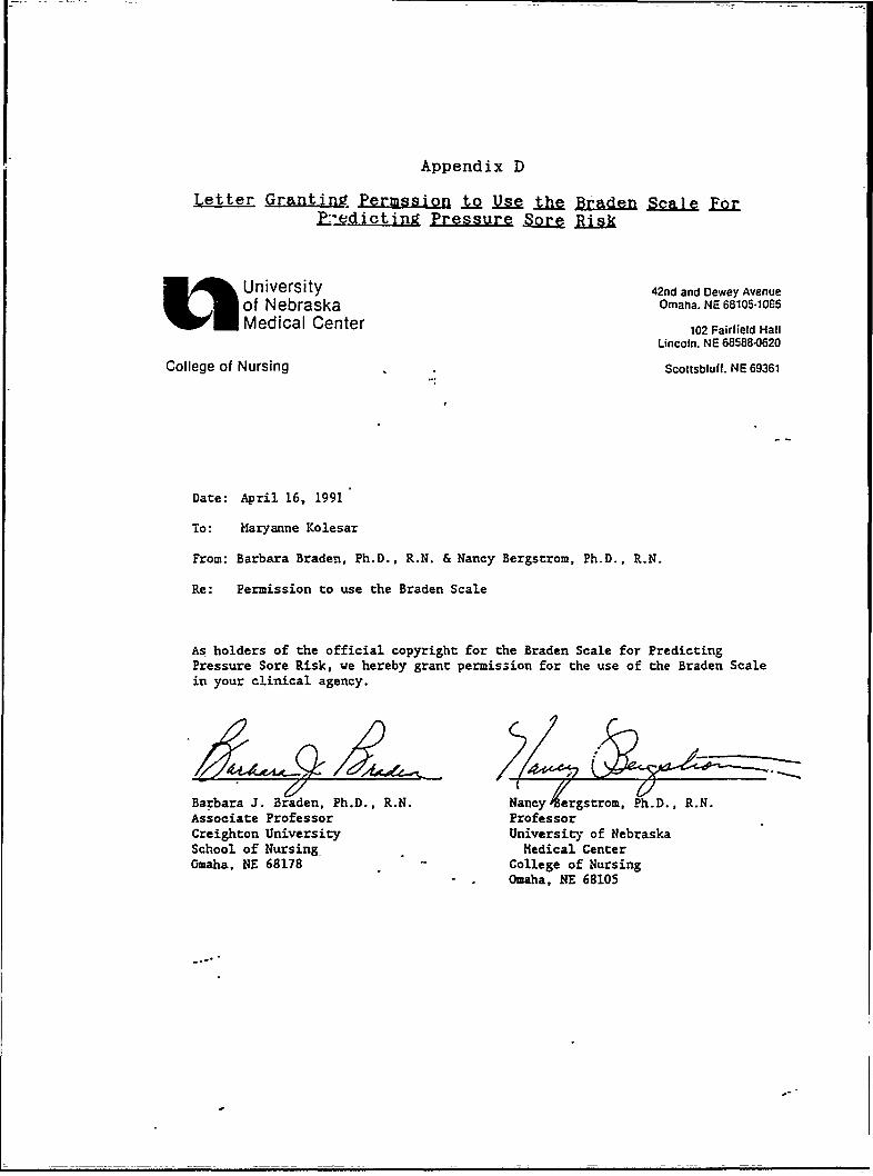
Appendix D
Letter Granting Permssion 1t e& th& Braden Scale FQZk:, _edc t in Pressure Sore R
L j University 42nd and Dewey Avenue
of Nebraska Omaha. NE 68105.1065
Medical Center 102 Fairfield HallLincoln. NE 68588-0620
College of Nursing Scottsbluff. NE 69361
Date: April 16, 1991
To: Maryanne Kolesar
From: Barbara Braden, Ph.D., R.N. & Nancy Bergstrom, Ph.D., R.N.
Re: Permission to use the Braden Scale
As holders of the official copyright for the Braden Scale for PredictingPressure Sore Risk, we hereby grant permission for the use of the Braden Scalein your clinical agency.
Barbara J. Braden, Ph.D., R.N. Nancy ergstrom. Ph.D., R.N.Associate Professor ProfessorCreighton University University of NebraskaSchool of Nursing_ Medical CenterOmaha, NE 68178 College of Nursing
Omaha, NE 68105

Appendix E
Letter Granting Permission t_ Utilize Ql.ical £a2i~ity L£foData Collection
TO: Maryanne Kolesar
FROM: Nursing-Research CommitteeDepartment of Nursing Service
RE: Proposed Study Titled:
nayG46 ggcf; ;ta' - ofCompletion-of Skin Assessment.and Doul ... ."' 0 *isk for Skin
Breakdown-in the Trauma PatientI~pr4 gr~twU.pa-aWlemic Shock
Requiring tMassive TransfusionResuscitation and Surgical Intervention
Your proposal has been approved for conduct within the Department of
Nursing. You are therefore, free to-proceed with your study within the
following guidelines:
1. Prior to initiating the study, you contact the Associate Director
of Nursing for-Nursing Education '-:'.to arrange completion
of appropriate mandatory orientation.
2. Inform the Associate Director of Nursing for Education
re: date data collected begins; date data
collection ends.
3. The results of-your study must be presented to the department of
nursing staff in a verbal presentation within 6 iiionths of comple-
tion of data collection.
Owned by the citzens o King Coun govrned by the Board of Trustees and a component
of the Warren G. Magnuson Health Sciences Center of the University of Washington

-87
LETTER OF AGREEMENT III R-1Attachment "D"Page-.2 of 2
TO:FROM: Nursing Research Committee
Department of Nursing Services
RE: Proposed Study Titled:
Analysis of the Extent of
Completion of Skin Assessmentand-Documentation of Risk for Skin
Breakdown in the Trauma Patient
4. A copy of the study's final report, including an abstract, shall
be submitted to the Department of Nursing Service wtthin-6 months
of the completion of the study.
5. -will not be identified by name--in the
study.
If you need clarification of any of the above, or-other information, your
contact person within the Nursing-Department is
(phone).
Assoa Directo(f Nulwng forEducatin
Date
SS:Jm4/22/87d/researchf/memo75