Efficiency ginkgolides and bilobalides for improving ... · in patients with bronchial asthma Key...
12
1 АСТМА ТА АЛЕРГІЯ, № 2 • 2016 UDC 616.248.1-085-084.001.5 Yu. I. Feshchenko, N. A. Prymushko, L. M. Kuryk, V. V. Kuts, O. I. Adamchuk, I. P. Turchyna, O. A. Kanarskiy, O. I. Krylach National Phthisiology and Pulmonology Institute named after F. H.Yanovskiy of the NAMS of Ukraine Efficacy of ginkgolides and bilobalides for prevention of decreased physical activity in patients with bronchial asthma Key words: bronchial asthma, physical activity, cardiorespiratory system, ginkgolides and bilobalides. So far, the problem of physical working capacity in pa- tients with bronchial asthma (BA) has become extremely topical, since most patients are young and physically active people. The issue of determining patients’ ability to achieve critical physical effort, specifically, anaerobic threshold (AT), is of particular interest. The term «physical working capacity» is used to denote potential human ability to ex- press maximum efforts under conditions of dynamic, static or mixed work. Anaerobic threshold is a point in physi- cal activity when insufficient delivery of oxygen to work- ing muscles triggers anaerobic energy supply mechanisms with generation of lactic acid, which leads to increased pro- duction of carbon dioxide (CO 2 ) and non-linear ventila- tion increase. The WHO finds physical working capacity studies ra- tional and helpful, especially when it comes to evaluation of organism’s functional reserves and differential diagnosis of cardiac activity disorders, design of sufficient and safe motion mode for the patients and development of physi- cal rehabilitation programs. The factors affecting the «physical working capacity» are: • respiratory function, including pulmonary mechan- ics and respiratory muscles work, as well as mechanisms of ventilation regulation; • ventilation-perfusion ratios, diffusion of oxygen; • heart function, including changes in heart rate and stroke volume; • control of peripheral circulation, i.e. the ability to dis- tribute blood flow in favour of working muscles; • muscle metabolism, including functions of oxidative and glycolytic enzymes. Muscle physiology itself is based on coordinated func- tioning of the respiratory, cardiovascular and muscular systems. Lack of one of these systems may lead to poor tolerance of physical activity. The study of functional safety margin is only possible in conditions of stress, because physical activity is a kind of stress that helps detecting the initial signs of pathology of pulmonary and cardiovascular systems, which are «masked» by reserve capacity of the or- ganism while resting. It is an established fact that in patients with bronchial asthma with reduced respiratory function (RF) physical working capacity is supported due to anaer- obic metabolism. Changes in performance of the respira- tory system affect efficiency of muscle activity by limiting oxygen supply to them. Anaerobic threshold is the indica- tor of this process. Its decline in patients with bronchial asthma is associated with the rising energy cost of breath- ing and decrease in respiratory ventilation. At maximum physical activity in patients with moderate persistent bronchial asthma, regardless of the phase of the disease, there is no effective performance of both pulmo- nary and cardiovascular systems. In response to physical activity the respiratory minute volume is increasing only through respiration rate, systolic blood pressure and heart rate are increasing excessively, consequently, the heart is unable to provide adequate minute volume of blood for covering energy consumption in the muscles, removal of excess lactic acid and maintaining homeostasis of the body. It has been established that in 100 % of patients with moderate persistent bronchial asthma in case of acute ex- acerbation of the disease the metabolic equivalent of task (MET) decreases on average to (4.8 ± 1.6) kcal/kg, the work performance level drops to (68.6 ± 4.7) %, and anaer- obic threshold lowers to (48.9 ± 3.2) %. Also there is an av- erage reduction of the oxygen cost of work performance © Yu. I. Feshchenko, N. A. Prymushko, L. M. Kuryk, V. V. Kuts, O. I. Adamchuk, I. P. Turchyna, O. A. Kanarskiy, O. I. Krylach, 2016
Transcript of Efficiency ginkgolides and bilobalides for improving ... · in patients with bronchial asthma Key...

1
АСТМА ТА АЛЕРГІЯ, № 2 • 2016
UDC 616.248.1-085-084.001.5
Yu. I. Feshchenko, N. A. Prymushko, L. M. Kuryk, V. V. Kuts, O. I. Adamchuk, I. P. Turchyna, O. A. Kanarskiy, O. I. KrylachNational Phthisiology and Pulmonology Institute named after F. H.Yanovskiy of the NAMS of Ukraine
Efficacy of ginkgolides and bilobalides for prevention
of decreased physical activity in patients with bronchial asthma
Key words: bronchial asthma, physical activity, cardiorespiratory system, ginkgolides and bilobalides.
So far, the problem of physical working capacity in pa-tients with bronchial asthma (BA) has become extremely topical, since most patients are young and physically active people. The issue of determining patients’ ability to achieve critical physical effort, specifically, anaerobic threshold (AT), is of particular interest. The term «physical working capacity» is used to denote potential human ability to ex-press maximum efforts under conditions of dynamic, static or mixed work. Anaerobic threshold is a point in physi-cal activity when insufficient delivery of oxygen to work-ing muscles triggers anaerobic energy supply mechanisms with generation of lactic acid, which leads to increased pro-duction of carbon dioxide (CO
2) and non-linear ventila-
tion increase.The WHO finds physical working capacity studies ra-
tional and helpful, especially when it comes to evaluation of organism’s functional reserves and differential diagnosis of cardiac activity disorders, design of sufficient and safe motion mode for the patients and development of physi-cal rehabilitation programs.
The factors affecting the «physical working capacity» are:• respiratory function, including pulmonary mechan-
ics and respiratory muscles work, as well as mechanisms of ventilation regulation;
• ventilation-perfusion ratios, diffusion of oxygen;• heart function, including changes in heart rate and
stroke volume;• control of peripheral circulation, i.e. the ability to dis-
tribute blood flow in favour of working muscles;• muscle metabolism, including functions of oxidative
and glycolytic enzymes.Muscle physiology itself is based on coordinated func-
tioning of the respiratory, cardiovascular and muscular
systems. Lack of one of these systems may lead to poor tolerance of physical activity. The study of functional safety margin is only possible in conditions of stress, because physical activity is a kind of stress that helps detecting the initial signs of pathology of pulmonary and cardiovascular systems, which are «masked» by reserve capacity of the or-ganism while resting. It is an established fact that in patients with bronchial asthma with reduced respiratory function (RF) physical working capacity is supported due to anaer-obic metabolism. Changes in performance of the respira-tory system affect efficiency of muscle activity by limiting oxygen supply to them. Anaerobic threshold is the indica-tor of this process. Its decline in patients with bronchial asthma is associated with the rising energy cost of breath-ing and decrease in respiratory ventilation.
At maximum physical activity in patients with moderate persistent bronchial asthma, regardless of the phase of the disease, there is no effective performance of both pulmo-nary and cardiovascular systems. In response to physical activity the respiratory minute volume is increasing only through respiration rate, systolic blood pressure and heart rate are increasing excessively, consequently, the heart is unable to provide adequate minute volume of blood for covering energy consumption in the muscles, removal of excess lactic acid and maintaining homeostasis of the body.
It has been established that in 100 % of patients with moderate persistent bronchial asthma in case of acute ex-acerbation of the disease the metabolic equivalent of task (MET) decreases on average to (4.8 ± 1.6) kcal/kg, the work performance level drops to (68.6 ± 4.7) %, and anaer-obic threshold lowers to (48.9 ± 3.2) %. Also there is an av-erage reduction of the oxygen cost of work performance
© Yu. I. Feshchenko, N. A. Prymushko, L. M. Kuryk, V. V. Kuts, O. I. Adamchuk, I. P. Turchyna, O. A. Kanarskiy, O. I. Krylach, 2016

2
АСТМА ТА АЛЕРГІЯ, № 2 • 2016
of (71.2 ± 2.1) %, deterioration of average oxygen (O2)
consumption effectiveness indices: V’O2 to (78.5 ± 2.5) %.
Merely (75.6 ± 6.2) % of patients reach the anaerobic threshold.
During remission of the disease in 46.7 % of patients with bronchial asthma the oxygen cost of work is significantly normalized to an average of (14.8 ± 1.5) ml/min/W, but in 53.3 % of patients no significant changes are observed, oxygen consumption in the metabolic equivalent of task (MET) units remains reduced in the group by 10.3 %, exer-cise tolerance (W) to (78.6 ± 4.3) % and (1.2 ± 0.1) W/kg, oxygen consumption peak (VO
2) to (88.4 ± 4.1) %. Merely
(86.7 ± 6.2) % of patients in the group reach the anaero-bic threshold.
Therefore, it is extremely important to develop and implement new prevention methods against the decline in physical activities with a view to improve the quality of life of patients with bronchial asthma.
The work is based on the method of prevention of reduc-tion of physical working capacity in patients with bronchial asthma as a prototype, which involves simultaneous admin-istration of basic treatment medications – inhaled corti-costeroids and short-acting β2-agonist – and prescription of bronchodilators (long-acting theophyllines) – Theopec, 600 mg daily for 1 month (see M.G.Gireeva. Anaerobic threshold and physical working capacity in patients with bronchial asthma [Text]: Author’s abstract of Doctoral Dissertation. 14.00.43: presented on 16.01.1991: approved on 16.07.1991 / Mariam Gireevna Gireeva. – Dagestan, 1991. – 23 p. – Bibliography. 23 p. – 200522022).
Yet, the drawback of this method is that prolonged and frequent use of long-acting theophyllines, especially in maximum daily dose, leads to unwanted side effects, namely: cardiotoxic effect (arrhythmia, angina spuria, in-creased frequency of angina attacks, tachycardia, fluctua-tions in blood pressure), as well as general weakness, de-creased performance and drowsiness.
The utility model is based on the task of development of a prevention method against the decline in physi-cal working capacity in patients with bronchial asthma, which uses a medicinal product based on ginkgolides and bilobalides against the background of standard treatment chosen for the period of remission to achieve an increase in metabolic and oxygen cost of work and level of its per-formance, improvement of oxygen consumption efficiency and muscle activity during physical activities, normaliza-tion of functioning of the cardiovascular system for im-provement of the quality of life of such patients.
The task is solved by additional prescription of a 90-day course of a medicinal product containing ginkgolides and bilobalides in the compendial dose and regimen against the background of administration of an inhaled glucocortico-steroid and a short-acting β2-agonist, according to the util-ity model during remission, which is a method of preven-tion of the decline in physical working capacity in patients with bronchial asthma.
The method is based on investigation of new data on identification of hidden pathological changes in cardio-respiratory system at maximum physical activity in patients
with BA, and their role in the disease during treatment with inhaled corticosteroid drugs. Bilobil Intens, a complex me-dicinal product based on flavonoid glycosides and terpene compounds (ginkgolides and bilobalides), was used for the claimed method as a medicinal product containing gink-golides and bilobalides. The active ingredients of the extract determine its specific pharmacological activity. The prod-uct is manufactured in the form of pink gelatine capsules filled with light to dark brown powder with visible dark par-ticles (registration No.UA/1234/01/03 of 06.05.2010, valid till 06.05.2015). The product improves cerebral blood flow and oxygen and glucose supply to the brain tissue, prevents platelet aggregation inhibiting platelet activating factor, di-lates blood vessels, improves blood circulation and protects cells and tissues from injuries caused by oxygen deficiency by regulation of metabolism. The product prevents devel-opment of toxic and traumatic brain oedema, age-related changes in the activity of cholinergic receptors and α2-adrenoreceptors. Flavonoids exhibit significant antioxidant effects. Bilobalides and ginkgolides have neuroprotective effects. The medicinal product improves blood rheology, sensory and cognitive functions of the brain and prevents deterioration of mental activity. The product is used for treatment of violation of cerebral blood flow and brain function (dementia syndrome in cases of primary degener-ative dementia, vascular dementia or their combinations), which is manifested in deterioration of memory and mental activity, melancholia and anxiety, dizziness, tinnitus, head-ache and sleep disorders, blood circulation disorders in the lower extremities in presence of chronic obliterating angi-opathy. The medicinal product can be taken without re-gard to food. The capsules should be swallowed with water.
The maximum therapeutic effect can be observed in 1 month. In order to achieve a sustainable effect Bilobil Intens shall be administered for at least 3 months, partic-ularly in elderly patients. After 3 months of treatment, jus-tification of further treatment shall be proven. The main contraindication for use is the individual drug intolerance. The adverse effect of the medicinal product may arise in the intestinal tract (rarely – disorders of the gastrointestinal tract), the nervous system (very rarely – headache), the immune system (very rarely – skin allergies (itching, red-ness and swelling).
There is no data on the use of medicinal products con-taining ginkgolides and bilobalides for prevention of phys-ical working capacity deterioration in patients with bron-chial asthma in the available literature.
The method is carried out as follows.BA patients are prescribed a medicinal product contain-
ing ginkgolides and bilobalides – 1 capsule 2 times a day after meals for 90 days, immediately after the end of treat-ment of exacerbation. All patients receive standard medical therapy chosen for remission period, which includes the use of an inhaled corticosteroids and a short-acting β2-agonist for reversal of asthma symptoms.
The cardiorespiratory stress test was used in order to de-termine the degree of exercise intolerance and basic mech-anisms of such disorders in patients with bronchial asthma (see Recommendations on the use of exercise testing

3
АСТМА ТА АЛЕРГІЯ, № 2 • 2016
in clinical practice [Text] / P.Palange [et al.] // Eur. Respir. J. – 2007. – Vol. 29. – P. 185–209). Ergospirometry makes it possible to quantify physical working capacity and func-tional status of the respiratory and cardiovascular systems (in-cluding tissue respiration) and their interaction. Ergometer ER/2 and Ergoselect 1000 LP Basic with automatic power dissipation regardless of pedalling speed were used for grad-uated exercise. The main parameters of pulmonary ventila-tion and gas exchange, as well as the heart rate and elec-trocardiogram were recorded automatically and processed by Ergopnevmotest OM/05-Ts and the Oxycon Pro ergo-spirometric system – Version JLAB 4.67, manufactured by VIASYS Healthcare, which consists of a pneumotach-ograph with an integrator, oxygen analyzers and an ECG recorder. The cycloergospirometric study was conducted subject to the general testing requirements specified for sub-maximal physical activity. Prior to the study, those medica-tions affecting the functional state of cardiorespiratory and nervous systems were cancelled. Smoking was prohibited for two or more hours prior to the study. Physical activity started no earlier than in 1 hour after meal. Absolute and relative contraindications for testing and conditions requir-ing special attention and care were taken into consideration based on the recommendations (see ERS School Courses ATS/ACCP Statement on Cardiopulmonary Exercise Testing [Text] // Am. J. Respir. Crit. Care Med. – 2006. – Vol. 167. – Р. 211 – 277). Temperature conditions – from +18°C to +25°C, light clothing, breathable and mois-ture absorbent, light shoes with hard soles on feet. Prior to the beginning of the study the patients were informed about the purpose and procedures of the motor test. Load capacity, maximum oxygen consumption, minute lung ventilation and heart rate were predicted based on extrap-olation method with a view to gender and anthropometric parameters of the patient under study. Physical working ca-pacity was defined in compliance with the Protocol RA – 150 – I B3 – BP2 – EC1. The work continued to refuse or was stopped after appearance of subjective and objective symptoms that limited further increase of the load: severe shortness of breath, achievement of submaximal heart rate, appearance of electrocardiographic signs of coronary insuf-ficiency. Deviation of pedalling rate from the given level (below 60 rpm) due to muscle weakness or lack of moti-vation for reaching the load limit was regarded as subjects’ withdrawal from further tests. The maximum level of exe-cuted load was estimated as the limit of functional capaci-ties of the body. The following parameters were evaluated:
• V’O2, mL/min – oxygen consumption in mL per min-
ute, measured at maximum load and at the level of anaer-obic threshold;
• VO2/kg, mL/min/kg – oxygen consumption in mL per
minute per kilogram of body weight, measured at the max-imum load and at the level of anaerobic threshold;
• BR, % – reserve ventilation at the maximum load;• HR, L/min – patient’s heart rate at the peak load;• HRR, L/min – patient’s heart rate reserve after reach-
ing the maximum load;• O2
/HR, mL – oxygen pulse, mL, observed at the max-imum load;
• SpO2, % – oxygen saturation measured at the maxi-
mum load;• BP, mm Hg – patient’s blood pressure at the peak load;• W – maximum achieved load in Watts;• MET – metabolic equivalent of task measured at the
maximum load.Each patient made an assessment of the reasons for termi-
nation of the test. Estimated indices were obtained through automatic calculation based on the developer’s method.
Here are specific examples of application of the method.Example 1 (based on the prototype method).Patient S., female, 49, case history No.1170, has
been treated at the Department of Obstructive Lung Diseases at the F.H.Yanovskiy National Phthisiology and Pulmonology Institute of the NAMS of Ukraine, SI, with the following diagnosis: moderate persistent III de-gree bronchial asthma,, exacerbation phase, VD II. The patient has been ill for more than 10 years. For the last 3 years the patient has been associating occurrence of ex-acerbations with hypothermia or the ARVI. The last ex-acerbation occurred 8 months ago. For the last 2 years the patient has been taking Flixotide 1,000 mg, 2 breaths daily, and Salbutamol as a short-acting bronchodila-tor. The patient was prescribed prolonged theophylline, Theopec, 1 capsule 2 times daily for 1 month in the course of the background treatment during remission. The pa-tient underwent clinical and instrumental examination prior to the beginning of the treatment, immediately upon termination of the course of treatment with Theopec and in 3 months after the treatment based on the prototype method.
The patient’s physical activity was decreased during ex-acerbations of BA, according to the cycloergospiromet-ric study data, in particular: the MET rate decreased to 4.5 kcal/kg, duration of the 3rd phase of cardiorespi-ratory stress test decreased to 6.3 minutes, oxygen cost of work dropped to 14.9 mL/min/kg, peak oxygen con-sumption (VO
2) reduced to 75.9 %, oxygen consumption
performance indices (O2) decreased to: V’O
2 to 82 %, V’O
2p
to 84 %, and the maximum oxygen consumption at the peak load (V’O
2max) to 95 %. The patient’s RER index de-
scriptive of the ratio of production of CO2 (VCO
2) to ox-
ygen consumption (VO2) was higher during exacerbation
of BA and amounted to 1.15 units, which confirmed hyper-ventilation during the test. The patient’s BR index assess-ing respiratory volumes was slightly reduced to 72 %, which is typical for bronchial asthma. The patient’s dO
2/dW index
descriptive of efficiency of muscle performance at the max-imum physical activity was reduced to 8.6 ml/min/W. The indices descriptive of the cardiovascular system perfor-mance at maximum physical activity were slightly invigo-rated, in particular: HR/VO
2 (in absolute terms and in per-
centage terms) were slightly higher – 6.9 beats/mL/kg, dHR/dO
2 – 75.6 beats/min/mL, VO
2/HR – 76 % (see
Table 1).Forced expiratory volume per 1 second was 63.8 %,
forced vital lung capacity was 72.5 %, peak expiratory flow rate was 70.5 %, the ratio of forced expiratory volume per 1 second to forced vital lung capacity was 70.6 %.

4
АСТМА ТА АЛЕРГІЯ, № 2 • 2016
The patient underwent repeated examination immedi-ately after the course of treatment with Theopec in the course of the background therapy. The following was estab-lished: there were no significant changes in spirogram speed performance, in particular: forced expiratory volume per 1 second was 68.3 %, forced vital lung capacity was 73.2 %, peak expiratory flow rate was 72.4 %, the ratio of forced expiratory volume per 1 second to forced vital lung capac-ity was 71.6 %. There were no significant changes in car-diorespiratory stress test indices, in particular: exercise tol-erance remained reduced to 5.5 kcal/kg, RW and PMA indices remained reduced as well – 0.9 W/kg and 75 %, respectively. The indices descriptive of the level of work performed remained unchanged (W) – 85 %, 1.9 W/kg, 159.8 W. The 3rd phase of the cardiorespiratory stress test lasted for 6.5 minutes. The indices descriptive of the oxygen cost of work remained reduced: V’O
2/kg to 5.3 mL/min/
kg, V’O2/kg, % -76.1 %, V’O
2 – 85 %, V’O
2p – 86 %,
V’O2max
– 90 %. The patient’s dO2/dW index descriptive
of the efficiency of muscle activity at the maximum phys-ical activity was reduced to 8.1 mL/min/W.
The patient’s indices descriptive of the cardiovascu-lar system performance at maximum physical activity af-ter the course of treatment with Theopec did not differ from those of the period of exacerbation: dHR/dO
2 –
72 beats/ml/min, VO2/HR – 6.7 beats/ml/kg, VO
2/HR –
78 %. SpO2 at maximum physical activity was 82 %, SBP
(systolic blood pressure) – 156 mm Hg, DBP (diastolic blood pressure) – 77 mm Hg.
In 3 months after discontinuation of Theopec the pa-tient underwent repeated examination. There were no sig-nificant changes in spirogram speed performance, in par-ticular: forced expiratory volume per 1 second was 71.3 %, forced vital lung capacity was 83.2 %, peak expiratory flow rate was 74.8 %, the ratio of forced expiratory volume per 1 second to forced vital lung capacity was 72.3 %. The in-dices descriptive of oxygen cost of the work performed re-mained unchanged, in particular: V’O
2/kg – 5.3 mL/kg and
76.2 %, V’O2 – 86 %, V’O
2p – 86 %, V’O
2max – 91 %.
The RER index – 1.01, MET – 5.2 kcal/kg, RW (relative workload) – 0.8 W/kg, PMA – 78 %. Duration of the 3rd phase of the test – 6.4 minutes. There were no significant changes in the indices descriptive of the work performed as well: W – 85 %, 1.8 W/kg and 157.6 W, dO
2/dW –
8.2 mL/min/W.The indices descriptive of effectiveness of cardiovas-
cular response to exercise did not differ from the indices of the exacerbation period: dHR/dO
2 – 71 beats/mL/min,
VO2/HR – 6.5 bps/mL/kg and 87 %, SBP – 158 mm Hg,
DBP – 78 mm Hg, SpO2 – 87 %.
Thus, the course of treatment resulted in achievement of:• a slight improvement in physical activity due to reserve
capacity of the cardiovascular and respiratory system for up to 2 months with a further deterioration.
Yet, the administered treatment led to adverse events – an increase in body weight by 3 kg per month of adminis-tration of the medicinal product, a slight increase in blood glucose level, dyspepsia and mild abdominal pain shortly after administration of the medicinal product (disappeared
Table 1. Cycloergospirometric test indices over time, patient S.
Indices
Patient S.
Exacer bation phase
immediately after
treatment based on the
prototype method
in 3 months after
treatment based on the
prototype method
1 2 3 4
Duration of the 3rd phase of the test (min)
6.3 6.5 6.4
V’O2/kg (mL/min/kg)
5.9 5.3 5.3
V’O2/kg (mL/min/kg), %
75.9 76.1 76.2
V’O2 ( %) 82 85 86
V’O2p, ( %) 86 86 86
V’O2max ( %) 85 90 91
RER (relative units)
1.15 1.01 1.01
BR (%) 72 73 75
BP (% of V’O2max) 38 37 35
W (%) 84 85 85
W (W/kg) 1.8 1.9 1.8
W (W) 156.3 159.8 157.6
dO2/dW (mL/min/W)
8.6 8.1 8.2
HR/VO2 (beats/mL/kg)
6.9 6.7 6.5
dHR/dO2 (beats/min/mL)
75.6 72 71
VO2/HR ( %) 76 78 78
SpO2 ( %) 85 82 87
MET (kcal/kg) 5.8 5.5 5.2
RW (W/kg) 0.8 0.9 0.8
PМА (%) 74 75 78
Assessment of dyspnoea based on Borg scale before the test (points)
0 0 0
Assessment of dyspnoea based on Borg scale after the test (points)
2 2 2
5
АСТМА ТА АЛЕРГІЯ, № 2 • 2016
independently in 2 days of administration), an increase of complaints about premature ventricular contraction (PVC), especially in the evening.
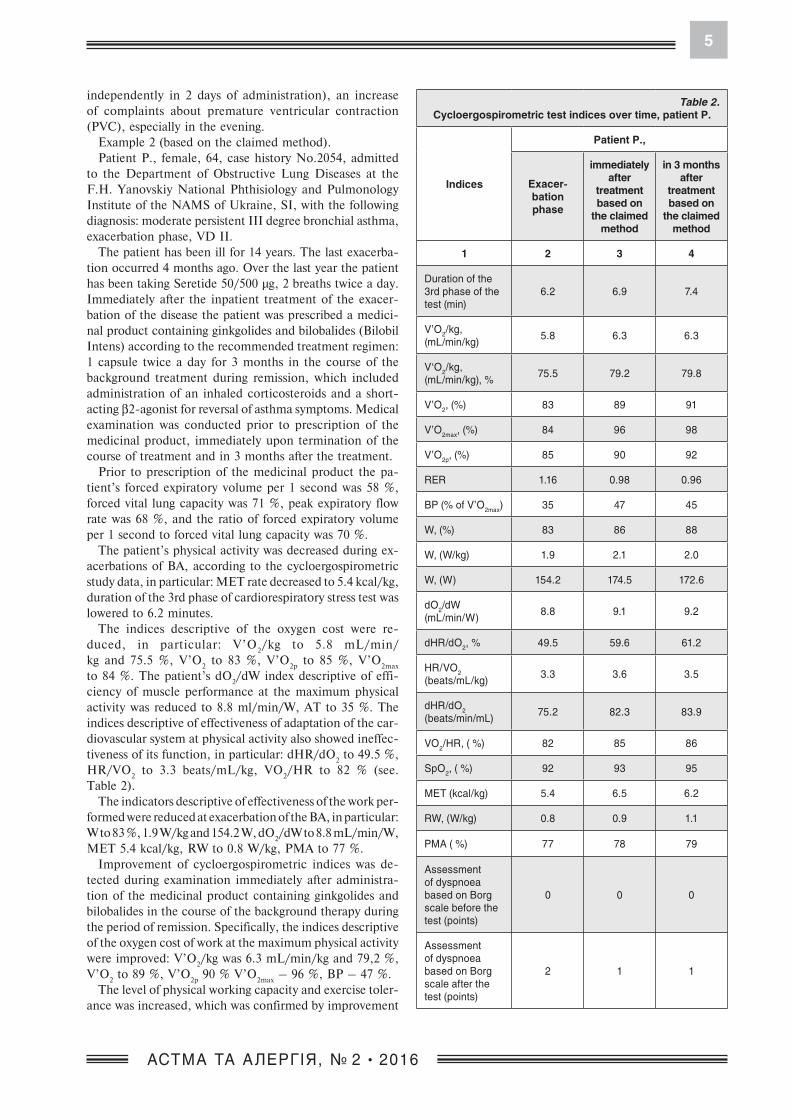
Example 2 (based on the claimed method).Patient P., female, 64, case history No.2054, admitted
to the Department of Obstructive Lung Diseases at the F.H. Yanovskiy National Phthisiology and Pulmonology Institute of the NAMS of Ukraine, SI, with the following diagnosis: moderate persistent III degree bronchial asthma, exacerbation phase, VD II.
The patient has been ill for 14 years. The last exacerba-tion occurred 4 months ago. Over the last year the patient has been taking Seretide 50/500 µg, 2 breaths twice a day. Immediately after the inpatient treatment of the exacer-bation of the disease the patient was prescribed a medici-nal product containing ginkgolides and bilobalides (Bilobil Intens) according to the recommended treatment regimen: 1 capsule twice a day for 3 months in the course of the background treatment during remission, which included administration of an inhaled corticosteroids and a short-acting β2-agonist for reversal of asthma symptoms. Medical examination was conducted prior to prescription of the medicinal product, immediately upon termination of the course of treatment and in 3 months after the treatment.
Prior to prescription of the medicinal product the pa-tient’s forced expiratory volume per 1 second was 58 %, forced vital lung capacity was 71 %, peak expiratory flow rate was 68 %, and the ratio of forced expiratory volume per 1 second to forced vital lung capacity was 70 %.
The patient’s physical activity was decreased during ex-acerbations of BA, according to the cycloergospirometric study data, in particular: MET rate decreased to 5.4 kcal/kg, duration of the 3rd phase of cardiorespiratory stress test was lowered to 6.2 minutes.
The indices descriptive of the oxygen cost were re-duced, in particular: V’O
2/kg to 5.8 mL/min/
kg and 75.5 %, V’O2 to 83 %, V’O
2p to 85 %, V’O
2max
to 84 %. The patient’s dO2/dW index descriptive of effi-
ciency of muscle performance at the maximum physical activity was reduced to 8.8 ml/min/W, AT to 35 %. The indices descriptive of effectiveness of adaptation of the car-diovascular system at physical activity also showed ineffec-tiveness of its function, in particular: dHR/dO
2 to 49.5 %,
HR/VO2 to 3.3 beats/mL/kg, VO
2/HR to 82 % (see.
Table 2).The indicators descriptive of effectiveness of the work per-
formed were reduced at exacerbation of the BA, in particular: W to 83 %, 1.9 W/kg and 154.2 W, dO
2/dW to 8.8 mL/min/W,
MET 5.4 kcal/kg, RW to 0.8 W/kg, PMA to 77 %.Improvement of cycloergospirometric indices was de-
tected during examination immediately after administra-tion of the medicinal product containing ginkgolides and bilobalides in the course of the background therapy during the period of remission. Specifically, the indices descriptive of the oxygen cost of work at the maximum physical activity were improved: V’O
2/kg was 6.3 mL/min/kg and 79,2 %,
V’O2 to 89 %, V’O
2p 90 % V’O
2max – 96 %, BP – 47 %.
The level of physical working capacity and exercise toler-ance was increased, which was confirmed by improvement
Table 2. Cycloergospirometric test indices over time, patient P.
Indices
Patient P.,
Exacerbation phase
immediately after
treatment based on
the claimed method
in 3 months after
treatment based on
the claimed method
1 2 3 4
Duration of the 3rd phase of the test (min)
6.2 6.9 7.4
V’O2/kg, (mL/min/kg)
5.8 6.3 6.3
V‘O2/kg, (mL/min/kg), %
75.5 79.2 79.8
V’O2, (%) 83 89 91
V’O2max, (%) 84 96 98
V’O2p, (%) 85 90 92
RER 1.16 0.98 0.96
BP (% of V’O2max) 35 47 45
W, (%) 83 86 88
W, (W/kg) 1.9 2.1 2.0
W, (W) 154.2 174.5 172.6
dO2/dW (mL/min/W)
8.8 9.1 9.2
dHR/dO2, % 49.5 59.6 61.2
HR/VО2 (beats/mL/kg)
3.3 3.6 3.5
dHR/dO2 (beats/min/mL)
75.2 82.3 83.9
VO2/HR, ( %) 82 85 86
SpO2, ( %) 92 93 95
MET (kcal/kg) 5.4 6.5 6.2
RW, (W/kg) 0.8 0.9 1.1
PМА ( %) 77 78 79
Assessment of dyspnoea based on Borg scale before the test (points)
0 0 0
Assessment of dyspnoea based on Borg scale after the test (points)
2 1 1

6
АСТМА ТА АЛЕРГІЯ, № 2 • 2016
of their indices: W up to 86 % and 174.5 W, 2.1 W/kg, dO
2/dW – 9.1 mL/min/W, MET – 6.5 kcal/kg, RW –
0.9 W/kg, PMA – 78 %.Also, there was improvement in the cardiovascular sys-
tem performance indices: dHR/dO2 – 82.3 %, HR/VO
2 –
3.6 beats/mL/kg, VO2/HR – 85 %. Forced expiratory vol-
ume per 1 second was 65 %, forced vital lung capacity was 76 %, peak expiratory flow rate was 72 %, the ratio of forced expiratory volume per 1 second to forced vital lung capacity was 74 %.
Stabilization of physical working capacity indices was detected during examination in 3 months after the course of medicinal product containing ginkgolides and bi-lobalides, namely Bilobil Intens, in the course of the back-ground therapy during the period of remission.
There was no drop in indices descriptive of the oxy-gen cost of work performed at maximum physical activ-ity: V’O
2/kg – 6.3 mL/min/kg and 79.8 %, V’O
2 – 91 %,
V’O2p
– 92 %, V’O2max
– 98 %, BP – 47 %. Effectiveness of the work performed and exercise tolerance remained stable, no deterioration was observed: W – 88 % and 172.6 W and 2.0 W/kg, dO
2/dW – 9.2 mL/min/W, MET –
6.2 kcal/kg, RW – 1.1 W/kg, PMA – 79 %.Cardiovascular system performance remained unchanged
as well: dHR/dO2 – 61.2 %, HR/VO
2 – 3.5 bps/mL/kg,
VO2/HR – 86 %. Forced expiratory volume per 1 second
was 69 %, forced vital lung capacity was 78 %, peak expi-ratory flow rate – 82 %, the ratio of forced expiratory vol-ume per 1 second to forced vital lung capacity was 80 %.
Thus, the course of the medicinal product containing ginkgolides and bilobalides in the course of the background therapy during the period of remission facilitated:
• improvement of efficiency of oxygen utilization by the body by increasing efficiency of its absorption at the time of maximum physical activity, which was confirmed by im-provement of indices: V’O
2/kg from 5.8 mL/min/kg and
75.5 % to 6.3 mL/min/kg and 79,8 %, V’O2 from 83 %
to 91 %, V’O2p
from 85 % to 92 %;• increase of exercise tolerance and level
of work performed, which is confirmed by improvement of indices: W from 83 % and 1.9 W/kg and 154.2 W, dO
2/dW from 8.8 mL/min/W, MET from 5.4 kcal/kg,
RW from 0.8 W/kg, PMA from 77 % to W – 88 % and 2.0 W/kg and 172.6 W, dO
2/dW to 9.2 mL/min/W, MET
to 6.2 kcal/kg, RW to 1.1 W/kg, PMA to 79 %;• improvement of the cardiovascular system perfor-
mance, which is confirmed by improvement of indices: dHR/dO
2 from 75.2 % to 83.9 %, VO
2/HR from 82 %
to 86 %.• no adverse events were observed.For the purpose of comparative assessment of the two
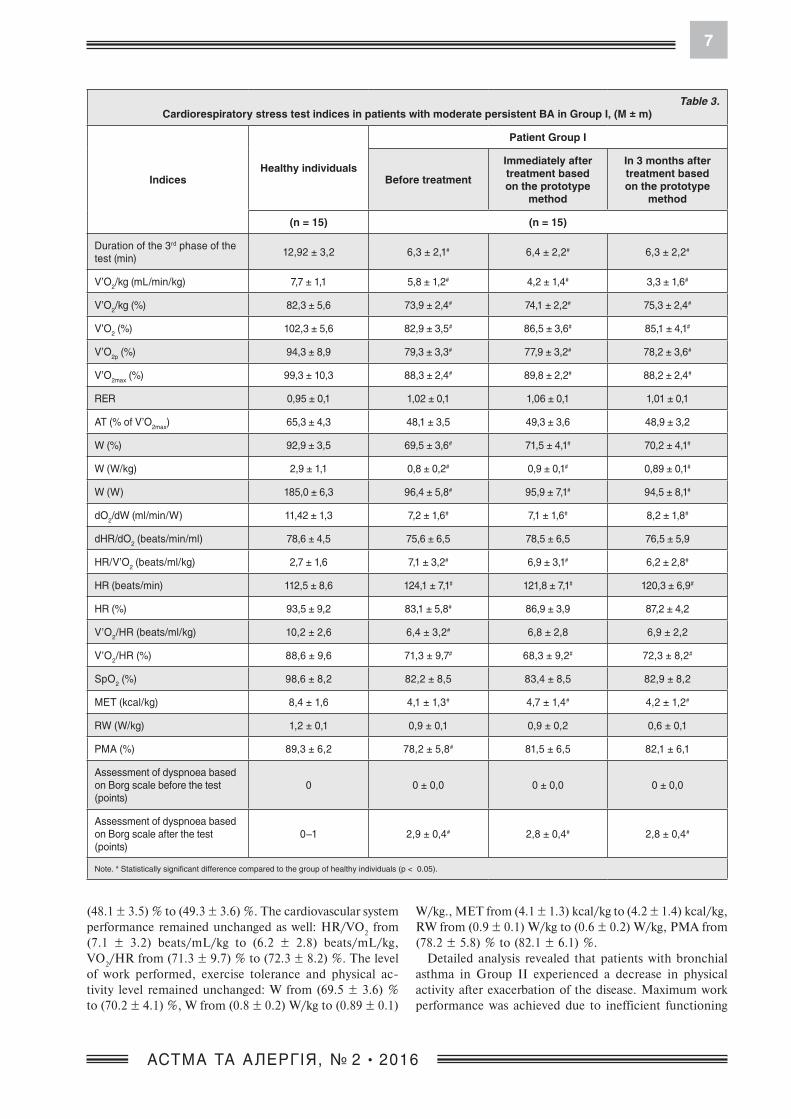
methods aimed at prevention of reduction of physical working capacity in patients with bronchial asthma, all pa-tients were divided into groups: the prototype method was used in 15 patients (Group I), the claimed method was used in 15 patients (Group II). There were 15 individuals in the group of healthy donors (control group). Age and gender composition of patients and the degree of severity of bron-chial asthma were comparable in all groups.
During sample comparison of the percentage of healthy individuals and the same number of bronchial asthma pa-tients it has been established that administration of Theopec as a part of combination treatment in patients with bron-chial asthma based on the prototype method resulted in a positive trend in the estimated indices.
No statistically significant difference compared to the be-ginning of the treatment was observed.
Detailed analysis revealed that patients with bronchial asthma in Group I experienced a decrease in physical ac-tivity after exacerbation of the disease. Maximum work performance is achieved due to inefficient functioning of the cardiovascular and pulmonary systems at the max-imum physical activity. Specifically, BA patients have lower oxygen supply during physical activity than healthy individuals as a result of inhibition of functional activ-ity of the respiratory system. This is confirmed by re-duced cycloergospirometric indices descriptive of the re-spiratory system function: V’O
2/kg to (5.8 ± 1.2) mL/min/
kg, V’O2/kg to (73.9 ± 2.4) %, V’O
2 to (82.9 ± 3.5) %,
V’O2p
(79.3 ± 3.3) %, V’O2max
to (88.3 ± 2.4) %, RER to (1.02 ± 0.1). The indices descriptive of the cardio-vascular system performance were reduced as well: dHR/dO
2 to (75.6 ± 6.5) %, HR/VO
2 to (7.1 ± 3.2) bps/mL/
kg, HR to (124.1 ± 7.1) beats/min and (83.1 ± 5.8) %, VO
2/HR to (6.4 ± 3.2) beats/mL/kg and (71.3 ± 9.7) %,
SBP to (184.2 ± 8.1) mm Hg, DBP to (70.5 ± 4.5) mm Hg, SpO
2 to (82.2 ± 8.5) %. As a result, exercise tolerance and
level of work performed were reduced: W to (69.5 ± 3.6) % and (0.8 ± 0.2) W/kg, (96.4 ± 5.8) W, dO
2/dW to (7.2 ± 1.6)
mL/min/W.After the preventive course of long-acting theophyl-
lines, in particular Theopec, with the background treat-ment in BA patients from Group I no significant changes were observed in the estimated indices compared with the beginning of the treatment: significant difference per-sisted compared to the group of healthy individuals in the following indices – V’O
2/kg from (5.8 ± 1.2) mL/min/
kg to (4.2 ± 1.4) mL/min/kg, V’O2/kg from (73.9 ± 2.4) %
to (74.1 ± 2.2) %, V’O2 from (82.9 ± 3.5) % to (86.5 ± 3.6) %,
V’O2p
from (79.3 ± 3.3) % to (77.9 ± 3.2) %, V’O2max
from (88.3 ± 2.4) % to (89.8 ± 2.2) %. There were no signif-icant changes in the cardiovascular system performance: VO
2/HR from (71.3 ± 9.7) % to (68.3 ± 9.2) %, HR/VO
2 from
(7.1 ± 3.2) beats/mL/kg to (6.9 ± 3.1) beats/mL/kg, SpO
2 from (82.2 ± 8.5) % to (83.4 ± 8.5) %. As a result, ex-
ercise tolerance, level of work performed and physical ac-tivity of patients treated with Theopec were reduced. In par-ticular: W changed from (69.5 ± 3.6) % to (71.5 ± 4.1) %, MET from (4.1 ± 1.3) kcal/kg to (4.7 ± 1.4) kcal/kg.
In 3 months after the treatment, the patients’ physi-cal activity remained lowered. The cardiovascular sys-tem performance during physical activity was ineffec-tive: V’O
2/kg from (5.8 ± 1.2) mL/min/kg to (3.3 ± 1,6)
mL/min/kg, V’O2/kg from (73.9 ± 2.4) % to (75.3 ± 2.4) %,
V’O2 from (82.9 ± 3.5) % to (85.1 ± 4.1) %, V’O
2p from
(79.3 ± 3.3) % to (78.2 ± 3.6) %, V’O2max
from (88.3 ± 2.2) % to (88.2 ± 2.4) %, the duration of the 3rd phase of the test from (6.3 ± 2.2) minutes to (6.3 ± 2.1) minutes, BP from
7
АСТМА ТА АЛЕРГІЯ, № 2 • 2016
(48.1 ± 3.5) % to (49.3 ± 3.6) %. The cardiovascular system performance remained unchanged as well: HR/VO
2 from
(7.1 ± 3.2) beats/mL/kg to (6.2 ± 2.8) beats/mL/kg, VO
2/HR from (71.3 ± 9.7) % to (72.3 ± 8.2) %. The level
of work performed, exercise tolerance and physical ac-tivity level remained unchanged: W from (69.5 ± 3.6) % to (70.2 ± 4.1) %, W from (0.8 ± 0.2) W/kg to (0.89 ± 0.1)
W/kg., MET from (4.1 ± 1.3) kcal/kg to (4.2 ± 1.4) kcal/kg, RW from (0.9 ± 0.1) W/kg to (0.6 ± 0.2) W/kg, PMA from (78.2 ± 5.8) % to (82.1 ± 6.1) %.
Detailed analysis revealed that patients with bronchial asthma in Group II experienced a decrease in physical activity after exacerbation of the disease. Maximum work performance was achieved due to inefficient functioning
Table 3. Cardiorespiratory stress test indices in patients with moderate persistent BA in Group I, (M ± m)
IndicesHealthy individuals
Patient Group I
Before treatment
Immediately after treatment based on the prototype
method
In 3 months after treatment based on the prototype
method
(n = 15) (n = 15)
Duration of the 3rd phase of the test (min)
12,92 ± 3,2 6,3 ± 2,1# 6,4 ± 2,2# 6,3 ± 2,2#
V’O2/kg (mL/min/kg) 7,7 ± 1,1 5,8 ± 1,2# 4,2 ± 1,4# 3,3 ± 1,6#
V’O2/kg (%) 82,3 ± 5,6 73,9 ± 2,4# 74,1 ± 2,2# 75,3 ± 2,4#
V’O2 (%) 102,3 ± 5,6 82,9 ± 3,5# 86,5 ± 3,6# 85,1 ± 4,1#
V’O2p (%) 94,3 ± 8,9 79,3 ± 3,3# 77,9 ± 3,2# 78,2 ± 3,6#
V’O2max (%) 99,3 ± 10,3 88,3 ± 2,4# 89,8 ± 2,2# 88,2 ± 2,4#
RER 0,95 ± 0,1 1,02 ± 0,1 1,06 ± 0,1 1,01 ± 0,1
AT (% of V’O2max) 65,3 ± 4,3 48,1 ± 3,5 49,3 ± 3,6 48,9 ± 3,2
W (%) 92,9 ± 3,5 69,5 ± 3,6# 71,5 ± 4,1# 70,2 ± 4,1#
W (W/kg) 2,9 ± 1,1 0,8 ± 0,2# 0,9 ± 0,1# 0,89 ± 0,1#
W (W) 185,0 ± 6,3 96,4 ± 5,8# 95,9 ± 7,1# 94,5 ± 8,1#
dO2/dW (ml/min/W) 11,42 ± 1,3 7,2 ± 1,6# 7,1 ± 1,6# 8,2 ± 1,8#
dHR/dO2 (beats/min/ml) 78,6 ± 4,5 75,6 ± 6,5 78,5 ± 6,5 76,5 ± 5,9
HR/V’O2 (beats/ml/kg) 2,7 ± 1,6 7,1 ± 3,2# 6,9 ± 3,1# 6,2 ± 2,8#
HR (beats/min) 112,5 ± 8,6 124,1 ± 7,1# 121,8 ± 7,1# 120,3 ± 6,9#
HR (%) 93,5 ± 9,2 83,1 ± 5,8# 86,9 ± 3,9 87,2 ± 4,2
V’O2/HR (beats/ml/kg) 10,2 ± 2,6 6,4 ± 3,2# 6,8 ± 2,8 6,9 ± 2,2
V’O2/HR (%) 88,6 ± 9,6 71,3 ± 9,7# 68,3 ± 9,2# 72,3 ± 8,2#
SpO2 (%) 98,6 ± 8,2 82,2 ± 8,5 83,4 ± 8,5 82,9 ± 8,2
MET (kcal/kg) 8,4 ± 1,6 4,1 ± 1,3# 4,7 ± 1,4# 4,2 ± 1,2#
RW (W/kg) 1,2 ± 0,1 0,9 ± 0,1 0,9 ± 0,2 0,6 ± 0,1
PМА (%) 89,3 ± 6,2 78,2 ± 5,8# 81,5 ± 6,5 82,1 ± 6,1
Assessment of dyspnoea based on Borg scale before the test (points)
0 0 ± 0,0 0 ± 0,0 0 ± 0,0
Assessment of dyspnoea based on Borg scale after the test (points)
0–1 2,9 ± 0,4# 2,8 ± 0,4# 2,8 ± 0,4#
Note. # Statistically significant difference compared to the group of healthy individuals (p < 0.05).

8
АСТМА ТА АЛЕРГІЯ, № 2 • 2016
of the cardiovascular and pulmonary systems at the maxi-mum physical activity. Specifically, BA patients had lower oxygen supply during physical activity than healthy in-dividuals. This was confirmed by reduced ergospiromet-ric indices descriptive of the respiratory system func-tion: V’O
2/kg to (70.9 ± 2.5) %, V’O
2 to (79.3 ± 3.1) %,
V’O2p
to (77.5 ± 3.1) %, V’O2max
to (79.6 ± 7.5) %, RER to (1.03 ± 0.1) %. There were changes in the cardiovascular
system’s adaptive capacity to physical activity: the amount of oxygen carried by the heart during maximum physical activity was reduced by restriction of its flow as a result of obstructive changes in the respiratory system, therefore, the amount of oxygen used for work performance was re-duced: dHR/dO
2 to (74.6 ± 5.1) %, HR/VO
2 to (6.9 ± 1.8)
beats/mL/kg, HR to (125.6 ± 7.2) beats/min and (82.1 ± 5.2) %, VO
2/HR to (72.6 ± 9.4) %. Consequently,
Table 4. Cardiorespiratory stress test indices in patients with moderate persistent BA in Group II, (M ± m)
IndicesHealthy individuals
Patient Group II
Before treatment
Immediately after treatment based on the prototype
method
In 3 months after treatment based on the prototype
method
(n = 15) (n = 15)
Duration of the 3rd phase of the test (min)
12,92 ± 3,2 6,4 ± 2,2# 8,2 ± 2,1* 8,3 ± 2,4*
V’O2/kg (mL/min/kg) 7,7 ± 1,1 5,6 ± 1,3# 7,2 ± 1,5* 7,3 ± 1,6*
V’O2/kg (%) 82,3 ± 5,6 70,9 ± 2,5# 92,3 ± 4,2* 92,3 ± 2,6*
V’O2 (%) 102,3 ± 5,6 79,3 ± 3,1# 98,3 ± 4,2* 91,6 ± 4,5*
V’O2p (%) 94,3 ± 8,9 77,5 ± 3,1# 85,6 ± 3,5* 86,3 ± 3,2*
V’O2max (%) 99,3 ± 10,3 79,6 ± 7,5# 92,3 ± 6,4* 91,6 ± 5,4*
RER (%) 0,95 ± 0,1 1,03 ± 0,1 1,01 ± 0,1 1,01 ± 0,1
AT (% of V’O2max) 65,3 ± 4,3 47,5 ± 3,6 48,3 ± 3,5 49,4 ± 2,9
W (%) 92,9 ± 3,5 69,2 ± 5,2# 92,9 ± 5,2* 92,8 ± 6,1*
W (W/kg) 2,9 ± 1,1 0,7 ± 0,2# 1,2 ± 0,1#* 1,3 ± 0,1#*
W (W) 185,0 ± 6,3 95,6 ± 6,8# 145,3 ± 6,3* 149,2 ± 8,2*
dO2/dW (mL/min/W) 11,42 ± 1,3 6,9 ± 1,8# 9,5 ± 1,8* 10,3 ± 2,1*
HR/V’O2 (beats/mL/kg) 2,7 ± 1,6 6,9 ± 1,8# 3,5 ± 1,3#* 4,1 ± 1,2#*
V’O2/HR (%) 88,6 ± 9,6 72,6 ± 9,4# 88,1 ± 9,4* 84,6 ± 8,1*
dHR/dO2 (beats/min/mL) 78,6 ± 4,5 74,6 ± 5,1 78,5 ± 6,5 76,5 ± 5,9
HR (beats/min) 112,5 ± 8,6 125,6,1 ± 7,2# 121,8 ± 7,1# 120,3 ± 6,9#
HR (%) 93,5 ± 9,2 82,1 ± 5,2# 86,9 ± 3,9 87,2 ± 4,2
SpO2 (%) 98,6 ± 8,2 92,6 ± 8,1 95,9 ± 8,1 97,2 ± 6,8
MET (kcal/kg) 8,4 ± 1,6 4,6 ± 1,8# 7,8 ± 1,5* 7,9 ± 1,5*
RW (W/kg) 1,2 ± 0,1 0,7 ± 0,1 1,1 ± 0,2 1,1 ± 0,1
PМА (%) 89,3 ± 6,2 75,6 ± 5,6# 88,6 ± 6,1* 86,1 ± 6,4
Assessment of dyspnoea based on Borg scale before the test (points)
0 0 ± 0,0 0 ± 0,0 0 ± 0,0
Assessment of dyspnoea based on Borg scale after the test (points)
0–1 3,2 ± 0,4# 2,0 ± 0,4# 1,4 ± 0,4*
Notes: # statistically significant difference compared to the group of healthy individuals (p < 0.05); * - statistically significant difference compared to the beginning of treatment (p < 0.05).

9
АСТМА ТА АЛЕРГІЯ, № 2 • 2016
exercise tolerance, load level and performed physical ac-tivity in patients were reduced: W to (69.2 ± 5.2) %, dO
2/dW to (6.9 ± 1.8) mL/min/W (see Table 4).
After treatment based on the claimed method the patients in this group had significantly normalized indices descrip-tive of the respiratory system performance: V’O
2/kg from
(70.9 ± 2.5) % to (92.3 ± 4.2) %, V’O2 from (79.3 ± 3.1) %
to (98.3 ± 4.2) %, V’O2p
from (77.5 ± 3.1) % to (85.6 ± 3.5) %, V’O
2max from (79.6 ± 7.5) % to (92.3 ± 6.4) %. There has
been a significant improvement in the following indi-ces descriptive of the cardiovascular system performance: HR/VO
2 from (6.9 ± 1.8) bps/mL/kg to (3.5 ± 1.3)
bps/mL/kg, VO2/HR from (72.6 ± 9.4) % to (88.1 ± 9.4) %.
Exercise tolerance indices have improved as well: W from (69.2 ± 5.2) % to (92.9 ± 5.2) %, dO
2/dW from (6.9 ± 1.8)
mL/min/W to (9.5 ± 1.8) mL/min/W.Significant difference persisted in 3 months after ad-
ministration of the medication based on ginkgolides and bilobalides in the course of the background treat-ment within the period of remission compared to the be-ginning of treatment in the following indices: duration of the 3rd phase of the test (6.4 ± 2.2) min to (8.3 ± 2.4) min, V’O
2/kg from (70.9 ± 2.5) % to (92.3 ± 2.6) %,
V’O2 from (79.3 ± 3.1) % to (91.6 ± 4.5) %, V’O
2p from
Table 5. Cardiorespiratory stress test indices in patients with moderate persistent BA in Groups I and II, (M ± m)
IndicesHealthy individuals
Group I – BA patients in 3 months based
on the prototype method
Group II – BA patients in 3 months based
on the claimed method
After treatment
(n = 15) (n = 15) (n = 15)
Duration of the 3rd phase of the test (min) 12,92 ± 3,2 6,4 ± 2,2* 8,2 ± 2,1
V’O2/kg (ml/min/kg) 7,7 ± 1,1 4,2 ± 1,4* 7,2 ± 1,5#
V’O2/kg (ml/min/kg), % 82,3 ± 5,6 74,1 ± 2,2* 92,3 ± 4,2#
V’O2 (%) 102,3 ± 5,6 86,5 ± 3,6* 98,3 ± 4,2#
V’O2p (%) 94,3 ± 8,9 77,9 ± 3,2* 85,6 ± 3,5
V’O2max (%) 99,3 ± 10,3 89,8 ± 2,2* 92,3 ± 6,4
RER (%) 0,95 ± 0,1 1,06 ± 0,1 1,01 ± 0,1
AT (% of V’O2max) 65,3 ± 4,3 49,3 ± 3,6 48,3 ± 3,5
W (%) 92,9 ± 3,5 71,5 ± 4,1* 92,9 ± 5,2#
W (W/kg) 2,9 ± 1,1 0,9 ± 0,1* 1,2 ± 0,1#
W (Bt) 185,0 ± 6,3 95,9 ± 7,1* 145,3 ± 6,3#*
dO2/dW (ml/min/W) 11,42 ± 1,3 7,1 ± 1,6* 9,5 ± 1,8#
dHR/dO2 (beats/min/ml) 78,6 ± 4,5 78,5 ± 6,5 78,4 ± 6,2
HR/V’O2 (bрs/ml/kg) 2,7 ± 1,6 6,9 ± 1,1* 3,5 ± 1,3#*
V’O2/HR (%) 88,6 ± 9,6 68,3 ± 9,2 88,1 ± 9,4#
HR/Vkg (beats/ml/kg) 9,2 ± 3,8 7,9 ± 3,9 8,6 ± 3,1
V’O2/HR (bрs/ml/kg) 10,2 ± 2,6 6,8 ± 2,8 10,8 ± 2,4
SpO2 (%) 98,6 ± 8,2 93,4 ± 8,5 95,9 ± 8,1
MET (kcal/kg) 8,4 ± 1,6 4,7 ± 1,4* 7,8 ± 1,5#
RW (W/kg) 1,2 ± 0,1 0,9 ± 0,2 1,1 ± 0,2
PМА (%) 89,3 ± 6,2 81,5 ± 6,5 88,6 ± 6,1
Assessment of dyspnoea based on Borg scale before the test* (points)
0 ± 0,0 0 ± 0,0 0 ± 0,0
Assessment of dyspnoea based on Borg scale after the test (points)
0–1 2,8 ± 0,4* 2,0 ± 0,4
Notes: # statistically significant difference between groups immediately after treatment (p < 0.05); * - statistically significant difference compared to the group of healthy individuals (p < 0.05).

10
АСТМА ТА АЛЕРГІЯ, № 2 • 2016
(77.56 ± 3.1) % to (86.3 ± 3.2) %, V’O2max
from (79.6 ± 7.5) % to (91.6 ± 5.4) %. Significant difference persisted in the cardio-vascular system performance indices compared to the beginning of treatment: HR/VO
2 from (6.9 ± 1.8)
beats/ml/kg to (10.3 ± 2.1) beats/mL/kg, VO2/HR from
(72.6 ± 9.4) % to (84.6 ± 8.1) %, MET from (4.6 ± 1.8) kcal/kg to (7.9 ± 1.5) kcal/kg. Significant difference also persisted in work performed indices compared to the begin-ning of treatment: W from (69.2 ± 5.2) % to (92.8 ± 6.1) %, dO
2/dW from (6.9 ± 1.8) mL/min/W to (10.3 ± 2.1)
mL/min/W. The positive tendency to normalization
in comparison with the beginning of treatment persisted in the rest of cardiovascular indices.
Significant difference in measured outcome was estab-lished by comparison of indices in both groups. Specifically, there was significant difference in the pulmonary system performance indices: V’O
2/kg, V’O
2 (see Table 5).
Significant difference was observed in the cardio-vas-cular system performance indices: HR/VO
2 to (6.9 ± 1.1)
beats/mL/kg in Group I and (3.5 ± 1.3) beats/mL/kg in Group II. VO
2/HR to (68.3 ± 9.2) % in Group I and
(88.1 ± 9.4) %, MET to (4.7 ± 1.4) kcal/kg in Group I and
Table 6.Cardiorespiratory stress test indices in patients with moderate persistent BA in Groups I and II, (M ± m)
IndicesHealthy individuals
Group I – BA patients based on the prototype
method
Group II – BA patients based on the claimed
method
in 3 months after treatment
(n = 15) (n = 15) (n = 15)
Duration of the 3rd phase of the test (min)
12,92 ± 3,2 6,3 ± 2,2# 8,3 ± 2,4
V’O2/kg (mL/min/kg) 7,7 ± 1,1 3,3 ± 1,6# 7,3 ± 1,6*
V’O2/kg (mL/min/kg), % 82,3 ± 5,6 75,3 ± 2,4# 92,3 ± 2,6*
V’O2 (%) 102,3 ± 5,6 85,1 ± 4,1# 91,6 ± 4,5
V’O2p (%) 94,3 ± 8,9 78,2 ± 3,6# 86,3 ± 3,2
V’O2max (%) 99,3 ± 10,3 88,2 ± 2,4# 91,6 ± 5,4
RER 0,95 ± 0,1 1,01 ± 0,1 1,01 ± 0,1
BR (%) 88,1 ± 6,2 73,2 ± 2,3# 79,6 ± 5,2
AT (%) 49,65 ± 4,3 48,9 ± 3,2 49,4 ± 2,9
SVc (mL) 8,4 ± 1,5 7,2 ± 1,4 7,6 ± 1,4
W (%) 92,9 ± 3,5 70,2 ± 4,1# 92,8 ± 6,1*
W (W/kg) 2,9 ± 1,1 0,89 ± 0,1# 1,3 ± 0,1*
W (W) 185,0 ± 6,3 94,5 ± 8,1# 149,2 ± 8,2*
dO2/dW (mL/min/W) 11,42 ± 1,3 8,2 ± 1,8# 10,3 ± 2,1
dHR/dO2 (beats/min/mL) 78,6 ± 4,5 76,5 ± 5,9 79,6 ± 5,2
HR/V’O2 (beats/mL/kg) 2,7 ± 1,6 6,2 ± 2,8# 4,1 ± 1,2#
V’O2/HR (beats/mL/kg) 10,2 ± 2,6 6,9 ± 2,2 10,4 ± 2,1
V’O2/HR (%) 88,6 ± 9,6 72,3 ± 8,2# 84,6 ± 8,1
MET (kcal/kg) 8,4 ± 1,6 4,2 ± 1,2# 7,9 ± 1,5*
RW (W/kg) 1,2 ± 0,1 0,6 ± 0,1 1,1 ± 0,1
PМА (%) 89,3 ± 6,2 82,1 ± 6,1 86,1 ± 6,4
Assessment of dyspnoea based on Borg scale before the test* (points)
0 ± 0,0 0 ± 0,0 0 ± 0,0
Assessment of dyspnoea based on Borg scale after the test (points)
0–1 2,8 ± 0,4# 1,4 ± 0,4*
Notes: * statistically significant difference between groups immediately after treatment (p < 0.05); # statistically significant difference compared to the group of healthy individuals (p < 0.05).

11
АСТМА ТА АЛЕРГІЯ, № 2 • 2016
(7.8 ± 1.5) kcal/kg. The indices descriptive of exercise tol-erance, level of work performed and patient’s activity were significantly different as well: W to (71.5 ± 4.1) % in Group I and (92.9 ± 5.2) % in Group II. dO
2/dW to (7.1 ± 1.6)
mL/min/W in Group I and (9.5 ± 1.8) mL/min/W. Yet, in Group I, which received treatment based on the proto-type method, there was a significant difference compared to the group of healthy individuals in the following indi-ces: the duration of the 3rd phase of the test, V´O
2/kg,
V’O2, V’O
2p, V’O
2max, V’O
2 (V-slope), V’CO
2 (V-slope),
BR, W, dO2/dW, HR/VО2, MET. The significant differ-
ence in the group treated based on the claimed method compared to the group of healthy individuals was observed in the following indices: W, HR/VO
2 (see Table 6).
In 3 months after the received treatment, significant dif-ference was observed between the groups in the follow-ing indices: V’O
2/kg to (75.3 ± 2.4) % in Group I and
to (92.3 ± 2.6) % in Group II, W to (70.2 ± 4.1) % in Group I and to (92.8 ± 6.1) % in Group II. SBP to (181.0 ± 7.2) mm Hg and (158.2 ± 8.6) mm Hg. MET to (4.2 ± 1.2) kcal/kg in Group I, to (7.9 ± 1.5) kcal/kg in Group II. In pa-tients with bronchial asthma in Group I, who were treated based on the prototype method, in 3 months after the re-ceived treatment the significant difference persisted com-pared to the group of healthy individuals in terms of efficiency of the pulmonary and cardiovascular systems performance: the duration of the 3rd phase of the test, V´O
2/kg, V’O
2,
V’O2p
, V’O2max
, V’O2 (V-slope), V’CO
2 (V-slope), BR, W,
dO2/dW, HR/VО2, HR, VO
2/HR, SBP, DBP, MET.
In patients in Group II, who were treated with the me-dicinal product based on ginkgolides and bilobalides against the background of standard treatment during the period of remission, in 3 months after treatment the indices of car-diorespiratory test did not differ from those in the group of healthy individuals. The difference was only in the HR/VO
2 index, which is representative of the amount
of oxygen carried due to cardiac function at the maximum physical activity.
Thus, administration of the medicinal product contain-ing ginkgolides and bilobalides in the course of the back-ground treatment within the period of remission of bron-chial asthma with a view to prevent the decline of physical working capacity in these patients makes it possible to:
• increase metabolic equivalent of task (MET) and level of work performed by an average of 32 %;
• improve patient’s muscle activity, level of accom-plished load and physical activity by 37 %;
• increase oxygen cost of work performed by an aver-age of 27 %
• improve average oxygen consumption (O2) effective-
ness indices: V’O2 by 36 % V’O
2p by 28 %, maximum ox-
ygen consumption at the peak load (V’O2max
) by 16 %;• reduce hyperventilation, improve effectiveness of car-
diovascular system performance by increasing oxygen pulse (VO
2/HR) by 24 %.
The proposed method can be recommended for wide-spread introduction into clinical practice in pulmonary in-stitutions.
Список літератури1. Фещенко, Ю. И. Современная стратегия ведения бронхиаль-
ной астмы [Текст] / Ю. И. Фещенко, Л. А. Яшина // Астма та алер-гія. – 2007. – № 3–4. – С. 8–11.
2. Фещенко, Ю. І. Особливості функціонального стану серцево-судинної системи у хворих на бронхіальну астму [Текст] / Ю. І. Фещенко, Л. М. Курик, О. А. Канарський // Матеріали міжнародної науково-практичної конференції на тему «Медична наука та практика» (м. Київ, 7−8 лютого 2014). – 2014. – № 1. – С. 48−52.
3. Feshchenko, Yu. I. Physical activity of patients suffering from a mild form of bronchial asthma [Text] / Y. І. Feshchenko, N. А. Prymushko, L. М. Kuryk, V. V. Kuts, O. I. Adamchuk, I. P. Turchyna, О. А. Kanarskyi, О. І. Krylach // Asthma and allergy. – 2014. – № 1. – P. 5–12.
4. Efficient Extraction of Ginkgolides and Bilobalides [Text] / K. Nakanishi, [еt al.] // J. Nat. Prod. – 2011. – № 5. – Р. 33–45.
5. Summative Interaction Summative Interaction between Astaxanthin, Ginkgobiloba Extract (EGb761) and Vitamin C in Suppression of Respiratory Inflammation: A Comparison with Ibuprofen [Теxt] / D. D. Haines, [et al.] // Phytotherapy Research. – 2011. – № 25. – Р. 128–136.
6. Efficient Extraction of Ginkgolides and Bilobalides from Ginkgo biloba leaves [Теxt] / D. Lichtblau, [et al.] // J. Nat. Prod. – 2002 – № 65. – Р. 150–154.
7. Montoro, P. Ginkgo biloba extracts: A Review of the Pharmacokinetics of the Active Ingredients. Institute of Pharmaceutical Chemistry. [Електронный ресурс]. – Режим доступу http://link.springer.com/html.
8. Van Beek, T. A. Chemical analysis and quality control of Ginkgo biloba leaves, extracts, and phyto pharmaceuticals [Теxt] / T. A. Van Beek // Journal of Chromatography. – 2009. –№ 11. – Р. 2002–2032.
9. Cornelia, A. Role of Ginkgo biloba In Suppression Of Asthma [Теxt] / A. Cornelia // Clinical Pharmacokinetics. – 2013. – № 52. – Р. 545–549.
10. Pharmaceutical Benefits of Ginkgo Biloba (Tree Of Life) [Теxt] / G. Singh1, [et al.] // J of Biomedical and Pharmaceutical Research. – 2013. – № 2. – Р. 15–21.
References1. Feshhenko YuI, Yashina LA. Sovremennaja strategija vedenija
bronhial’noj astmy (Current management of bronchial asthma). Astma ta alergіja. 2007;3–4: 8 –11.
2. Feshchenko YuІ, Kurik LM, Kanars’kiy OA. Osoblivostі funktsіonal’nogo stanu sertsevo sudinnoї sistemi u khvorikh na bronkhіal’nu astmu (Features of the functional state of the car-diovascular system in patients with asthma). Materіali mіzhnarodnoї naukovo praktichnoї konferentsії na temu «Medichna nauka ta prak-tika»;2014 Nov 7−8 Kyiv;1:48−52.
3. Feshchenko YuI, Prymushko NА, Kuryk LМ, Kuts VV, Adamchuk OI, Turchyna IP, Kanarskyi ОА, Krylach ОІ. Physical activity of patients suffering from a mild form of bronchial asthma. Asthma and allergy. 2014;1:5–12.
4. Nakanishi K, et al. Efficient Extraction of Ginkgolides and Bilobalides. J Nat Prod. 2011;5:33–45.
5. Haines DD, et al. Summative Interaction Summative Interaction between Astaxanthin, Ginkgobiloba Extract (EGb761) and Vitamin C in Suppression of Respiratory Inflammation: A Comparison with Ibuprofen. Phytotherapy Research. 2011;25:128–136.
6. Lichtblau D, et al. Efficient Extraction of Ginkgolides and Bilobalides from Ginkgo biloba leaves. J Nat Prod. 2002;65:150–154.
7. Montoro P. Ginkgo biloba extracts: A Review of the Pharmacokinetics of the Active Ingredients. Institute of Pharmaceutical Chemistry. Available from: http://link.springer.com/html.
8. Van Beek TA. Chemical analysis and quality control of Ginkgo biloba leaves, extracts, and phyto pharmaceuticals. Journal of Chromatography. 2009;11:2002–2032.
9. Cornelia A. Role of Ginkgo biloba In Suppression Of Asthma. Clinical Pharmacokinetics. 2013;52:545–549.
10. Singh G, et al. Pharmaceutical Benefits of Ginkgo Biloba (Tree Of Life). J of Biomedical and Pharmaceutical Research. 2013;2:15–21.
11. Singh M, et al. Phyto pharmacological Potential of Ginkgo bi-loba: A Review. J of Pharmacy Research 2012;5:28–35.

12
АСТМА ТА АЛЕРГІЯ, № 2 • 2016
11. Phyto-pharmacological Potential of Ginkgo biloba: A Review [Теxt] / M. Singh, [et al.] // J of Pharmacy Research 2012. – № 5. – Р. 28–35.
12. Research progress on polysaccharides from Ginkgo bi-loba [Теxt] / L. He, [et al.] // Journal of Medicinal Plants Research.– 2012. – № 6. – Р. 171–176.
13. Ginkgo extractEGb761 confers neuroprotection by reduction of glutamate release in ischemic brain, [Теxt] / A. Mdzinarishvili, [et al.] // Journal of Pharmacy and Pharmaceutical Sciences. – 2012. – № 5. – Р. 94–102.
14. Про затвердження клінічних протоколів надання ме-дичної допомоги за спеціальністю «Пульмонологія» [Текст]: Наказ МОЗ України № 128 від 19.03.2007 р. – Київ, 2007. – 146 с. On approval of clinical protocols of medical care in the spe-cialty «Pulmonology». (Decree of MOH of Ukraine № 128 from 19.03.2007)
15. Фещенко, Ю. И. Особенности бронхиальной астмы у боль-ных с метаболическим синдромом [Текст] / Ю. И. Фещенко, Л. А. Яшина // Здоров’я України. – 2014. – № 2 (26). – С. 6–8.
16. Kuryk, L. М. Efficiency ginkgolides and bilobalides in complex cor-rection of erythrocyte homeostasis in asthma patients [Теxt] / L. М. Kuryk // Астма та алергія. – 2014. – № 2. – С. 12–18.
17. Курик, Л. М. Вплив гінкголідів та білобалідів на ре-ологічну властивість крові у хворих на бронхіальну астму [Текст] / Л. М. Курик, О. І. Адамчук, О. А. Канарський // Матеріали міжнародної науково-практичної конференції на тему «Забезпечення здоров’я нації та здоров’я особистості як пріори-тетна функція держави» (м. Одеса, 21−22 лютого 2014). – 2014. – № 1. – С. 40−44.
18. Курик, Л. М. Клініко-функціональна ефективність препа-ратів, до складу яких входять гінкголіди та білобаліди у комплек-сному лікуванні хворих на бронхіальну астму [Текст] / Л. М. Курик, О. А. Канарський, О. І. Крилач // Матеріали міжнародної на-уково-практичної конференції на тему «Перспективні на-прямки розвитку сучасних медичних та фармацевтичних наук» (м. Дніпропетровськ, 14−15 березня 2014). – 2014. – № 1. – С. 53−57.
19. Бабич, П. Н. Применение современных статистических методов в практике клинических исследований. Сообщение третье. Отношение шансов: понятие, вычисление, интерпре-тация [Текст] / П. Н. Бабич, А. В. Чубенко, С. Н. Лапач // Український медичний часопис. – 2005. – № 2. – С. 113–119.
20. Лапач, С. Н. Статистические методы в медико-биологиче-ских исследованиях с использованием Excel [Текст] / С. Н. Лапач, А. В. Чубенко, П. Н. Бабич. – К.: Морион, 2001. – 320 с.
12 He L, et al. Research progress on polysaccharides from Ginkgo biloba. Journal of Medicinal Plants Research.2012;6:Р. 171–176.
13. Mdzinarishvili A, et al. Ginkgo extractEGb761 con-fers neuroprotection by reduction of glutamate release in isch-emic brain. Journal of Pharmacy and Pharmaceutical Sciences. 2012;5:94–102.
14. Nakaz MOZ Ukraїni № 128 vіd 19.03.2007r. Pro zat-verdzhennya klіnіchnikh protokolіv nadannya medichnoї dopo-mogi za spetsіal’nіstyu «Pul’monologіya» (Decree of MOH of Ukraine № 128 from 19.03.2007. On approval of clinical protocols of medical care in the specialty «Pulmonology». Kyiv: 2007. 146 p.
15. Feshchenko YuI, Yashina LA. Osobennosti bronkhial’noy astmy u bol’nykh s metabolicheskim sindromom (Features of asthma in patients with metabolic syndrome). Zdorov’ya Ukraїni. 2014:2 (26):6–8.
16. Kuryk L. М. Efficiency ginkgolides and bilobalides in com-plex correction of erythrocyte homeostasis in asthma patients. Астма та алергія. 2014;2:12–18.
17. Kurik LM, Adamchuk OІ, Kanars’kiy OA. Vpliv gіnkgolіdіv ta bіlobalіdіv na reologіchnu vlastivіst’ krovі u khvorikh na bronkhіal’nu astmu (Impact of ginkgolides and bilobalidiv on the rheological properties of blood in patients with asthma). Materіali mіzhnarodnoї naukovo praktichnoї konferentsії na temu «Zabezpechennya zdorov’ya natsії ta zdorov’ya osobistostі yak prіoritetna funktsіya derzhavi»;2014 Febr 21−22 Odesa;1:40−44.
18. Kurik LM, Kanars’kiy OA, Krilach OІ. Klіnіko-funktsіonal’na efektivnіst’ preparatіv, do skladu yakikh vkhodyat’ gіnkgolіdi ta bіlobalіdi u kompleksnomu lіkuvannі khvorikh na bronkhіal’nu astmu (Clinical and functional efficacy of drugs, which include ginkgolides and bilobalidy in treatment of patients with asthma). Materіali mіzhnarodnoї naukovo praktichnoї konferentsії na temu «Perspektivnі napryamki rozvitku suchasnikh medichnikh ta farmatsevtichnikh nauk»;2014 Mar 14−15 Dnіpro;1:53−57.
19. Babich PN, Chubenko AV, Lapach SN. Primenenie sovre-mennyy statisticheskikh metodov v praktike klinicheskikh issledo-vaniy. Soobshchenie tret’e. Otnoshenie shansov: ponyatie, vychisle-nie, interpretatsiya (The use of modern statistical methods in the practice of clinical research. Message three. The odds ratio: defi-nition, calculation, interpretation). Ukraїns’kiy medichniy chaso-pis. 2005;2:113–119.
20. Lapach SN, Chubenko AV, Babich PN. Statisticheskie metody v mediko biologicheskikh issledovaniyakh s ispol’zovaniem Excel (Statistical methods in biomedical research using Excel). Kyiv: Morion;2001. 320 p.