Principles of Management of Impacted Teeth Part-2
15
1 Oral Surgery Fourth Stage Dr.Anas Hammad Lec.6 Principles of Management of Impacted Teeth Part-2 ROOT MORPHOLOGY CLASSIFICATION SYSTEMS FOR UPPER THIRD MOLAR IMPACTION REMOVAL OF OTHER IMPACTED TEETH SURGICAL PROCEDURE PERIOPERATIVE PATIENT MANAGEMENT Root Morphology Just as the root morphology of the erupted tooth has a major influence on the degree of difficulty of a closed extraction. Root morphology plays a major role in determining the degree of difficulty of the removal of an impacted tooth. Several factors must be considered when assessing the morphologic structure of the root. The first consideration is the length of the root. As discussed before, the optimal time for the removal of an impacted tooth is when the root is one third to two thirds formed. When this is the case, the ends of the roots are blunt (Figure 1) Figure 1; Roots that are two thirds formed, which are less difficult to remove than if fully formed If the tooth is not removed during the formative stage and the entire length of the root develops, the possibility increases for abnormal root
Transcript of Principles of Management of Impacted Teeth Part-2

1
Oral Surgery
Fourth Stage Dr.Anas Hammad
Lec.6
Principles of Management of Impacted Teeth
Part-2
ROOT MORPHOLOGY CLASSIFICATION SYSTEMS FOR UPPER THIRD MOLAR IMPACTION
REMOVAL OF OTHER IMPACTED TEETH SURGICAL PROCEDURE PERIOPERATIVE PATIENT MANAGEMENT
Root Morphology
Just as the root morphology of the erupted tooth has a major influence on the degree of difficulty of a closed extraction. Root morphology plays a
major role in determining the degree of difficulty of the removal of an impacted tooth. Several factors must be considered when assessing the
morphologic structure of the root. The first consideration is the length of the root. As discussed before, the optimal time for the removal of an impacted tooth is when the root is one
third to two thirds formed. When this is the case, the ends of the roots are blunt (Figure 1)
Figure 1; Roots that are two thirds formed, which are less difficult to remove than if
fully formed
If the tooth is not removed during the formative stage and the entire length of the root develops, the possibility increases for abnormal root

2
morphology and for fracture of the root tips during extraction or the root tips impeding root delivery.
If the root development is limited (i.e., less than one third complete), the tooth is often more difficult to remove because it tends to roll in its
socket like a marble, which prevents routine elevation (Figure 2).
Figure 2; Lack of root development. If extraction is attempted, the crown will often roll around in the socket, making it difficult to remove
The next factor to be assessed is whether the roots are fused into a
single, conical root (Figure 3) or whether they are separate and distinct roots. The fused, conical roots are more straightforward to remove than are widely separated roots.
Figure 3; Fused roots with conical shape.

3
The curvature of the tooth roots also plays a role in the difficulty of the extraction. Severely curved or dilacerated roots are more difficult to
remove than are straight or slightly curved roots (see Figure 4)
Figure 4; Divergent roots with severe curvature. Such roots are more difficult to
remove.
The surgeon should carefully examine the apical area of impacted teeth
on the radiograph to assess the presence of small, abnormal, and sharply hooked roots that will probably fracture if the surgeon does not give them
special consideration. However, if the roots of a mesioangular impaction are straight or curved mesially, the roots commonly fracture if the tooth is not sectioned before being delivered (First case for tooth sectioning). The total width of the
roots in the mesiodistal direction should be compared with the width of the tooth at the cervical line. If the tooth root width is greater, the
extraction will be more difficult. More bone must be removed, or the tooth should be sectioned before extraction (second case for
sectioning).
Finally, the surgeon should assess the periodontal ligament space. The wider the periodontal ligament space, typically the easier the tooth is to
remove (Figure 5). However, older patients, especially those over age 40 years, tend to have a much narrower periodontal ligament space that increases the difficulty
of the extraction.

4
Figure 5; Wide periodontal ligament space. The widened space makes extraction
process less difficult.
Size of the Follicular Sac The size of the follicle around the impacted tooth can help determine the
difficulty of the extraction. If the follicular sac is wide (almost cystic in size), much less bone must be removed, which makes the tooth more straightforward to extract (Figure 6). (Young patients are more likely to
have large follicles, which is another factor that makes extractions less complex in younger patients).
If the follicular space around the crown of the tooth is narrow or nonexistent, the surgeon must create space around the crown increasing the difficulty of the procedure and, usually, the time required to remove
the tooth.
Figure 6; Large follicular sac. When the space of the sac is large, the amount of bone
removal required is decreased.

5
Density of Surrounding Bone
Bone density is best determined by the patient’s age. Patients who are 18 years of age or younger have bone densities favorable for tooth
removal. The bone is less dense, is more likely to be pliable, and expands which allows the socket to be expanded by elevators or by luxation forces applied to the tooth itself. Conversely, patients who are older than age
35 years have much denser bone with decreased flexibility and ability to
expand.
Contact with Mandibular Second Molar
To remove the third molar safely without injuring the second molar, the surgeon must be cautious with pressure from elevators or with the bur when removing bone. However, if the tooth is a distoangular or horizontal
impaction, it is frequently in direct contact with the adjacent second molar.
If the second molar has caries or a large restoration or has been endodontically treated, the surgeon must take special care not to fracture the restoration or a portion of the carious crown (Figure 7). The
patient should be forewarned of this possibility.
Figure 7; The occlusal surface of horizontal impacted third molar is usually
immediately adjacent to the root of the second molar, which endontically treated with
big restoration.
Relationship to Inferior Alveolar Nerve
One of the recognised complications of removing impacted mandibular third molars is damage to the inferior alveolar nerve. Impacted
mandibular third molars frequently have roots that are superimposed on

6
the inferior alveolar canal on radiographs. Although the canal is usually on the buccal aspect of the tooth, it is still in proximity to
the roots. Although the exact mechanism of nerve damage is still up for
debate, other than tooth extraction, there are 3 main theories in the
literature: 1. needle trauma Between 3% and 7% the needle has come into
contact with the nerve fascicles “electric shock feeling”,
2. intraneural hematoma the needle may pierce an intraneural
blood vessel and cause a hematoma. Hemorrhage would compress the nerve fibres, causing reactive fibrosis and scar formation, thus
applying further pressure to the nerve fibres. The magnitude of the pressure would determine the extent of the injury
3. and anesthetic toxicity neurotoxicity of the local anesthetic
solution. articaine and prilocaine having a higher incidence because of their higher concentration. Possible mechanisms
include intrafascicular injection and the production of aromatic alcohols in the vicinity of the nerve
If the root ends of the tooth appear to be close to the inferior alveolar canal on a radiograph, the surgeon must take special care to avoid injuring the nerve (Figure 8), cone-beam CT scans makes preoperative
assessment of the root and canal relationship easier to view, helping guide surgical decisions.
Figure 8; A, Radiographic view of the mandibular third molar that suggests proximity
to the inferior alveolar nerve, B, Hole through the root of the third molar seen in the radiograph after removal. During removal, the inferior alveolar neurovascular bundle
was severed. (Courtesy of Dr. Edward Ellis III.)

7
Nature of Overlying Tissue
This classification is the system used by most dental insurance companies and is the one by which the surgeon charges for the services.
The dental insurance companies separate types of third molar impactions into three categories. The three types of impactions are:
1. soft tissue,
2. partial bony, and
3. full bony.
Although this classification is extensively used (for upper & lower third molar impactions), it frequently has no relationship to the difficulty of
the extraction or the likelihood of complications. The parameters of angulation, ramus relationship, root morphology,
and patient age are more relevant to treatment planning than the system used by third-party dental insurers. The surgeon must use all of the information available to determine the difficulty of the proposed surgery.
Classification Systems For Upper Third Molar Impaction The classification systems for the maxillary impacted third molar are essentially the same as for the impacted mandibular third molar.
However, several distinctions and additions must be made to assess more accurately the difficulty of removal during the treatment planning phase of the procedure.

8
Concerning angulation, the three types of maxillary third molars are (Figure 9, A, B, C), are as the following:
1. Vertical impaction,
2. Distoangular impaction 3. Mesioangular impaction.
Figure 9; A, Vertical impaction of maxillary third molar. This angle accounts for 63% of
impactions. B, Distoangular impaction of maxillary third molar. This angle accounts for 25% of impactions. C, Mesioangular impaction of maxillary third molar. This angle
accounts for 12% of impactions.
Rarely, other positions such as a transverse, inverted, or horizontal
position are encountered; these unusual positions account for less than 1% of impacted maxillary third molars.
Vertical and distoangular impactions are the less complex to remove, whereas mesioangular impactions are the most difficult (exactly the opposite of impacted mandibular third molars).
The amount of the bone overlies the mesioangular upper third molar and the access to remove; both factors make the removal more difficult than vertical and distoangular impaction.
Most maxillary third molars are angled toward the buccal aspect of the alveolar process; this makes the overlying bone in that area thin and,
therefore, easy to remove or expand. While the palatal aspect angulation of impacted teeth are more difficult to extract, cause the greater amount of bone removal to access the
impacted tooth and an approach from the palatal aspect risks injury to nerves and vessels of the palatine foramina.
If a more palatal position is determined by clinical examination, the surgeon must anticipate a longer, more involved procedure.

9
The most common factor that causes difficulty with maxillary third molar removal is a thin, nonfused root with erratic curvature (Figure 10).
Figure 10; The maxillary third molar has the most
erratic and variable root formation of all teeth.
The majority of maxillary third molars have fused roots that are conical.
Preoperative radiographical examination is very important to specify the followings:
1. Unusual root pattern 2. Periodontal ligament space (decrease as the patient ages) 3. Follicular space (broad space easier extraction and vice versa)
4. Bone density 5. Relationship to adjacent second molar
6. The maxillary sinus is commonly in intimate contact with the roots of molars; and, frequently, the maxillary third molar actually forms a portion of the posterior sinus wall, which may result in
maxillary sinus postoperative complications such as sinusitis or an oroantral fistula.
The use of elevators is common in the removal of maxillary third molars,
the surgeon must be aware of the existence of large restorations or caries in the adjacent second molar. Injudicious use of elevators can result in the fracture of restorations or brittle crowns of teeth.
Finally, in maxillary third molar removal the tuberosity of the posterior
maxilla can be fractured. This is true even when the third molar is erupted or if an erupted second molar is the most distal remaining tooth.

10
The possibility of maxillary tuberosity fracture increase in case of:
1. Dense and nonelastic bone exists, as in older patients 2. Large pneumatized maxillary sinus makes the surrounding
alveolar bone thin and more susceptible to fracture when excessive force is applied
3. A root morphology that has divergent roots or multirooted large
bulbous roots requires greater force to remove and increases the likelihood of tuberosity bone fracture
4. Mesioangular impactions increase the possibility of fractures
5. The surgeon uses excessive force to elevate the tooth
Removal Of Other Impacted Teeth The maxillary canine is the most common impacted tooth after
mandibular and maxillary third molars. it must be determined whether the tooth is positioned labially, toward the palate, or in the middle of the alveolar process.
When assessing the impacted maxillary canine for removal, the surgeon’s most important assessment is of the buccolingual position of the tooth, if
the tooth is on the palatal aspect or in the intermediate buccolingual position, it is much more difficult to remove. When a buried canine is positioned in such a way that orthodontic
manipulation can assist the proper positioning, the tooth can be exposed and bracketed.
Similar considerations are necessary for other impactions such as those of mandibular premolars and supernumerary teeth. The supernumerary
tooth in the midline of the maxilla, called a mesiodens, is almost always found on the palate and should be approached from the palatal direction
for removal.
Surgical Procedure The principles and steps for removing impacted teeth are the same as for other surgical extractions. Five basic steps make up the technique:
1. Adequate exposure of the area of the impacted tooth. This means that the reflected soft tissue flap must be of an adequate
dimension to allow the surgeon to retract the soft tissue and perform the necessary surgery without seriously damaging the
flap.

11
2. Assess the need for bone removal and to remove a sufficient amount of bone to expose the tooth for any needed sectioning and
delivery. 3. If needed, divide the tooth with a bur to allow the tooth to be
extracted without removing unnecessarily large amounts of bone. Purchase points may also be placed at this step.
4. The sectioned or unsectioned tooth is delivered from the alveolar
process with the appropriate elevators. 5. Finally, bone in areas of elevation is smoothed with a bone file; the
wound is thoroughly irrigated with a sterile, physiologic solution;
and the flap is reapproximated with sutures.
The typical surgical extraction of a tooth or tooth root requires the removal of a relatively small amount of bone. However, when an impacted tooth (especially a mandibular third molar) is extracted, the
amount of bone that must be removed to deliver the tooth can be substantially greater.
Impacted teeth frequently require sectioning, whereas other types of tooth extractions do not. Although erupted maxillary and mandibular molars are occasionally divided for removal, it is not a routine step in the
extraction of these teeth. However, with impacted mandibular third molars, the surgeon is required to divide the tooth in a substantial majority of patients. The surgeon must be able to balance the degree of
bone removal and sectioning.
Essentially, all impacted teeth can be removed without sectioning if a large amount of bone is removed. However, the removal of excessive amounts of bone unnecessarily prolongs the healing period and may
result in a weakened jaw. Therefore, the surgeon should remove most bony impacted mandibular third molars only after sectioning them. The surgeon must remove an adequate amount of bone and section the
tooth into a reasonable number of pieces, both to hasten healing and to minimize the time of the surgical procedure.
Step 1: Reflecting Adequate Flaps for Accessibility
The surgeon must reflect an adequate mucoperiosteal flap. The reflection must be of a dimension adequate to allow the placement and stabilization of retractors and instruments for the removal of bone. In
most situations, the envelope flap is the preferred technique. The envelope flap is quicker to suture and heals better than the three
cornered Flap
12
However, if the surgeon requires greater access to the more apical areas of the tooth, which might stretch and tear the envelope flap, the surgeon
should consider using a three-cornered flap.
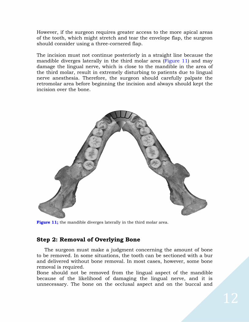
The incision must not continue posteriorly in a straight line because the mandible diverges laterally in the third molar area (Figure 11) and may damage the lingual nerve, which is close to the mandible in the area of
the third molar, result in extremely disturbing to patients due to lingual nerve anesthesia. Therefore, the surgeon should carefully palpate the retromolar area before beginning the incision and always should kept the
incision over the bone.
Figure 11; the mandible diverges laterally in the third molar area.
Step 2: Removal of Overlying Bone
The surgeon must make a judgment concerning the amount of bone to be removed. In some situations, the tooth can be sectioned with a bur
and delivered without bone removal. In most cases, however, some bone removal is required. Bone should not be removed from the lingual aspect of the mandible
because of the likelihood of damaging the lingual nerve, and it is unnecessary. The bone on the occlusal aspect and on the buccal and

13
distal aspects, down to the cervical line of the impacted tooth, should be removed initially.
The amount of bone that must be removed varies with the following factors:
1. the depth of the impaction, 2. the morphology of the roots, 3. the angulation of the tooth
Step 3: Sectioning the Tooth
The direction in which the impacted tooth should be divided or sectioned depends primarily on the angulation of the impacted tooth and any root curvature.
The tooth is sectioned only three fourths of the way toward the lingual
aspect not completely to avoid lingual nerve injury. A straight elevator is inserted into the slot made by the bur and rotated to split the tooth.
Step 4: Delivery of the Sectioned Tooth with Elevator
The tooth is delivered from the alveolar process with dental elevators.
In the mandible the most frequently used elevators are the straight elevator, the paired Cryer elevators, or the Crane pick. An important
difference between the removal of an impacted mandibular third molar and of a tooth elsewhere in the mouth is that almost no luxation of the tooth occurs for the purpose of expansion of the buccal or linguocortical
plate. Instead, bone is removed, and teeth are sectioned to prepare an unimpeded pathway for delivery of the tooth.
Application of excessive force may result in unfavorable fracturing:
1. of the tooth, 2. of excessive buccal bone, 3. of the adjacent second molar,
4. or possibly of the entire mandible. Delivery of maxillary third molars is accomplished with small straight
elevators, which distobuccally luxate the tooth. Some surgeons prefer angled elevators such as Potts, Miller, or Warwick elevators, which aid in
gaining access to the impacted tooth, and force is applied to displace the tooth in the distobuccal direction.
14
The surgeon should have a finger on the tuberosity of the maxilla (especially if the impaction is mesioangular) so that if a fracture does
occur, steps can be taken to salvage the tuberosity of the maxilla by maintaining the soft tissue attachments.
Step 5: Preparing for Wound Closure
The closure of the incision made for an impacted third molar is usually a primary closure. A bone file is used to smooth any sharp, rough edges of bone, particularly where an elevator was in bony contact.
The surgeon should next direct attention to removing all particulate bone chips and debris from the wound. This is done with vigorous irrigation with sterile saline. Special care should be taken to irrigate thoroughly
under the reflected soft tissue flap. The surgeon should check for adequate hemostasis. Bleeding can occur
from a vessel in the flap, from the bone marrow that has been cut with a bur, or from the inferior alveolar vessels. Specific bleeding points should be controlled if they exist. If brisk generalized ooze is seen after the
sutures are placed, the surgeon should apply firm pressure with a small, moistened gauze pack.
Many surgeons deliver an antibiotic such as tetracycline into the sockets of lower third molars to help prevent osteitis sicca (dry socket).
PERIOPERATIVE PATIENT MANAGEMENT
The removal of impacted third molars is a surgical procedure that is
usually associated with a great deal of patient anxiety. As a result, surgeons who routinely remove impacted third molars commonly
recommend to their patients some type of profound anxiety control such as intravenous deep sedation or ambulatory general anesthesia.
The use of long-acting local anesthetics should be considered in the mandible. These anesthetics provide the patient with a pain-free period of 6 to 8 hours, during which prescriptions can be filled and analgesics
taken. Enough doses should be prescribed to last for at least 3 or 4 days. Combinations of codeine, codeine congeners, or oxycodone with aspirin
or acetaminophen are commonly used. Nonsteroidal anti-inflammatory drugs such as ibuprofen may be of value for patients.
Intravenous administration of a glucocorticoid steroid provides sufficient anti-inflammatory activity to greatly limit edema and to minimize the swelling, the single administration of 8-mg dexamethasone (long acting
steroid) before surgery is relatively common. This drug can then be

15
continued in an oral dose of 0.75 to 1.25 mg twice a day for 2 to 3 days to continue edema control.
It is common to prescribe antibiotics for a few days after surgery. In case of pre-existing pericoronitis or periapical abscess.
The use of a topical antibiotic such as tetracycline has been scientifically shown to greatly lower the incidence of osteitis sicca (dry socket) in mandibular molar extraction sites. Placing one fourth of the contents
of a 250-mg capsule into the socket is adequate to give the desired
protection.
The use of ice packs or packages of frozen peas on the face to help prevent postoperative swelling, Patients who have had mandibular third
molars surgically removed frequently have mild to moderate trismus. The trismus gradually resolves, and the ability to open the mouth should return to normal by 7 to 10 days after surgery.
GOOD LUCK
Contemporary Oral and Maxillofacial Surgery, Sixth Edition – Hupp, James R