Impacted Teeth Part b
18
IMPACTED 3 RD MOLAR REMOVAL PRE OPERATIVE ASSESSMENT
-
Upload
mohsin-habib -
Category
Documents
-
view
222 -
download
0
Transcript of Impacted Teeth Part b
8/2/2019 Impacted Teeth Part b
http://slidepdf.com/reader/full/impacted-teeth-part-b 1/18
IMPACTED 3RD MOLARREMOVAL
PRE OPERATIVE ASSESSMENT
8/2/2019 Impacted Teeth Part b
http://slidepdf.com/reader/full/impacted-teeth-part-b 2/18
WHO IS GOING TO DO IT
• General dental surgeon / Referral
• Oral surgeon
who has the;1. experience or trained in.
2. facilities
3. know possible complications –
intraoperative & post operative
4. Choice of anesthesia i.e. L.A or G.A
8/2/2019 Impacted Teeth Part b
http://slidepdf.com/reader/full/impacted-teeth-part-b 3/18
Pre operative assessment
CLINICAL ASSESSMENT
• General factors (Hx )
• Local factors1. Acute inflammation
2. Mouth opening (clinical access Poor
Good - External oblique ridge –
guide for access
3. Second molar
4. Missing 1st molar
8/2/2019 Impacted Teeth Part b
http://slidepdf.com/reader/full/impacted-teeth-part-b 4/18
Pre operative assessment cont1.
Radiographic Assessment
• Peri apical view
•
Bite wing• Occlusial view.
• Lateral oblique.
• OPG.
8/2/2019 Impacted Teeth Part b
http://slidepdf.com/reader/full/impacted-teeth-part-b 5/18
Pre operative assessment cont2.
Interpretation of standardized intra oral radiograph
1. Access:
2. Position & depth:
3. Root pattern:
4. Shape of crown:
5. Texture of the investing bone:
6. Position & root pattern of the 2nd molar:
7. Inferior dental canal
8/2/2019 Impacted Teeth Part b
http://slidepdf.com/reader/full/impacted-teeth-part-b 6/18
Classification
A. Relation ship of tooth to the ramus & 2nd molar.
Class I.
Class II.
Class III.
B. Relationship of 3rd molar in bone.
Position A.
Position B.
Position C.
8/2/2019 Impacted Teeth Part b
http://slidepdf.com/reader/full/impacted-teeth-part-b 7/18
WINTERS CLASSIFICATION
• Long axis of 3rd molar in relation to longaxis of 2nd molar.
• Describes 3rd molar impactions as – vertical, Mesio-angular, Disto-angular &Horizontal.
• Howe modified it using the Winter Lines – importance of the vertical depth of 3rd molar.
8/2/2019 Impacted Teeth Part b
http://slidepdf.com/reader/full/impacted-teeth-part-b 8/18
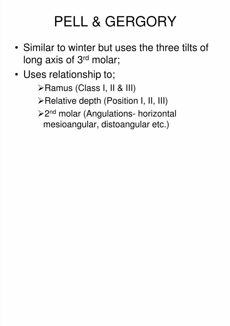
PELL & GERGORY
• Similar to winter but uses the three tilts oflong axis of 3rd molar;
• Uses relationship to;
Ramus (Class I, II & III)
Relative depth (Position I, II, III)
2nd molar (Angulations- horizontal
mesioangular, distoangular etc.)
8/2/2019 Impacted Teeth Part b
http://slidepdf.com/reader/full/impacted-teeth-part-b 9/18
8/2/2019 Impacted Teeth Part b
http://slidepdf.com/reader/full/impacted-teeth-part-b 10/18
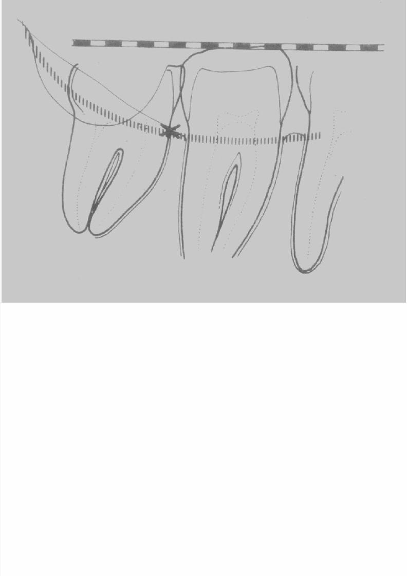
MESIO & DISTO-ANGULAR IMPACTIONS
8/2/2019 Impacted Teeth Part b
http://slidepdf.com/reader/full/impacted-teeth-part-b 11/18
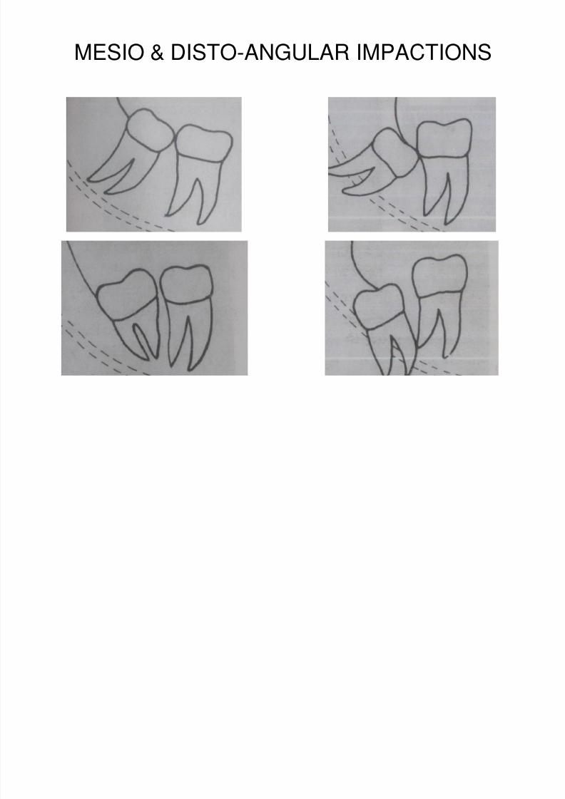
HORIZONTAL IMPACTIONS
8/2/2019 Impacted Teeth Part b
http://slidepdf.com/reader/full/impacted-teeth-part-b 12/18
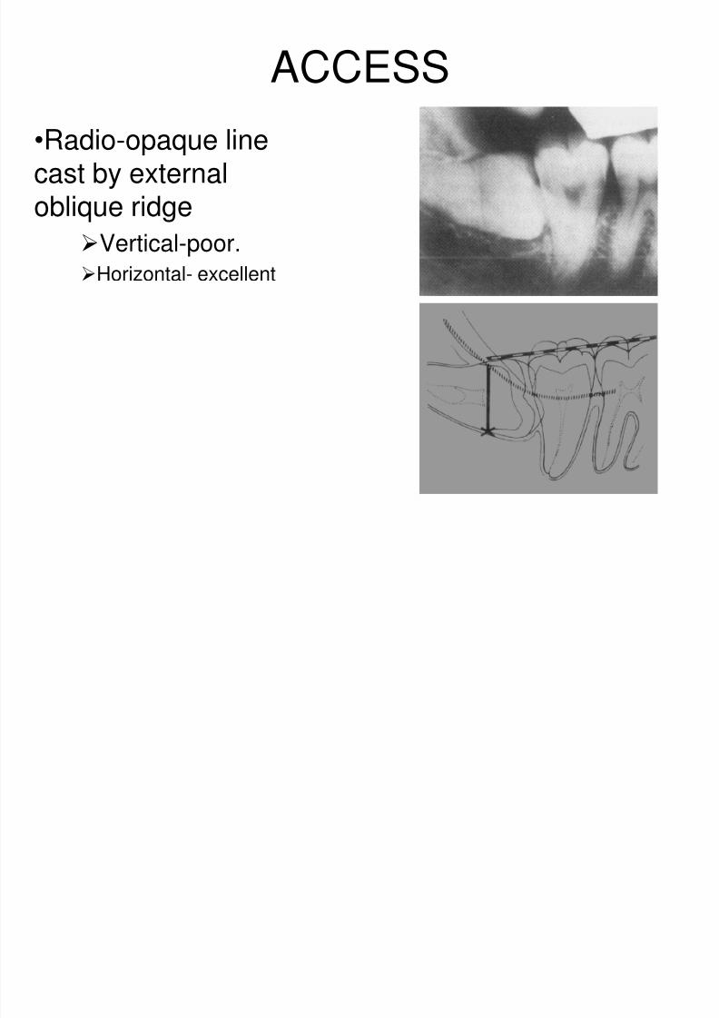
ACCESS
•Radio-opaque linecast by externaloblique ridge
Vertical-poor.
Horizontal- excellent
8/2/2019 Impacted Teeth Part b
http://slidepdf.com/reader/full/impacted-teeth-part-b 13/18
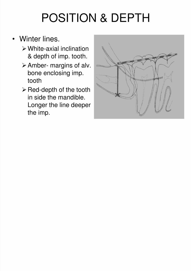
POSITION & DEPTH
• Winter lines.
White-axial inclination& depth of imp. tooth.
Amber- margins of alv.bone enclosing imp.tooth
Red-depth of the tooth
in side the mandible.Longer the line deeperthe imp.
8/2/2019 Impacted Teeth Part b
http://slidepdf.com/reader/full/impacted-teeth-part-b 14/18
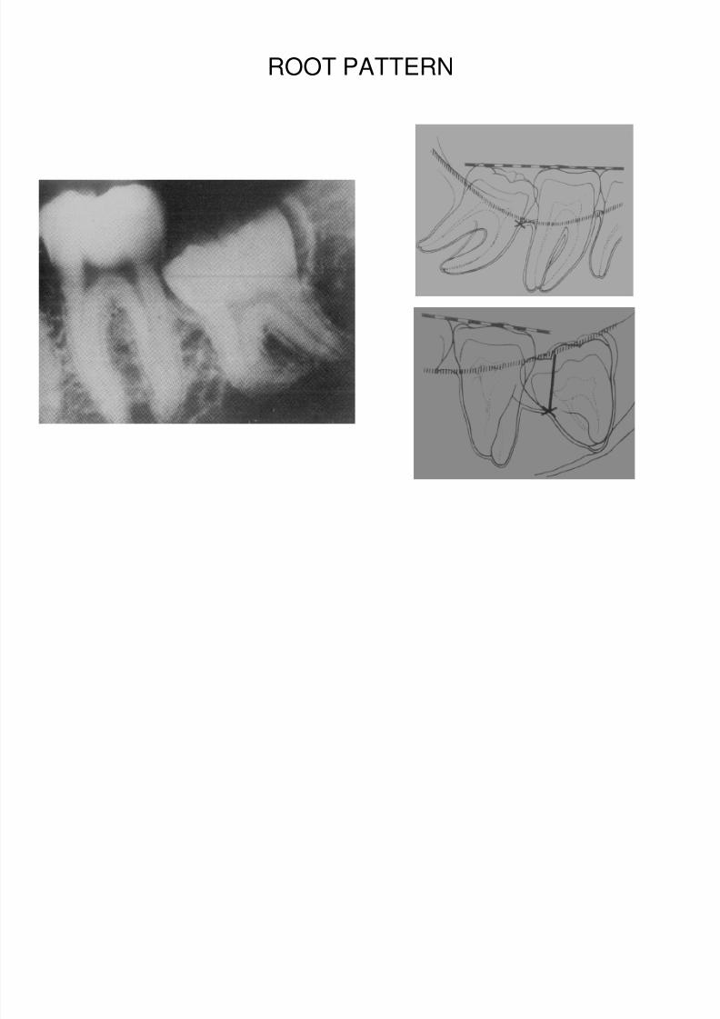
ROOT PATTERN
8/2/2019 Impacted Teeth Part b
http://slidepdf.com/reader/full/impacted-teeth-part-b 15/18
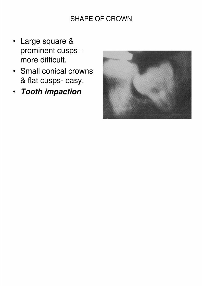
SHAPE OF CROWN
• Large square &prominent cusps – more difficult.
• Small conical crowns& flat cusps- easy.
• Tooth impaction
8/2/2019 Impacted Teeth Part b
http://slidepdf.com/reader/full/impacted-teeth-part-b 16/18
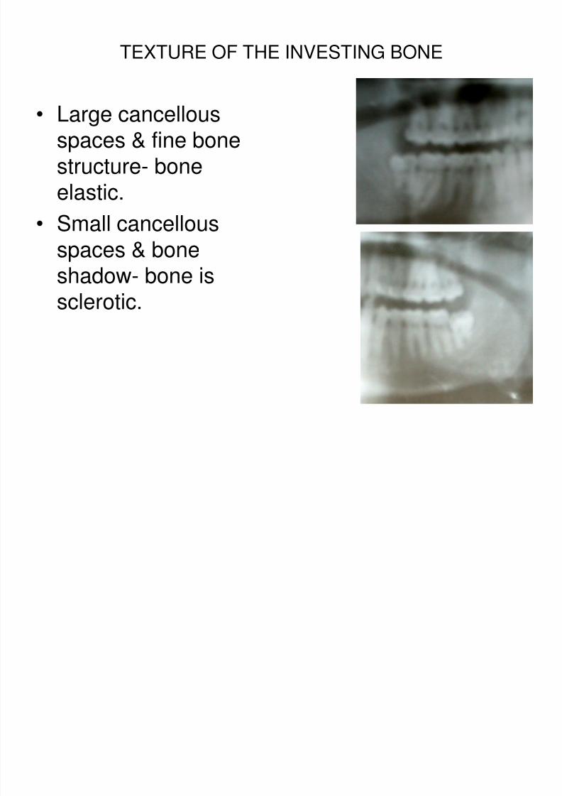
TEXTURE OF THE INVESTING BONE
• Large cancellousspaces & fine bonestructure- bone
elastic.• Small cancellous
spaces & boneshadow- bone is
sclerotic.
8/2/2019 Impacted Teeth Part b
http://slidepdf.com/reader/full/impacted-teeth-part-b 17/18
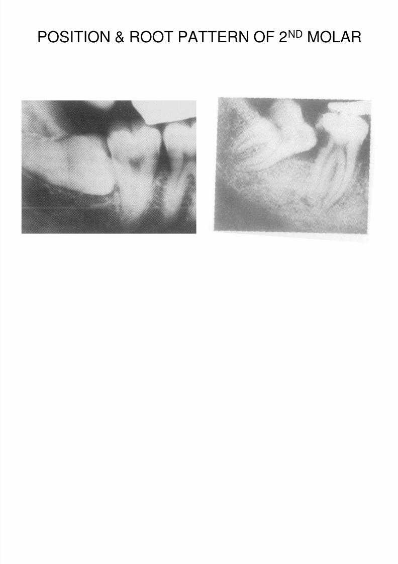
POSITION & ROOT PATTERN OF 2ND MOLAR
8/2/2019 Impacted Teeth Part b
http://slidepdf.com/reader/full/impacted-teeth-part-b 18/18
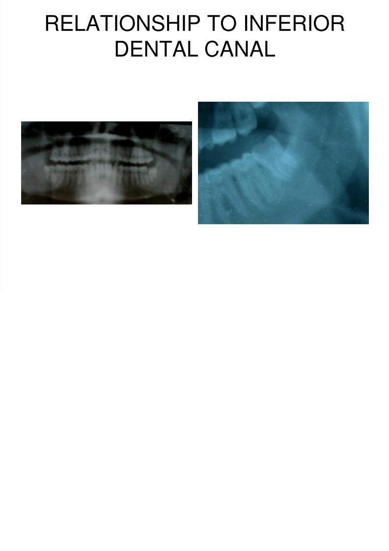
RELATIONSHIP TO INFERIORDENTAL CANAL