Extrusion of Impacted Teeth - uniklinik-duesseldorf.de · Extrusion of Impacted Teeth ... Position...
7
Transcript of Extrusion of Impacted Teeth - uniklinik-duesseldorf.de · Extrusion of Impacted Teeth ... Position...
T reatment of impacted teeth usually comprises three phases: surgical exposure and bonding
of an attachment, eruption of the impacted tooth by application of an extrusive force, and three-dimensional orthodontic alignment.1-3 The force needed to extrude an impacted tooth often pro-duces side effects such as intrusion of the adjacent teeth or even canting of the occlusal plane.4 Stable anchorage is essential to minimize these effects.
In recent years, skeletal anchorage with mini-implants has become increasingly popular because of its versatility, minimal invasiveness, and low cost.5-9 This article presents several clinical cases in which Benefit* mini-implants with interchange-able abutments10 (Fig. 1) were placed in the ante-rior palate to serve as anchorage for extrusion and alignment of impacted teeth.
Depending on the anchorage needs of the case and the location of the teeth to be moved, various types of implant mechanics can be used.
Single Implant: Direct Anchorage
These simple mechanics are usually em -ployed to extrude a single impacted tooth. Since implants with wider diameters have greater stabil-ity,11,12 a single mini-implant with dimensions of 2.3mm × 9mm or 2.3mm × 11mm is inserted in the anterior palate. A bracket abutment with a pre-ligated .016" × .022" TMA** sectional wire is screwed to the head of the mini-implant (Fig. 2). The impacted tooth is surgically exposed, and an
attachment fitted with a gold chain is bonded to the tooth. To generate an extrusive force, the sec-tional wire is bent toward the chain and attached with a ligature.
© 2012 JCO, Inc.
Extrusion of Impacted Teeth Using Mini-Implant Mechanics
MANUEL NIENKEMPER, DDS, MSC BENEDICT WILMES, DDS, MSC, PHDGUDRUN LÜBBERINK, DDS, MSCBJÖRN LUDWIG, DMD, MSDDIETER DRESCHER, DDS, MSC, PHD
150 JCO/MARCH 2012
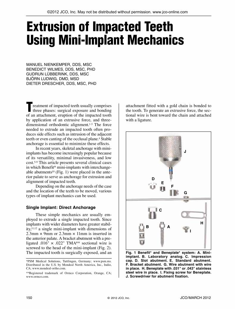
Fig. 1 Benefit* and Beneplate* system: A. Mini-implant. B. Laboratory analog. C. Impression cap. D. Slot abutment. E. Standard abutment. F. Bracket abutment. G. Wire abutment with wire in place. H. Beneplate with .031" or .043" stainless steel wire in place. I. Fixing screw for Beneplate. J. Screwdriver for abutment fixation.
*PSM Medical Solutions, Tuttlingen, Germany; www.psm.ms. Distributed in the U.S. by Mondeal North America, Inc., Indio, CA; www.mondeal-ortho.com.
**Registered trademark of Ormco Corporation, Orange, CA; www.ormco.com.
AB
C
DE
F
G
H
I
J
©2012 JCO, Inc. May not be distributed without permission. www.jco-online.com

Tandem Implants: Direct Anchorage
In cases with greater anchorage needs, two 2mm-diameter mini-implants can be combined. A stable connection between the mini-implants is cru-cial. Although a composite bridge can be con-structed, the composite may fail when subjected
to force. A better solution is to ligate the mini-implants together with a wire segment or to con-nect the abutments with laser-welded stainless steel wires. More individualized treatment is possible with the use of a rigid plate such as a Beneplate*13
(Fig. 1H). A superelastic wire can be ligated to a bracket integrated with the plate, or a prefabri-
VOLUME XLVI NUMBER 3 151
Dr. Lübberink Dr. LudwigDr. Wilmes Dr. DrescherDr. Nienkemper
Dr. Nienkemper is an Instructor, Dr. Wilmes is an Associate Professor, Dr. Lübberink is an Assistant Professor, and Dr. Drescher is Professor and Head, Department of Orthodontics, University of Düsseldorf, Moorenstrasse 5, 40225 Düsseldorf, Germany. Dr. Wilmes is also a Visiting Professor, Department of Orthodontics, University of Alabama at Birmingham School of Dentistry, and the developer of the Benefit system. Dr. Ludwig is a Contributing Editor of the Journal of Clinical Orthodontics; an Instructor, Department of Orthodontics, University of Homburg, Saar, Germany; and in the private practice of orthodontics in Traben-Trarbach, Germany. E-mail Dr. Nienkemper at [email protected].
Fig. 2 Single implant: direct anchorage. A. .016" × .022" TMA** sectional wire with activation bend ligated to bracket head of implant abutment and attached to gold chain bonded to impacted tooth. B. 9-year-old male patient with impacted upper central incisor before treatment. C. After exposure of impacted central incisor, abutment with TMA sectional wire screwed to head of mini-implant. D. Patient after eight months of incisor extrusion.
DCB
A

152 JCO/MARCH 2012
Extrusion of Impacted Teeth Using Mini-Implant Mechanics
cated plate with an .031" integrated wire can be used (Fig. 3).
Single Implant: Indirect Molar Anchorage
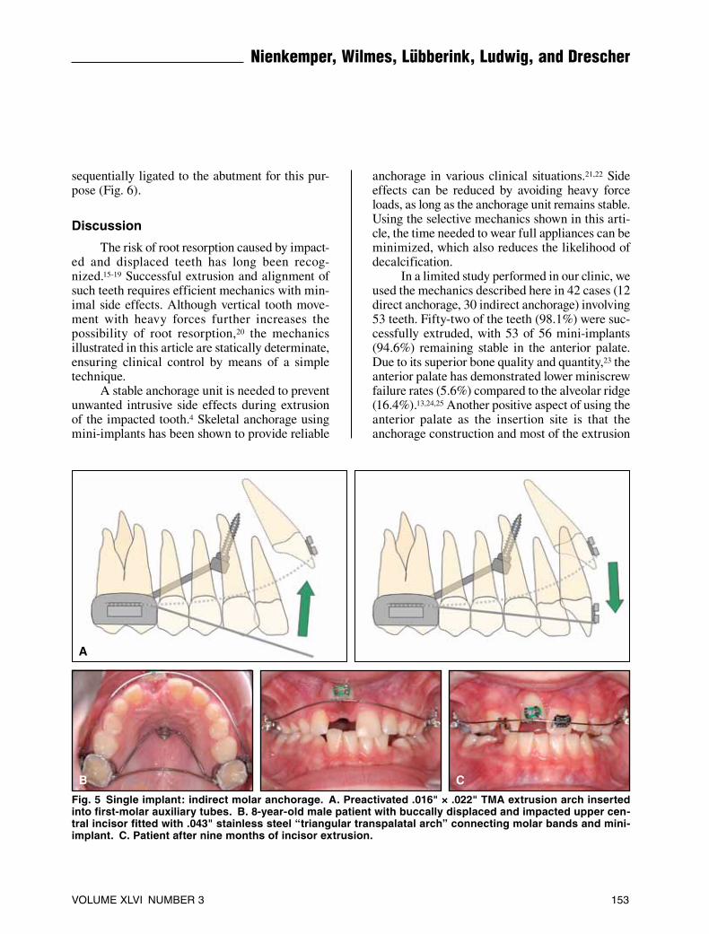
To prevent mesial tipping and intrusion of the molars during extrusion of anterior teeth, a pre-fabricated abutment with an .031" stainless steel wire can be used to create a skeletally anchored trans palatal arch (Fig. 4). If a tooth is buccally displaced, the forces must be applied from the buccal, using an .016" × .022" TMA extrusion arch inserted into the first-molar auxiliary tubes (Fig. 5A). Perhaps the most stable transpalatal-anchor-age construction is a “triangular transpalatal arch” of .043" stainless steel wire, connecting the molar bands and the mini-implant (Fig. 5B,C). The ad -ditional transverse connector prevents buccal or
palatal tipping of the molars, resulting in complete immobilization of these teeth.
Adjustment of Force Vectors
By varying the length of wire or preactivat-ing it in a mesial or distal direction, the line of force can be adapted to the individual clinical situation. Since these mechanics can be classified as statically determinate, measurement and adjust-ment of the applied force are straightforward. To extrude an upper central incisor, a force of 20cN is typically applied; for extrusion of an upper canine, 25cN is generally adequate.9
The versatility of abutments or plates with brackets is a great advantage when a force adjust-ment is needed during extrusion to prevent root damage to the adjacent teeth or to correct the posi-tion of the erupted tooth. Different wires can be
Fig. 4 Single implant: indirect mo -lar anchorage. A. 13-year-old fe -male patient with impacted upper canines. Molar tipping and intru-sion prevented during canine ex -trusion by using transpalatal bar formed from .031" stainless steel wire, pre-welded to implant abut-ment; extrusive forces provided by two preactivated .016" × .022" TMA sectional wires inserted into addi-tional lingual tubes on molar bands. B. Patient after four months of canine extrusion.
A B
Fig. 3 Tandem implants: direct anchorage. A. 53-year-old male patient with retained and dystopic upper left third molar. B. Two mini-implants coupled with Beneplate; preactivated .031" stainless steel wire used to create extrusive force. C. Patient after seven months of third-molar extrusion.
CBA
sequentially ligated to the abutment for this pur-pose (Fig. 6).
Discussion
The risk of root resorption caused by impact-ed and displaced teeth has long been recog-nized.15-19 Successful extrusion and alignment of such teeth requires efficient mechanics with min-imal side effects. Although vertical tooth move-ment with heavy forces further increases the possibility of root resorption,20 the mechanics illustrated in this article are statically determinate, ensuring clinical control by means of a simple technique.
A stable anchorage unit is needed to prevent unwanted intrusive side effects during extrusion of the impacted tooth.4 Skeletal anchorage using mini-implants has been shown to provide reliable
anchorage in various clinical situations.21,22 Side effects can be reduced by avoiding heavy force loads, as long as the anchorage unit remains stable. Using the selective mechanics shown in this arti-cle, the time needed to wear full appliances can be minimized, which also reduces the likelihood of decalcification.
In a limited study performed in our clinic, we used the mechanics described here in 42 cases (12 direct anchorage, 30 indirect anchorage) in volving 53 teeth. Fifty-two of the teeth (98.1%) were suc-cessfully extruded, with 53 of 56 mini-implants (94.6%) remaining stable in the anterior palate. Due to its superior bone quality and quantity,23 the anterior palate has demonstrated lower miniscrew failure rates (5.6%) compared to the alveolar ridge (16.4%).13,24,25 Another positive as pect of using the anterior palate as the insertion site is that the anchorage construction and most of the extrusion
VOLUME XLVI NUMBER 3 153
Nienkemper, Wilmes, Lübberink, Ludwig, and Drescher
Fig. 5 Single implant: indirect molar anchorage. A. Preactivated .016" × .022" TMA extrusion arch inserted into first-molar auxiliary tubes. B. 8-year-old male patient with buccally displaced and impacted upper cen-tral incisor fitted with .043" stainless steel “triangular transpalatal arch” connecting molar bands and mini-implant. C. Patient after nine months of incisor extrusion.
CB
A
154 JCO/MARCH 2012
Extrusion of Impacted Teeth Using Mini-Implant Mechanics
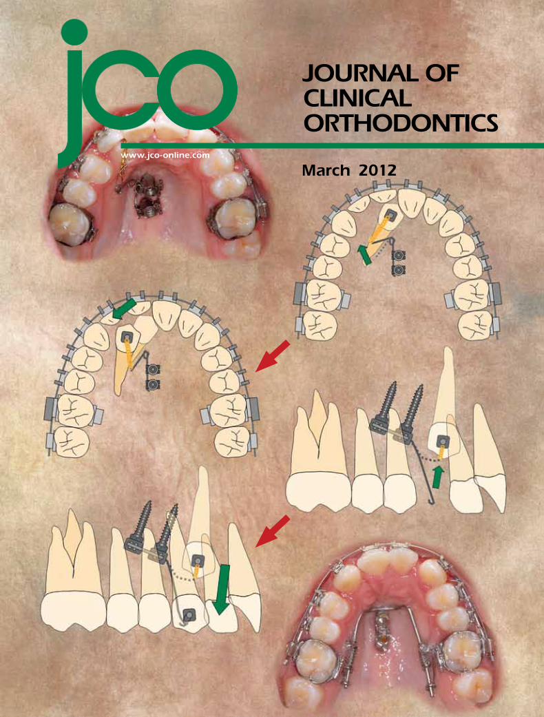
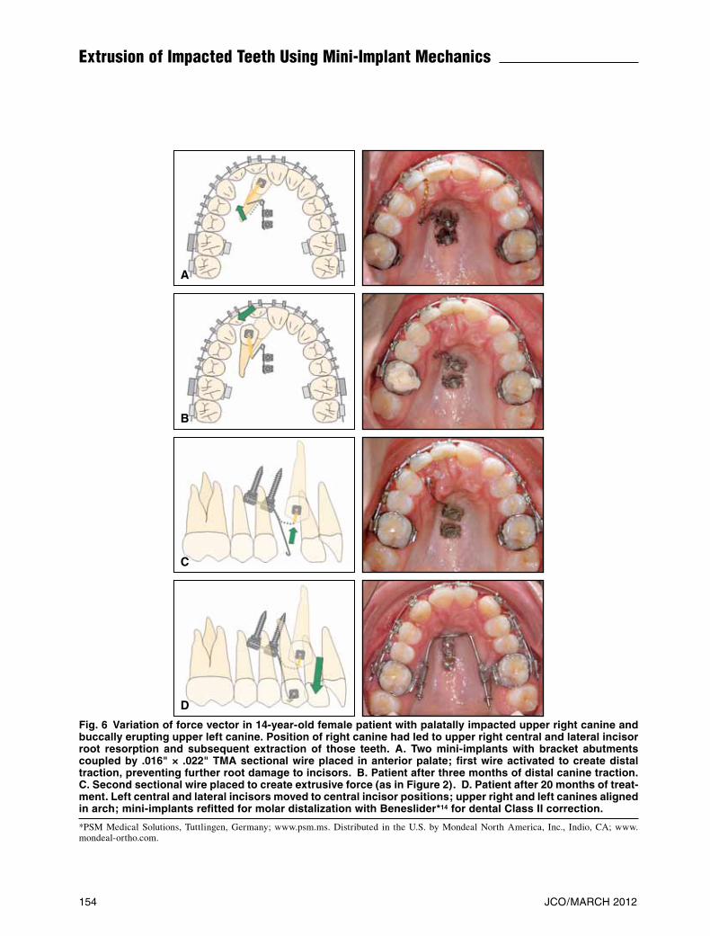
Fig. 6 Variation of force vector in 14-year-old female patient with palatally impacted upper right canine and buccally erupting upper left canine. Position of right canine had led to upper right central and lateral incisor root resorption and subsequent extraction of those teeth. A. Two mini-implants with bracket abutments coupled by .016" × .022" TMA sectional wire placed in anterior palate; first wire activated to create distal traction, preventing further root damage to incisors. B. Patient after three months of distal canine traction. C. Second sectional wire placed to create extrusive force (as in Figure 2). D. Patient after 20 months of treat-ment. Left central and lateral incisors moved to central incisor positions; upper right and left canines aligned in arch; mini-implants refitted for molar distalization with Beneslider*14 for dental Class II correction.
*PSM Medical Solutions, Tuttlingen, Germany; www.psm.ms. Distributed in the U.S. by Mondeal North America, Inc., Indio, CA; www.mondeal-ortho.com.
D
C
B
A
appliance are nearly invisible extraorally.Since mini-implants inserted in the anterior
palate do not interfere with the dental roots, they can be used in the mixed dentition. In cases with severely palatally displaced teeth, however, radio-graphic location is always advisable prior to implant insertion. Adjusting the line of force during the extrusion phase is crucial to prevent root damage.
The prefabricated components of the Benefit/Beneplate system allow fabrication without weld-ing or soldering. In many cases, the appliance can be fitted at the chair. Standard constructions pro-viding either direct or indirect anchorage are now available. Using the appropriate interchangeable abutment, the mechanics can be easily adapted to each patient’s biomechanical requirements.
REFERENCES
1. Hattab, F.N.; Rawashdeh, M.A.; and Fahmy, M.S.: Impaction status of third molars in Jordanian students, Oral Surg. Oral Med. Oral Pathol. Oral Radiol. Endod. 79:24-29, 1995.
2. Bishara, S.E.: Clinical management of impacted maxillary ca -nines, Semin. Orthod. 4:87-98, 1998.
3. Becker, A.: The Orthodontic Treatment of Impacted Teeth, Martin Dunitz, Ltd., London, 1998.
4. Kokich, V.G. and Mathews, D.P.: Surgical and orthodontic management of impacted teeth, Dent. Clin. N. Am. 37:181-204, 1993.
5. Costa, A.; Raffaini, M.; and Melsen, B.: Miniscrews as ortho-dontic anchorage: A preliminary report, Int. J. Adult Orthod. Orthog. Surg. 13:201-209, 1998.
6. Melsen, B. and Costa, A.: Immediate loading of implants used for orthodontic anchorage, Clin. Orthod. Res. 3:23-28, 2000.
7. Wilmes, B.: Fields of application of mini-implants, in Innovative Anchorage Concepts: MiniImplants in Orthodontics, ed. B. Ludwig, S. Baumgaertel, and S.J. Bowman, Quin-tessence, Berlin, 2008.
8. Kanomi, R.: Mini-implant for orthodontic anchorage, J. Clin. Orthod. 31:763-767, 1997.
9. Wilmes, B. and Drescher, D.: Vertical periodontal ligament distraction—a new method for aligning ankylosed and dis-placed canines, J. Orofac. Orthop. 70:213-223, 2009.
10. Wilmes, B. and Drescher, D.: A miniscrew system with inter-changeable abutments, J. Clin. Orthod. 42:574-580, 2008.
11. Wilmes, B.; Rademacher, C.; Olthoff, G.; and Drescher, D.: Parameters affecting primary stability of orthodontic mini-implants, J. Orofac. Orthop. 67:162-174, 2006.
12. Wilmes, B.; Ottenstreuer, S.; Su, Y.Y.; and Drescher, D.: Impact of implant design on primary stability of orthodontic mini-implants, J. Orofac. Orthop. 69:42-50, 2008.
13. Wilmes, B.; Drescher, D.; and Nienkemper, M.: A miniplate system for improved stability of skeletal anchorage, J. Clin. Orthod. 43:494-501, 2009.
14. Wilmes, B. and Drescher, D.: Application and effectiveness of the Beneslider: A device to move molars distally, World J. Orthod. 11:331-340, 2010.
15. Alqerban, A.; Jacobs, R.; Lambrechts, P.; Loozen, G.; and Willems, G.: Root resorption of the maxillary lateral incisor caused by impacted canine: A literature review, Clin. Oral Investig. 13:247-255, 2009.
16. Amlani, M.S.; Inocencio, F.; and Hatibovic-Kofman, S.: Lateral incisor root resorption and active orthodontic treat-ment in the early mixed dentition, Eur. J. Paediat. Dent. 8:188-192, 2007.
17. Arens, D.E.: An alternative treatment for the severely resorped maxillary lateral incisor: A sequela of ectopic eruption, J. Endod. 21:95-100, 1995.
18. Ericson, S. and Kurol, J.: Incisor resorption caused by maxil-lary cuspids. A radiographic study, Angle Orthod. 57:332-346, 1987.
19. Ericson, S. and Kurol, J.: Incisor root resorptions due to ecto-pic maxillary canines imaged by computerized tomography: A comparative study in extracted teeth, Angle Orthod. 70:276-283, 2000.
20. Han, G.; Huang, S.; Von den Hoff, J.W.; Zeng, X.; and Kuijpers-Jagtman, A.M.: Root resorption after orthodontic intrusion and extrusion: An intraindividual study, Angle Orthod. 75:912-918, 2005.
21. Yao, C.C.; Lai, E.H.; Chang, J.Z.; Chen, I.; and Chen, Y.J.: Comparison of treatment outcomes between skeletal anchor-age and extraoral anchorage in adults with maxillary dento-alveolar protrusion, Am. J. Orthod. 134:615-624, 2008.
22. Upadhyay, M.; Yadav, S.; and Patil, S.: Mini-implant anchor-age for en-masse retraction of maxillary anterior teeth: A clinical cephalometric study, Am. J. Orthod. 134:803-810, 2008.
23. Kang, S.; Lee, S.J.; Ahn, S.J.; Heo, M.S.; and Kim, T.W.: Bone thickness of the palate for orthodontic mini-implant anchor-age in adults, Am. J. Orthod. 131(4 suppl):S74-81, 2007.
24. Stanford, N.: Mini-screws success rates sufficient for ortho-dontic treatment, Evid. Based Dent. 12:19, 2011.
25. Schätzle, M.; Männchen, R.; Zwahlen, M.; and Lang, N.P.: Survival and failure rates of orthodontic temporary anchorage devices: A systematic review, Clin. Oral Impl. Res. 20:1351-1359, 2009.
VOLUME XLVI NUMBER 3 155
Nienkemper, Wilmes, Lübberink, Ludwig, and Drescher