Principles of Management of Impacted Teeth Part-1
12
1 Oral Surgery Fourth Stage Dr.Anas Hammad Lec.6 Principles of Management of Impacted Teeth Part-1 Guidelines; INTRODUCTION INDICATIONS FOR REMOVAL OF IMPACTED TEETH CONTRAINDICATIONS FOR REMOVAL OF IMPACTED CLASSIFICATION SYSTEMS FOR MANDIBULAR THIRD MOLAR IMPACTIONS Introduction The term un-erupted teeth include impacted teeth and teeth that are in the process of erupting. An impacted tooth is one that fails to erupt into the dental arch within the usual range of expected time. The tooth becomes impacted because adjacent teeth, dense overlying bone, excessive soft tissue, or a genetic abnormality prevents eruption. Teeth most often become impacted because of total length of the alveolar bone arch is smaller than the total length of the tooth arch. The most common impacted teeth are maxillary and mandibular third molars, followed by maxillary canines and mandibular premolars. The third molars are the most frequently impacted because they are the last teeth to erupt; therefore, they are the most likely to have inadequate space for eruption. In the anterior maxilla, canine usually erupts after the maxillary lateral incisor and maxillary first premolar. If space is inadequate to allow eruption, the canine becomes impacted or erupts labial to the dental arch. In the anterior mandible, a similar situation affects the mandibular premolars because they erupt after the mandibular first molar and the mandibular canine. As a general rule, all impacted teeth should be removed unless removal is contraindicated. Removal of impacted teeth becomes more difficult with advancing age of the patient.
Transcript of Principles of Management of Impacted Teeth Part-1

1
Oral Surgery
Fourth Stage Dr.Anas Hammad
Lec.6
Principles of Management of Impacted Teeth
Part-1
Guidelines;
INTRODUCTION
INDICATIONS FOR REMOVAL OF IMPACTED TEETH
CONTRAINDICATIONS FOR REMOVAL OF IMPACTED
CLASSIFICATION SYSTEMS FOR MANDIBULAR THIRD MOLAR
IMPACTIONS
Introduction
The term un-erupted teeth include impacted teeth and teeth that are
in the process of erupting. An impacted tooth is one that fails to erupt into the dental arch within the usual range of expected time. The tooth becomes impacted because adjacent teeth, dense overlying bone,
excessive soft tissue, or a genetic abnormality prevents eruption. Teeth most often become impacted because of total length of the alveolar
bone arch is smaller than the total length of the tooth arch. The most common impacted teeth are maxillary and mandibular third molars, followed by maxillary canines and mandibular premolars. The
third molars are the most frequently impacted because they are the last teeth to erupt; therefore, they are the most likely to have inadequate
space for eruption. In the anterior maxilla, canine usually erupts after the maxillary lateral incisor and maxillary first premolar. If space is inadequate to allow
eruption, the canine becomes impacted or erupts labial to the dental arch. In the anterior mandible, a similar situation affects the mandibular premolars because they erupt after the mandibular first molar and the
mandibular canine. As a general rule, all impacted teeth should be removed unless removal is contraindicated. Removal of impacted teeth becomes more
difficult with advancing age of the patient.

2
The dentist should typically not recommend that impacted teeth be left in place until they cause difficulty. If the impacted teeth are left in place
until problems arise, the patient may experience the following: 1. an increased incidence of local tissue morbidity,
2. loss of or damage to adjacent teeth and bone, 3. and potential injury to adjacent vital structures. 4. surgery is more likely to be complicated and hazardous because
the patient may have compromising systemic diseases, 5. the surrounding bone becomes more dense, 6. and more fully formed roots may grow near structures such as the
inferior alveolar nerve or the maxillary sinus.
Indications for Removal of Impacted Teeth
The average age for completion of the eruption of the third molar is age
20 years, although eruption may continue in some patients until age 25 years. During normal development, the lower third molar begins in a
horizontal angulation, and changes from horizontal to mesioangular to vertical. Failure of rotation from the mesioangular to the vertical direction is the
most common cause of lower third molars becoming impacted. The second major factor is that inadequate room exists in the alveolar process anterior to the anterior border of the ramus to allow the tooth to
erupt into position. At age 18 or 19 years, if the diagnosis for inadequate room for functional
eruption can be made, then the asymptomatic third molar can be removed, and the long-term periodontal health of the second molar will be optimized, because soft tissue and bone tissue healing will occur at its
maximal level. The procedure is easier to perform in younger patients because bone is
less dense and root formation is incomplete. The ideal time for removal of impacted third molars is when the roots of teeth are one third formed and before they are two thirds formed, usually during the mid- to late
teenage years, between ages 17 and 20 years. The indications for removal of impacted teeth are as follow;
1. Prevention of Periodontal Disease
2. Prevention of Dental Caries 3. Prevention of Pericoronitis
4. Prevention of Root Resorption 5. Impacted Teeth under a Dental Prosthesis 6. Prevention of Odontogenic Cysts and Tumors
7. Treatment of Pain of Unexplained Origin 8. Prevention of Jaw Fractures
9. Facilitation of Orthodontic Treatment 10. Optimal Periodontal Healing

3
Contraindications for Removal of Impacted Teeth All impacted teeth should be removed unless specific
contraindications justify leaving them in position. When the potential benefits outweigh the potential complications and risks, the procedure should be performed. Similarly, when the risks are greater than the
potential benefits, the procedure should be deferred. Contraindications for the removal of impacted teeth primarily involve the
patient’s physical status. 1. Extremes of Age
The most common contraindication for the removal of impacted teeth is
advanced age. As patient ages, the bone becomes highly calcified and, therefore, less flexible and less likely to bend under the forces of tooth
extraction. The result is that more bone must be surgically removed to elevate the tooth from its socket. Similarly, as patient ages, they respond less favorably and with more
postoperative sequelae. An 18-year-old patient may have 1 or 2 days of discomfort and swelling after the removal of an impacted tooth, whereas a similar procedure may result in a 4- or 5-day recovery period for a 50-
year-old patient. Finally, if a tooth has been retained in the alveolar process for many
years without periodontal disease, caries, or cystic degeneration, it is unlikely that these unfavorable sequelae will occur. Therefore, in an older patient (usually over age 35 years) with an impacted tooth that shows no
signs of disease and that has a radiographically detectable layer of overlying bone, the tooth should not be removed (Figure 1). The dentist
caring for the patient should check the impacted tooth radiographically every 1 or 2 years to ensure that no adverse sequelae have occurred.
Figure 1; Impacted maxillary right third molar in a 63-year-old patient. This molar
should not be extracted because it is deeply embedded and no signs of disease are
present

4
If the impacted tooth shows signs of cystic formation or periodontal disease involving the adjacent tooth or the impacted tooth, if it is a single
impacted tooth underneath prosthesis with thin overlying bone, or if it becomes symptomatic as the result of infection, the tooth should be
removed.
2. Compromised Medical Status
Frequently, compromised medical status and advancing age go hand in hand. The removal of impacted tooth may be contraindicated by the
patient’s compromised medical status. The surgical removal of asymptomatic impacted tooth must be
viewed as elective. However, if the tooth becomes symptomatic, the surgeon should consider working with the patient’s physician to plan removal of the tooth with
minimal operative and postoperative medical sequelae.
3. Probable Excessive Damage to Adjacent Structures If the impacted tooth lies in an area in which its removal may seriously
jeopardize adjacent nerves, teeth, or previously constructed bridges, it may be prudent to leave the tooth in place. When the dentist makes the decision not to remove a tooth, the reasons must be weighed
against potential future complications. However, in the case of the older patient with no signs of impending
complications and for whom the probability of such complications is low, the impacted tooth should not be removed.
CLASSIFICATION SYSTEMS FOR MANDIBULAR THIRD MOLAR IMPACTIONS
Removal of impacted teeth can be relatively straightforward or extremely
difficult, even for the experienced surgeon. To determine the degree of difficulty preoperatively, the primary factor determining the difficulty of
the removal is accessibility Accessibility is determined by adjacent teeth or other structures impairing access or the extraction delivery pathway.
This includes 1. assessing the ease of exposing the tooth, 2. assessing the ease of preparing a pathway for its delivery,
3. assessing the ease of preparing a purchase point

5
The majority of classification schemes are based on an analysis of a radiograph;
1. The panoramic radiograph; shows the most accurate picture of the total anatomy of the region and is the radiograph of choice for
planning removal of impacted third molars 2. A well-positioned periapical radiograph; all parts of the impacted
tooth are visible along with important adjacent anatomy
3. A cone-beam computed tomography (CBCT) Scan; actually show the relationship of the roots to the canal
I- Relationship to Angulation
The determination of the angulation of the long axis of the impacted third molar with respect to the long axis of the adjacent second molar is
the most common classification system. According to the degree of inclination the difficulty for molar removal can
be evaluated, but this classification system not enough to fully interpret struggle of impacted taking away. Mesioangular impaction is the mini difficult tooth to remove, as shown in
(Figure 2). This type of impaction is the most commonly seen, making up approximately 43% of all impacted teeth.
Figure 2; A, Mesioangular impaction—most common and easiest impaction to remove,
B, mesioangular impaction is usually in proximity to the second molar.
When the long axis of the third molar is perpendicular to the second molar, the impacted tooth is considered horizontal (Figure 3). It is more
difficult to remove compared with mesioangular impaction. Horizontal impactions occur in approximately 3% of all mandibular impactions.

6
Figure 3; A, Horizontal impaction—uncommon and more difficult to remove than
mesioangular impaction. B, The occlusal surface of horizontal impacted third molar is
usually immediately adjacent to the root of the second molar, which often produces
early severe periodontal disease.
In vertical impaction, the long axis of the impacted tooth runs parallel to the long axis of the second molar. This impaction occurs approximately 38% of all impactions, and is considered third in ease of
removal (Figure 4).
Figure 4; A, Vertical impaction—second most common impaction and second most
difficult to remove. B, Vertical impaction is frequently covered on its posterior aspect
with bone of anterior ramus of mandible.
Finally, distoangular impaction involves the tooth with the most
difficult angulation for removal (Figure 5). In distoangular impaction, the long axis of the third molar is distally or posteriorly angled away from the second molar, and its removal requires significant surgical intervention.

7
Distoangular impactions occur approximately 6% of all impacted third molars.
Figure 5; Distoangular impaction—uncommon and most difficult of the four types to
remove. B, The occlusal surface of distoangular impaction is usually embedded in the ramus of the mandible and requires significant bone removal for extraction.
Erupted third molars may be in a distoangular position. When this occurs, these teeth are much more difficult to remove compared with other erupted teeth. The reason is that the third molar’s mesial root is
very close to the root of the second molar. When approaching lower third molars, the possible presence of a high-riding lingual nerve still makes a buccal approach appropriate, even
when the tooth is inclined toward the lingual aspect. Rarely, a transverse impaction occurs, that is, the tooth erupting in an absolutely horizontal position in the buccolingual direction. The occlusal surface of the tooth can face the buccal or lingual direction. To determine
buccal or lingual version accurately, the dentist must take a perpendicular occlusal radiograph or obtain a CBCT scan. However, this determination is usually not necessary because the surgeon can make
this identification early in the operation, and the buccal or lingual position of the tooth does not greatly influence the approach to the
surgery.
II- Relationship to Anterior Border of Ramus
The Pell and Gregory classification is another method for
classifying impacted mandibular third molars is based on the amount of
impacted tooth that is covered with the bone of the mandibular ramus, also referred to as Pell and Gregory classes 1, 2, and 3. For this
classification, it is important that the surgeon carefully examine the relationship between the tooth and the anterior part of the ramus.

8
If the mesiodistal diameter of the crown is completely anterior to the anterior border of the mandibular ramus, it is in a class 1 relationship. If
the tooth is angled in a vertical direction, the chances for the tooth to erupt into a normal position are good (Figure 6).
Figure 6; Pell and Gregory class 1 impaction. The mandibular third molar has
sufficient anteroposterior room ( anterior-to-anterior border of ramus) to erupt.
If the tooth is positioned posteriorly so that approximately one half is covered by the ramus, the relationship of the tooth with the ramus is
class 2. In the class 2 situation, the tooth cannot erupt completely because a small shelf of bone overlies the distal portion of the tooth (Figure 7).
Figure 7; Pell and Gregory class 2 impaction. Approximately half is covered by the
anterior portion of the ramus of the mandible.
Class 3 relationship between the tooth and ramus occurs when the tooth
is located completely within the mandibular ramus (Figure 8). The class 3 relationship provides the least accessibility and, thus, presents the
greatest difficulty.

9
Figure 8; Pell and Gregory class 3 impaction. The impacted third molar is completely embedded in the bone of the ramus of the mandible.
III- Relationship to the Occlusal Plane
This classification system was also suggested by Pell and Gregory and
is called the Pell and Gregory A, B, and C classification In this classification. The depth of the impacted tooth compared with the height
of the adjacent second molar provides the next classification system for determining the difficulty of impaction removal. The degree of difficulty is measured by the thickness of overlying bone;
that is, the degree of difficulty increases as the depth of the impacted tooth increases.
class A impaction is one in which the occlusal surface of the impacted tooth is level or nearly level with the occlusal plane of the second molar (Figure 9).
class B impaction involves an impacted tooth with an occlusal surface between the occlusal plane and the cervical line of the second molar (Figure 10).
Finally, the class C impaction is one in which the occlusal surface of the impacted tooth is below the cervical line of the second molar (Figure 11).

10
Figure 9; Pell and Gregory class A impaction. The occlusal plane of the impacted tooth
is at the same level as the occlusal plane of the second molar.
Figure 10; Pell and Gregory class B impaction. The occlusal plane of the impacted tooth
is between the occlusal plane and the cervical line of the second molar.
Figure 11; Pell and Gregory class C impaction. The impacted tooth is below the cervical
line of the second molar

11
Summary The three classification systems discussed above are used in conjunction to determine the difficulty of an extraction. For example, a
mesioangular impaction with a class 1 ramus and a class A depth is usually straightforward to remove (Figure 12). However, as the ramus relationship changes to a class 2 and the depth of the impaction
increases to a class B, the degree of difficulty becomes much greater. A horizontal impaction with a class 2 ramus relationship and a class B
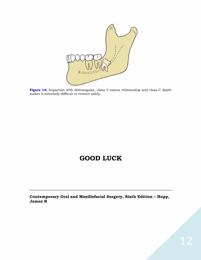
depth is a moderately difficult extraction, and one that most experienced general practitioners do not want to attempt (Figure 13). Finally, the most difficult of all impactions is a distoangular impaction with a class 3
ramus relationship at a class C depth. Even specialists view removing this tooth as a surgical challenge (Figure 14).
Figure 12; Mesioangular impaction with class 1 ramus relationship and class A depth.
All three classifications make it the easiest type of impaction to remove.
Figure 13; Horizontal impaction with class 2 ramus relationship and class B depth
makes it moderately difficult to extract.
12
Figure 14; Impaction with distoangular, class 3 ramus relationship and class C depth
makes it extremely difficult to remove safely.
GOOD LUCK
Contemporary Oral and Maxillofacial Surgery, Sixth Edition – Hupp,
James R