ORIGIN AND DISTRIBUTION OF THE TRACTUS SOLITARIUS IN ...
34
AUTHOR'S ABSTRACT OF THIS PAPER ISSUED F? THE DIDLIOGRAPEOC SERVICE, JANIIARY 29 Reprinted from TeE JOURNAL OP COMPARATIVE NED/tOLOG I", Vol 35, No, 2, February, 1923 ORIGIN AND DISTRIBUTION OF THE TRACTUS SOLITARIUS IN THE GUINEA PIG WILLIAM F. ALLEN Department of Anatomy of the University of Oregon Medical School, Portland, Oregon :NINETEEN FIGURES CONTENTS Introductory 171 Review of the literature 172 Material and methods 173 Operations 174 Microscopical technique 176 Tractus solitarius (Pars nervi facialis) 177 Tractus solitarius (Pars nervi glossopharyngei) 184 Tractus solitarius (Pars nervi vagi) 188 Tractus solitarius proper 192 Summary and general considerations 198 Literature cited 202 INTRODUCTORY Several years ago the writer began a series of experiments and studies on the guinea pig's brain to determine if perchance the central connections of the general visceral and gustatory systems in mammals were comparable to those found by Herrick and Johnston in fishes. In brief, Herrick's description of the gustatory system of the carp, catfish and Amblystoma may be summarized as follows: Both gustatory and general visceral sensory impulses are con- ducted by the VII, IX and X nerves to facial and vagal lobes of the medulla. From these primary centers two descending secondary gustatory tracts follow the spinal V tract to the region of the commissural nuclei and two ascending secondary gustatory tracts accompany the spinal V tracts to two superior secondary gustatory nuclei, situated in the isthmus, ventrally to the line 171
Transcript of ORIGIN AND DISTRIBUTION OF THE TRACTUS SOLITARIUS IN ...

AUTHOR'S ABSTRACT OF THIS PAPER ISSUED
F? THE DIDLIOGRAPEOC SERVICE, JANIIARY 29Reprinted from TeE JOURNAL OP COMPARATIVE
NED/tOLOG I", Vol 35, No, 2, February, 1923
ORIGIN AND DISTRIBUTION OF THE TRACTUSSOLITARIUS IN THE GUINEA PIG
WILLIAM F. ALLEN
Department of Anatomy of the University of Oregon Medical School,Portland, Oregon
:NINETEEN FIGURES
CONTENTSIntroductory 171Review of the literature 172Material and methods 173
Operations 174Microscopical technique 176
Tractus solitarius (Pars nervi facialis) 177Tractus solitarius (Pars nervi glossopharyngei) 184Tractus solitarius (Pars nervi vagi) 188
Tractus solitarius proper 192Summary and general considerations 198Literature cited 202
INTRODUCTORY
Several years ago the writer began a series of experiments andstudies on the guinea pig's brain to determine if perchance thecentral connections of the general visceral and gustatory systemsin mammals were comparable to those found by Herrick andJohnston in fishes.
In brief, Herrick's description of the gustatory system of thecarp, catfish and Amblystoma may be summarized as follows:Both gustatory and general visceral sensory impulses are con-ducted by the VII, IX and X nerves to facial and vagal lobes ofthe medulla. From these primary centers two descendingsecondary gustatory tracts follow the spinal V tract to the regionof the commissural nuclei and two ascending secondary gustatorytracts accompany the spinal V tracts to two superior secondarygustatory nuclei, situated in the isthmus, ventrally to the line
171

172 WILLIAM F. ALLEN
of fusion of the valvula cerebelli and the cerebellum. Theprincipal tertiary fibers arising from the superior nuclei pass tothe inferior lobes. Herrick notes also the possibility of a relayto lower centers by way of the tractus lobo-bulbaris. Johnstonin a number of papers and in his book describes a tractus lobo-epistriaticus or tractus palli connecting the hypothalamus (inferiorlobes) with the telencephalon. On p. 174 of his book Johnstonfigures visceral fibers from the spinal cord entering the secondaryvisceral tract.
My own studies on this system in mammals, based largely onMarchi preparations, will be reported in three parts. The resultsof the first phase of the subject will be incorporated in this paperand will include a description of the distribution of the primarygustatory and general visceral fibers in the medulla of the guineapig. This will be followed by a description of the course of thesecondary visceral fibers taking origin from the primary visceralnucleus, namely, the nucleus tractus solitarii, and thirdly by astudy of the basal cerebellar nuclei, emboliformis and globosuswith a view of determining their relationships.
REVIEW OF LITERATURE
Complete summaries of the early work on the tractus solitariusand its components have been given by Koelliker and VanGehuchten. As a result of these studies, dating from Stilling toKoelliker the tractus solitarius was found to be a continuoustract extending from the entrance of the VII nerve to the decussa-tion of the pyramids, and to be made up of sensory fibers fromthe VII, IX and X nerves. Koelliker noted the close proximityof the radix sensibilis nervi facialis to the radix decendens nervivestibularis. Also he observed the nucleus tractus solitarii extend-ing the entire length of the tractus solitarius from the radixsensibilis nervi facialis to the decussatio pyramidum.
As a result of a study of some Marchi sections of a brain from apatient who had a lesion causing the loss of taste and tactilesensations on the left half of the tongue, Wallenberg traced adegenerated fasciculus, cephalad, from the tractus solitarius at thelevel of the sensory root of the glossopharyngeal nerve, to the

TRACTUS SOLITAIIIUS IN THE GUINEA PIG 173
bend of the motor root of the facial nerve. Since only normalfibers were found in the sensory roots of the facial and glosso-pharyngeal nerves, he concluded that these degenerated fiberscame from the trigeminal nerve.
In some surgical cases involving the removal of the ganglionsemilunare, Cushing noted that there was no loss of taste in thedistal end of the tongue, and concluded therefrom, that the chorda,tympani must carry gustatory sensations from the region of thetongue through the ganglion geniculi. Furthermore some experi-mental work by Hunt, demonstrated that tactile sensationsmay be present in the tip of the tongue after severing the nervuslingualis, centrally, to the exit of the chorda tympani.
Bruce in 1898, had an opportunity of studying a Marchi seriesof a human medulla in which a tumor, after having destroyed thetrigeminal nerve, completely severed the glossopharyngeal nervein the jugular foramen, without injuring the vagus. His seriesdemonstrated degenerated glossopharyngeal fibers entering theupper end of the tractus solitarius, which disappeared, caudally,by terminating in the gelatinous substance belonging to the vagusnerve; not a single fiber was traced to the dorsal nucleus or thenucleus ambiguus. Bruce concluded that the upper part of thetractus solitarius belongs to the glossopharyngeal nerve and thelower two-thirds to the vagus nerve. His figure 2, which isfrom the spinal medulla region. demonstrates a few glossopharyn-geal fibers in the tractus solitarius.
Van Gehuchten has given us a most comprehensive work on thenerve components of the tractus solitarius in the rabbit, and myown investigation on this phase of the problem is largely a repeti-tion of this work extended a little further along some lines.A separate review of Van Gehuchten's work will not be includedfor the reason that • requent reference will be made to it through-out the text.
MATERIAL AND METHODS
The reasons for selecting the guinea pig for these investigationswere essentially the same as those set forth in a previous paper(Jour. Comp. Neur., vol. 30, p. 176). For this study it was

174 WILLIAM F. ALLEN
advisable to have at least three Marchi series of the medulla,showing in one series, on one side, the radix sensibilis nervifacialis in degeneration; a second series, in which the radix sen-sibilis nervi glossopharyngei of the same side was in degeneration;and a third series having the radix sensibilis nervi vagi also ofthe same side in degeneration. In fact, I had a number of Marchiseries of each of the three components of the left tractus solitarius,one series of a rabbit's medulla where the fibers of the left radixsensibilis nervi glossopharyngei et vagi were degenerated, and anumber of series where the tractus solitarius had been cut atdifferent levels. There were also available many Marchi series,which demonstrated the neighboring tracts, such as, the radixdescendens nervi vestibularis, radix spinalis nervi trigemini,tractus spino-cerebellaris dorsalis et ventralis, lemniscus medialis,corpus restirforme, brachium conjunctivum, fasciculus longi-tudinalis medialis, and various motor roots in dengeneration.Then for comparison, the writer had a number of Weigert, Nissl,Golgi, and Cajal series of normal guinea pig's brains.
Operations: The general method of conducting the operationswas the same as was described in detail in the paper cited above.For the sake of uniformity, all of the lesions were made on theleft side.
In the experiments designed to produce degeneration of thefacialis portion of the tractus solitarius (no. 136 and others) anincision was made through the skin, immediately behind andbelow the left ear. The nervus facialis was carefully dissectedto its entrance or exit from the foramen stylomastoideum; fromwhence-it was jerked out with a hemostat, after the techniqueemployed by Van Gehuchten; this carried out about an inch ofthe nerve from the foramen, including the ganglion geniculi.In other operations, equally good results were obtained, thoughwith more difficulty, by dissecting out the nervus facialis to andincluding its ganglion, with the aid of a small pair of bone forceps.
The operation intended to bring about the degeneration of theglossopharyngeal component of the tractus solitarius was by farthe most difficult of all the operations to perform, due partly tothe close proximity of the nervus glossopharyngeus to the vagus

TEACTUS SOLITARIUS IN THE GUINEA PIG 175
and its ganglia and partly to the general inaccessibility of thenervus glossopharyngeus in curving around behind the tympanicbulla. The skin incision was made a little median to the angle ofthe left mandibula. After reflecting the skin, the salivary andlymph glands were separated from the musculus masseter,bringing into view the posterior belly of the in. digastricus andthe nervus hypoglossus traversing its median surface. Carefullyseparating the nerve from the m. digastricus and expanding theopening exposes the nervus glossopharyngeus crossing the outersurface of the m. stylopharyngeus, which can be followed withsome difficulty along the median ventral surface of the tympanicbulla in front of the nervus vagus and ganglion nodosum to theforamen jugulare. After carefully separating the nervus glosso-pharyngeus from the vagus, the nervus glossopharyngeus andganglia were jerked from the foramen without injuring the vagusat or above its ganglia.
The general technique employed in destroying the sensoryroot fibers of the nervus vagus was identical to that used fordestroying the glossopharyngeal sensory root fibers. It was,however, much easier to accomplish for the reason that it was notnecessary to go in so deep. All that was necessary was to followthe nervus vagus to the ganglion nodosum and to sever the rootfibers centrally to the ganglion, without injuring the glossopharyn-geal root fibers or its ganglia.
In order to sever the tractus solitarius I made a median inci-sion through the skin of the occipital and neck region. Afterturning the skin to either side, the median incision was con-tinued through the muscles of the neck to the depth ofthe median occipital crest and dorsal arch of the atlas. Nextthe capitus muscles were severed close to the base of the skulland turned to either side, which exposes the membrane coveringthe foramen occipitale magnum. With a small pair of boneforceps some of the squamus portion of the os occipitale wasclipped off in front of the occipital foramen on the right or oppositeside from which the lesion is to be made, after which the freecephalic border of the membrane covering the foramen occipitalemagnum was seized with a small pair of curved forceps, and

176 WILLIAM F. ALLEN
turned backward at the same time cutting it free from the skulland atlas with a very small scalpel. This brings into view theentire dorsal surface of the medulla up to the cerebellum. Finallythe cerebellum was gently lifted up from the medulla and ashallow lesion was made through the left tractus solitarius,being careful not to injure any of the dorsal tracts or nucleiapproximating it. For making this lesion I filed down a pieceof A in. steel wire in the form of a chisel and bent it at an angleof about 45°, parallel to, and about one inch from the blade.
Microscopical technique. Ordinarily the animal was killed onthe 14th day after the operation. According to my experiencewith the Marchi method, the maximum degeneration appears inmammals about the 11th day, and it is best not to let the animallive more than 14 days after the operation, for the reason thatretrograde degeneration will take place from the central motornuclei, as a result of the chromatolysis of their cells, which willassume considerable proportions by the 18th day. Upon removalthe brain was placed in a dish of water, where the cerebralhemispheres were severed and discarded, but leaving the corpusstriatum intact with the brain stem. This portion of the brain wasthen placed in a 3 per cent solution of potassium bichromate for4 days, from whence it was removed after a superficial hardening,to strip off the dorsal part of the cerebellar cortex and to cut upinto transverse blocks of about 5 or 6 mm. in thickness. (Ifthis procedure, which was necessary for the complete infiltrationof osmic acid, was delayed much longer, there was danger of thebrain stem becoming brittle and crumbling in making thesesections.) These blocks were returned to a 3 per cent solution ofpotassium bichromate for a period of about 2 weeks to completethe infiltration; from which they were removed to a solutioncomposed of 30 cc. of 3 per cent potassium bichromate plus 10 cc.of 1 per cent osmic acid, for about 3 weeks. Finally these blockswere thoroughly washed in water, dehydrated, thoroughly infil-trated with celloidin, cut 50 microns in thickness, and mountedserially.

TRACTUS SOLITAR/US IN THE GUINEA PIG 177
TRACTUS SOLITARITIS (PARS NFRVI FACIALIS)
A comparison of figure 3 with Van Gehuchten's figure disclosesa close resemblance in the guinea pig and the rabbit of thepassage of the radix sensibilis nervi facialis or nerve of Wrisbergthrough the medulla to form the tractus solitarius. About theonly difference is that the VIII root of the guinea pig separatesinto four instead of three bundles or rootlets in traversing dorso-ventrally, through the dorsal portion of the radix spinalis nervitrigemini (Sp.V.) and its sensory nucleus (Sp.V.N .) to reunitemedially of the dorso-medial corner of the radix spinalis nervitrigemini in forming the tractus solitarius (T.S.). From thedorsal reconstruction, figure, 1A, it is obvious that the four
EXPLANATION OF FIGURES
Figures 1 and 2 are from graphic reconstructions, the remainder are fromportions of transverse sections, or in a few instances, from a combination ofseveral adjacent sections.
The graphic reconstructions were made after a method given in an earlierpaper (Quart. Journ. Micro. Sri., vol. 59, pp. 352-3). In each case the mediansagittal plane of the medulla was chosen for the projection line (P–L). The.marginal scale is in terms of centimeters, each division equalling 1 cm. In thesefigures the degenerated components of the tractus solitarius are represented byparallel lines and motor nuclei as irregular coarse dots. The limits of sensorynuclei may be indicated by outlines or their cells ma y be shown as circles. Theboundaries of normal nerve roots entering the tractus solitarius, the radix spinalisnervi trigemini and tract Z are drawn in outline; while the borders of the radixdescendens nervi vestibuli are shown by very fine dotted lines.
All of the drawings from the transverse sections were made with the aid of aprojection drawing apparatus. In these figures degenerated fibers appear asblack dots; other structures were drawn in sepia or in case of the sensory nucleiof the IX and X nerves were left in white.
ABBREVIATIONS
B., In fig. 1A, most cephalic nervusfacialis fibers and collaterals to thenucleus tractus solitarii. In fig. 1B,most cephalic nervus glossopharyn-geus fibers and collaterals to thenucleus tractus solitarii. In fig.1C, most cephalic nervus vagusfibers and collaterals to the nucleustractus solitarii
C. C., canalis centralisC. F., commissural fibers
Corn. N., nucleus commissuralis organglion commissurale
Cor. Sp. D., tractus cortico-spinalisdorsalis or dorsal pyramidal tract
C. Trap., corpus trapezoideumGun. N., nucleus fasciculi cuneati or
nucleus of BurdachD. Car. N., nucleus nervi cochlearis
dorsalis or dorsal cochlear nucleusDec. Per., clecussatio pyramidum or
decussation of the pyramids

178 WILLIAM F. ALLEN
D. I. Arc., fibrae arcuate dorsales ordorsal internal arcuate fibers
D. N., nucleus nervi vestibularis later-ails or Deiters nucleus
E. Arc., fibrae arcuatae externae orexternal arcuate fibers
F. R., formatio reticularisF. R. L., formatio reticularis lateralisF. R, M., formatio reticularis medialisGrad!. N., nucleus faseiculi gracilis or
nucleus of CoilI. rc,, fibrae arcuata,e internae or
internal arcuate fibersInt. S. N., nucleus intercalatus
StaderiniLon. M., lemniscus medialis or median
lemniscusMon. N., nucleus Monakow of Winkler's
descriptionsOf. I., nucleus olivaris inferior or
inferior oliveP-L., projection line in reconstructions
1 and 2P. S. N., nucleus parasolitariusPyr., tractus cortico-spinalis or pyramisRes. B., corpus restiformeRes. N., nucleus proprius corporis
restiformisS., tractus solitarius fibers to nucleus
motorius dorsalis nervi vagiSp. V., tractus spinalis nervi trigemini
or radix spinalis nervi VSp. V. N., nucleus tractus spinalis
nervi trigemini or substantia gela-tinosa trigeminal root
St. Ac., striae acusticae or medullaresT., In fig. 1A, termination of facialis
fibers in the tractus solitarius. Infig. 113, termination of glossopharyn-geal fibers in the tractus solitarius
T. S., tractus or fasciculus solitariusT. S. VII., tractus solitarius (pars
nervi facialis)T. S. IX., tractus solitarius (pars
nervi glossopharyngei)T. S. X., tractus solitarius (pars nervi
vagi)T. S. N., nucleus tractus solitariiTub. Ac. tubercultun acusticum
V. Coc. N., nucleus nervi cochlearventralis or ventral cochlear nuclei
Ves. Crb., direct fibrae vestibulae-certbello
Ves. Sp. N., nucleus nervi vestibularispinalis
Z., tractus ZVII, radix sensibilis nervi facialis o
nerve of WrisbergVIIa-d., rootlets of radix sensibili
nervi facialisVII M., radix motorius nervi
(pars secunda)VII M. N., nucleus motorius nervi
facialisVII T. S., In fig. 113, tractus solitarius
(pars nervi facialis)VIII Ves., radix nervi vestibularisVIII Ves, D., radix descendens nervi
vestibularis and tractus arcuatusRussell
IX, radix sensibilis nervi glosso-pharyngei
IXa-d, rootlets of radix sensibilisnervi glossopharyngei
IXd. N., nucleus motorius dorsalisnervi glossopharyngei
IX M., radix motorius nervi glosso-pharyngei
IX S., nucleus sensibilis nervi glosso-pharyngei
Xd. N., nucleus motorius dorsalis nervivagi
X M., radix motorius nervi vagiXv. N., nucleus ventralis nervi vagi or
nucleus ambiguusX1 a-d, rootlets of the first radix
sensibilis nervi vagiX2, second radix sensibilis nervi vagiXy a-b, rootlets of the second radix
sensibilis nervi vagiXY, third radix sensibilis nervi vagiX4, fourth radix sensibilis nervi vagiXI N., nucleus nervi accessorii or
caudal continuation of the nucleusmotorius dorsalis nervi vagi
XII, radix nervi hypoglossiXII N., nucleus nervi hypoglossi

9 11 la 15 11 is is 17 14 19 20 ,e 22 ZS 2'• 7.5 .215 17 23 29 30 51 32 33 39 S 17 3a 31 ♦0 41 12 13
P L
2
Fig. 1 Graphic reconstruction showing the distribution of the components of the left trac-tus solitarius of a guinea pig viewed from above. X 10. A. Shows the distribution of degener-ated fibers resulting from the extirpation of the left nervus facialis. centrally to the ganglion
geniculi, without injuring the nervus acusticus (Ex. 136). B. Illustrates the course ofdegenerated fibers resulting from pulling out the left nervus glossopharyngeus centrally to its ganglia, without injury to the vagus (Ex. 156).C. Demonstrates the distribution of degenerated fibers caused from severing the left nervus vagus centrally to the ganglion nodosum, withoutinjury to the glossopharyngeal roots or ganglia (Ex. 121). •
Fig. 2. Graphic reconstruction showing the arrangement of the sensory roots of the vagus in a different guine .,./, pig (Ex. 132 ) from the onefrom which figure 1C was drawn; introduced to show individual variation of the vagus roots. X 6.6.

1 80 WILLIAM F. ALLEN
rootlets (a-d) are situated one above the other and that theyassume a slightly caudal direction in penetrating the medulla.In agreement with Koelliker and Van Gehuchten these rootletspursue a parallel but ventral course to a number of radix nervivestibularis bundles, and might readily be confused with themif the two were not differentiated by the Marchi stain.
Especial note should be made of the close association of thefacial and vestibular roots and to the many direct fibrae vestibu-lae-cerebello (fig. 3, Ves.Crb.) entering the cerebellum, and to thefact that no degenerated radix sensibilis nervi facialis fibers areseen going to the cerebellum in this series or in any other serieswhere the facialis fibers were in degeneration. In fact, no degen-erated fibers were observed going to the cerebellum when allthe components of the tractus solitarius (VII, IX and X sensoryroots) were in degeneration. There is no branching of the facialissensory fibers into superior and inferior branches as is charac-teristic of the sensory fibers of the trigeminus. All make a sharpcaudal bend and continue in a general caudal direction. Thedegenerated particles of myelin are very minute, which is prob-ably indicative of finely medullated sheaths.
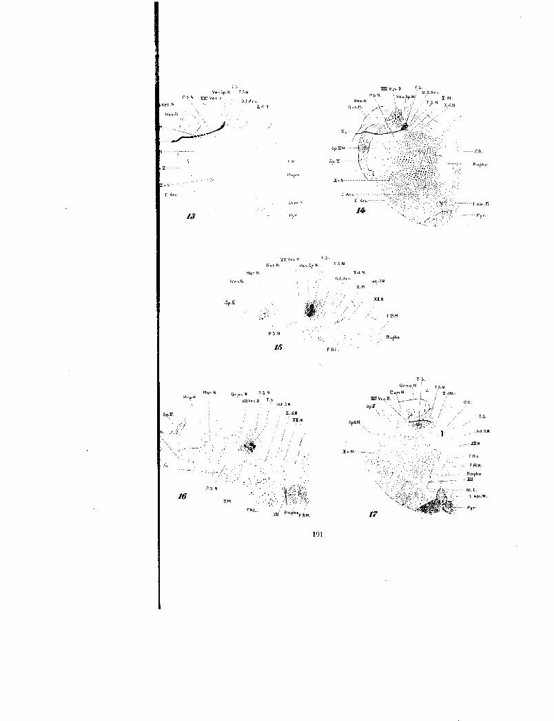
Figures 3 to and including 6 are from sections of the same guinea pig series asreconstruction lA (Ex. 136) taken at various intervals from higher to lower levelsintended to illustrate the distribution and relationships of the left radix sensibilisnervi facialis.
Fig. 3 Left half of a transverse section at the level of the facial and vestibularnerve roots, on which were projected the degenerated fibers of the left radixsensibilis nervi facialis shown in the previous three sections and the followingfour sections. X 4.
Fig. 4 Rather highly magnified drawing of the left tractus solitarius from asection of the same series as figure 3, taken a short distance from the origin ofthe tractus solitarius. In reconstruction IA this section would be situated in the7th cm. plane. X 45.
Fig. 5 Left half of a transverse section from the 9th cm. plane inreconstruction IA. X 4.
Fig. 6 Left tractus solitarius region from the same series as figure 3 at thelevel of the radix sensibilis nervi glossopharyngei. X 18.
Fig. 7 Reconstruction and projection of the four rootlets of the left radixsensibilis nervi glossopharyngei, taken from five sections of the same series asfigure 1B and drawn on the most median section. In this series (no. 156) theleft nervus glossopharyngeus was jerked out of its foramen so as to include bothof its ganglia. X 4.

.rilE J01_71:NAL 1OL. 35, xo. 2
TS.Yrr

182 WILLIAM F. ALLEN
The general course of the facialis portion of the tractus soli-tarius of the guinea pig conforms very closely to Van Gehuchten'sdescription of the rabbit, namely, it is in no sense a solitary tract,being at first closely associated with the radix spinalis nervitrigemini and later with the radix descendens nervi vestibularis.Reconstruction IA demonstrates the tractus solitarius as takingorigin at the 6th cm. plane; then for 3 cm. it passes rapidly mesad,and maintains this general position for some distance caudad.A transverse section through the 9th cm. plane (fig. 5) portraysthe tractus solitarius (T.S.) situated ventrally in the ventro-median corner of the radix descendens nervi vestibularis. Toacquire this median position the tractus solitarius graduallycrossed the ventral border of the vestibular root. At its origin(fig. 4) the tractus solitarius is oval in section, but in the 9th cm.plane (fig. 5) it has become greatly flattened horizontally.
Differing from Van Gehuchten's description of the rabbit, thetractus solitarius of the guinea pig is accompanied from its verybeginning by a terminal sensory nucleus, which in figure 4 is asmall light area (T .S.N .), situated medially and ventrally to thetractus solitarius. At the level of the 9th cm. plane in figure lAthis nucleus (fig. 5) broadens out laterally under the tractussolitarius, and gradually increases in size caudally until at thelevel of the entrance of the IX sensory root (fig. 6) it has assumedenormous proportions. Here it is more or less continuous witha larger ventral nucleus, the nucleus parasolitarius (P.S.N .).
At its origin (figs. lAand 4) the nucleus tractus solitarii receivesa few degenerated facialis fibers. These are probably collateralsrather than terminal fibers. On a level with the 9th cm. planein reconstruction lA (fig. 5) there are many degenerated fibersin the nucleus tractus solitarii. The maximum number ofdegenerated facialis fibers found in this nucleus occurs in theregion of the entrance of the IX sensory root (fig. 6). From thislevel caudal, the number gradually decreases until very few arepresent at the level of the entrance of the first root of the X nerve.Before the IX root is received, the tractus solitarius gives off atleast two bundles of fibers for the nucleus tractus solitarii. Thefirst of these bundles is a dorsal fasciculus, which leaves the tractus

TRACTUS SOLITARIUS IN TIIE GUINEA PIG 183
solitarius at the 7.5th cm. plane in figure lA and extends about2 cm. in this reconstruction before ending in the nucleus tractussolitarii. This bundle is shown in transverse section in figure 5,where it would be undifferentiated from the bundles of the radixdescendens nervi vestibularis, but for the Marchi stain. Thesecond bundle is given off laterally and ventrally from the tractussolitarius at the level of the 9th cm. plane in figure 1A.
Two places on the facialis portion of the tractus solitarius areof especial interest, namely, the entrance of the IX and the firstX sensory roots. Inasmuch as their mode of entrance is almostidentical, a description of the IX will suffice for the X. Figure 6demonstrates the normal glossopharyngeal fibers crossing underthe degenerated facialis fibers of the tractus solitarius, in part theyintermingle with the facialis fibers, but chiefly they add to thewidth and height of the tractus solitarius, making it crescent-shaped at the ventro-median corner of the radix descendensnervi vestibularis. Contrarily, Van Gehuchten on page 9 of hissecond paper and in figure 20 of the same paper, represents theIX sensory root as forming a fasciculus, lateral to the nerve ofWrisberg.
No more degenerated facialis fibers appear in the tractussolitarius or its nucleus at the level of the entrance of the secondradix sensibilis nervi vagi than are present on the opposite ornormal side. The writer would place their ending at the pointmarked (T) in figure 1A; which will be seen to be some distancecephalad of the termination of these fibers in the tractus soli-tarius of the rabbit as determined by Van Gehuchten. The dif-ference in levels of the ending of the facialis fibers in the tractussolitarius in the guinea pig and the rabbit may be accounted forby the fact that, since the nucleus tractus solitarii began muchfurther cephalad in the guinea pig, it consequently might beexpected to terminate further cephalad, thereby bringing about anearlier ending of the tractus solitarius fibers.

184 WILLIAM F. ALLEN
TRACTUS SOLITARIUS (PARS NERVI GLOSSOPITARYNCIEI)
It is apparent from a superficial examination of the radixsensibilis nervi glossopharyngei in reconstructions 1B and 7 thatthere is a close resemblance to the radix sensibilis nervi facialis,but many differences from Van Gehuchten's figure 4 of the IXsensory root of the rabbit. Like the facialis root, the degeneratedparticles of myelin are very small, indicative of minute medullarysheaths.
Transverse reconstruction 7 shows the radix sensibilis nerviglossopharyngei of the guinea pig breaking up into three smallbundles upon entering the medulla. Before penetrating thetractus spinalis nervi trigemini, the most dorsal bundle separatesinto two; so that there are at least four distinct bundles or root-lets of degenerated radix sensibilis nervi glossopharyngei fibers•(IXa-d) traversing the dorsal portion of the tractus spinalis nervitrigemini (Sp.V .) and its sensory nucleus (Sp.V.N .). The mostdorsal of these bundles, rootlet (a), crosses the ventral surface ofthe radix descenders nervi vestibularis (VIII Ves. P.). Root-lets (b) and Cc) unite and their common stem joins dorsally with(a), forming a common bundle that soon receives rootlet (d)from below. This common fasciculus curves around the ventraland median surfaces of the normal facialis fibers of the tractussolitaries, in part to intermingle with them, but mainly to con-tinue caudad, median and dorsal to them.
Since Van Gehuchten's figure 4 shows such a different arrange-ment of the rootlets of the IX sensory root within the medulla,it seemed advisable to make a reconstruction of this root from arabbit's series where the IX and X nerves were in degeneration.'This reconstruction (fig. 8) demonstrates an arrangement of fourrootlets (a-d) more closely resembling the disposition shown inreconstruction (fig. 7) than Van Gehuchten's figure 4. The chiefpoint of difference in the rabbit, as shown in figure 8, consists ofan earlier union of rootlets (a-c) and the consequent elongation ofthe combined bundle. It should be recorded that a small portionof the delicate rootlet (d) appears in a number of sections, so thatit is highly probable that this rootlet would have been over-looked if a reconstruction had not been made.

TRACTUS SOUTAR-WS IN THE GUINEA PIG 185
Throughout the entire course of the glossopharyngeal portionof the tractus solitarius, the tractus solitarius continues to occupythe ventro-median corner of the radix descendens nervi vesti-bularis. Caudally, it gradually acquires a more dorsal position,to become located medially, in the dorso-ventral corner of thevestibular root. A highly magnified drawing (fig. 9) of the lefttractus solitarius from a section immediately behind the entranceof the radix sensibilis nervi glossopharyngei, demonstratesdegenerated glossopharyngeal fibers (IX T .8 .) median to thenormal facialis fibers (VII T.S.), ventrally, and median to theradix descendens nervi vestibularis fibers, dorsally. It is im-possible in this section to discriminate between facialis anddescending vestibular bundles, but after examining a similarsection from a series where the facialis portion of the tractussolitarius was in degeneration, it is safe to affirm that thosebundles designated as (VII T.S.) in figure 9 are facialis bundlesof the tractus solitarius. At the level of the entrance of the firstradix sensibilis nervi vagi (fig. 10) the normal fibers of this root(Xi ) are clearly shown below and median to the degeneratedglossopharyngeal fibers (T.S. IX). Some of the vagus fibersapparently intermingle with the glossopharyngeal fibers but themajority retain their ventral and median positions.
A section taken from the level of the entrance of the third radixsensibilis nervi vagi demonstrates but few degenerated glosso-pharyngeal fibers in the tractus solitarius or in its nucleus. Asection taken immediately caudal of the entrance of the fourth.radix sensibilis nervi vagi exhibits no more degenerated glosso-pharyngeal fibers in the left tractus solitarius or its nucleus thanare present on the normal or right side. In the guinea pig the-writer would place the caudal ending of the glossopharyngealfibers, both in the tractus solitarius and its sensory nucleus,opposite the entrance of the third radix sensibilis nervi vagi in.the tractus solitarius (fig. 1B, T.). This is considerably cephaladof Van Gehuchten's level for the termination of these fibers inthe rabbit.
In addition to the previous notes on the termination of theglossopharyngeal fibers in the nucleus tractus solitarii it should

186 W ILLIAM F. ALLEN
be recorded that a few degenerated glossopharyngeal fibers werefound in this nucleus opposite the entrance of this root (figs. I Band 7, T.S.N.), and at the level of the entrance of the first vagusroot (fig. 10, T.S.N.) they were very abundant.
In another Marchi series (No. 161) where a lesion had severedthe IX sensory root in its course through the tractus spinalisnervi trigemini, without injuring the tractus solitarius or theX sensory roots, the distribution of the IX sensory fibers in themedulla, is identical to those series in which the IX nerve wassevered centrally to the ganglia.
It is clear from all sections taken from the region of the entranceof the various sensory roots of the X nerve (fig. 10) that the grayarea (white in the figures), situated below and median to thetractus solitarius, and known generally as the dorsal nucleus ofthe glossopharyngeal and vagus nerves, has assumed such pro-portions and complications that it merits separate consideration.Van Gehuchten described it on p. 186 as a clear oval spot,traversed by fasciculi belonging to the acoustic nuclei. These
Fig. 8 Similar projection to figure 7, but from a rabbit series (no. 143), wherethe nervus glossopharyngeus had been severed centrally to its ganglia. To obtainthis reconstruction of the radix sensibilis nervi glossopharyngei it was necessaryto utilize eleven sections and project them on a drawing of the most mediansection. X 4.
Fig. 9 From the same guinea pig series as figures 113 and 7 taken through theleft tractus solitarius a short distance after the receipt of the left radix sensibilisnervi glossopharyngei. X 45.
Fig. 10 Portion of a transverse section from the same series as figure 7, includ-ing the union of the first radix sensibilis nervi vagi with the tractus solitarius.X 18.
Figs. 11-19 are from transverse sections of a guinea pig's medulla (Ex. 121),where the vagus had been severed centrally to the ganglion nodosum withoutinjuring the glossopharyngeal roots or ganglia.
Fig. 11 A reconstruction of the rootlets of the first left radix sensibilis nervivagi taken from eleven different sections and projected on a drawing of the mostmedian section. X 4.
Fig. 12 Reconstruction and projection of the second left radix sensibilisnervi vagi taken from seven transverse sections of the same series as figures inand 11 and projected on a drawing of the most median section. X 4.
Fig. I2A More highly magnified view of the left tractus solitarius taken froma section of the same series as above, directly below the level of the second leftradix sensibilis nervi vagi. X 45.

81- 7
RaPhe
„s- Len, Ft
b •
5p.VM1/
xI12
T.S.N.
12A
sr. Y. NJ...,
SITS( Ve s. D. -
10
187

188 WILLIAM F. ALLEN
fibers were said to divide the gray mass into two parts: an internalmass of small cells, the motor nucleus; and an external part, theterminal cells of the tractus solitarius. Van Gehuchten's sen-sory nucleus can readily be subdivided into central and pe-riphereal areas. The former is composed only of small cellspossessing little or no Nissl substance and receives few or nodegenerated fibers from the tractus solitarius. The latter or morelateral area is situated mainly median and ventral to the tractussolitarius. It includes a few medium-sized cells and numerousdegenerated fibers from the tractus solitarius in addition to thesmall cells described for the more median area. According tovon Monakow these larger cells, which contain Nissl substance,are undoubtedly the cells of origin of the secondary visceraltract to the thalamus. It is possible, then, to divide the so-calledklorsal glossopharyngeal and vagus nucleus into three parts:A median area, the nucleus motorius dorsalis nervi vagi l a lateralarea approximating the tractus solitarius, the nucleus tractussolitarii; and an intermediate area composed only of small cells,which may function as a visceral reticular nucleus.
TRACTUS SOLITARIUS (PARS NERVI VAGI)
As was noted by Van Gehuchten, the passage of the vagussensory fibers through the medulla to the tractus solitarius isvery different from either the glossopharyngeal or the facial.Instead of being composed of a single fasciculus or root, geparating
more or less into several bundles or rootlets, the vagus fibers enterthe tractus solitarius through several distinct fasciculi or roots.The relative number of sensory fibers of the X nerve, whichcontains few, if any, taste fibers is much greater than the com-bined VII and IX fibers. Also the degenerated particles of myelinare much coarser, indicating thicker medullated sheaths.
According to my interpretation of the vagus sensory complex,there are at least four distinct fasciculi or roots entering thetractus solitarius in the guinea pig. In series 121, the first radix
That this nucleus was in reality a motor nucleus was determined by section-ing the IX and X nerves peripherally to their ganglia and studying this nucleusfor chromatolysis and also by tracing retrograde motor fibers to it.

TRACTUS SOLITARIUS IN THE GUINEA PIG 189
sensibilis nervi vagi (reconstructions 1C and 11) consists of fourbundles or rootlets (Xia-X id). The first three rootlets (fig. 1C,Xia-X,c) are very closely associated and are situated in about thesame vertical plane. In figure 11 rootlets (X ia-Xic) pass dorso-ventrally, one above the other, through the fibrae arcuataeexternae (E. Arc.), to penetrate the dorsal portion of the tractusspinalis nervi trigemini (Sp.V.) and its sensory nucleus (Sp.V.N.).The most.dorsal of these roots (X ia) continues across the ventralborder of the radix descendens nervi vestibularis (VIII Ves.D.).Rootlets (X ib and Xi() unite in the dorsal part of the nucleustractus spinalis nervi trigemini and the common stem continuesdorso-medially, to unite dorsally with rootlet (X ia) a shortdistance from the tractus solitarius. Rootlet (Xid), which willlater be demonstrated to be a somewhat irregular bundle, issituated in figure 1C midway between rootlets (Xia-c) androotlet (X2a), and when about to reach the tractus solitarius, itbends cephalad and dorsad to join the combined trunk of rootlets(Xia-Xic). This common fasciculus is shown in figure 11 passingunder the normal glossopharyngeal fibers of the tractus solitarius(T.S.) to constitute the beginning of the vagus portion of thetractus solitarius, obviously situated median and ventrally to theglossopharyngeal portion of the tractus solitarius.
The second radix sensibilis nervi vagi appears as two medium-sized bundles (X 2a and X2b) in the dorsal reconstruction, figure1C, situated one behind the other and pursuing an almost mediancourse. Transverse reconstruction (fig. 12) demonstrates root-let (X2b) considerably ventrad as well as caudad of rootlet (X2a).It occupies almost the same horizontal plane as rootlet (Xid).Reconstructions 1C and 12 show these two rootlets uniting in acommon fasciculus close to the tractus solitarius, which in turncrosses under the tractus solitarius. It is apparent from figure12a that the tractus solitarius is composed mainly of degeneratedvagus fibers from the first two roots (T.S. X), immediately behindthe entrance of the second vagus root and that the few remainingglossopharyngeal fibers (T.S. IX) are situated laterally to thevagus fibers. At the level of figure 12 there are but few degener-ated vagus fibers in the nucleus tractus solitarii (T.S.N.). Amotor root (X. M.) will be seen ventrally to rootlet (X2b).

190 WILLIAM F. ALLEN
The third radix sensibilis nervi vagi is a large fasciculus in thisseries (figs. 1C and 13, X:,) situated caudad to (X 21)). Since itscourse is directly median, it can be followed in one section (fig. 13)traversing the fibrae arcuatae externae (E.Arc.), the dorsal partof the tractus spinalis nervi trigemini (Sp. V.) and its sensorynucleus (Sp.V.N.); thence across the ventral surface of the radixdescendens nervi vestibularis (VIII Ves.D.) and the tractussolitarius (T.S.), to acquire a position in the tractus solitariusmedian and ventral to the degenerated fibers of the first twosensory roots of the vagus. It is obvious that there are but fewdegenerated vagus fibers in the nucleus tractus solitarii (T.S.N.)at this level. The fourth and last radix sensibilis nervi vagi(figs. 1C and 14, X4) is also a single bundle in this series, butdiffers from the preceding root in being notably smaller andassuming a more caudal direction in its median course to join thetractus solitarius. As a result only a part of it appeared in anyone section and it was necessary to utilize three sections to obtainfigure 14. It will be seen that (X4) pursues a more dorsal coursethan root (X5), but terminates in the tractus solitarius in likemanner.
It is reasonable to suppose that there would be some, if not con-siderable, variation in the arrangement and number of the rootsand rootlets of the sensory part of the vagus. To determine theamount of individual variation, a dorsal reconstruction wasmade of the sensory roots of the vagus from another guinea pig's
Fig. 13 Left half of a transverse section from the same series as figure 11at the level of the entrance of the third radix sensibilis nervi vagi. X 4.
Fig. 14 Reconstruction of the fourth left radix sensibilis nervi vagi from threesections of the same series as figures IC and 11 projected on a drawing of themiddle section. X 4.
Fig. 15 Portion of the left half of a transverse section from the same seriesas figure 11, situated in the 30th cm. plane in reconstruction 1C. X 12.
Fig. 16 Left tractus solitarius and adjacent structures from the same seriesas figure 11, taken from the 32.5 cm. plane in figure IC or about four sectionscephalad of the passage of the ventriculus quartos into the census centralis.X 12.
Fig. 17 Left half of a transverse section through the spinal portion of themedulla. In reconstruction 1C this section would lie in the same plane thattractus (Z) crosses the tractus solitarius. X 4.
lot
T5N
ILI Arch.r1
Mon h
Gr„, N T 5 N
f) T.
TFU-.
X..101
n oPheF.km,
Xv-N
P.S.N ffir VesU
Res .N
Ros.k=.
F. Arc
'a
Mon. N
P N
16

192 WILLIAM F. ALLEN
series (no. 132), in which the nervus vagus had likewise beensevered centrally to the ganglion nodosum. This reconstruction(fig. 2) when compared with figure 1C discloses surprisingly littlevariation. The same roots and rootlets are present and a similararrangement is maintained; some irregularity, however, occurs inconnection with rootlet (X id). This rootlet will be seen infigure 2 to be closely associated with the other rootlets of thisroot during the first part of its course through the medulla, butupon approaching the tractus solitarius, it bends caudally toenter the tractus solitarius directly in front of the second radixsensibilis nervi vagi. Future investigation may reveal it best toconsider this rootlet as a separate root of the vagus sensorycomplex. In figure 2 the two rootlets of the second radix sensi-bilis nervi vagi (X2a and X2b) are in about the same verticalplane, in place of (X2b) being situated at a lower level as in figure1C. Also in figure 2 the roots (X,) and (X 4) are about the samesize, instead of (X 3) being much the larger.
Throughout the general area of the passage of the sensory vagusroots through the tractus spinalis nervi trigemini there arepossibly a few more degenerated fibers in the left tract than in theright, but no degenerated fibers were observed leaving the sensoryroots of the vagus to enter the tractus spinalis nervi trigemini.Possibly if the nervus vagus had been severed centrally to theganglion jugulare, some degenerated general cutaneous fiberswould have appeared in the tractus spinalis nervi trigemini.The nucleus parasolitarius is of considerable size in this region,but no degenerated tractus solitarius fibers were distributed to it.There was no bunching of the motor roots in this area, as is charac-teristic of the sensory roots.
Tractus solitarius proper: Caudally to the entrance of thefourth and last radix sensibilis nervi vagi, the tractus solitarius(pars nervi vagi) as it is from this point caudad, is a true solitarytract. This tract gradually assumes a more median position,is constantly giving off fibers and collaterals until its supply isexhausted in the neighborhood of the decussatio pyramidum,and can conveniently be divided into three parts for descriptivepurposes.

TRACTUS SOLITARIUS IN THE GUINEA PIG 193
The first of these areas is shown in figure 1C to extend from the29th to the 32d planes. In this region, the tractus solitarius(T.S.), its sensory nucleus (T.S.N.) and the nucleus motoriusdorsalis nervi vagi (Xd.N.) are gradually acquiring a moremedian position in passing caudad. it is a locality where manydegenerated fibers are given off to the nucleus tractus solitarii(T.S.N.), and at the center of this area, extending between the29.5th and the 31st cm. planes, some degenerated tractus soli-tarius fibers or collaterals continue medially into the nucleusmotorius dorsalis nervi vagi (S). A transverse section takenthrough the center of this area (fig. 15) discloses the tractus,solitarius to be a compact cylindrical bundle, excepting theextreme lateral portion, which is beginning to display a tendencyto break up into smaller bundles. It is obviously well separatedfrom the ventro-median corner of the radix descendens nervivestibularis (VIII Ves.D.) by a portion of the nucleus tractussolitarii. The descending vestibular tract has become greatlyreduced in size. A number of fibrae arcuatae dorsales (D.I.Arc.),cross above, below and even through the tractus solitarius.Many degenerated tractus solitarius fibers and collaterals arepresent in the nucleus tractus solitarii (T.S.N.), which at thislevel not only surrounds the tractus solitarius, but extends somelittle distance medially and dorso-medially. Practically all ofthe area between the tractus solitarius and the nucleus motoriusdorsalis nervi vagi contains degenerated fibers, and a string ofdegenerated fibers extends into the motor nucleus (Xd.N.).There are also some degenerated fibers in a radix motorius nervivagi (X M.) and in the ventro-median part of its nucleus, causedfrom the chromatolysis of the motor cells, resulting from severingthe nervus vagus. No degenerated tractus solitarius fibers wereseen in the nucleus parasolitarius (P.S.N.) or in the nucleusnervi vestibularis spinalis (Ves.Sp.N.).
The second or middle area of the true tractus solitarius is short,extending from the 32d to the 33d cm. planes in figure 1C. Itis characterized by its abrupt median turn, and by its breakingup laterally into many small bundles. Since the 33d cm. planein figure 1C marks the level of the termination of the ventriculus

194 WILLIAM F. ALLEN
quartus into the canalis spinalis, this marked median turn of thetractus solitarius simply conforms to the general contour of themedulla rapidly tapering down into the medulla spinalis. Figure16, which is from the 32.5 cm. plane in figure 1C, at the levelwhere the medulla is about to enter the medulla spinalis, dem-onstrates very clearly the migration of all structures medially.The nucleus rnotorius dorsalis nervi vagi (Xd.N.) is near themedian sagittal plane, dorsad of the nucleus nervi hypoglossi(XL/ N.). The radix descendens nervi vestibularis (VIII Ves.D.)has been reduced to a very small tract, which from a Marchiseries after a cerebellar lesion, is shown to be composed almostentirely of tractus arcuatus Russell or fasciculus cerebello-bulbarius fibers at this level. The tractus solitarius has beenseparated still further from the radix descendens nervi vesti-bularis by the increase in size of the nucleus tractus solitarii,laterally. It is broken up laterally into many small bundles,which branch and rebranch to terminate, dorsally, ventrallyand medially, but especially dorsally in the nucleus tractussolitarii (T.S.N.). This nucleus has assumed considerable pro-portions about the tractus solitarius, dorsally and medially.No degenerated tractus solitarius fibers were found in the nucleusrnotorius dorsalis nervi vagi (Xd.N.), but a few retrogradedegenerated motor fibers were present in this nucleus and in aradix motorius nervi vagi (X.M.).
The third and last area of the true tractus solitarius is an areaof considerable importance that is confined solely to the medullaspinalis. Here the tractus solitarius pursues only a slightlymedian course; it is rapidly breaking up into small bundles,which soon exhaust themselves in the nucleus tractus solitariiand neighboring nuclei.
A level of especial interest is shown in figure 17, which is fromthe 34.5 cm. plane in figure 1C and through the transverse armof a tract designated as tractus (Z). In this region the nucleusrnotorius dorsalis nervi vagi (Xd.N.) is lateral to the canaliscentralis, more or less separated from the nucleus nervi hypoglossi(XII N.), below, by the nucleus intercalatus Staderini (Int.S.N.).Dorsal and lateral will be seen the tractus solitarius (T.S.),

TRACTUS SOLITARIUS IN THE GUINEA PIG 195
here completely isolated, circular in outline and composed of anumber of small scattered bundles of degenerated fibers. Numer-ous degenerated tractus solitarius fibers are present in the nucleustractus solitarii (T.S.N.), situated mainly median and dorso-median of the tractus solitarius. This nucleus has maintainedits original size, but occupies a more median position, overlyingthe nucleus motorius dorsalis nervi vagi to some extent, and it isperfectly clear that degenerated tractus solitarius fibers extendinto the dorso-lateral corner of this motor nucleus (fig. 17, Xd.N.and indicated by S in fig. 1C). Also in the rabbit series degener-ated fibers appear in the dorso-lateral corner of the nucleus motor-izes dorsalis nervi vagi at this level. The tractus solitarius and itssensory nucleus is bounded dorsally and laterally in figure 17 bythe nucleus fasciculi gracilis (Grac.N.), the nucleus fasciculicuneati (Cun.N.) and by the radix descendens nervi vestibularis(VIII Ves.D.), here reduced to a small fasciculus composedmainly of tractus arcuatus Russell fibers.
A tract, which might readily be taken for a part of the tractussolitarius in Weigert sections of this region, is a bundle designatedpreviously as tractus (Z), for the reason that its relationshipshave not been fully established. It apparently takes originfrom the nucleus cuneatus or nucleus proprius corporis restiformis,the level varying somewhat in different individuals. As shown infigures 1C and 17, it bends abruptly caudad after crossing thetractus solitarius dorsally. Then for some distance it pursuesa caudal course, dorso-mesad, of the tractus solitarius, to ulti-mately bend ventrad, parallel with a bundle of cortico-spinalisdorsalis fibers, with which it decussates, and apparently entersthe pyramis rather than the lemniscus medialis. If so, it isprobably a descending tract. Sometimes this tract is presenton both sides , but often it is confined to one side, and mayconsist of one or more bundles. To determine accurately thecourse and relationships of this tract it should be studied from aseries showing this tract in degeneration. It is possible thatfuture study may show tractus (Z) to be the same as Kosakaand Yagita's fasciculus solitario-spinalis of the dog or Hirose'sbulbospinale tract of the rabbit.

196 WILLIAM F. ALLEN
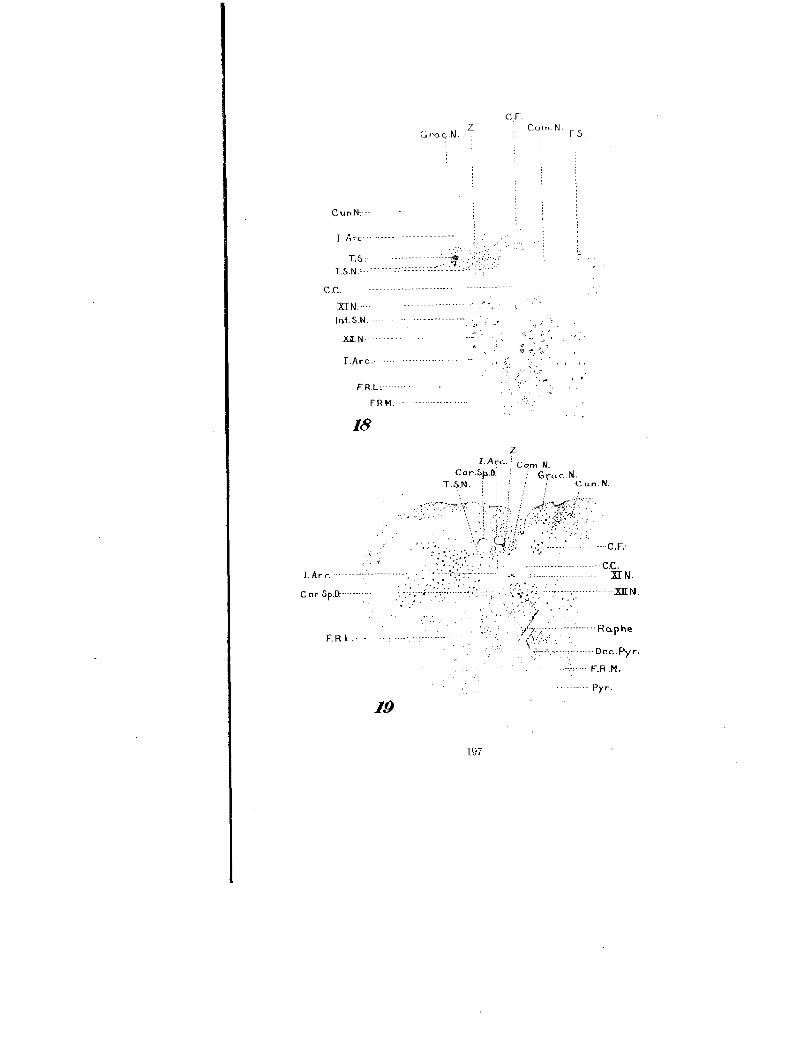
The remainder of the tractus solitarius is largely concerned withits termination in the nucleus commissuralis or ganglion com-rnissurale. Figure 18, which is from a level immediately in frontof the 37th cm. plane in figure 1C, demonstrates the tractussolitarius (T.S.) directly ventrad of a bundle of fibrae arcuateinternae (I.Arc.) and a little lateral and ventral to tractus (Z).It is reduced to a few small bundles of degenerated fibers. Theterminal sensory nucleus of the tractus solitarius (T.S.N.) notonly surrounds the tract at this level, but extends inward to themid-line to meet the corresponding nucleus of the opposite side.This common nucleus is usually termed the nucleus commissuralisor ganglion commissurale (T.S.N. or Com.N.). From figure 18it is apparent that this nucleus receives a very rich supply ofdegenerated tractus solitarius fibers. It is also obvious that veryfew of these degenerated fibers cross to the opposite side as corn-rnissural fibers (C.F.). Most of these fibers are shown in figure1C. In this series the commissural fibers were mainly normalfibers, taking origin apparently from the nuclei of the generalreticular substance; for in another Marchi series, where the retic-ular substance was destroyed at this level, there were manymore degenerated commissural fibers. In the rabbit series not asingle degenerated tractus solitarius fiber was seen crossing to theopposite side in the region of the nucleus commissuralis, and nocommissural fibers are shown in Van Gehuchten's figure 18, whichis a section from this region. In fact, the entire number ofcommissural fibers is relatively small in mammals, as was demon-strated from a Marchi series where the entire nucleus corn-missuralis was severed longitudinally on one side. At this levelthe nucleus commissuralis is situated considerably dorsad of thenucleus nervi accessorii or caudal continuation of the nucleusmotorius dorsalis nervi vagi (XI N.) and no degenerated tractussolitarius fibers were observed near this,nucleus.
Fig. 18 Dorso-central portion of a transverse section through the medullaspinalis from the same series as figure 11. This section passes through the nucleuscommissuralis and in figure 1C would lie in the plane (C. F.). X 18.
Fig. 19 Left half of a transverse section from the same series as above at alevel of the decussatio pyramidum, situated in the 39th , cm. plane in figure 1C.X 6.
---
C .C.xr N.
............. 2aL N.
ClGra.c.N.
z corn N C.S.
18
I.Arc.z,'
Cor.Sp.D.Cun.N.
Pyr.
19
197
I Arc-.
T.S.T.S.N.'---- -
C .C.
I.Arc.
-
ERM.--
I.Ar c.

198 WILLIAM F. ALLEN
From the region of the 34th cm. plane in figure 1 C, caudad, the-tractus solitarius (T.S.) becomes reduced from very small bundlesto individual fibers. In figure 19, which is from the 39th cm. planein figure IC, through the decussatio pyramidum, there are a few de-generated tractus solitarius fibers in the caudal end of the nucleuscommissuralis (Com.N.) and a few in the region occupied by thetractus solitarius in more cephalic sections (T.S.N.). The de-generated fibers in the area of the nucleus commissuralis terminatein the third or fourth section caudal to figure 19, while the morelateral area of degenerated tractus solitarius fibers continues afew sections further caudad in the general reticular area. Infigure 19, tractus (Z), a bundle of fibrae arcuatae internae (I. Are.)and a bundle of tractus cortico-spinalis dorsalis fibers (Cor.Sp.D.)will be seen between these two areas of degenerated tractus soli-tarius fibers and the nucleus fasciculi gracilis and the fasciculicuneati. No degenerated fibers are present in the nucleus nerviaccessorii, which is situated lateral to the canalis centralis anddorsal to the nucleus nervi hypoglossi. Both the fibrae arcuataeinternae and the tractus cortico-spinalis dorsalis are decussating,and since both are normal fibers it is difficult to discriminate be-tween them. Tractus (Z) does not make its ventral bend until alower level is reached.
SUMMARY AND GENERAL CONSIDERATIONS
As in other mammals, the tractus solitarius in the guinea pigis made up of descending sensory fibers from the VII, IX and Xnerves. It begins at the level of the entrance of the radixsensibilis nervi facialis and extends caudally to the level of thedecussatio pyratnidum. This tract permits of a division intotwo almost equal parts at about the entrance of the thirdor fourth radix sensibilis nervi vagi. The cephalic portion isembedded in the ventro-lateral corner of the radix descendensnervi vestibularis, is an area for the reception of sensory rootfibers and distributes both gustatory and general visceral sensa-tions to a primary sensory nucleus. On the other hand, thecaudal portion is isolated from the radix descendens nervi vesti-bularis, receives no sensory roots and distributes no gustatory

TRACTUS SOLITARITIS IN THE GUINEA PIG 199
fibers to its sensory nucleus. Caudally the tractus solitariusbreaks up into small bundles, which terminate in the nucleustractus solitarii.
Two of the three components of the tractus solitarius, namely,the radix sensibilis nervi facialis and the radix sensibilis nerviglossopharyngei, are very similar; both separate into at least fourbundles or rootlets in traversing the medulla, to reunite in acommon fasciculus that gives rise to, or enters the tractus soli-tarius. No facialis fibers appear in the tractus solitarius caudalto the entrance of the first radix sensibilis nervi vagi, and noglossopharyngeal fibers are present in the tractus solitariuscaudal to the entrance of the third radix sensibilis nervi vagi.
In the guinea pig four distinct sensory fasciculi or roots of theX nerve were recognized. The first two are composed of fromtwo to four bundles or rootlets and are readily comparable tothe sensory roots of the VII and IX nerves; while the last twoconsist of one bundle each.
The various roots entering into the formation of the tractussolitarius, cross ventrally the previously formed portion of thetractus solitarius and for the most part continue caudally medianto it. This signifies that the VII fibers could be separatedlaterally from the tractus solitarius with little injury to the IXfibers, and likewise the IX with little injury to the first root ofthe X. It was recorded, however, that there was some inter-mingling of these root fibers.
None of the sensory fibers of the VII, IX or X (having cellbodies in the ganglion nodosum) nerves are distributed anywherebut to the tractus solitarius.
If the relative size of degenerated myelin is any indication of thecorresponding thickness of medullary sheaths, the VII and IXsensory fibers would be finely medullated and the X would becoarse.
In the guinea pig the nucleus tractus solitarii extends through-out the entire length of the tractus solitarius. At first it is alight area situated ventro-medially to the tractus solitarius, whichsoon expands, joining other nuclear masses in the. formation ofthe so-called dorsal vagus nucleus. This oval nuclear mass
200 WILLIAM F. ALLEN
can be divided into three parts: A median mass of cells, thenucleus motorius dorsalis nervi 61ossopharyngei et vagi; a lateralmass of cells next to the tracti solitarius, the nucleus tractussolitarii; and an intermediate mass of small cells may representa visceral reticular nucleus for short relays in the medulla. Cau-dally to the entrance of the last sensory root of the vagus thenucleus tractus solitarii assumes enormous proportions, dorsallyand medially, to ultimately unite with its fellow in the spinalportion of the medulla in forming the nucleus comraissuralis.
It was noted that the maximum number of facialis tractussolitarius fibers are dispensed to the nucleus tractus solitariiopposite the entrance of the IX sensory root, and that none arefound opposite the entrance of the first vagus sensory root.Between the entrance of the IX and first X sensory roots,numerous glossopharyngeal tractus solitarius fibers are distributedto the nucleus tractus solitarii, but none appear caudal to theentrance of the third vagus sensory root. Very few vagus tractussolitarius fibers are present in the nucleus tractus solitarii oppositethe entrance of the vagus sensory roots. Caudad, these fibersbecome very abundant, especially in the nucleus commissuralis.
In at least two places vagus fibers or collaterals are distributedfrom the tractus solitarius to the nucleus motorius dorsalis nervivagi (fig. 1, S.). One of these areas is in the caudal part of themedulla and the other is in the medulla spinalis. In many otherplaces the neurons of this motor nucleus would not have topossess very long dendrites to come in contact with fibers fromthe tractus solitarius.
No fibers or collaterals from the tractus solitarius were seen inthe nucleus nervi hypoglossi, in the nucleus parasolitarius, or inthe nucleus nervi vestibularis spinalis.
A so-called tractus (Z), which may be the same as Kosaka andYagita's fasciculus solitario-spinalis or Hirose's bulbospinaletract, was briefly mentioned on account of its intimate associationwith the tractus solitarius in the medulla spinalis region.
Since practically all, if not all, of the taste fibers are carriedby the VII and IX nerves, and these fibers have ended in thetractus solitarius at the level of the entrance of the third radix

TRAcrus SOLITARITTS IN THE GUINEA PIG 201
sensibilis nervi vagi, it is evident that no part of the tractussolitarius of the guinea pig, caudal of this level, is concerned withconducting taste sensory impulses. It must be associated withgeneral visceral and possibly muscle sense. Visceral or smoothmuscle sense will be included under general visceral sensations.Such endings have been described by Ploshko between the smoothmuscle bundles of the trachea ; by Carpenter in the longitudinalmuscle of the stomach and intestine, and by Larsell in the smoothmuscle of the bronchial tree. Parts or all of the nucleus tractussolitarii, cephalad of the entrance of the third radix sensibilisnervi vagi into the tractus solitarius, must also relay nervousimpulses of general visceral and possibly muscle sense in additionto those of taste.
The description of the entrance, disposition and distributionof the VII, IX and X nerves in the tractus solitarius suggests anoriginal segmental grouping of these roots, corresponding to theexisting segmental arrangement of the motor roots throughoutthe medulla. Apparently certain segmentally arranged sensoryroots have been massed in three places and joined with littleintermingling, in forming the tractus solitarius, which extendscaudad to the level of the first cervical nerve. To reproduce theoriginal primitive segmental condition, all that would be necessarywould be to scatter the sensory roots of the VII, IX and X nervesinto the intervals between and caudal of these roots, where nosensory root fibers are penetrating the medulla.
If any reduction has occurred in the sensory system of the me-dulla in mammals, it is in connection with the somatic and notthe visceral system.
As a result of this study the writer has been impressed with thepossibilities of the caudal part of the medulla and the medullaspinalis as a region for correlating the vital activities of the body.

202 WILLIAM F. ALLEN
LITERATURE CITED
Anrf;xs EAPPERS, C. U. 1920 Die Vergleichende Anatomic des Nervensy,stemsder Wirbeltiere und des Menschen. 2 Abschnitt. Haarlem.
AUTORE, P. 1915 Morphologic e svilluppo del nucleo dorsale del vago in Susscopha. Monit. Zool. Ital. An. 26, p. 134.
13nmAiii, N. 1922 Studi comparitivi sulla struitura del Ithombencefalo. Az-ch.Ital. di Anat. e di Emb. Vol. 19, fasc. 1-2, pp. 122-291,
BERKELBACH VAN DER SPRENKEL, H. 1915 The central relations of the cranialnerves of Silimns glanis and Mormyrus caschive. Jour. (ortip. Ncur.,vol. 25, pp. 5-63.
Box, S. T. 1915 Die Entwicklung der Ifirmierven und ihrer Zentralen lla.hnen.Folic Neuro-biol., Bd. 9, S. 475-565.
BREGMANN, L. E, 1892 Ueber experimentelle aufsteigende Degenerationmotorischer und sensibiler Hirnnerven, Arb. d. lost. f. Anat. it.Physiol., Wien.
BRUCE, A. N. 1898 On the dorsal or so-called sensory nucleus of the glossa-pharyngeal nerve and nuclei of origin of the trige,minal nerve. Brain,vol. 21, pp. 383-387.
CARPENTER, F. W. 1918 Nerve endings of sensory type in the muscular coatof the stomach and small intestine. Jour. Comp. Neur., vol. 29,pp. 553-559.
CUSHING, H. 1903 The taste fibers and their independence of the nervestrigeminus. Johns Hopkins Hosp. Bull., vol. 14, pp. 71-78.
HELD, H. 1892 Die Endigungsweise der sensibilen Nerven im Gehirn. Arch.f. Anat. u. Entwicklungsgeschichte, S. 33-40.
HERRICK, C. J. 1905 The central gustatory paths in the brains of bony fishes..Jour. Comp. Neur., vol. 15, pp. 375-456.1907 On the commissura infima of the brains of fishes. Anat. Rec.,vol. 1, p. 88.1908 On the commissura infima and its nuclei in the brains of fishes.Jour. Comp. Neur., vol. 18, pp. 409-431.1914 The medulla oblongata of larval Amblystoma. Jour. Comp.Neur., vol. 24, pp. 343-427.
HInosE, K. 1016 Uber eine bulbospinale Bahn. Point Neuro-biol., Bd. 10,S. 371-382.
His, W. 1887 Die Entwicklung der ersten Nervenbahnen beirn menschlichenEmbryo. Arch. 1. Anat. u. Physiol., S. 367-378.
Holm, H. 1893 Die Anatomic und Pathologic des dorsalen Vaguskerns. Vir-chow's Arch., S. 78-120.
HUNT, J. H. 1909 The sensory system of the facial nerve, and itssymptomatology. Journ. Nerv. and Mental Disease, vol. 36, pp.321-350.
JOHNSTON, J. B. 1901 The brain of Acipenser. Zool. Jahrb., Bd. 15, pp. 1-204.1906 Nervous system of vertebrates. Philadelphia.1902-1912 Various papers on the telencephalon of fishes. Jour.Comp. Neur.
KOELLIKER, A. 1896 Handbuch der Gewebelehre des Meoschen. Leipzig.

14 -1" le
TRACTIJS SOLITARI1JS 1N THE GUINEA PIG 203
E.- OMSTAMM Una IIINDERLANC 1910 Der Nucleus intermedius sensibilis alstIrsprung einer gekreurt aufsteigenden Balm (Viszeralbahn?). Monet-schr, Psych. Neurol., Bd. 28, S. 82-83. Also, Neurol. Centrabl., Bd.29, S. 663.
KosAKA, K. 1909 Ueber die Vaguskerne des Hundes. Neurol..ROSANA UND YAturit 1905 Experiinentelte Untersuchungen fiber den Unsprung
des N. vagus und die centralen Endigen der dem Plexus nodosusentstammenden sensibilen Vagusfasern, sowie fiber den Verlauf ihrersekundaren Bahn. Shinkeiguku Zasshi, Tokyo, Bd. 4, S. 29-49.
LARSELL, 0. 1922 The ganglia, plexuses and nerve terminations of the mamma-lian lung and the pleura pulmonalis. Jour. Comp. Neur., vol. 35,pp. 97-132.
MOLHANT, M. 1910 Le nerf vague, etude anatomique et experirnentale. LeNevraxe, T, 11, pp. 137-249.
MaLLGAARD, H 1911 Eine inorpologische Studie ilher den Nervenkomplexvago-glossopharyngeo-accessorius. Skand. Arch. 1. Physiol., Bd. 25,S. 69-80.1912 Studicn Utter das respiratorische Nervensystcm bei den Wirbel-tieren. As above, Bd. 26, S. 315-383. Folia Neuro-biol., Bd. 6,S. 622-623.
Mucum, N. 1893 Der Nucleus dorsalis und der sensorischc Kern des Nervusglossopharyngeus. Centrabl. f. Nervenh. u. Psych., Ed. 4, S. 212-217.
OBETISTEINER, H. 1912 Anleitung beim Stadium des Baues der nervOsenZentralorgane. Leipzig.
PLoscrixo, A. 1897 Die Nervenendigungen und Ganglien der Respirations-organe. Anat. Anz., Bd. 13, S. 12-22.
POPE, F. M. 1889 Thrombosis of vertebral artery pressing on glossopharyngealnerve; unilateral loss of taste at back of tongue. Brit. Med. Journ.,vol. 2, pp. 1148-1149.
RAMON Y CA.JAL, S. 1911 Histologic du systême nerveux de l'homme et desvertebras, Paris, T. 2.
RRINELIART, D. A. 1918 The nervus facialis of the albino rat. Jour. Comp.Neur., vol. 30, pp. 81-126.
ROLLER, C. F. W. 1881 Der centrale Verlauf des Nervus glossopharyngeus.Der Nucleus lateralis medius. Arch. f. Mikr. Anat., Bd. 19, S. 347-383.
SoLoreoNsoRN, H. 1888 Ueber den Weg der Geschrnacksfasern zum Gehirn.Reviewed, Neurol. Centrabl. Bd. 7, S. 295-296.
STAGE-aim, R. 1896 LIbicazione e rapporti di alcuni nuclei di sostanza grigiadella midolla allungata. Internat. Monatschr. f. Anat. u. Physiol.,Bd. 13, S. 326-357.
STILLING 1842 Ueber die Textur der Medulla oblongata. Quoted by VanGehuchten.
TRICOMI-ALLEGRA, G. 1903 Suite connessioni bulbari del nervo vago,d. patol. nerv., Firenze.
TUMOERLAKA, R. 1916 Konsekutive Veranderungen eines kleinen Hordes indem ventro-lateralen Thalamuskern und das demselben zugrundeliegende klinische Psychiatrische en Neurologische Bladen.Amsterdam.

204
WILLIAM P. ALLEN
TURNER, W. A. 1895 The central connections and relations of the trigeminal,vago-glossopharyngeal, vago-accessory, and hypoglossal nerves.Jour, Anat. and Physiol., vol. 29, pp. 1-15.1897 Note on the course of the fibers of taste. Edin. Med. Journ,,vol. 2, pp. 2111-262.
TURNER AND BULLOCK 1894 Observations upon the central relations of thevago-glossopharyngeal, vago-accessory, and hypoglossal nerves, froma study of a case of bulbar paralysis. Brain, vol. 17, pp. (193-709.
VAN GEHIICH TEN, A. 1900 Le nerf intermediaire. de Wrisberg. Le N4vraxe,T. 1, pp. 5-12.1900 Le faisceau solitaire. Le Nevraxe, T. 1, pp. 173-195.1906 Anatomie du systême nerveux de rho/nine. Louvain.
VON LENHOSSliK, M. 1894 Das Ganglion geniculi Nervi facialis und seineVerbindungen. Quoted by Van Gehuchten.
VON MON A K 0 W , C. 1913 Zur Kenntnis der Grosshirnanteile (Vago-glosso-pharyngeusschleife, Fasc. bulbo-thal. II). Neurol. Centrabl., Bd. 32,S. 331-333.
WALLENBERG, A. 1898 Des dorsale Gebiet der spinalen Trigeminuszwurzelund seine Beziehungen zum solitaren Biludei beim Mcnschen. Deuche.Zeitschr. f. Nervenheilkunde.1907 Die kaudale Endigung der bulbo-spinalen Wurzeln des Trigern-inus, Vestibularis, und Vagus beim Frosche. Anat. Ariz., Bd. 30.
WEED, L. H. 1913 Reconstruction of the nuclear masses in the rhomben-cephalon. Anat. Rec., vol. 7, pp. 447-148.
Winsois,:, J. G. 1905 The structure and function of the taste buds of the larynx.Brain, vol. 28, pp. 339-357.
WINKLER AND POTTER 1911 An anatomical guide to experimental researches.on the rabbit's brain, A series of 40 frontal sections. Amsterdam.1914 Same of the cat. Thirty-five frontal sections. Amsterdam.
Y A GITA, K. 1910 Experimental Untersuchungen fiber den Ursprung des Nervusfacialis. Anat. Anz., Bd. 37, S. 195.1914 Einige Experimente an dem Nervus petrosus superficialis major.zur Bestimmung des Ursprungsgebietes des Nerven. Folic Neuro-biol., Bd. 8, S. 361-382.
Z IEHEN, T. 1913 Anatomie des Centralnervensystems. II Abtheilung, Tell 1,,Jena.


![[1b] Tractus Urinaria 2003](https://static.fdocuments.us/doc/165x107/577cd84d1a28ab9e78a0e6b2/1b-tractus-urinaria-2003.jpg)