![[1b] Tractus Urinaria 2003](https://static.fdocuments.us/doc/165x107/577cd84d1a28ab9e78a0e6b2/1b-tractus-urinaria-2003.jpg)
ON THE STRUCTURE OF THE DIGESTIVE TRACTUS IN ... · segments of the digestive tractus, in...
6
Analele Ştiinţifice ale Universităţii „Al. I. Cuza” Iaşi, s. Biologie animală, Tom LIV, 2008 ON THE STRUCTURE OF THE DIGESTIVE TRACTUS IN HYPOPHTHALMICHTHYS MOLITRIX Gabriela VASILE, Gianina COMĂNESCU, Elena CIORNEA and Costică MISĂILĂ “Al. I. Cuza” University Iaşi, Faculty of Biology, Bd. Carol I 20A, 700505 Iaşi, Romania, [email protected]; [email protected]; [email protected] Abstract. The paper discusses some histological aspects of the digestive tractus in its various segments (anterior post-esophagus intestine, anterior distal intestine, median and posterior intestine), which have been correlated with both age and main type of food for the species under study. The experimental results obtained put into evidence some variation in the form of the mucosa folds (simpler in one summer-old individuals and more complex in the four summer-old ones), along with variations in the thickness of the mucosa and muscularis. Keywords: digestive tractus, mucosa, muscularis, silver carp. Rezumat. Structura tractusului digestiv la Hypophthalmichthys molitrix. Lucrarea de faţă prezintă aspectele histologice ale tractusului digestiv la Hypophthalmichthys molitrix în diferitele sale segmente (intestin anterior postesofagian, intestin anterior distal, intestin mediu şi intestin posterior), aspecte care au fost corelate cu vârsta şi cu tipul predominant de hrană a speciei luate în studiu. Rezultatele experimentale au evidenţiat o variaţie de formă a cutelor mucoasei (fiind mai simple la exemplarele în vârstă de o vară şi mai complexe la exemplarele în vârstă de patru veri), precum şi o variaţie în ceea ce priveşte grosimea mucoasei şi a musculoasei. Cuvinte cheie: tractus digestiv, tunică mucoasă, tunică musculoasă, sânger. Introduction As known, the cyprinids are species having no stomach (in the structure of the digestive tractus distinguishing three distinct regions parts, namely: anterior intestine (post-esophagus and distal), median and posterior intestine), its absence being compensated by a long intestine, which exceeds several times the length of the fish body, thus granting a larger surface for the absorbance of the nutritive elements at intestinal level, as well as a complete utilization of the food, in parallels with a longer retention time of the aliments, once they are subjected to a prolonged enzymatic and, especially, protease action (Stăncioiu et al., 2006; Apetroaei, 2007). As the studies devoted to the morpho-structural aspects of the digestive tractus in Asian cyprinids are quite few and very synthetic (Berry & Low, 1970; Bremer, 1978; Trevisan, 1979; Athikesavan et al., 2006), the present investigation analyzes such aspects in Hypophthalmichthys molitrix (silver carp) of various stages, i.e., one and four summer- old individuals, for evidencing some possible intraspecific (age-dependent) similarities or differences. Material and Methods The digestive tube has been taken over form 5 Hypophthalmichthys molitrix individuals of various ages, namely: one- and four-old summer ones, the digestive tractus being unfolded along its whole length, after which rectangularly-shaped fragments were taken over and processed by methods of histological techniques; this, the nuclei were colored with hemalaune, while the cytoplasm - with eosine (Diaconiţă et al., 1953; Humason, 1962; Martoja & Martoja, 1967). The thickness of the mucosa and muscularis
Transcript of ON THE STRUCTURE OF THE DIGESTIVE TRACTUS IN ... · segments of the digestive tractus, in...
Analele Ştiinţifice ale Universităţii „Al. I. Cuza” Iaşi, s. Biologie animală, Tom LIV, 2008
ON THE STRUCTURE OF THE DIGESTIVE TRACTUS IN HYPOPHTHALMICHTHYS MOLITRIX
Gabriela VASILE, Gianina COMĂNESCU, Elena CIORNEA and
Costică MISĂILĂ “Al. I. Cuza” University Iaşi, Faculty of Biology, Bd. Carol I 20A, 700505 Iaşi, Romania,
[email protected]; [email protected]; [email protected]
Abstract. The paper discusses some histological aspects of the digestive tractus in its various segments (anterior post-esophagus intestine, anterior distal intestine, median and posterior intestine), which have been correlated with both age and main type of food for the species under study. The experimental results obtained put into evidence some variation in the form of the mucosa folds (simpler in one summer-old individuals and more complex in the four summer-old ones), along with variations in the thickness of the mucosa and muscularis. Keywords: digestive tractus, mucosa, muscularis, silver carp. Rezumat. Structura tractusului digestiv la Hypophthalmichthys molitrix. Lucrarea de faţă prezintă aspectele histologice ale tractusului digestiv la Hypophthalmichthys molitrix în diferitele sale segmente (intestin anterior postesofagian, intestin anterior distal, intestin mediu şi intestin posterior), aspecte care au fost corelate cu vârsta şi cu tipul predominant de hrană a speciei luate în studiu. Rezultatele experimentale au evidenţiat o variaţie de formă a cutelor mucoasei (fiind mai simple la exemplarele în vârstă de o vară şi mai complexe la exemplarele în vârstă de patru veri), precum şi o variaţie în ceea ce priveşte grosimea mucoasei şi a musculoasei.
Cuvinte cheie: tractus digestiv, tunică mucoasă, tunică musculoasă, sânger.
Introduction As known, the cyprinids are species having no stomach (in the structure of the
digestive tractus distinguishing three distinct regions parts, namely: anterior intestine (post-esophagus and distal), median and posterior intestine), its absence being compensated by a long intestine, which exceeds several times the length of the fish body, thus granting a larger surface for the absorbance of the nutritive elements at intestinal level, as well as a complete utilization of the food, in parallels with a longer retention time of the aliments, once they are subjected to a prolonged enzymatic and, especially, protease action (Stăncioiu et al., 2006; Apetroaei, 2007).
As the studies devoted to the morpho-structural aspects of the digestive tractus in Asian cyprinids are quite few and very synthetic (Berry & Low, 1970; Bremer, 1978; Trevisan, 1979; Athikesavan et al., 2006), the present investigation analyzes such aspects in Hypophthalmichthys molitrix (silver carp) of various stages, i.e., one and four summer-old individuals, for evidencing some possible intraspecific (age-dependent) similarities or differences.
Material and Methods The digestive tube has been taken over form 5 Hypophthalmichthys molitrix
individuals of various ages, namely: one- and four-old summer ones, the digestive tractus being unfolded along its whole length, after which rectangularly-shaped fragments were taken over and processed by methods of histological techniques; this, the nuclei were colored with hemalaune, while the cytoplasm - with eosine (Diaconiţă et al., 1953; Humason, 1962; Martoja & Martoja, 1967). The thickness of the mucosa and muscularis

Gabriela Vasile et al.
was measured by the micrometric method, based on the utilization of a micrometric objective thin plate lamina and of an ocular micrometric lamina (Mureşan et al., 1974).
Results and Discussion
In one-summer old Hypophthalmichthys molitrix representatives, the post-esophagus part (from the immediate vicinity of the esophagus) is relatively well developed, with a high mucosa, with deep plaits (some cylindrical in shape, others large, bifurcated, trapezoidal), with a typical intestinal epithelium - i.e., prismatic, with a striated plateau and mucosa Goblet cells. The larger corione, occurring under the epithelium, appears percolated by lymphocytes which, here and there, penetrate the epithelium, too, migrating even in the respective lumen (Fig. 1a). One may even observe the presence of a lymphoid nodule (Fig. 1b).
a. b.
Figure 1. Cross-section through the anterior post-esophagus intestine of one summer-old Hypophthalmichthys molitrix: a. assembly, 100x; b. lymphoid nodule detail,
200x (original photos).
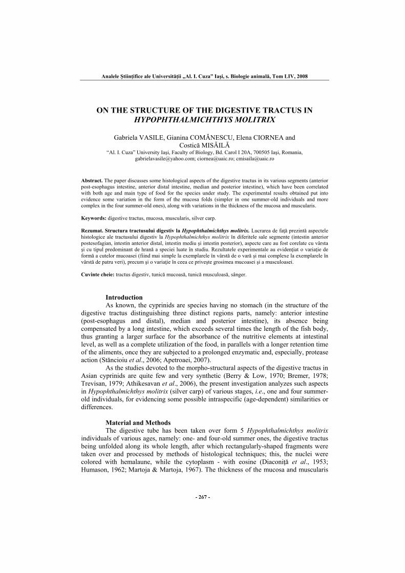
For the 10x ocular - 10x objective combination, the mucosa has a total thickness of 875 μm. As to the muscularis, it appears - for the same combination - as relatively well-developed, with a thickness of 562.5 μm, formed of a layer of circularly-arranged, 500 μm thick, smooth muscular fibers, and of an external layer of longitudinal muscular wave-shaped fibers, with a thickness of 62.5 μm. As to the anterior post-esophagus intestine of four summer-old silver carp individuals, the mucosa tunic shows a very high prismatic epithelium, about 75 μm thick, very rich in mucosa Goblet cells, and quite a varied aspect of the mucosa folds in this area. Comparatively with the superior vertebrates (the mammalians), one might say that they occur as “folds”, once they involve not only the corione but also the muscularis part of the mucosa and the submucosa tunic, there existing, on the surface of this “folds”, other smaller folds to be possibly compared with some “villi”, once they involve only the corione (Fig. 2a).
The total thickness of the mucosa is of 1375 μm, while the muscularis is also well-developed, with a thickness of 750 μm. Mention should be again made here of the fact that that some lymphoid infiltrations may be observed on the thickness of the mucosa; more than that, here and there, the epithelium appears slightly eroded (Fig. 2b).
Analele Ştiinţifice ale Universităţii „Al. I. Cuza” Iaşi, s. Biologie animală, Tom LIV, 2008
a. b.
Figure 2. Cross-section through the anterior post-esophagus intestine of four summer-old Hypophthalmichthys molitrix: a. assembly, 100x; b. detail with lymphoid infiltrations, 200x)
(original photos). .
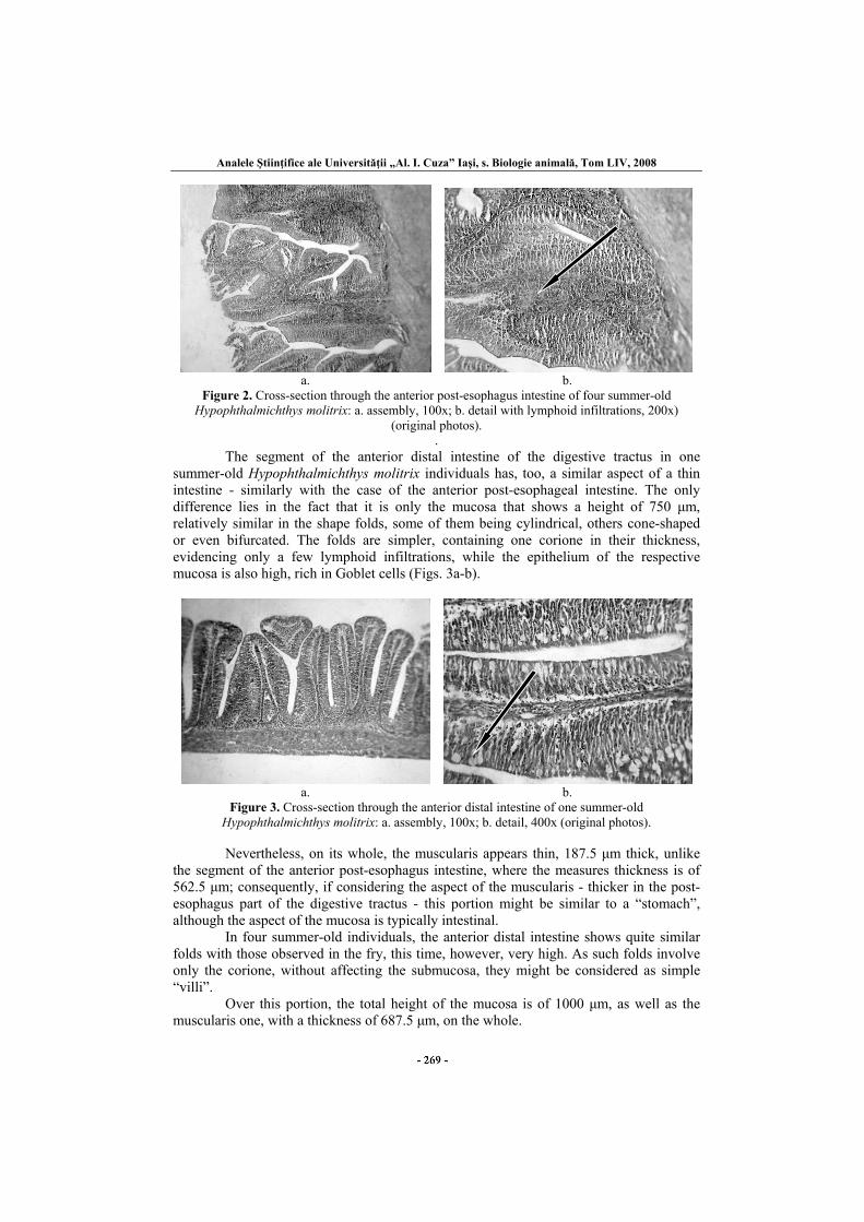
The segment of the anterior distal intestine of the digestive tractus in one summer-old Hypophthalmichthys molitrix individuals has, too, a similar aspect of a thin intestine - similarly with the case of the anterior post-esophageal intestine. The only difference lies in the fact that it is only the mucosa that shows a height of 750 μm, relatively similar in the shape folds, some of them being cylindrical, others cone-shaped or even bifurcated. The folds are simpler, containing one corione in their thickness, evidencing only a few lymphoid infiltrations, while the epithelium of the respective mucosa is also high, rich in Goblet cells (Figs. 3a-b).
a. b.
Figure 3. Cross-section through the anterior distal intestine of one summer-old Hypophthalmichthys molitrix: a. assembly, 100x; b. detail, 400x (original photos).
Nevertheless, on its whole, the muscularis appears thin, 187.5 μm thick, unlike the segment of the anterior post-esophagus intestine, where the measures thickness is of 562.5 μm; consequently, if considering the aspect of the muscularis - thicker in the post-esophagus part of the digestive tractus - this portion might be similar to a “stomach”, although the aspect of the mucosa is typically intestinal. In four summer-old individuals, the anterior distal intestine shows quite similar folds with those observed in the fry, this time, however, very high. As such folds involve only the corione, without affecting the submucosa, they might be considered as simple “villi”. Over this portion, the total height of the mucosa is of 1000 μm, as well as the muscularis one, with a thickness of 687.5 μm, on the whole.
Gabriela Vasile et al.
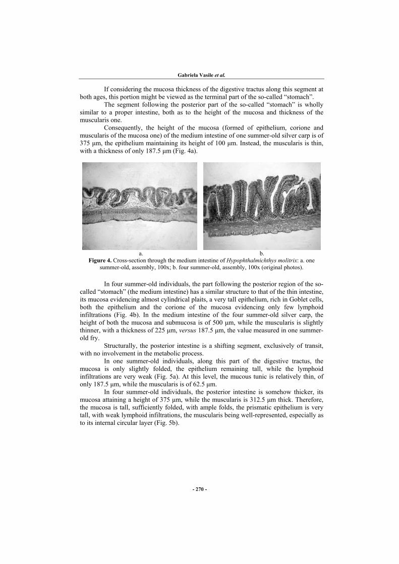
If considering the mucosa thickness of the digestive tractus along this segment at both ages, this portion might be viewed as the terminal part of the so-called “stomach”. The segment following the posterior part of the so-called “stomach” is wholly similar to a proper intestine, both as to the height of the mucosa and thickness of the muscularis one. Consequently, the height of the mucosa (formed of epithelium, corione and muscularis of the mucosa one) of the medium intestine of one summer-old silver carp is of 375 μm, the epithelium maintaining its height of 100 μm. Instead, the muscularis is thin, with a thickness of only 187.5 μm (Fig. 4a).
a. b.
Figure 4. Cross-section through the medium intestine of Hypophthalmichthys molitrix: a. one summer-old, assembly, 100x; b. four summer-old, assembly, 100x (original photos).
In four summer-old individuals, the part following the posterior region of the so-called “stomach” (the medium intestine) has a similar structure to that of the thin intestine, its mucosa evidencing almost cylindrical plaits, a very tall epithelium, rich in Goblet cells, both the epithelium and the corione of the mucosa evidencing only few lymphoid infiltrations (Fig. 4b). In the medium intestine of the four summer-old silver carp, the height of both the mucosa and submucosa is of 500 μm, while the muscularis is slightly thinner, with a thickness of 225 μm, versus 187.5 μm, the value measured in one summer-old fry. Structurally, the posterior intestine is a shifting segment, exclusively of transit, with no involvement in the metabolic process. In one summer-old individuals, along this part of the digestive tractus, the mucosa is only slightly folded, the epithelium remaining tall, while the lymphoid infiltrations are very weak (Fig. 5a). At this level, the mucous tunic is relatively thin, of only 187.5 μm, while the muscularis is of 62.5 μm.
In four summer-old individuals, the posterior intestine is somehow thicker, its mucosa attaining a height of 375 μm, while the muscularis is 312.5 μm thick. Therefore, the mucosa is tall, sufficiently folded, with ample folds, the prismatic epithelium is very tall, with weak lymphoid infiltrations, the muscularis being well-represented, especially as to its internal circular layer (Fig. 5b).
Analele Ştiinţifice ale Universităţii „Al. I. Cuza” Iaşi, s. Biologie animală, Tom LIV, 2008
a. b.
Figure 5. Cross-section through the posterior intestine of Hypophthalmichthys molitrix: a. one summer-old, assembly, 200x; b. four summer-old, assembly, 100x (original photos).
0
200
400
600
800
1000
1200
1400
μm
ant. post-esoph. int. ant . distal int . medium int . posterior int.
0+ 3+
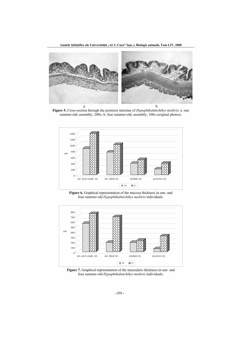
Figure 6. Graphical representation of the mucosa thickness in one- and
four summer-old Hypophthalmichthys molitrix individuals.
0
100
200
300
400
500
600
700
800
μm
ant. post-esoph. int . ant . distal int. medium int . posterior int .
0+ 3+
Figure 7. Graphical representation of the muscularis thickness in one- and
four summer-old Hypophthalmichthys molitrix individuals.
Gabriela Vasile et al.
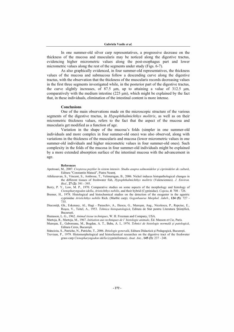
In one summer-old silver carp representatives, a progressive decrease on the thickness of the mucosa and muscularis may be noticed along the digestive tractus, evidencing higher micrometric values along the post-esophagus part and lower micrometric values along the rest of the segments under study (Figs. 6-7).
As also graphically evidenced, in four summer-old representatives, the thickness values of the mucosa and submucosa follow a descending curve along the digestive tractus, with the observation that the thickness of the muscularis records decreasing values in the first three segments investigated while, in the posterior part of the digestive tractus, the curve slightly increases, of 87.5 μm, up to attaining a value of 312.5 μm, comparatively with the medium intestine (225 μm), which might be explained by the fact that, in these individuals, elimination of the intestinal content is more intense.
Conclusions
One of the main observations made on the microscopic structure of the various segments of the digestive tractus, in Hypophthalmichthys molitrix, as well as on their micrometric thickness values, refers to the fact that the aspect of the mucosa and muscularis get modified as a function of age. Variation in the shape of the mucosa’s folds (simpler in one summer-old individuals and more complex in four summer-old ones) was also observed, along with variations in the thickness of the muscularis and mucosa (lower micrometric values in one summer-old individuals and higher micrometric values in four summer-old ones). Such complexity in the folds of the mucosa in four summer-old individuals might be explained by a more extended absorption surface of the intestinal mucosa with the advancement in age.
References Apetroaei, M., 2007. Creşterea peştilor în sistem intensiv. Studiu asupra salmonidelor şi ciprinidelor de cultură,
Editura "Constantin Matasă", Piatra Neamţ. Athikesavan, S., Vincent, S., Ambrose, T., Velmurugan, B., 2006. Nickel induces histopathological changes in
the different tissues of freshwater fish, Hypophthalmichthys molitrix (Valenciennes). J. Environ. Biol., 27 (2): 391 - 395.
Berry, P. Y., Low, M. P., 1970. Comparative studies on some aspects of the morphology and histology of Ctenopharyngodon idella, Aristichthys nobilis, and their hybrid (Cyprinidae). Copeia, 4: 708 - 726.
Bremer, H., 1978. Histological and histochemical studies on the detection of the esogaster in the agastric cyprinidae Aristichthys nobilis Rich. (Marble carp). Gegenbaurus Morphol. Jahrb., 124 (5): 727 - 735.
Diaconiţă, Gh., Eskenasy, Al., Hagi - Paraschiv, A., Iliescu, G., Mureşan, Aug., Nicolescu, P., Repciuc, E., Roşca, V., Teitel, A., 1953. Tehnica histopatologică, Editura de Stat pentru Literatura Ştiinţifică, Bucureşti.
Humason, L. G., 1962. Animal tissue techniques, W. H. Freeman and Company, USA. Martoja, R., Martoja, M., 1967. Initiation aux techniques de l’ histologie animale, Éd. Masson et Cie, Paris. Mureşan, E., Gaboreanu, M., Bogdan, A. T., Baba, A. I., 1974. Tehnici de histologie normală şi patologică,
Editura Ceres, Bucureşti. Stăncioiu, S., Patriche, N., Patriche, T., 2006. Ihtiologie generală, Editura Didactică şi Pedagogică, Bucureşti. Trevisan, P., 1979. Histomorphological and histochemical researches on the digestive tract of the freshwater
grass carp Ctenopharyngodon idella (cypriniformes). Anat. Anz., 145 (3): 237 - 248.