Anatomía y Cambio Histofisiológicos de La Glandula Pituitaria
Upload
erickmattosCategory
view
213download
0
8/10/2019 Molecular Morfgenesis de la glandula Tiroidea.pdf
http://slidepdf.com/reader/full/molecular-morfgenesis-de-la-glandula-tiroideapdf 1/20
Please cite this article in press as: Fagman, H., Nilsson, M., Morphogenesis of the thyroid gland. Mol. Cell. Endocrinol. (2010),
doi:10.1016/j.mce.2009.12.008
ARTICLE IN PRESSGModel
MCE-7389; No.of Pages20
Molecular and Cellular Endocrinology xxx (2009) xxx–xxx
Contents lists available at ScienceDirect
Molecular and Cellular Endocrinology
j o u r n a l h o m e p a g e : w w w . e l s e v i e r . c o m / l o c a t e / m c e
Review
Morphogenesis of the thyroid gland
Henrik Fagman a,∗, Mikael Nilsson b
a Istituto di Ricerche Genetiche “Gaetano Salvatore” (IRGS), Biogem scarl.,Via Camporeale, 830 31 Ariano Irpino, Italyb Department of Medical Chemistry and Cell Biology, Institute of Biomedicine, Sahlgrenska Academy at University of Gothenburg, Göteborg, Sweden
a r t i c l e i n f o
Keywords:
Thyroid gland
Thyroid dysgenesis
C-cellsDevelopment
Endoderm
a b s t r a c t
Congenital hypothyroidism is mainlydue to structuraldefects of thethyroid gland,collectivelyknown as
thyroiddysgenesis. Thetwo most prevalentforms of this condition areabnormal localization of differen-
tiated thyroid tissue (thyroid ectopia) and total absence of the gland (athyreosis). The clinical picture of
thyroid dysgenesis suggests that impaired specification, proliferation and survival of thyroid precursor
cells and lossof concerted movement of these cells in a distinct spatiotemporal pattern are major causes
of malformation. In normal development the thyroid primordium is firstdistinguished as a thickeningof
theanterior foregut endoderm at thebase of theprospectivetongue. Subsequently, this group of progeni-
tors detaches from the endoderm, moves caudally and ultimately differentiates into hormone-producing
units, the thyroid follicles, at a distant location from the site of specification. In higher vertebrates later
stages of thyroid morphogenesis are characterized by shape remodeling into a bilobed organ and the
integration of a second type of progenitors derived from the caudal-most pharyngeal pouches that will
differentiate into C-cells. The present knowledge of thyroid developmental dynamics has emerged from
embryonicstudies mainlyin chicken, mouseand morerecentlyalso in zebrafish. Thisreviewwill highlight
the key morphogenetic steps of thyroid organogenesis and pinpoint which crucial regulatory mecha-
nisms are yet to be uncovered. Considering the co-incidence of thyroid dysgenesis and congenital heart
malformations the possible interactions between thyroid and cardiovascular development will also be
discussed.
© 2009 Elsevier Ireland Ltd. All rights reserved.
Contents
1. Introduction . . . . . . . . . . . . . . . . . . . . . . . . . . . . . . . . . . . . . . . . . . . . . . . . . . . . . . . . . . . . . . . . . . . . . . . . . . . . . . . . . . . . . . . . . . . . . . . . . . . . . . . . . . . . . . . . . . . . . . . . . . . . . . . . . . . . . . . . . . 00
1.1. Congenital hypothyroidism . . . . . . . . . . . . . . . . . . . . . . . . . . . . . . . . . . . . . . . . . . . . . . . . . . . . . . . . . . . . . . . . . . . . . . . . . . . . . . . . . . . . . . . . . . . . . . . . . . . . . . . . . . . . . . . . . . 00
1.2. Thyroid dysgenesis . . . . . . . . . . . . . . . . . . . . . . . . . . . . . . . . . . . . . . . . . . . . . . . . . . . . . . . . . . . . . . . . . . . . . . . . . . . . . . . . . . . . . . . . . . . . . . . . . . . . . . . . . . . . . . . . . . . . . . . . . . . 00
1.3. Normal thyroid development . . . . . . . . . . . . . . . . . . . . . . . . . . . . . . . . . . . . . . . . . . . . . . . . . . . . . . . . . . . . . . . . . . . . . . . . . . . . . . . . . . . . . . . . . . . . . . . . . . . . . . . . . . . . . . . . 00
2. Animal models of thyroid development . . . . . . . . . . . . . . . . . . . . . . . . . . . . . . . . . . . . . . . . . . . . . . . . . . . . . . . . . . . . . . . . . . . . . . . . . . . . . . . . . . . . . . . . . . . . . . . . . . . . . . . . . . . . . 00
3. Endoderm origin of thyroid progenitor cells . . . . . . . . . . . . . . . . . . . . . . . . . . . . . . . . . . . . . . . . . . . . . . . . . . . . . . . . . . . . . . . . . . . . . . . . . . . . . . . . . . . . . . . . . . . . . . . . . . . . . . . . 00
3.1. Specification and commitment of cell fate—general aspects . . . . . . . . . . . . . . . . . . . . . . . . . . . . . . . . . . . . . . . . . . . . . . . . . . . . . . . . . . . . . . . . . . . . . . . . . . . . . . . . 00
3.2. Fate-mapping of thyroid progenitors in mouse endoderm . . . . . . . . . . . . . . . . . . . . . . . . . . . . . . . . . . . . . . . . . . . . . . . . . . . . . . . . . . . . . . . . . . . . . . . . . . . . . . . . . . 00
3.3. Fate-mapping of thyroid progenitors in zebrafish . . . . . . . . . . . . . . . . . . . . . . . . . . . . . . . . . . . . . . . . . . . . . . . . . . . . . . . . . . . . . . . . . . . . . . . . . . . . . . . . . . . . . . . . . . . 00
3.4. Nodal signaling and early regulation of thyroid size . . . . . . . . . . . . . . . . . . . . . . . . . . . . . . . . . . . . . . . . . . . . . . . . . . . . . . . . . . . . . . . . . . . . . . . . . . . . . . . . . . . . . . . . 00
4. Positioning of the thyroid primordium . . . . . . . . . . . . . . . . . . . . . . . . . . . . . . . . . . . . . . . . . . . . . . . . . . . . . . . . . . . . . . . . . . . . . . . . . . . . . . . . . . . . . . . . . . . . . . . . . . . . . . . . . . . . . . 00
5. Induction of a thyroid fate . . . . . . . . . . . . . . . . . . . . . . . . . . . . . . . . . . . . . . . . . . . . . . . . . . . . . . . . . . . . . . . . . . . . . . . . . . . . . . . . . . . . . . . . . . . . . . . . . . . . . . . . . . . . . . . . . . . . . . . . . . . 00
5.1. Role of FGF and BMP signaling in early thyroid development . . . . . . . . . . . . . . . . . . . . . . . . . . . . . . . . . . . . . . . . . . . . . . . . . . . . . . . . . . . . . . . . . . . . . . . . . . . . . . . 00
5.2. Is thyroid mor ph ogenesis coordin ated with h ea rt development? . . . . . . . . . . . . . . . . . . . . . . . . . . . . . . . . . . . . . . . . . . . . . . . . . . . . . . . . . . . . . . . . . . . . . . . . . . 00
5.3. A role of endothelial cells in early thyroid development? . . . . . . . . . . . . . . . . . . . . . . . . . . . . . . . . . . . . . . . . . . . . . . . . . . . . . . . . . . . . . . . . . . . . . . . . . . . . . . . . . . . 00
6. Regulation of thyroid budding . . . . . . . . . . . . . . . . . . . . . . . . . . . . . . . . . . . . . . . . . . . . . . . . . . . . . . . . . . . . . . . . . . . . . . . . . . . . . . . . . . . . . . . . . . . . . . . . . . . . . . . . . . . . . . . . . . . . . . . 00
6.1. Proliferation versus recruitment of progenitors . . . . . . . . . . . . . . . . . . . . . . . . . . . . . . . . . . . . . . . . . . . . . . . . . . . . . . . . . . . . . . . . . . . . . . . . . . . . . . . . . . . . . . . . . . . . . 00
6.2. Cytoskeletal remodeling . . . . . . . . . . . . . . . . . . . . . . . . . . . . . . . . . . . . . . . . . . . . . . . . . . . . . . . . . . . . . . . . . . . . . . . . . . . . . . . . . . . . . . . . . . . . . . . . . . . . . . . . . . . . . . . . . . . . . 00
6.3. Role of Fgf10 . . . . . . . . . . . . . . . . . . . . . . . . . . . . . . . . . . . . . . . . . . . . . . . . . . . . . . . . . . . . . . . . . . . . . . . . . . . . . . . . . . . . . . . . . . . . . . . . . . . . . . . . . . . . . . . . . . . . . . . . . . . . . . . . . . 00
∗ Corresponding author.
E-mail address: [email protected] (H. Fagman).
0303-7207/$ – see front matter © 2009 Elsevier Ireland Ltd. All rights reserved.
doi:10.1016/j.mce.2009.12.008
8/10/2019 Molecular Morfgenesis de la glandula Tiroidea.pdf
http://slidepdf.com/reader/full/molecular-morfgenesis-de-la-glandula-tiroideapdf 2/20
Please cite this article in press as: Fagman, H., Nilsson, M., Morphogenesis of the thyroid gland. Mol. Cell. Endocrinol. (2010),
doi:10.1016/j.mce.2009.12.008
ARTICLE IN PRESSGModel
MCE-7389; No.of Pages20
2 H. Fagman, M. Nilsson / Molecular and Cellular Endocrinology xxx (2009) xxx–xxx
7. Survival and growth of thyroid progenitors depend on a thyroid-specific signature of transcription factors . . . . . . . . . . . . . . . . . . . . . . . . . . . . . . . . . . . . . 00
7.1. Nkx2-1 . . . . . . . . . . . . . . . . . . . . . . . . . . . . . . . . . . . . . . . . . . . . . . . . . . . . . . . . . . . . . . . . . . . . . . . . . . . . . . . . . . . . . . . . . . . . . . . . . . . . . . . . . . . . . . . . . . . . . . . . . . . . . . . . . . . . . . . . 00
7.2. Pax8 . . . . . . . . . . . . . . . . . . . . . . . . . . . . . . . . . . . . . . . . . . . . . . . . . . . . . . . . . . . . . . . . . . . . . . . . . . . . . . . . . . . . . . . . . . . . . . . . . . . . . . . . . . . . . . . . . . . . . . . . . . . . . . . . . . . . . . . . . . . 00
7.3. Foxe1 . . . . . . . . . . . . . . . . . . . . . . . . . . . . . . . . . . . . . . . . . . . . . . . . . . . . . . . . . . . . . . . . . . . . . . . . . . . . . . . . . . . . . . . . . . . . . . . . . . . . . . . . . . . . . . . . . . . . . . . . . . . . . . . . . . . . . . . . . 00
7.4. Hhex . . . . . . . . . . . . . . . . . . . . . . . . . . . . . . . . . . . . . . . . . . . . . . . . . . . . . . . . . . . . . . . . . . . . . . . . . . . . . . . . . . . . . . . . . . . . . . . . . . . . . . . . . . . . . . . . . . . . . . . . . . . . . . . . . . . . . . . . . . 00
7.5. Nkx2-1, Pax8, Foxe1 and Hhex form a regulatory network in thyroid progenitor cells . . . . . . . . . . . . . . . . . . . . . . . . . . . . . . . . . . . . . . . . . . . . . . . . . . . . 00
8. Embryonic thyroid migration—active, passive or both? . . . . . . . . . . . . . . . . . . . . . . . . . . . . . . . . . . . . . . . . . . . . . . . . . . . . . . . . . . . . . . . . . . . . . . . . . . . . . . . . . . . . . . . . . . . . 00
8.1. Arguments favoring active migration of thyroid progenitor cells . . . . . . . . . . . . . . . . . . . . . . . . . . . . . . . . . . . . . . . . . . . . . . . . . . . . . . . . . . . . . . . . . . . . . . . . . . . 00
8.2. Positional change of the thyroid primordium due to differential growth of surrounding tissues . . . . . . . . . . . . . . . . . . . . . . . . . . . . . . . . . . . . . . . . . . 00
8.3. Thyroid movement related to remodeling of embryonic vessels . . . . . . . . . . . . . . . . . . . . . . . . . . . . . . . . . . . . . . . . . . . . . . . . . . . . . . . . . . . . . . . . . . . . . . . . . . . . 009. Late thyroid morphogenesis—the bilobation process . . . . . . . . . . . . . . . . . . . . . . . . . . . . . . . . . . . . . . . . . . . . . . . . . . . . . . . . . . . . . . . . . . . . . . . . . . . . . . . . . . . . . . . . . . . . . . . 00
9.1. Clinical features of thyroid hemiagenesis . . . . . . . . . . . . . . . . . . . . . . . . . . . . . . . . . . . . . . . . . . . . . . . . . . . . . . . . . . . . . . . . . . . . . . . . . . . . . . . . . . . . . . . . . . . . . . . . . . . . 00
9.2. Embryonic vessels may act as guidin g t racks for bilateral thyr oid growth . . . . . . . . . . . . . . . . . . . . . . . . . . . . . . . . . . . . . . . . . . . . . . . . . . . . . . . . . . . . . . . . . 00
9.3. Nkx2-1 an d Pa x8 gene dosage cell-autonomously affects th yroid bilobation . . . . . . . . . . . . . . . . . . . . . . . . . . . . . . . . . . . . . . . . . . . . . . . . . . . . . . . . . . . . . . 00
10. Role of neural crest in thyroid morphogenesis . . . . . . . . . . . . . . . . . . . . . . . . . . . . . . . . . . . . . . . . . . . . . . . . . . . . . . . . . . . . . . . . . . . . . . . . . . . . . . . . . . . . . . . . . . . . . . . . . . . . . . 00
11. Development of lateral thyroid primordia and C-cell precursors . . . . . . . . . . . . . . . . . . . . . . . . . . . . . . . . . . . . . . . . . . . . . . . . . . . . . . . . . . . . . . . . . . . . . . . . . . . . . . . . . . 00
11.1. Factors regulating the formation of ultimobranchial bodies . . . . . . . . . . . . . . . . . . . . . . . . . . . . . . . . . . . . . . . . . . . . . . . . . . . . . . . . . . . . . . . . . . . . . . . . . . . . . . . 00
11.2. The C-cell origin—neural crest or endoderm? . . . . . . . . . . . . . . . . . . . . . . . . . . . . . . . . . . . . . . . . . . . . . . . . . . . . . . . . . . . . . . . . . . . . . . . . . . . . . . . . . . . . . . . . . . . . . . 00
12. Concluding remarks and future directions . . . . . . . . . . . . . . . . . . . . . . . . . . . . . . . . . . . . . . . . . . . . . . . . . . . . . . . . . . . . . . . . . . . . . . . . . . . . . . . . . . . . . . . . . . . . . . . . . . . . . . . . . . 00
Note added in proof . . . . . . . . . . . . . . . . . . . . . . . . . . . . . . . . . . . . . . . . . . . . . . . . . . . . . . . . . . . . . . . . . . . . . . . . . . . . . . . . . . . . . . . . . . . . . . . . . . . . . . . . . . . . . . . . . . . . . . . . . . . . . . . . . . 00
Acknowledgements . . . . . . . . . . . . . . . . . . . . . . . . . . . . . . . . . . . . . . . . . . . . . . . . . . . . . . . . . . . . . . . . . . . . . . . . . . . . . . . . . . . . . . . . . . . . . . . . . . . . . . . . . . . . . . . . . . . . . . . . . . . . . . . . . . 00
R e f e r e n c e s . . . . . . . . . . . . . . . . . . . . . . . . . . . . . . . . . . . . . . . . . . . . . . . . . . . . . . . . . . . . . . . . . . . . . . . . . . . . . . . . . . . . . . . . . . . . . . . . . . . . . . . . . . . . . . . . . . . . . . . . . . . . . . . . . . . . . . . . . . . 00
1. Introduction
1.1. Congenital hypothyroidism
Congenital hypothyroidism (CH) is the most common disorder
of the endocrine system among newborns. The incidence of CH
affecting 1/3000–4000 is relatively constant globally (Toublanc,
1992), indicating that it is a distinct entity from hypothyroidism
acquired by environmental influence as iodine deficiency. Irre-
spective of the cause, dwarfism and severe intellectual disability
are the predominant features of children with untreated CH. This
underscores the fundamental importance of thyroid homeostasis
for somatic growth and development of the central nervous sys-
tem in the infant (Morreale de Escobar, 2001). Hence, delayed
onset of thyroid hormone replacement therapy by only a few
weeksafter birthis associatedwith reduced development of mental
functions later on in life (Klein et al., 1972). Fortunately, neona-
tal screening programs for early detection of CH have drastically
improved the prognosis for these children which when properly
substituted with thyroxin will reach the same intellectual capacity
as matched control individuals including healthy siblings (Arnold
et al., 1981). Nevertheless, there might still be a risk that CH with
delayed onset due to thyroid hypoplasia is undiagnosed by neona-
tal screening. It is also evident that despite optimal treatment a
subset of children with CH never develops normally due to the
fact that the missing factor(s) of importance for normal thyroid
organogenesis is required for the embryonic development of the
brain and other organs independently of thyroid hormone (Krude
et al., 2002). Moreover, congenital heart disease is overrepresentedamong childrenwith thyroiddysgenesis, suggesting a developmen-
tal relationship between the thyroid and the cardiovascular system
(Olivieri et al., 2002).
1.2. Thyroid dysgenesis
In approximately 15% of CH cases thyroid hormone production
fails in a structurally normally developed gland. This condition,
referred to as dyshormonogenesis, is due to autosomal recessive
mutations of key molecules of thyroid hormone synthesis, e.g.
thyroperoxidase causing inborn errors of thyroid function. It is a
relatively well-characterized entity of thyroid disorders and will
therefore not be further discussed in this overview; for a recent
review see Park and Chatterjee (2005). In the majority of chil-
dren with CH the thyroid displays structural malformations. This
constitutes a heterogeneous group of developmental defects with
considerable phenotypic variations (Van Vliet, 2003). The most
severe form is the complete lack of thyroid tissue named thyroid
agenesis or athyreosis. The gland may also be smaller than nor-
mally. If it still has a normal position the condition is known as
thyroid hypoplasia. If thyroid tissue has an aberrant location along
the migratory path of the primordium it is called thyroid ectopia.
From a pathophysiological point of view these conditions are col-
lectively known as thyroid dysgenesis.
The prevalence of the different forms of thyroid dysgenesis
varies dependingon the diagnostic modality used for thyroid imag-
ing. 99Tc scintigraphyis considered more sensitive than ultrasound
to detect small remnants, although it requires that the thyroid
tissue is terminally differentiated and accumulates the radioiso-
tope (Kreisner et al., 2003). In a systematic scintigraphic survey
of patients with permanent CH thyroid ectopia was found in 61%,
athyreosis in 16%, hemiagenesis in less than 1% whereas 22%
of the cases displayed a structurally normal or even hyperplas-
tic thyroid (Devos et al., 1999). Similar numbers were detected
in a study where the diagnosis was based on a combination of
scintigraphy and ultrasonography. Interestingly, in this study thy-
roid ectopia was found only in 14% by ultrasonography indicating
that small ectopic thyroids avoid detection and are instead classi-
fied as athyreosis. Nevertheless, ultrasonography likely provides a
more precise assessment of thyroid morphology when the gland
is in the normal location (Bubuteishvili et al., 2003). Hypopla-
sia of an orthotopic, bilobed gland has been reported to accountfor at most 5% of cases and thyroid hemiagenesis for less than
0.5% (Van Vliet, 2003). As hemiagenesis is occasionally detected in
asymptomatic individuals (Shabana et al., 2000; Korpal-Szczyrska
et al., 2008) it could perhaps be questioned if it should really
be included in the spectrum of disorders underlying CH (see
below).
1.3. Normal thyroid development
The thyroid gland in higher vertebrates is a composite organ
formed by fusion of three anlagen that develop from the anterior
foregut. The thyroid diverticulum from which the thyroid follicu-
lar cells derive emerges in the midline of the prospective pharynx
8/10/2019 Molecular Morfgenesis de la glandula Tiroidea.pdf
http://slidepdf.com/reader/full/molecular-morfgenesis-de-la-glandula-tiroideapdf 3/20
Please cite this article in press as: Fagman, H., Nilsson, M., Morphogenesis of the thyroid gland. Mol. Cell. Endocrinol. (2010),
doi:10.1016/j.mce.2009.12.008
ARTICLE IN PRESSGModel
MCE-7389; No.of Pages20
H. Fagman, M. Nilsson / Molecular and Cellular Endocrinology xxx (2009) xxx–xxx 3
close to the tongue base. The median anlage merges with a pair
of lateral primordia, anatomically discussed as the ultimobranchial
bodies (UB) that bring the parafollicular C-cells to the thyroid. This
developmental process follows a sequence of morphogenetic steps
that are similar in humans and mice, the most widely used animal
model of thyroid dysgenesis (De Felice and Di Lauro, 2007). Briefly,
foregut endoderm cells specified to a thyroid fate first assemble
as a placode in the pharyngeal floor. At the molecular level, these
cells can be distinguished by the combined expression of a set of
transcription factors: Nkx2-1 (formerly known as TTF-1), Foxe1
(formerly known as TTF-2), Pax8 and Hhex (Plachov et al., 1990;
Lazzaro et al., 1991; Zannini et al., 1997; Thomas et al., 1998 ). As
will be discussed in more detail below, these transcription factors
play a fundamental role not only in the formation of the thyroid
bud, but are also important for the functional differentiation of the
gland in late development and postnatally. The growing midline
thyroid primordium buds off from the pharyngeal floor and moves
caudally along the anterior neck region. Thereafter, following bilat-
eral expansion of the embryonic thyroid tissue, fusion with the UB
takes place. Incorporation of the UB designates early lobe forma-
tion, whereas the mid-portion of the median primordium remains
as the isthmus connecting the thyroid lobes across the midline.
By this the final shape of the gland is established, accompanied
by folliculogenesis and terminal differentiation of progenitor cellsto hormone-producing thyrocytes that express thyrotropin recep-
tor (TSHR), sodium-iodide symporter (NIS) and thyroglobulin (TG)
(Lazzaro et al., 1991; Postiglione et al., 2002). Notably, as evidenced
by genetic deletion experiments TSH, the main regulatorof thyroid
function, does not participate in the embryonicmorphogenesis and
growth of the gland (Postiglione et al., 2002).
The causal events leading to thyroid malformations and CH
in humans are in most cases unknown. The variable features of
thyroid dysgenesis suggest that multiple defects interfering with
fundamentally different developmental processes that regulate
specification of thyroid progenitor cells, growth and relocaliza-
tion of primordia and bilobation, respectively, are involved. Here,
we summarize by a temporal approach the current knowledge of
key steps in thyroid morphogenesis and mechanisms of dysgene-sis based on mainly experimental findings. When appropriate this
will be discussed in the broader context of organ outgrowth along
the embryonic gut tube and the molecules involved in endoderm
patterning. We also tryto identifyunsolvedissues of thyroid devel-
opment that warrant further studies. For a detailed discussion on
the genetics of thyroid dysgenesis in human patients with CH the
reader is referred to excellent recent reviews (De Felice and Di
Lauro, 2004; Castanet et al., 2007; Deladoey et al., 2007).
2. Animal models of thyroid development
Research on thyroid development has traditionally been con-
ducted mainly using the mouse (Figs. 1 and 3) and chick asexperimental models. More recently, along with the growing
awareness of the zebrafish as a powerful model system in develop-
mental biology (Pyati et al., 2007), several novel mechanisms have
also been discovered in studies on the zebrafish thyroid homologue
(Fig. 2 and Table 1). However, although the biochemical machinery
required for thyroid hormones synthesis is evolutionary well con-
served and the basic structure of the thyroid functional unit, the
follicles, is found in all vertebrates (Capen, 2000), there are dis-
tinct differences in thyroid anatomy among rodent, avian and fish
species thatneed to be considered in comparative evaluations. Also
the species-specific timing of key developmental steps needs to be
taken into account when results from different organisms are dis-
cussed (Table 2) (O’Rahilly, 1983; De Felice and Di Lauro, 2004;
Fagman et al., 2006; Alt et al., 2006b).
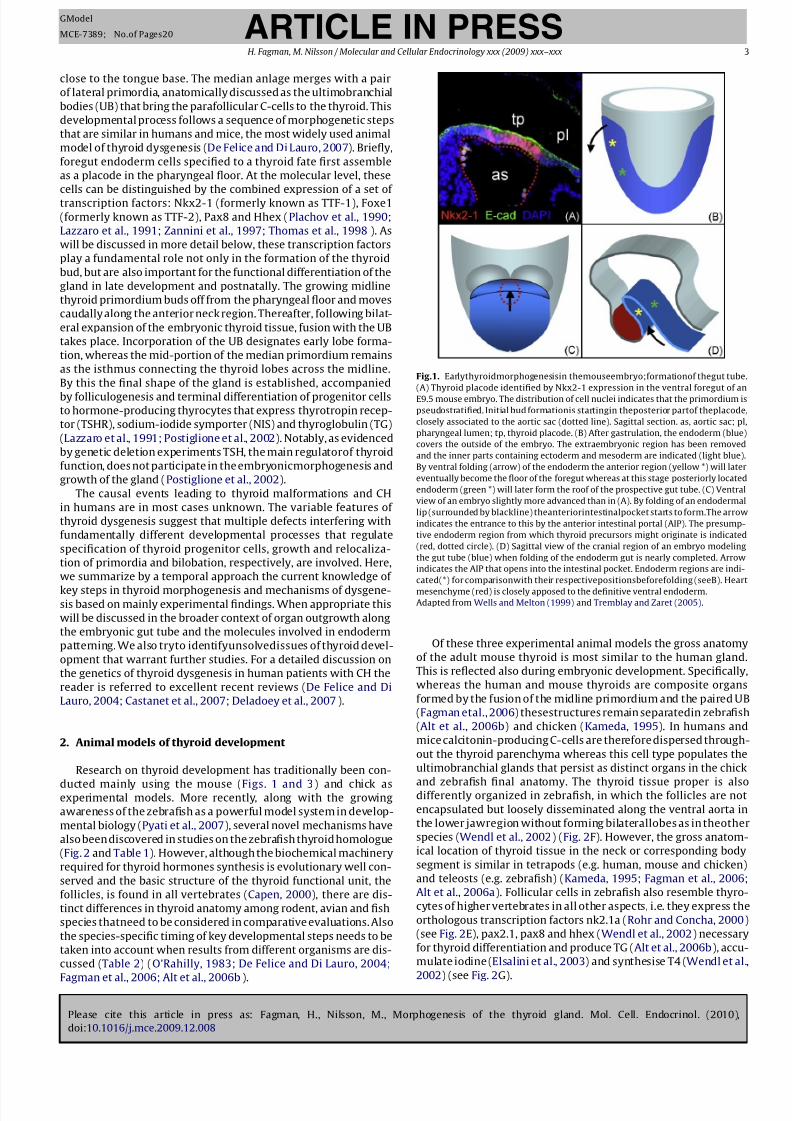
Fig.1. Earlythyroidmorphogenesisin themouseembryo;formationof thegut tube.
(A) Thyroid placode identified by Nkx2-1 expression in the ventral foregut of an
E9.5 mouse embryo. The distribution of cell nuclei indicates that the primordium is
pseudostratified. Initial bud formationis startingin theposterior partof theplacode,
closely associated to the aortic sac (dotted line). Sagittal section. as, aortic sac; pl,
pharyngeal lumen; tp, thyroid placode. (B) After gastrulation, the endoderm (blue)
covers the outside of the embryo. The extraembryonic region has been removed
and the inner parts containing ectoderm and mesoderm are indicated (light blue).
By ventral folding (arrow) of the endoderm the anterior region (yellow *) will later
eventually become the floor of the foregut whereas at this stage posteriorly located
endoderm (green *) will later form the roof of the prospective gut tube. (C) Ventral
view of an embryo slightly more advanced than in (A). By folding of an endodermal
lip (surrounded by blackline) theanteriorintestinalpocket starts to form.The arrow
indicates the entrance to this by the anterior intestinal portal (AIP). The presump-
tive endoderm region from which thyroid precursors might originate is indicated
(red, dotted circle). (D) Sagittal view of the cranial region of an embryo modeling
the gut tube (blue) when folding of the endoderm gut is nearly completed. Arrow
indicates the AIP that opens into the intestinal pocket. Endoderm regions are indi-
cated(*) for comparisonwith their respectivepositionsbeforefolding (seeB). Heart
mesenchyme (red) is closely apposed to the definitive ventral endoderm.Adapted from Wells and Melton (1999) and Tremblay and Zaret (2005).
Of these three experimental animal models the gross anatomy
of the adult mouse thyroid is most similar to the human gland.
This is reflected also during embryonic development. Specifically,
whereas the human and mouse thyroids are composite organs
formed by the fusion of the midline primordium and the paired UB
(Fagman etal., 2006) thesestructures remain separatedin zebrafish
(Alt et al., 2006b) and chicken (Kameda, 1995). In humans and
mice calcitonin-producing C-cells are therefore dispersed through-
out the thyroid parenchyma whereas this cell type populates the
ultimobranchial glands that persist as distinct organs in the chick
and zebrafish final anatomy. The thyroid tissue proper is alsodifferently organized in zebrafish, in which the follicles are not
encapsulated but loosely disseminated along the ventral aorta in
the lower jawregion without forming bilaterallobes as in theother
species (Wendl et al., 2002) (Fig. 2F). However, the gross anatom-
ical location of thyroid tissue in the neck or corresponding body
segment is similar in tetrapods (e.g. human, mouse and chicken)
and teleosts (e.g. zebrafish) (Kameda, 1995; Fagman et al., 2006;
Alt et al., 2006a). Follicular cells in zebrafish also resemble thyro-
cytes of higher vertebrates in all other aspects, i.e. they express the
orthologous transcription factors nk2.1a (Rohr and Concha, 2000)
(see Fig. 2E), pax2.1, pax8 and hhex (Wendl et al., 2002) necessary
for thyroid differentiation and produce TG (Alt et al., 2006b), accu-
mulate iodine (Elsalini et al., 2003) and synthesise T4 (Wendl et al.,
2002) (see Fig. 2G).
8/10/2019 Molecular Morfgenesis de la glandula Tiroidea.pdf
http://slidepdf.com/reader/full/molecular-morfgenesis-de-la-glandula-tiroideapdf 4/20
Please cite this article in press as: Fagman, H., Nilsson, M., Morphogenesis of the thyroid gland. Mol. Cell. Endocrinol. (2010),
doi:10.1016/j.mce.2009.12.008
ARTICLE IN PRESSGModel
MCE-7389; No.of Pages20
4 H. Fagman, M. Nilsson / Molecular and Cellular Endocrinology xxx (2009) xxx–xxx
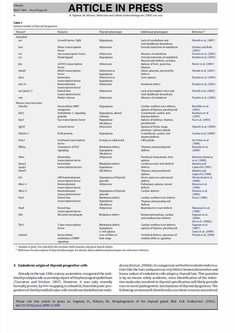
Table 1
Animal models of thyroid dysgenesis.
Mutanta Features Thyroid phenotype Additional phenotypes Referenceb
Zebrafish
ace Growth factor; Fgf8 Hypoplasia Lack of cerebellum and
mid-hindbrain-boundrary
Wendl et al. (2007)
bon Mixer transcription
factor
Athyreosis Overall reduction of endoderm Elsalini and Rohr
(2003)
cas Sox transcription factor Athyreosis Absence of endoderm Elsalini et al. (2003)
cyc Nodal ligand Hypoplasia Overall reduction of endoderm Elsalini et al. (2003)Neural tube defects, cyclopia
fau GATA5 transcription
factor
Athyreosis Aplasia of liver, pancreas,
thymus
Reiter et al. (2001)
hand2 bHLH transcription
factor
Athyreosis or
hypoplasia
Heart, pharynx, pectoral fin
defects
Wendl et al. (2007)
hhex homeobox
transcription factor
Athyreosis or
hypoplasia
Liver aplasia Elsalini et al. (2003)
nk2.1a Homeodomain
transcription factor
Athyreosis Forebrain defect Elsalini et al. (2003)
noi (pax2.1) Paired-box
transcription factor
Athyreosis Lack of pronephric duct and
mid-hindbrain-boundrary
Wendl et al. (2002)
oep Nodal cofactor Athyreosis Absence of endoderm Elsalini et al. (2003)
Mouse (mus musculus)
Chordin Extracellular BMP
antagonist
Hypoplasia Cardiac outflow tract defects,
aplasia of thymus, parathyroid
Bachiller et al.
(2003)
Edn1 Endothelin-1; signaling
peptide
Hypoplasia, absent
isthmus
Craniofacial, cardiac and
thymus defects
Kurihara et al.
(1995)Eya1 Eya transcription factor Hypoplasia Aplasia of kidneys, thymus,
parathyroid
Xu et al. (2002)
UB defects
Fgf10 Growth factor Athyreosis Aplasia of limbs, lungs,
pituitary, salivary glands
Ohuchi et al. (2000)
Fibulin-1 ECM protein Hypoplasia Craniofacial, cardiac and
thymus defects
Cooley et al. (2008)
Foxe1 Forkhead transcription
factor
Ecto pia o r athy re os is Clef t palate De Felice et al.
(1998)
FRS2˛ Transducer of FGF
signaling
Bilobation defect,
hypoplasia
Thymus and parathyroid
defects
Kameda et al.
(2009)
UB defects
Hhex Homeobox
transcription factor
Athyreosis Forebrain truncations, liver
aplasia
Martinez Barbera
et al. (2000)
Hoxa3 Homeobox
transcription factors
Bilobation defect,
hypoplasia
Cardiovascular and skeletal
defects
Manley and
Capecchi (1995)Hoxb3
Hoxd3 UB defects Thymus and parathyroid
aplasia
Manley and
Capecchi (1998)Isl1 LIM homeodomain
transcription factor
Hypoplasia of thyroid
placode
Heart, pancreas and neural
defects
Westerlund et al.
(2008)
Nkx2-1 Homeodomain
transcription factor
Athyreosis Pulmonary aplasia, neural
defects
Kimura et al.
(1996)
Nkx2-5 Homeodomain
transcription factor
Hypoplasia of thyroid
placode
Cardiac defects Dentice et al.
(2006)
Pax3 Paired-box
transcription factor
Bilobation defect,
hypoplasia
Cardiac outflow tract defects Franz (1989)
UB defects
Thymus and parathyroid
defects
Pax8 Paired-box
transcription factor
Athyreosis Reproductive tract defects Mansouri et al.
(1998)
Shh Secreted morphog en Bil obation defect Holoprosencephaly, cardiac
and outflow tract defects
Fagman et al.
(2004)
Alt et al. (2006a)
Tbx1 T-box transcription
factor
Bilobation defect,
hypoplasia
Cardiac outflow tract defects,
aplasia of thymus, parathyroid
Fagman et al.
(2007)
C-cell aplasia Lania et al. (2009)
twisted Extracellular
modulator of BMP
signaling
Loss of Hhex at
bud-stage
Vertebral defects, spectrum of
midline defects, agnathia
Petryk et al. (2004)
a Symbol or gene. For zebrafish this includes both mutants and gene knock-down.b Reference for description of thyroid phenotype, for details about additional phenotypes see references therein.
3. Endoderm origin of thyroid progenitor cells
Already in the late 19th century anatomists recognized the mid-
linethyroidplacode as an integralpart of thepharyngeal epithelium
(Tourneux and Verdun, 1897). However, it was only recently
formally proven, by fate-mapping in zebrafish, thatembryonic pro-
genitorsof thethyroidfollicular cells stemfrom thedefinitive endo-
derm(Altetal.,2006b). In comparison withotherendodermderiva-
tives like the liver andpancreas very littleis knownaboutwhen and
howa subset of endoderm cells adoptsa thyroid fate. This question
is by no means solely academic, since identification of the induc-
tive molecules involved in thyroid specification will likely provide
cues to novel pathogenetic mechanisms of thyroid dysgenesis. The
following sectionswill thereforediscuss these issuesin moredetail.
8/10/2019 Molecular Morfgenesis de la glandula Tiroidea.pdf
http://slidepdf.com/reader/full/molecular-morfgenesis-de-la-glandula-tiroideapdf 5/20
Please cite this article in press as: Fagman, H., Nilsson, M., Morphogenesis of the thyroid gland. Mol. Cell. Endocrinol. (2010),
doi:10.1016/j.mce.2009.12.008
ARTICLE IN PRESSGModel
MCE-7389; No.of Pages20
H. Fagman, M. Nilsson / Molecular and Cellular Endocrinology xxx (2009) xxx–xxx 5
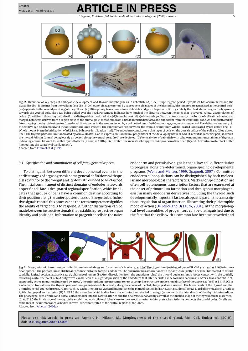
Fig. 2. Overview of key steps of embryonic development and thyroid morphogenesis in zebrafish. (A) 1-cell stage, zygote period. Cytoplasm has accumulated and the
blastodisc (bd) is distinct from the yolk sac (ys). (B) 16-Cell stage, cleavage period. By subsequent cleavages of the blastodisc, blastomeres are generated at the animal pole
(an) opposite to the vegetal pole (veg)of the yolk sac. (C) 50%-epiboly, transitionbetween blastula and gastrula periods. During epiboly the blastoderm progressively spreads
towards the vegetal pole, like a cap being pulled over the head. Percentage indicates how much of the distance between the poles that is covered. A local accumulation of
cells at (*)will form theembryonic shield that distinguishes thedorsal side (d)fromthe ventral (v)of theembryo.Gastrulationoccursby involution of cells at theblastoderm
margin. Ectoderm derives from a region close to the animal pole, mesoderm from a broad intermediate area and endoderm from the equatorial zone. As demonstrated by
fate-mapping the thyroid originates from dorsal blastomeres in the area encircled by a red dotted line. (D) 6-Somite stage, segmentation period. The definitive anatomy of the embryo can be discerned and the optic primordium is evident. The approximate region where the thyroid primordium will be located is indicated by red dotted line. (E)
Whole mount in situ hybridization of nk2.1a at 24 h post-fertilization (hpf). The endoderm constitutes a thin layer of cells on the dorsal surface of the yolk sac (blue dotted
line). The thyroid primordium is indicated by arrow. Rostral nk2.1a expression is in neural progenitors of the developing brain. (F) Adult zebrafish (anterior part) in which
the thyroid follicles (green) being loosely dispersed along the ventral aorta (red) are depicted. (G) Ventral view of zebrafish with whole mount immunostaining of thyroxin
indicating accumulationof T4 in thethyroidfollicles (arrow) at 120hpf.Red dottedline indicatesthe approximate position of theheart (h)and theventralaorta, black dotted
lines outline the ceratohyal cartilages (ch).
Adapted from Kimmel et al. (1995).
3.1. Specification and commitment of cell fate—general aspects
To distinguish between different developmental events in the
earliest stages of organogenesis some general definitions with spe-
cial reference to the foregut and its derivatives need to be clarified.
The initial commitment of distinct domains of endoderm towardsa specific cell fate is designated regional specification, which impli-
cates that groups of cells have a common destiny according to
their position along the anteroposterior axis of the guttube. Induc-
tive signals control this process and the term competence signifies
the ability of target cells to respond. A further distinction can be
made between instructive signals that establish prospective organ
identity and positional information to progenitor cells in the naïve
endoderm and permissive signals that allow cell differentiation
to progress along pre-determined, organ-specific developmental
programs (Wells and Melton, 1999; Spagnoli, 2007). Committed
endoderm subpopulations can be distinguished by both molecu-
lar and morphological characteristics. Markers of specification are
often cell-autonomous transcription factors that are expressed atthe onset of primordium formation and throughout morphogen-
esis; in many endoderm derivatives including the thyroid such
developmentally important factors alsoparticipatein thetranscrip-
tional regulation of organ function, illustrating their pleiotrophic
mode of action (De Felice and Di Lauro, 2004). At the morpholog-
ical level assemblies of progenitors can be distinguished due to
the fact that the cells with a common fate become crowded and
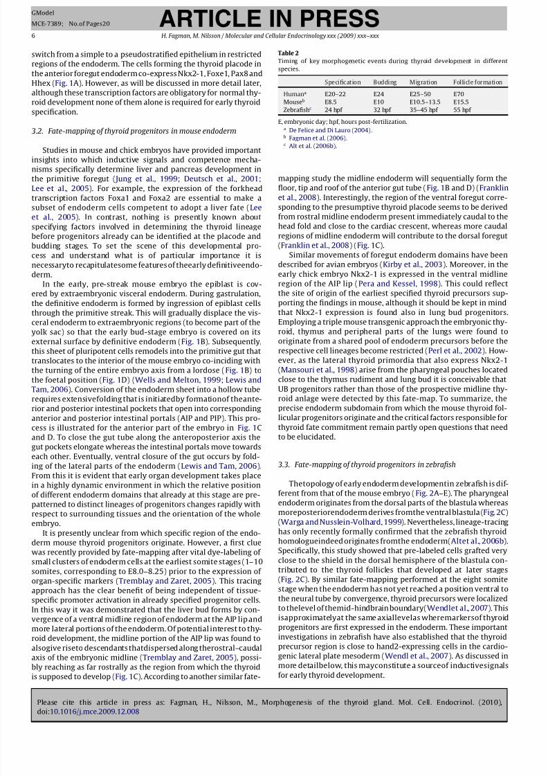
Fig. 3. Dissociationof themouse thyroid budfrom theendoderm andformation ofa bilobed gland.(A) Thethyroidbud (evidenced by redNkx2-1 staining)at E10.5 ofmouse
development. The primordium is still broadly connected to the foregut endoderm. The bud maintains association with the aortic sac (dotted line) that has started to retract
caudally. Sagittal section. as, aortic sac; pl, pharyngeal lumen. (B) After dissociation from the endoderm (blue) the thyroid bud transiently looses contact with the caudally
retracting aorta. The point of bud outgrowth can be seen as a slight depression of the endoderm that later persists as the foramen caecum (*). After a transient phase of
supposedly active migration (indicated by arrow), the primordium (green) comes to rest as a cap-like structure on the cranial surface of the aortic sac (red) at E11.5. (C) In
a schematic, frontal view the thyroid primordium (green) extends bilaterally along the course of the 3rd pharyngeal arch arteries. The lateral ends of the thyroid and the
ultimobranchial bodies (brown) are approaching eachother (arrow). Dotted lineindicatesthe planeof section in (B).Ao, aorta; D, dorsal aorta; 3, 3rd pharyngealarch arteries;
4, 4th pharyngeal arch arteries. (D) At E13.5 the ultimobranchial bodies have made contact and started to merge (arrow) with the lateral ends of the thyroid primordium.
The pharyngeal arch arteries and dorsal aorta remodel into the carotid arteries and the final vascular anatomy as well as the bilobed shape of the thyroid can be discerned.
(E) At E18.5 the final shape of the thyroid is established with bilateral lobes close to the carotid arteries. A thin, pretracheal isthmus connects the caudal poles. C-cells and
remnants of the ultimobranchial bodies (brown) are concentrated to the central regions of the lobes.
Adapted from Alt et al. (2006a).
8/10/2019 Molecular Morfgenesis de la glandula Tiroidea.pdf
http://slidepdf.com/reader/full/molecular-morfgenesis-de-la-glandula-tiroideapdf 6/20
Please cite this article in press as: Fagman, H., Nilsson, M., Morphogenesis of the thyroid gland. Mol. Cell. Endocrinol. (2010),
doi:10.1016/j.mce.2009.12.008
ARTICLE IN PRESSGModel
MCE-7389; No.of Pages20
6 H. Fagman, M. Nilsson / Molecular and Cellular Endocrinology xxx (2009) xxx–xxx
switch from a simple to a pseudostratified epithelium in restricted
regions of the endoderm. The cells forming the thyroid placode in
the anterior foregut endoderm co-express Nkx2-1, Foxe1, Pax8 and
Hhex (Fig. 1A). However, as will be discussed in more detail later,
although these transcription factors are obligatory for normal thy-
roid development none of them alone is required for early thyroid
specification.
3.2. Fate-mapping of thyroid progenitors in mouse endoderm
Studies in mouse and chick embryos have provided important
insights into which inductive signals and competence mecha-
nisms specifically determine liver and pancreas development in
the primitive foregut ( Jung et al., 1999; Deutsch et al., 2001;
Lee et al., 2005). For example, the expression of the forkhead
transcription factors Foxa1 and Foxa2 are essential to make a
subset of endoderm cells competent to adopt a liver fate (Lee
et al., 2005). In contrast, nothing is presently known about
specifying factors involved in determining the thyroid lineage
before progenitors already can be identified at the placode and
budding stages. To set the scene of this developmental pro-
cess and understand what is of particular importance it is
necessaryto recapitulatesome features of theearly definitiveendo-
derm.In the early, pre-streak mouse embryo the epiblast is cov-
ered by extraembryonic visceral endoderm. During gastrulation,
the definitive endoderm is formed by ingression of epiblast cells
through the primitive streak. This will gradually displace the vis-
ceral endoderm to extraembryonic regions (to become part of the
yolk sac) so that the early bud-stage embryo is covered on its
external surface by definitive endoderm (Fig. 1B). Subsequently,
this sheet of pluripotent cells remodels into the primitive gut that
translocates to the interior of the mouse embryo co-inciding with
the turning of the entire embryo axis from a lordose (Fig. 1B) to
the foetal position (Fig. 1D) (Wells and Melton, 1999; Lewis and
Tam, 2006). Conversion of the endoderm sheet into a hollow tube
requires extensivefolding that is initiatedby formationof theante-
rior and posterior intestinal pockets that open into correspondinganterior and posterior intestinal portals (AIP and PIP). This pro-
cess is illustrated for the anterior part of the embryo in Fig. 1C
and D. To close the gut tube along the anteroposterior axis the
gut pockets elongate whereas the intestinal portals move towards
each other. Eventually, ventral closure of the gut occurs by fold-
ing of the lateral parts of the endoderm (Lewis and Tam, 2006).
From this it is evident that early organ development takes place
in a highly dynamic environment in which the relative position
of different endoderm domains that already at this stage are pre-
patterned to distinct lineages of progenitors changes rapidly with
respect to surrounding tissues and the orientation of the whole
embryo.
It is presently unclear from which specific region of the endo-
derm mouse thyroid progenitors originate. However, a first cluewas recently provided by fate-mapping after vital dye-labeling of
small clusters of endoderm cells at the earliest somite stages (1–10
somites, corresponding to E8.0–8.25) prior to the expression of
organ-specific markers (Tremblay and Zaret, 2005). This tracing
approach has the clear benefit of being independent of tissue-
specific promoter activation in already specified progenitor cells.
In this way it was demonstrated that the liver bud forms by con-
vergence of a ventral midline region of endoderm at the AIP lip and
more lateral portions of the endoderm. Of potential interest to thy-
roid development, the midline portion of the AIP lip was found to
alsogive riseto descendants thatdispersed along therostral–caudal
axis of the embryonic midline (Tremblay and Zaret, 2005), possi-
bly reaching as far rostrally as the region from which the thyroid
is supposed to develop (Fig. 1C). According to another similar fate-
Table 2
Timing of key morphogenetic events during thyroid development in different
species.
Specification Budding Migration Follicle formation
Humana E20–22 E24 E25–50 E70
Mouseb E8.5 E10 E10.5–13.5 E15.5
Zebrafishc 24 hpf 32 hpf 35–45 hpf 55 hpf
E, embryonic day; hpf, hours post-fertilization.a De Felice and Di Lauro (2004).b Fagman et al. (2006).c Alt et al. (2006b).
mapping study the midline endoderm will sequentially form the
floor, tip and roof of the anterior gut tube (Fig. 1B and D) (Franklin
et al., 2008). Interestingly, the region of the ventral foregut corre-
sponding to the presumptive thyroid placode seems to be derived
from rostral midline endoderm present immediately caudal to the
head fold and close to the cardiac crescent, whereas more caudal
regions of midline endoderm will contribute to the dorsal foregut
(Franklin et al., 2008) (Fig. 1C).
Similar movements of foregut endoderm domains have been
described for avian embryos (Kirby et al., 2003). Moreover, in the
early chick embryo Nkx2-1 is expressed in the ventral midlineregion of the AIP lip (Pera and Kessel, 1998). This could reflect
the site of origin of the earliest specified thyroid precursors sup-
porting the findings in mouse, although it should be kept in mind
that Nkx2-1 expression is found also in lung bud progenitors.
Employing a triple mouse transgenic approach the embryonic thy-
roid, thymus and peripheral parts of the lungs were found to
originate from a shared pool of endoderm precursors before the
respective cell lineages become restricted (Perl et al., 2002). How-
ever, as the lateral thyroid primordia that also express Nkx2-1
(Mansouri et al., 1998) arise from the pharyngeal pouches located
close to the thymus rudiment and lung bud it is conceivable that
UB progenitors rather than those of the prospective midline thy-
roid anlage were detected by this fate-map. To summarize, the
precise endoderm subdomain from which the mouse thyroid fol-licular progenitors originate and the critical factors responsible for
thyroid fate commitment remain partly open questions that need
to be elucidated.
3.3. Fate-mapping of thyroid progenitors in zebrafish
Thetopology of early endoderm developmentin zebrafish is dif-
ferent from that of the mouse embryo (Fig. 2A–E). The pharyngeal
endoderm originates from the dorsal parts of the blastula whereas
moreposteriorendoderm derives fromthe ventral blastula (Fig. 2C)
(Warga and Nusslein-Volhard, 1999). Nevertheless, lineage-tracing
has only recently formally confirmed that the zebrafish thyroid
homologueindeed originates fromthe endoderm(Altet al., 2006b).Specifically, this study showed that pre-labeled cells grafted very
close to the shield in the dorsal hemisphere of the blastula con-
tributed to the thyroid follicles that developed at later stages
(Fig. 2C). By similar fate-mapping performed at the eight somite
stage when the endoderm has not yet reached a position ventral to
the neural tube by convergence, thyroid precursors were localized
to thelevel of themid-hindbrain boundary(Wendlet al., 2007). This
isapproximatelyat the same axiallevelas wheremarkersof thyroid
progenitors are first expressed in the endoderm. These important
investigations in zebrafish have also established that the thyroid
precursor region is close to hand2-expressing cells in the cardio-
genic lateral plate mesoderm (Wendl et al., 2007). As discussed in
more detailbelow, this mayconstitute a sourceof inductivesignals
for early thyroid development.
8/10/2019 Molecular Morfgenesis de la glandula Tiroidea.pdf
http://slidepdf.com/reader/full/molecular-morfgenesis-de-la-glandula-tiroideapdf 7/20
Please cite this article in press as: Fagman, H., Nilsson, M., Morphogenesis of the thyroid gland. Mol. Cell. Endocrinol. (2010),
doi:10.1016/j.mce.2009.12.008
ARTICLE IN PRESSGModel
MCE-7389; No.of Pages20
H. Fagman, M. Nilsson / Molecular and Cellular Endocrinology xxx (2009) xxx–xxx 7
3.4. Nodal signaling and early regulation of thyroid size
At the molecular level, formation of the definitive endoderm
and its further development in vertebrates are regulated by a well-
conserved signaling network where members of the Nodal family
play a superior role (Tam et al., 2003). Again, much of the signaling
pathways have been elucidated in mouse and zebrafish embryo
models. The crucial importance of Nodal signaling is evident in
zebrafish mutants of the Nodal cofactor one-eyed-pinhead (oep)
in which the endoderm is not specified and a gut tube fails to
form. Consequently, no thyroid primordium can be found in this
mutant (Elsalini et al., 2003). Considering the global effect of Nodal
on endoderm development it can be anticipated that also muta-
tions of downstream components of Nodal signaling affect thyroid
morphogenesis. Indeed, the thyroid anlage is completely absent
in cas and bon mutants (Elsalini et al., 2003), in which activation
of Sox17, a downstream effector of Nodal crucial for the expres-
sion of endoderm specific genes is disrupted (Aoki et al., 2002;
Tam et al., 2003; Sinner et al., 2004). It is probable that the thy-
roid phenotype merely is secondary to the global disturbance of
endoderm formation rather than being thyroid-specific. However,
thyroid specification may indeed be more specifically sensitive to
disruption of Nodal-signaling components, suggested by findings
in zebrafish mutants of the GATA homologue faust ( fau), which isa downstream target of Nodal that activates Sox17. In this mutant
nk2.1 (the Nkx2-1 ortholog) expression is lost in the region of the
prospective thyroidprimordium whereas at thesame timeearly gut
tube formation is only mildly affected (Reiter et al., 2001). Notably,
whereas ablation of Sox17 in mouse embryos leads to increased
apoptosis in the foregut endoderm, specification and initial mor-
phogenesis of the thyroid takes place also in the absence of Sox17
(Kanai-Azuma et al., 2002). This suggests thatGATA signaling influ-
ences thyroid progenitors by additional mechanisms.
In cyclops (cyc ) mutants, targeting one of the zebrafish Nodal
ligands, the thyroid primordium is specified but smaller than in
wildtype embryos, presumably reflecting an overall reduction of
pharyngeal endoderm(Elsaliniet al.,2003). Intriguingly,in latethy-
roid development the number of follicles is reduced in this mutant.This suggests that the final thyroid size is constrained by the num-
ber of cells initially recruited to a thyroid fate. Other examples
of thyroid hypoplasia associated with a reduced size of the early
primordium are the hand2 hypomorphic allele (hanc99) and fgf8
mutant (ace) in zebrafish (Wendl et al., 2007) and the Tbx1 null
mouse embryo (Lania et al., 2009), the effects of which will be
discussed in further detail below. Direct evidence for this novel
concept of developmental regulation comes from a recent study
demonstrating that the number of progenitor cells in the pancre-
atic budis limiting final organ size (Stanger et al., 2007). In contrast,
embryonic liver growth seems to be able to fully compensate for
a reduction of the hepatic progenitor pool, possibly reflecting the
inborn regenerative capacity of the adult liver. Notably, in the
absence of goitrogenous stimuli the thyroid regenerates poorly if alarge portion is surgically excised (Clark et al., 1976). It may thus
be speculated that a reduction of the precursor cell number ini-
tially specified determines the total growth capability of thyroid
tissue and the final size of the gland. Such a mechanism could pos-
siblyunderlie thyroidhypoplasia as a cause of hypothyroidism (Van
Vliet, 2003).
4. Positioning of the thyroid primordium
This section will discuss another but yet related aspect of early
thyroid development, namely the anatomical localization of the
definitiveprimordium and factors that areenvisaged to play a deci-
sive role. Formation of the mouse primitive gut tube is completed
after turning of the embryoat approximately E8.5. At this stage the
endoderm appears as a homogenous monolayered epithelium yet
lacking obvious signs of specialization at the prospective sites of
organ primordium outgrowth. However, the journey towards ulti-
mate fate decision of pluripotent progenitors is initiated already
as the definitive endoderm exits the primitive streak (Wells and
Melton, 1999). The earliest recruited endodermal cells tend to
contribute to theforegutwhereascells recruitedlater willpreferen-
tially occupy the mid- and hindguts (Lawson and Pedersen, 1987).
The endoderm is thus probably loosely regionalized before the dif-
ferentorganbuds start to develop along the anteroposterior axis of
the gut. The mechanisms by which this positional identity of pro-
genitor cells is established in the endoderm are largely unknown,
although signals from adjacent embryonic tissues, e.g. mesoderm
and notochord appear to be important in restricting the emerging
progenitor domains (Wells and Melton, 2000).
Mouse thyroid precursors come to populate a region of the ven-
tral endoderm of the anterior foregut that anatomically forms the
pharyngeal floor. So far no factors regulating the recruitment of
cells to the prospective thyroid placode in mouse embryos have
been identified. In zebrafish,retinoicacid (RA) impacts on theearly
regionalization of the endoderm and abrogation of RA signaling
leads to posterior expansion of anterior cell fate. This influences
also the position of the thyroid primordium, identified by nk2.1aand hhex expression, which is shifted posteriorly (Stafford and
Prince, 2002). IncreasedRA activity, obtainedby exposure to exoge-
nously added RA, conversely results in an expansion of posterior
endoderm cell fate. Interestingly, this is accompanied by a loss of
the thyroid expression domain of hhex, supposedly due to a reas-
signment of positional identity (Stafford and Prince, 2002). These
observations in zebrafish are most likely due to positional regula-
tion of thyroid progenitors rather than being the result of altered
thyroid fate of pluripotent endoderm. Also in mouse embryos,
the thyroid primordium develops in a region of low endogenous
RA activity indicated by the absence of Raldh2 expression and
RA-reporter gene activation (Desai et al., 2004). In line with this,
blocking of RA signaling in mouse embryoexplants at a stage when
initial patterning is already established does not influence the pro-gression of early thyroid development, whereas at the same time
RA-dependent lung bud induction is lost. Notably, in this situation
the Nkx2-1 expression is markedly attenuated in the prospective
lung bud but unaffected in the thyroid primordium (Desai et al.,
2004). This suggests differential requirements of RA signaling for
Nkx2-1 expression in these adjacent precursor fields.
In the chick embryo FGF-mediated patterning of the endoderm
might be of relevance to thyroid positioning. FGF4 from the meso-
derm regulates positional identity along the gut tube as indicated
by the expression of Sprouty1/2 in the prospective hindgut. Expo-
sure toexogenous FGF4 results inan anteriorshift of theseposterior
markers and repression of Hhex and Nkx2-1 in the anterior endo-
derm (Dessimoz et al., 2006). Conversely, inhibition of the FGF
receptor signaling pathway up-regulates and shifts Hhex poste-riorly. As FGF2 and FGF8 are unable to reproduce this effect it is
likely specific to FGF4. Since Hhex is required for the thyroid budto
develop normally(MartinezBarberaet al.,2000) FGF4 can thus pos-
siblymodulate the territory where thyroid progenitors assemble at
least partly by regulation of Hhex.
Exclusion of morphogenetic signals may also be of importance
for endoderm patterning and positioning of organ buds. One of the
bestexamplesis the locally repressedexpressionof Sonic hedgehog
(Shh) in the endoderm region from which the dorsal pancreas bud
forms. This is regulated by FGF2 and activin from the nearby noto-
chord, whichthus creates a boundary to pancreatic specification of
pluripotent endodermprogenitors(Hebroket al., 1998). The signifi-
cance is illustrated by the factthat more widespread Shh repression
leads to ectopic pancreatic differentiation (Kim and Melton, 1998)
8/10/2019 Molecular Morfgenesis de la glandula Tiroidea.pdf
http://slidepdf.com/reader/full/molecular-morfgenesis-de-la-glandula-tiroideapdf 8/20
Please cite this article in press as: Fagman, H., Nilsson, M., Morphogenesis of the thyroid gland. Mol. Cell. Endocrinol. (2010),
doi:10.1016/j.mce.2009.12.008
ARTICLE IN PRESSGModel
MCE-7389; No.of Pages20
8 H. Fagman, M. Nilsson / Molecular and Cellular Endocrinology xxx (2009) xxx–xxx
and that targeted over-expression of Shh in the prospective pan-
creasprimordium partly diverts the lineage program of progenitors
towards an intestinal fate (Apelqvist et al., 1997). That Shh might
play a similar role in thyroid development by restricting the endo-
derm adopting a thyroid fate is suggested by the finding that Shh
is strongly expressed in the adjacent foregut epithelium but not at
all in the Nkx2-1-expressing progenitor cells forming the thyroid
placode (Fagman et al., 2004; Parlato et al., 2004; Moore-Scott and
Manley, 2005). However, the size of the early thyroid bud in Shh
deficientmiceis comparable tothatof thewildtype anlage (Fagman
et al., 2004; Parlato et al., 2004). Instead Shh may prevent ectopic
differentiation of thyroid cells in more distant parts of the foregut
andits derivatives as the prospective trachea (Fagmanet al., 2004).
The thyroid phenotype in Shh null mice as an animal model of thy-
roid dysgenesis will be further commented on below. Clearly, the
exact role of Shh in thyroid development is far from completely
understood. Notably, Shh is able to diffuse over considerable dis-
tances (Gritli-Linde et al., 2001) and although Shh is not expressed
by endoderm cells committed to a thyroid fate these might still be
targets of Shh morphogenetic signals since they express the Shh
receptor Ptc1 (Washington Smoak et al., 2005). The importance of
this morphogen as a key regulator of endoderm patterning and gut
organogenesishas recently beenexcellently reviewed,highlighting
the profound and diverse activities of Shh throughout embryonicdevelopment and beyond (van den Brink, 2007).
5. Induction of a thyroid fate
Accumulating evidence indicates that the different endoderm
cell lineages are induced by instructive signals from surrounding
embryonic tissues. As already pointed out, the anterior foregut
endoderm is in close apposition to the pre-cardiac mesoderm that
gives a spatial foundation forreciprocal interactions(see Fig.1D).By
tissue recombination experiments it has for long been appreciated
that that liver development depends on soluble factors produced
by the cardiac mesoderm (Douarin, 1975; Gualdi et al., 1996; Rossi
et al., 2001). Induction of a liver cell fate wasproven to be mediatedby FGF1 andFGF2 from this sourcewhereas subsequent outgrowth
of the liver bud from the endoderm depends on FGF8 ( Jung et al.,
1999). As the thyroid primordium develops from pharyngeal endo-
derm in close proximity to the visceral mesoderm that forms the
secondaryheart field it is conceivable thatthyroidinductive signals
may originate there. Also, this opens up the possibility that athyre-
osis could be due to a variety of aberrations leading to disturbed
juxtapositioning of the prospective thyroid field to the source of
inductive signals or to inappropriate levels of these rather than to
impaired function of a thyroid-specific master-gene.
5.1. Role of FGF and BMP signaling in early thyroid development
In mouse explant co-cultures, cardiac mesoderm induces theexpression of Nkx2-1 in ventral endoderm isolated at E7.5–8 (2–5
somites) (Serls et al., 2005). Also downstream targets of Nkx2-1
specific to the lung (surfactant protein C) and the thyroid (TG) are
induced.As previously describedfor the liver ( Jung et al., 1999), this
capacity of cardiac mesoderm is reproduced by exogenous FGF1 or
FGF2. Interestingly, the action is dose-dependent with lower FGF
concentrations inducing liver genes like albumin whereas higher
doses are required for Nkx2-1 expression. As FGF1 and 2 are able
to induce Nkx2-1 also in dorsal midgutendoderm the signal seems
to be instructivefor a widespread domainof pluripotent endoderm
progenitors (Serls et al., 2005).
The first direct evidence for a role of FGF in the induction of a
thyroid fate comes from studies in zebrafish (Wendl et al., 2007).
Exposure of early embryos to an inhibitor of FGF at a stage prior
to specification leads to lack of thyroid primordium development
without global defects in endoderm patterning. A similar pheno-
type is found in embryos mutant for the transcription factor hand2
that has been suggested to be upstream of FGF (Abe et al., 2002).
This effect is most likely non-cell-autonomous to the thyroid but
due to hand2 activity in the cardiac lateral plate mesoderm jux-
taposed to the thyroid anlage. The thyroid phenotype in hand2
mutants is partly rescued by implantation of FGF (1, 2, 8) soaked
beads in close apposition to the endoderm. Importantly, the posi-
tionof therescued thyroid primordium is similar to thatof wildtype
embryos, suggesting a permissiverather thaninstructiverole of the
FGF signal that is downstream or in parallel to hand2. The impor-
tance of FGF-signaling is further supported by the finding that ace
mutants deficient for FGF8 display severely hypoplastic thyroid
primordia (Wendl et al., 2007). This maycorrespond to the permis-
sive role of FGF8 in liver morphogenesis after a liver fate has been
induced in progenitor cells by FGF1 and FGF2 ( Jung et al., 1999).
Recent work in mouse models has substantiated and further
elaborated these findings. Disruption of the Shp2-binding sites
of the FGF receptor (FGFR) docking protein FRS2 causes thyroid
hypoplasia (Kamedaet al., 2009), indicating thatembryonic thyroid
growth is influenced by FGFR signaling. In view of the significant
impact genetic deletion of Tbx1, a transcriptional regulator of FGFs
(Vitelli et al., 2002), has on thyroid size in late morphogenesis(Fagman et al., 2007), early thyroid development was investigated
in embryos where Tbx1 was either generally deleted or specifically
targeted in the mesoderm (Mesp1Cre/+;Tbx1 fl/−) (Lania et al., 2009).
This showed that proliferation in the E8.5 foregut endoderm and
the number of cells in the thyroid placode is reduced by meso-
dermal ablation of Tbx1 suggesting a diminished thyroid precursor
cell pool size. These effect are largely mimicked by deletion of Fgf8 in the Tbx1 expression domain (Tbx1Cre/+;Fgf8 fl/−). Conversely,
over-expression of Fgf8 from the Tbx1 locus on a Tbx1 deficient
background (Tbx1Fgf8/−) rescues the thyroid size defect that is oth-
erwise severely hypoplastic in Tbx1 null embryos (Fagman et al.,
2007). An FGF8 signal that depends on Tbx1 likely emanates from
the mesoderm of the secondary heart field that is close to the thy-
roid primordium at early developmental stages. However, it is notyetclearif endodermal cells arethe directtargetsof this Fgf8 signal
as deletion of FGFR1 and FGFR2 in the endoderm does not produce
a thyroid defect (Lania et al., 2009).
Taken together, studies in zebrafish and mice firmly establish
that FGF-signaling is crucial to early thyroid development and that
the cardiac mesoderm is a probable source of inductive and per-
missive signals (Wendl et al., 2007; Lania et al., 2009). However,
the exact role of FGF in early stages of thyroid development needs
to be further clarified. This also accounts for bone morphogenetic
protein (BMP) signaling. BMPs and FGFs secreted from the septum
transversum mesenchyme workin concert to promote competence
of hepatic precursors as well as morphogenesis of the liver bud
(Rossi et al., 2001). It remains to be elucidated whether BMPs play
a similar role in early thyroid development. So far, the only indica-tion in this direction comes from observations in mice deficient
in Twisted, a context dependent modulator of BMP function. In
Twisted null embryos the expression of Hhex is diminished in the
liver primordium and cannot be detected in the presumptive thy-
roid primordium (Petryket al., 2004). BMP might thuscontribute to
Hhex-dependent development of the thyroid bud(the role of Hhex
will be further discussed below).
5.2. Is thyroid morphogenesis coordinated with heart
development?
Cardiac defects are overrepresented among children with thy-
roid dysgenesis (Olivieri et al., 2002). This together with the close
spatial relationship between the heart and thyroid primordial tis-
8/10/2019 Molecular Morfgenesis de la glandula Tiroidea.pdf
http://slidepdf.com/reader/full/molecular-morfgenesis-de-la-glandula-tiroideapdf 9/20
Please cite this article in press as: Fagman, H., Nilsson, M., Morphogenesis of the thyroid gland. Mol. Cell. Endocrinol. (2010),
doi:10.1016/j.mce.2009.12.008
ARTICLE IN PRESSGModel
MCE-7389; No.of Pages20
H. Fagman, M. Nilsson / Molecular and Cellular Endocrinology xxx (2009) xxx–xxx 9
sues suggest the possibility of common patterning traits in early
morphogenesis. There are several observations of concomitant car-
diac and thyroid defects also in experimental studies, supporting
this notion. Zebrafish embryos deficient in FGF8 display both car-
diac ventricle malformations (Reifers et al., 2000) and thyroid
hypoplasia (Wendl et al., 2007). The action of FGF8 on heart devel-
opment is probably mediated by the transcriptional activities of
Nkx2.5 and GATA4 in responding cardiac progenitor cells (Reifers
et al., 2000). Interestingly, Nkx2.5 is expressed in the pharyngeal
endoderm encompassing the thyroid primordium (Biben et al.,
2002) and Nkx2.5 mouse mutants exhibit a rudimentary thyroid
along with the cardiac defect (Dentice et al., 2006). Moreover, cer-
tain GATA4 sensitive enhancer elements of the mouse Nkx2.5 gene
are able to target expression specifically to both the heart and thy-
roid(Lien et al., 1999). Thissuggeststhat Nkx2.5 might be regulated
by FGF8 also in thyroid progenitors.
Another example of a developmentally important transcription
factor expressed in both thyroid and heart progenitor cells is Isl1
(Westerlund et al., 2008). Heart development is severely disturbed
in Isl1 deficient mouse embryos involving defective reciprocal
signaling between the cardiac mesoderm andthe pharyngeal endo-
derm (Cai et al., 2003). The thyroid bud is smaller in Isl1 null
mutant embryos (Westerlund et al., 2008). This may be the result
of decreased cell survival in the pharyngeal endoderm that is nor-mally supported by Isl1 (Cai et al., 2003). The mutant is embryonic
lethalat E10.5 dueto failure of thecardiovascular systemto further
develop. A possible additional role of Isl1 in thyroid organogen-
esis, as suggested by persisting expression of Isl1 in progenitor
cells during migration of primordia (Westerlund et al., 2008), is
presently not possible to appreciate. Nevertheless, these findings
suggest that co-incidental thyroid and heart malformations may
arise in parallel by loss or otherwise impaired function of com-
mon signalingpathways. Alternatively, one structure could depend
on correct signaling from the other for its early morphogenesis
to proceed normally. Of particular interest, avian cardiogenesis is
induced by anterior endoderm-derived signals (Schultheiss et al.,
1995; Lough and Sugi, 2000), suggesting that reciprocal interac-
tions between adjacent germ layers are important in the foregutregion. Whether this involves the thyroid progenitor zone, which
constitutes a relatively large portion of the endoderm facing the
secondary heart field and the presumptive cardiac outflow tract, is
an open intriguing question.
5.3. A role of endothelial cells in early thyroid development?
In addition to cardiac mesoderm much attention has been paid
to the possible involvement of embryonic vessels and particularly
endothelial cells as a source of inductive signals for endoderm-
derived organs. The best examples of this are found in pancreatic
and liver development. In a classical experiment, removal of the
dorsal aorta located close to the emerging dorsal pancreas was
found to block the expression of pancreas markers in the endo-derm (Lammert et al., 2001). Furthermore, tissue recombination
experiments showed that endothelial cells are able to induce a
pancreatic fate in isolated endoderm (Lammert et al., 2001). Simi-
lar results were at the same time provided for liver organogenesis
(Matsumoto et al., 2001). Together, this showed for the first time
that vessels not only provide metabolic substances, but also play
an active role in organ development. Using Flk1 deficient mice in
which endothelial cells do not develop it has subsequently been
demonstrated thatinitialpancreas induction occurs but emergence
of the pancreatic bud fails (Yoshitomi and Zaret, 2004). Of note,
the thyroid placode and the subsequent bud outgrowth are estab-
lishedin closecontact with theaorticsac. Infact,all Nkx2-1positive
thyroid progenitors assemble in the pharyngeal endoderm directly
apposing the aortic sac endothelium before budding take place
(Fig. 1A) (Fagman et al., 2006). This raises the question whether
factors generated in the vessel wall or provided by the local circu-
lation, as shownfor the embryonicpancreas (Edsbaggeet al., 2005),
may influence early thyroid development as well. However, avail-
able experimental data clearly indicate that thyroid specification
does not require a vascular signal. Specifically, in zebrafish Scl or
vegf mutants in which the vascular patterning of the pharyngeal
region including the thyroid specification zone of the endoderm is
severely disturbed thyroid progenitors are readily specified. Also
in cloche mutants lacking all vascular progenitors of the anterior
trunk the thyroid primordium is present (Altet al., 2006a). Thatthis
also may account for the embryonic mouse thyroid is suggested by
observations of a thyroid rudiment present in Tie2 mutants lacking
endothelial cells (Postiglione and di Lauro, personal communica-
tion). As will be highlighted later on, the nearness to embryonic
vessels is instead likely important for later stages of thyroid devel-
opment (Alt et al., 2006a; Fagman et al., 2007).
6. Regulation of thyroid budding
Assembly of thyroid progenitors in the pharyngeal endoderm is
soon followed by budding of the primordium as a whole. Budding
probably occurs concomitantly with the recruitment of new pro-genitor cells to the growing anlage (Smuts et al., 1978), signified
by the thyroid placode becoming increasingly crowded with cells
leading to a pseudostratified epithelium. As this process proceeds
the thyroid bud, or diverticulum, forms a caudally protruding, cup-
shaped evagination of the midline pharyngeal endoderm (Romert
and Gauguin, 1973; Fagman et al., 2006) (Fig. 3A). Eventually, the
descending bud detaches from the overlying endoderm by gradual
thinning and disintegration of the thyroglossal duct, which thus is
a transient embryonic structure.
Thyroid budding probably follows similar morphogenetic traits
as other budding organs of the endoderm and it can be assumed
thatsome common principles regarding theactionof the regulatory
molecules areat hand. However, unlike budding of,e.g. thesalivary
glands, lungs and pancreas, which develop as iterative generationsof branches maintaining physical contact with the region of pri-
mary bud emergence, the connection of the thyroid bud dissociates
completely andrapidly fromits site of origin. How bud detachment,
shared also by the parathyroid and thymus primordia, is regulated
is a yet largely unexplored aspect of thyroid development. Below
is discussed what we know to date and what can be learned from
studies on other budding organs.
6.1. Proliferation versus recruitment of progenitors
A locally enhanced proliferation of progenitor cells has been
considered an important driving force for bud outgrowth in sev-
eral primordial organs (Michael and Davies, 2004). Accordingly, the
endoderm of thelungshowsan increasedproliferation rate (Goldinet al., 1984) which is evident once the bud is partially formed and
not before (Nogawa et al., 1998). In contrast, an unexpectedly low
proliferation rate characterizes the early thyroid primordium in
mouse embryos, signified by the observation that both the pla-
code and the emerging bud contain only few BrdU positive cells
indicative of DNA synthesis (S-phase of cell cycle) as compared to
the immediate neighbouring endoderm and adjacent mesoderm
(Fagman et al., 2006). In fact, the fractionof cycling thyroid progen-
itors equals that of other highly proliferating tissues in the anterior
embryo first when the primordium has detached from the endo-
derm and started migration.
The cellular dynamics of thyroid bud formation has previously
been studied in greater detail in chick embryos. Following [3H]
thymidine incorporation it was demonstrated that the labeling
8/10/2019 Molecular Morfgenesis de la glandula Tiroidea.pdf
http://slidepdf.com/reader/full/molecular-morfgenesis-de-la-glandula-tiroideapdf 10/20
Please cite this article in press as: Fagman, H., Nilsson, M., Morphogenesis of the thyroid gland. Mol. Cell. Endocrinol. (2010),
doi:10.1016/j.mce.2009.12.008
ARTICLE IN PRESSGModel
MCE-7389; No.of Pages20
10 H. Fagman, M. Nilsson / Molecular and Cellular Endocrinology xxx (2009) xxx–xxx
index is significantly lower in the thyroid placode than in the adja-
cent endoderm (Smuts et al., 1978). The labeling pattern further
suggested that the bud size expands by annexation of proliferating
endoderm from outside the placode so that the central regions of
the bud consists of the first integrated progenitor cells that display
a lower rate of proliferation. The flow of cells from the periphery
into the evaginating primordium has been further corroborated by
detailed analysis of cell movements (Hilfer et al., 1990; Kinebrew
and Hilfer, 2001). Together, data from mouse and chicken studies
thus suggest that the endoderm portion engaged in the specifica-
tion of thyroid progenitors is larger than that appreciated by the
restricted expression of, e.g. Nkx2-1 in the thyroid placode and
early bud.
The mechanism of budding related to intraepithelial move-
ments of proliferating precursor cells was recently investigated in
the liver bud (Bort et al., 2006). This showed that the prehepatic
epithelium becomes pseudostratified by a mechanism resembling
asymmetric cell division originally identified in neuroepithelial
cells (Gotz and Huttner, 2005). In this process called “interkinetic
nuclear migration” cycling cellsundergo S-phase whenthe nucleus
has a more basal position and proceed into G2/M phase when it
moves apically towards the luminal surface of the epithelium. The
resulting cleavage at mitosis makes the daughter cells distribute
asymmetrically relative to the plane of the epithelial sheet and,importantly, inherit apical and basal constituents that may reflect
a first sign of differentiation towards distinct cell fates. A similar
pattern was noticed for the liver, pancreatic and lung buds (Bort et
al., 2006) suggesting that this may be a general feature of budding
organogenesis in the endoderm.
Pseudostratification possibly involving interkinetic nuclear
migration is also evident in thyroid placode and early bud for-
mation. [3H] thymidine incorporation in the chick thyroid bud
indicates that DNA synthesis occurs in the basal nuclear layers
whereas mitosis is completed in the apical layers (Smuts et al.,
1978). The mouse thyroid placode is also pseudostratified before
the bud is formed. However, the essential lack of BrdU positive
nuclei in mouse thyroid progenitors at the placode and budding
stages (Fagman et al., 2006) does not clearly fit with the modelof asymmetric cell division as a driving force of budding. Possibly,
mouse thyroid precursor cells have passed through S-phase in a
synchronized fashion before entering the placode and Nkx2-1 is
expressed. That the initiation of thyroid bud morphogenesis prob-
ably is differently regulated than in the liver is further suggested
fromobservationsin Hhex nullmouse embryos.In mutantsthe hep-
aticendodermremains as a columnar, simple epitheliumindicating
that Hhex is required for interkinetic nuclear migration and liver
bud formation (Bort et al., 2006). In contrast, although thyroid bud
outgrowth fails the thyroid placode is readily pseudostratified in
the absence of Hhex (Parlato et al., 2004). Irrespective of the mech-
anism, pseudostratification may be required but is not sufficient to
initiate thyroid budding.
6.2. Cytoskeletal remodeling
Contraction of the cortical cytoskeleton present in the apical
portion of epithelial cells is believed to generate a purse-string
action that promotes bud morphogenesis (Wessells et al., 1971;
Kiehart et al., 2000). This may be true also for the thyroid pla-
code in which prominent cytoskeletal bundles are concentrated
under the apical surfaces of the progenitor cells (Shain et al., 1972;
Romert and Gauguin, 1973). Indeed, in vitro experiments on iso-
lated foregut endoderm suggest that contraction of these bundles
is a morphogenetic force that contributes to thyroid evagination
(Hilferet al., 1977). The significance of cytoskeletal modificationsin
budding processes is further supported by the finding that branch-
ing morphogenesis of the ureteric bud is inhibited by blocking the
myosin activating Rho kinase ROCK (Michael et al., 2005). Recent
results from real-time imaging indicate that the apical constriction
of epithelial invaginations in Drosophila are more complex than a
simple purse-string mechanism and involves pulsed contractions
of an actin–myosin network that are stabilized by activity of the
transcription factors snail and twist (Martin et al., 2008). Thus,
cytoskeletal rearrangements participating in organ bud formation
should be considered a highly coordinated event under the con-
trol of specific developmentally important regulatory signals. This
further highlights the possibility that defective cytoskeletal regu-
lation might be a novel mechanism of thyroid dysgenesis yet to be
investigated.
6.3. Role of Fgf10
The molecular regulation of thyroid bud progression once initi-
ated is largely unknown. However, since the thyroid and lung buds
both express Nkx2-1 that is of crucial importance to further organ
development, it is likely that at least partly shared mechanisms
operate and that elucidation of a putative regulatory network in
the thyroid may be inspired by present knowledge of embryonic
lung morphogenesis. As recently reviewed (Kimura and Deutsch,
2007) this is controlled by an intricate network of endoderm- and
mesoderm-derived signals comprising, e.g. BMP4 produced by theendoderm tips of the lung buds and FGF10 secreted by the sur-
rounding mesenchyme that act in concert to drive further budding
and branching. Earlier studies on embryo explants have demon-
strated that FGF10 is capable of inducing lung budding with only
a modest effect on the proliferation of progenitor cells (Park et al.,
1998). More recent expression analysis in the embryonic lung has
shown that FGF10 induces genes associated with cell rearrange-
ment and migration but not genes involved in cell cycle regulation
(Lu et al., 2005). The possible relevance for thyroid development
is suggested from studies on FGF10 in several transgenic mouse
models; expression of a soluble, dominant-negative form of the
preferred FGF10 receptor, FGFR2IIIb, causes agenesis or dysgene-
sis of a wide range of organs including the thyroid and the lungs
(Celli et al., 1998) and FGF10 null mutant embryos lack both thethyroid and lungs (Ohuchi et al., 2000). However, a drawback is
that in these studies only late embryos (E18.5) or newborns were
analysed, which precludes any conclusions on the time window
whenFGF10 is required during thyroid morphogenesis or its mech-
anism. For example, FGF10 acts as a survival factor in developing
epithelia (Sala et al., 2006) and lack of FGF10 might thus result in
defective development due to increased cell death by apoptosis.
Nevertheless, recent findings pointing out Nkx2-1 as a mediator of
FGF10 morphogenetic signaling in the lung bud (Xing et al., 2008)
is of particular interest as Nkx2-1 is an early progenitor marker
in both lung and thyroid. Interestingly, over-expression of FGF10
induces ectopic expression of Nkx2-1and budding from theesoph-
agus endoderm (Sakiyama et al., 2003), suggesting a mechanistic
role in early development.The possibility that FGF10 in thethyroid
primordium regulates Nkx2-1 therefore needs to be investigated.
7. Survival and growth of thyroid progenitors depend on a
thyroid-specific signature of transcription factors
The combined expression of Nkx2-1, Pax8, Foxe1 and Hhex is
specific to the developing thyroid (De Felice and Di Lauro, 2004);
although each of these transcription factors exerts distinct roles
in other embryonic tissues it is only in thyroid progenitor cells
they cooperate to drive organogenesis. Their importance is fur-
ther emphasized by their impact on functional differentiation of
follicular cells and regulation of thyroid-specific genes. Mice defi-
cient of Nkx2-1, Pax8, Foxe1 or Hhex are well-studied models of
athyreosis, i.e. the primordium is correctly specified but eventually
8/10/2019 Molecular Morfgenesis de la glandula Tiroidea.pdf
http://slidepdf.com/reader/full/molecular-morfgenesis-de-la-glandula-tiroideapdf 11/20
Please cite this article in press as: Fagman, H., Nilsson, M., Morphogenesis of the thyroid gland. Mol. Cell. Endocrinol. (2010),
doi:10.1016/j.mce.2009.12.008
ARTICLE IN PRESSGModel
MCE-7389; No.of Pages20
H. Fagman, M. Nilsson / Molecular and Cellular Endocrinology xxx (2009) xxx–xxx 11
regresses leading to a lack of thyroid tissue in late stages of devel-
opment (Kimura et al., 1996; Mansouri et al., 1998; De Felice et al.,
1998; Martinez Barbera et al., 2000). These transcriptionfactors are
thus not individually required for thyroid specification or early bud
formation. Instead, experimental data suggests that progenitor cell
recruitment and survival leading to enlargement of the thyroid bud
depend on the concerted action of Nkx2-1, Pax8, Foxe1 and Hhex.
CH patients with mutations of the respective genes have indeed
been identified (De Felice and Di Lauro, 2004), albeit they seem to
be relatively rarely occurring (Al Taji et al., 2007). Germ line dele-
tions of these transcription factors in animal models have provided
important information on thyroid dysgenesis as a genetic disease.
This will be summarized in the following paragraphs.
7.1. Nkx2-1
Mice deficient of Nkx2-1 are stillborn due to pulmonary dys-
plasia (Kimura et al., 1996). The defect is related to the fact that
Nkx2-1, as in the thyroid, is expressed in progenitor cells from
the time of bud formation and onwards throughout lung devel-
opment, regulating sequentially branching morphogenesis (Minoo
et al., 1995) and alveolar maturation (Bohinski et al., 1994). The
thyroid defect is also severe, as the thyroid bud is hypoplastic and
regresses completely by E12–13 (Kimura et al., 1999; Parlato et al.,2004). However, formation of the thyroid placode and subsequent
pseudostratification of the earlybud takeplace seeminglynormally
(Parlato et al., 2004), indicating that Nkx2-1 is required to com-
plete budding. In zebrafish embryos loss of the Nkx2-1 ortholog
nk2.1a leads to disturbed thyroid development in a fashion similar
tothatdemonstratedin themouse mutant(Elsaliniet al.,2003). The
mechanism by which the thyroid primordium degenerates in the
absence of Nkx2-1has not been investigatedin detail, althoughthe
presence of fragmented nuclei in Nkx2-1 deficient progenitor cells
suggests involvement of an apoptotic process (Kimura et al., 1999).
This possibility is supported by observations of an increased num-
berof TUNEL positive cells in theUB epithelium deficient of Nkx2-1
(Kusakabe et al., 2006). It was also recently shown that inhibition
of Nkx2-1 expression by RNAi induces decreased proliferation andincreased apoptosis in lung cancer cell lines (Tanaka et al., 2007).
Taken together, this suggests that Nkx2-1 is a cell-autonomous
factor to thyroid and lung progenitor cells during development.
In humans Nkx2-1 haploinsufficiency gives rise to a syndrome
characterized by thyroid hypoplasia, choreoathetosis and pul-
monary disease (Krude et al., 2002; Pohlenz et al., 2002). The
neurological symptoms cannot be rescued by thyroxin, illustrat-
ing the importance of Nkx2-1 transcriptional activity in forebrain
development. Miceheterozygousfor Nkx2-1 display mildhypothy-
roidism with elevated serum levels of TSH. Of interest, the thyroid
phenotype of Nkx2-1 haploinsufficiency depends on the genetic
background (Pohlenz et al., 2002). As will be further discussed
below, this indicates the existence of modifier alleles that is also
evident in thyroid dysgenesis of polygenic origin (Amendola et al.,2005).
7.2. Pax8
Pax8 null mice are born at an expected Mendelian frequency
but with a low body weight already at birth. Growth retardation
becomes increasingly pronounced and only 20% of the pups sur-
vive until three weeks of age (Mansouri et al., 1998; Friedrichsen
et al., 2003). T3 and T4 are undetectable (Friedrichsen et al.,
2003) and TSH strongly elevated (Friedrichsen et al., 2004), indi-
cating that these animals suffer from CH. The cause of death
is evidently hypothyroidism since Pax8 mutants are rescued by
supplementary thyroxin treatment (Friedrichsen et al., 2004).
Thyroxin-substituted Pax8 deficient animals are nevertheless ster-
ile, most probably due to a primary developmental defect in the
reproductive system (Mittag et al., 2007; Wistuba et al., 2007).
The thyroid defect is similar to that of the Nkx2-1 knockout
mouse as embryos lacking Pax8 show a normal thyroid placode
(Parlato et al., 2004) but the bud is diminutive and by E12.5 thy-
roid progenitors can no longer be detected (Mansouri et al., 1998).
The precise mechanism of thyroid dysgenesis in these animals
is unknown but may involve loss of cells by apoptosis, as previ-
ously suggested (Parlato et al., 2004). Indeed, a protective role of
Pax genes against apoptosis is suggested from in vitro studies of
tumour cells (Muratovska et al., 2003). The mild extra-thyroidal
phenotype makes the Pax8 null mutant an attractive animal model
for further investigation of the molecular and cellular mechanisms
leading to athyreosis by regression of the thyroid budand CH. Pax8
is also a preferable thyroid marker to experimentally distinguish
progenitors destined to a follicular fate; the other three transcrip-
tion factors (Nkx2-1, Foxe1 and Hhex) are in addition expressed in
other regions of the endoderm, the UB and the lung bud.
Zebrafishorthologs of the pax2/5/8 paraloggroup arealso crucial
to thyroid development. Pax8 and pax2.1 are co-expressed in the
thyroid anlage, and the thyroid gland is absent even though spec-
ification does occur in pax2.1 mutants (noi) (Wendl et al., 2002).
In the mouse deletion of Pax2 does not affect thyroid development
(Wendlet al., 2002). Pax2.1 maythusholda role in zebrafishthyroiddevelopment similar to that of Pax8 in the mouse.
In humans heterozygous Pax8 mutations are occasionally iden-
tified in CH patients with variable thyroid phenotypes, mostly
characterized by hypoplasia of the gland (Macchia et al., 1998).
7.3. Foxe1
Foxe1 deficient mice are born at an expected ratio but die in
the early postnatal period, probably due to a severe cleft palate (De
Felice et al., 1998). No orthotopic thyroid is present and hormone
analysis reveals severe CH. In early thyroid morphogenesis a rudi-
mentary budis formedbut itsfate is variable:in 50% of the embryos
the thyroid bud retains a position at the base of the prospective
tongue, present also in late development (E15.5), whereas in theremainder cases no thyroid is found at all. It has therefore been
proposed that Foxe1 specifically regulates the migration of the
mouse thyroid primordium after detachment of the bud, besides
supporting progenitor cell survival (De Felice et al., 1998), which
willbe discussed in further detail below(migrationsection). In con-
trast, no thyroid phenotype has been identified after knock-down
of a recently characterized zebrafish Foxe1 ortholog (see below)
(Nakada et al., 2009). The variable phenotype of the Foxe1 knock-
out is interesting, considering the fact that athyreosis and ectopia
may be present among relatives in familiar forms thyroid dysgen-
esis. Even though the mechanism in these cases might not directly
involve Foxe1, this suggests that different thyroid malformations
may share a common primary genetic cause (Castanet et al., 2001).
Homozygous Foxe1 mutations havebeen identified as a likely causalevent in two pairs of siblings with the Bamforth-Lazarus syndrome
in which CH due to athyreosis is a prominent feature (Clifton-Bligh
et al., 1998).
7.4. Hhex
Reflecting the widespread expression of Hhex in early organo-
genesis Hhex null mouse embryos show multiple malformations
and are not viable after E15.5. Most strikingly, the liver is absent
and the forebrain truncated, the severity of which grades the
phenotype from I (severe) to III (mild). In the initial descrip-
tion of class I Hhex mutants Nkx2-1 and Foxe1 expression were
missing in the endoderm of the prospective thyroid primordium
(Martinez Barbera et al., 2000), suggesting that Hhex might play
8/10/2019 Molecular Morfgenesis de la glandula Tiroidea.pdf
http://slidepdf.com/reader/full/molecular-morfgenesis-de-la-glandula-tiroideapdf 12/20
Please cite this article in press as: Fagman, H., Nilsson, M., Morphogenesis of the thyroid gland. Mol. Cell. Endocrinol. (2010),
doi:10.1016/j.mce.2009.12.008
ARTICLE IN PRESSGModel
MCE-7389; No.of Pages20
12 H. Fagman, M. Nilsson / Molecular and Cellular Endocrinology xxx (2009) xxx–xxx
a role in thyroid specification. However, it should be noted that
these embryos display a general deficiency in anterior endoderm
(Martinez Barbera et al., 2000) and the defect is therefore likely
not thyroid-specific. Thyroid specification readily occurs in type II
and III mutants, although the primordium is severely hypoplas-
tic and the cells do not organize into a distinct bud (Parlato et
al., 2004). Hhex is thus required for thyroid bud formation and
likely also influences the survival of progenitor cells. Whether this
is due to a direct action of Hhex on target genes or is mediated
by Pax8 and Foxe1, that are down-regulated in the absence of
Hhex (Parlato et al., 2004) (see discussion below), is currently not
clear.
In zebrafish depleted of hhex thyroid specification occurs but
further development fails(Elsalini et al.,2003), similar tothe mouse
phenotype. The developmental stage and mechanismby which this
defectoccurs areyet to be demonstrated. Of particular interest,late
thyroid development is influenced by hhex dosage in zebrafish, i.e.
over-expressionof hhexincreases thefinal sizeof thegland (Elsalini
etal.,2003). Asthe thyroid placodein this situationwasfound not to
be enlarged,this suggeststhat hhexregulates cellproliferationafter
the pool of endoderm progenitors destined to a thyroid fate has
been specified. This is of fundamental interest taking into account
the recent findings in mouse that the number of progenitors that
populatesthe placode determines the embryonic thyroidsize andiscontrolled by Tbx1 expressed in the mesoderm (Lania et al., 2009).
Hhex thus appears to be a regulator of thyroid size in later stages
of organogenesis, at least in zebrafish.
7.5. Nkx2-1, Pax8, Foxe1 and Hhex form a regulatory network in
thyroid progenitor cells
Dissecting the individual roles of Nkx2-1, Pax8, Foxe1 andHhex
in thyroid development is complicated by the fact that they form a
network of reciprocal cross-interactions. This probably occurs both
at thelevel of promoterbinding andby physicalinteractionwith the
other transcription factor(s). In vitro data demonstrate that binding
sites and transactivating capacity of Nkx2-1 (Puppin et al., 2003)
and Pax8 (Puppin et al., 2004) are present in the Hhex promoter.Pax8 interacts physically with Nkx2-1 (Di Palma et al., 2003) and
activates the transcription of Foxe1 (D’Andrea et al., 2006). In addi-
tion, Hhex (Puppin et al., 2003) and Nkx2-1 (D’Andrea et al., 2006)
autoregulate their own promoters. The relevance of this transcrip-
tional network to thyroid development has been demonstrated in
both mouse (Parlato et al., 2004) and zebrafish (Wendl et al., 2002;
Elsalini et al., 2003). Interestingly, the transcriptional regulation
appears to change as the thyroid primordium progresses from one
developmental stage to another. In the placode, when either one
of Nkx2-1, Pax8 or Hhex is deleted the expression of the other two
transcription factors remains unaltered; only Foxe1 is already at
this stage under the control of Pax8 (Parlato et al., 2004). However,
when budding proceeds both Foxe1 and Hhex are down-regulated
in Nkx2-1 or Pax8 deficient embryos and the expression of Pax8and Foxe1 is lost in Hhex null embryos (Parlato et al., 2004). Foxe1
seemingly holds a lower position in this network since the expres-
sion of the other transcription factors maintain in Foxe1 deficient
progenitors (De Feliceet al., 1998; Parlato et al., 2004). The interde-
pendence of Nkx2-1, Pax8 and Hhex makes it difficult to determine
whether the disappearance of the thyroid bud in mutant embryos
reveals a direct regulation of cell proliferation or survival mecha-
nisms by the individual factor or if the effect is secondary to loss of
function of other components of the network.
Several lines of evidence suggest that a similar network of cell-
autonomous transcription factors regulate thyroid development in
zebrafish. Thyroid specification occurs when either nk2.1a, hhex
(Elsalini et al., 2003) or pax2.1 (Wendl et al., 2002) are deleted, sig-
nified by the expression of the other factors in progenitor cells.
However, although hhex and pax2.1 are initially not affected when
nk2.1a is knocked down both factors subsequently disappear, sug-
gesting a requirement of nk2.1a for their sustained expression
(Elsalini et al., 2003). Likewise, in pax2.1 mutant embryos nk2.1a
andhhex are expressed in the thyroid placode butin later develop-
ment both markers are lost (Wendl et al., 2002). A time-dependent
lossof nk2.1a andpax2.1also characterizes thethyroidprimordium
deficient of hhex (Elsalini et al., 2003). If these findings in zebrafish
reflect a direct regulation by one factor of the others at later stages
of development or merely is the result of the disappearance of
thyroid tissue is not clear. The advantages of the zebrafish as an
experimental tool and the apparent conservation of the regulatory
network make it an attractive model for further studies of these
molecular interactions in thyroid development. However, when
comparingdatafrommouseand zebrafishit shouldbe kept inmind
that the temporal progression of thyroid morphogenesis and func-
tional differentiation is clearly different between these species. In
themouse growth of primordialcells occurs forseveral daysand the
final shape of the gland is manifest before differentiation towards
a mature thyrocyte phenotype is evident. In contrast, functional
differentiation is rapid and largely precedes growth of the thyroid
primordium in the zebrafish. This probably reflects the demand
of zebrafish embryos to become independent of maternal thyroid
hormone supply already in earlydevelopment(Elsalini et al., 2003).
8. Embryonic thyroid migration—active, passive or both?
The developing thyroid in higher vertebrates undergoes a sig-
nificant change in position, from the site of origin at the base of the
prospectivetongue to theinferior neck atthe level of thelarynx and
proximal trachea. This process is generally described as migration
of the thyroid primordium (Fig. 3B). A corresponding transloca-
tion takes place in zebrafish embryos although the thyroid tissue
does not assemble into an encapsulated organ (Alt et al., 2006a). In
the human fetus defective or incomplete caudal movement of the
thyroid may be responsible for several clinically important con-
ditions, e.g. an ectopic lingual thyroid or thyroglossal duct cysts(Sprinzl et al., 2000). Whether embryonic thyroid migration is an
active process involving cell-autonomous mechanisms decisive for
a migrating phenotype of the individual thyroid progenitor cells
or merely reflects a positional change secondary to the growth
and gross rearrangement of adjacent tissues and structures in the
anterior part of the embryo is a matter of debate. Although not
necessarily mutually exclusive, possible mechanisms of active or
passive movement will be discussed separately in the following.
8.1. Arguments favoring active migration of thyroid progenitor
cells
Epithelial cellstypically acquire migrating capacityby activation
of distinct signaling pathways and gene expression changes lead-ing to epithelial-to-mesenchymal transition (EMT), a phenomenon
commonly recognized in embryonic development (Baum et al.,
2008). EMT is characterized by the adoption of a spindle-shaped
morphology and a switch in the expression of cadherin adhesion
molecules. Down-regulation of E-cadherin has thus been linked to
the induction of EMT whereas neoexpression of N-cadherin pro-
motes the acquisition of an invasive phenotype (Wheelock et al.,
2008). In thyroid development, when the bud disconnects and
migrates caudally, the thyroid progenitors move as a coherent
group of seemingly tightly connected cells that retain epithelial
characteristics (Fagman et al., 2006) (Fig. 3A and B). In fact, the
expressionof E-cadherin is maintained throughout all stages of thy-
roid morphogenesis, and the cells do not ever express N-cadherin
(Fagman et al., 2003). Together, this strongly argues against the
8/10/2019 Molecular Morfgenesis de la glandula Tiroidea.pdf
http://slidepdf.com/reader/full/molecular-morfgenesis-de-la-glandula-tiroideapdf 13/20
Please cite this article in press as: Fagman, H., Nilsson, M., Morphogenesis of the thyroid gland. Mol. Cell. Endocrinol. (2010),
doi:10.1016/j.mce.2009.12.008
ARTICLE IN PRESSGModel
MCE-7389; No.of Pages20
H. Fagman, M. Nilsson / Molecular and Cellular Endocrinology xxx (2009) xxx–xxx 13
involvement of EMT in embryonic thyroid migration. However,
this does not rule out that individual progenitor cells participate
in the process by active migration. In fact, the concept of collec-
tive cell migration, implicating that firm cell-to-cell adhesion does
not impair but on the contrary is a prerequisite for the move-
ment of groups of cells, has gained increasing attention (Friedl and
Gilmour, 2009). An illustrating example of this kind of migration is
the relocalization process of border cells during Drosophila oogen-
esis. These cells delaminate from the anterior follicular epithelium
and migrate posteriorly as a coherent cluster towards the oocyte.
Genetic analysis combined with live imaging have revealed that
each cell of the cluster reads out the gradient of an attracting
guidance cue but that the direction of movement is encoded by
the collectively generated net vector of the cluster (Rorth, 2007;
Bianco et al., 2007). By this attractive model follows that adhesion
is nota constraint formigration butis instead necessaryto transmit
the net vector of directed movement to the trailing cells and pre-
vent the cluster from tearing apart (Friedl and Gilmour, 2009). The
observation that the thyroid bud, after it has pinched off from the
endoderm,has a leading edge that points in themigration direction
towards the descending aortic sac (Fagman et al., 2006) is compat-
ible with the idea that collective migration might apply also to the
embryonic thyroid.
The strongest argument in favor of active cell migration in thy-roid morphogenesis comes from studies on Foxe1 mutant mouse
embryos in which the thyroid bud is either absent or remains con-
tiguous with the pharyngeal endoderm (De Felice et al., 1998).
Still, evidence that this is due to a cell-autonomous, direct func-
tion in thyroid cell migration has been lacking. However, a recent
study provides proof in the direction of such a role of Foxe1 in
the detachment and caudal movement of the bud (Parlato et al.,
2004). By knock-in of Foxe1 into the Nkx2-1 locus of Foxe1 defi-
cient mice a role of Foxe1 in the adjacent endoderm where it is also
expressed could be ruled out but instead that the observed res-
cuedthyroid phenotype depends on Foxe1expressedin the thyroid
primordium itself. Formally, this does not exclude the possibility
that Foxe1 is critical at a stage immediately preceding an actual
migration process. For example, Foxe3 is necessary for the sep-aration of the lens vesicle from the ectoderm of the prospective
cornea (Blixt et al., 2000; Medina-Martinez et al., 2005), suggest-
ing a more general function of Foxe members in the delamination
of budding epithelia during embryogenesis. It is thus possible
that Foxe1 primarily regulates the growth and detachment of
the thyroid bud from the endoderm whereas further movement
relies on non-cell-autonomous morphogenetic mechanisms. Nev-
ertheless, irrespective of the mechanism Foxe1 likely generates a
cell-autonomous signal, by activating yet unknown target genes,
which either is permissive to or directly execute embryonic thyroid
migration in higher mammals.
Inthisrespectit isinterestingto shortly commenton Foxe1 from
a phylogenetic point of view. The Ci-FoxE gene of Ciona intestinalis
and the amphioxus AmphiFoxE4 gene have been considered ances-tral to Foxe1, Foxe2 and Foxe3 in higher vertebrates (Yu et al., 2002)
even though this notion has recently been questioned (Nakada
et al., 2009). FoxE genes are expressed in the thyroid homolog of
the adult endostyle that remains contiguous with the pharyngeal
endoderm (Ogasawara and Satou, 2003; Hiruta et al., 2005). It has
therefore been hypothesized that a function of a FoxE gene arose in
the endostyle already in urochordates to regulate thyroid-specific
gene expression before a putative morphogenetic function devel-
oped (Hiruta et al., 2005). In Xenopus, a FoxE1 gene homologous to
murine Foxe1 is expressed in the presumptive pituitary and thy-
roid that later buds off from the maternal germ layer ( El-Hodiri
et al., 2005). Following a gene-duplication event, Foxe1 might thus
have acquired an additional, morphogenetic role in higher verte-
brates to promote migration of the developing thyroid. A Foxe1
ortholog (foxe1) in zebrafish was recently described and found to
be expressed in the pharyngeal endoderm and developing thy-
roid. However, unlike mouse Foxe1 null embryos knock-down of
zebrafish foxe1 caused no alteration of thyroid morphology but
other craniofacialdefects.The authors suggest thatthe roleof Foxe1
in mammalian thyroid morphogenesis might have been acquired
during evolution or that a foxe1 paralog or other foxe genes pos-
sibly compensate for loss of foxe1 function in zebrafish thyroid
development (Nakada et al., 2009).
8.2. Positional change of the thyroid primordium due to
differential growth of surrounding tissues
An alternative mechanismto active migration of progenitor cells
is that the thyroid primordium shifts position as a consequence
of differential growth of adjacent embryonic tissues and the over-
all elongation of the neck. By this follows that after delamination
only the relative position of the embryonic thyroid would change
as it thus passively moves in a caudal direction with respect to the
position of its point of evagination. Support of this possibility was
provided in earlier studies in chick embryos in which the disper-
sionpatternof beads implantedaroundthe thyroidbud wasstudied
(Knospe et al., 1991). However, as all structures of a body that are
not attached to the surface seem to move away from it as the bodyexpands it is difficult from a methodological point of view to elu-
cidate what is actually moving. The principle could be illustrated
by placing two marks on an elastic cord and pulling the ends. This
causes the marks to move apart, not as a consequence of migra-
tion, but secondary to elongation of the cord. Recently, use of more
stable, centrally located reference points in computerized recon-
structions of serially sectioned embryonic tissues have been used
to overcome this problem. Applying this approach it has been pro-
posedthat the positional change of thethyroidduringdevelopment
depends on differential growth of the embryo as a whole rather
than on active migration(Gasser, 2006). The fact that patients with
ectopically located sublingual thyroid mostly have a normal neck
anatomy might be taken as an argument against a contribution of
this mechanism. However, as pointed out before a sublingual thy-roid may be due to a failure of the bud to correctly dissociate from
the pharyngeal floor rather than indicating a true migration defect.
8.3. Thyroid movement related to remodeling of embryonic
vessels
The thyroid primordiumis closely associatedwith the aortic sac,
representing the distal segment of the embryonic cardiac outflow
tract (OFT), when it progresses through the budding and migration
stages (Fagman et al., 2006). When the OFT is transposed into the
thoracic cavity as the heart assumes its final position this offers
a potential “piggyback” mechanism by which the embryonic thy-
roid is carried or rather pulled downwards the neck to the level
where bilobation later occurs (see Fig. 3A and B). In fact, a rela-tionship between thyroid migration and the development of the
cardiovascular tree was proposed already in the 1980s by Hilfer
and Brown (1984). The hypothesis is supported by the occasional
reports of ectopic thyroid tissue present in or close to the heart
and ascendingaorta(Casanovaet al., 2000;Williamset al., 2002). It
is assumed that this represents thyroid progenitors that are car-
ried into the thoracic cavity by the retracting OFT. In contrast,
ectopic thyroid tissue occasionally found in abdominal locations
(Strohschneider et al., 1993; Eyuboglu et al., 1999; Shiraishi et al.,
1999; Gungoret al., 2002;Venditti et al., 2007) likelyariseby meta-
plasia of mostly endoderm-derived epithelia in adult life and thus
unrelated to embryonic movement of thyroid progenitors. As will
be discussed in more detail concerning the mechanisms of thyroid
bilobation, several mouseand zebrafishmutantswith defectivecar-
8/10/2019 Molecular Morfgenesis de la glandula Tiroidea.pdf
http://slidepdf.com/reader/full/molecular-morfgenesis-de-la-glandula-tiroideapdf 14/20
Please cite this article in press as: Fagman, H., Nilsson, M., Morphogenesis of the thyroid gland. Mol. Cell. Endocrinol. (2010),
doi:10.1016/j.mce.2009.12.008
ARTICLE IN PRESSGModel
MCE-7389; No.of Pages20
14 H. Fagman, M. Nilsson / Molecular and Cellular Endocrinology xxx (2009) xxx–xxx
diovascular development also present a thyroid phenotype (Alt et
al., 2006a; Fagman et al., 2007). Moreover, a possible association
between heart and thyroid is illustrated by the clinical associa-
tion of cardiac malformations and thyroid dysgenesis (Olivieri et
al., 2002). It should be noted, however, that most patients with a
sublingual thyroid suggestive of a migration defect do not display
overt cardiac defects and when investigated display no anatomical
anomalies of the heart and great vessels (Olivieri et al., 2002; Al
Taji et al., 2007). If pathogenetic, impaired interaction with vessels
therefore must be subtle, affectingfurther developmentonly of the
thyroid.
9. Late thyroid morphogenesis—the bilobation process
In the mouse embryo the median and lateral thyroid anlagen
undergo major morphogenetic rearrangements between E12–14,
which leads to the definitive shaping of the gland characterized
by left and right lobes connected across the midline by the isth-
mus portion (Fig. 3E). The sequence of events has been thoroughly
investigated (Fagman et al., 2006). By E12.5 the growing median
primordium has adopted the shape of a horizontal bar that follows
the course of the third pharyngeal arch arteries (Fig. 3C). Subse-
quently, the distal ends of the primordium approach the pairedUB, and starting at E13.5 these tissues gradually coalesce by which
the UB are overgrown and eventually enclosed by the thyroid pro-
genitors destined to a follicular fate (Fig.3D and E). Both primordial
tissues thus contribute to theprospectivethyroid lobes. Theappar-
ent precision by which the anlagen find each other from first being
widely separatedboth in the transverse plane and along the antero-
posterior axis of the embryo (Fagman et al., 2006) suggests that
fusion is a finely regulated process, presumably involving mutually
attractive forces. Supporting this notion, embryos deficient of Hox
genes or Pax3 show defective thyroid bilobation along with lack
of fusion or agenesis of one or both UB ( Franz, 1989; Manley and
Capecchi, 1995, 1998). Also, in a model of defective FGF-signaling
the lack of one thyroid lobe is prevalent and frequently associated
with the lack of a UB on the ipsilateral side ( Kameda et al., 2009).Yet, fusion of the midline thyroid and the UB does not seem to be
necessary for bilobation to occur. In chick embryos the UB develop
into distinct organs, the ultimobranchial glands that remain sepa-
ratedfrom thethyroidprimordium throughout morphogenesisand
later. Still, thechick thyroidhas twolobes, thatare widely separated
and not connected by an isthmus (Kameda, 1995). In the turtle on
the other hand, the thyroid has the shape of a single, unlobulated
disc located in the midline close to the aortic arch whereas UB are
present bilaterally (Clark, 1971). Deletion of Pax9 leads to impaired
UB formation in early mouse development (Peters et al., 1998) but
mutants have no thyroid bilobation defect (H. Peters, personalcom-
munication). From this it can be concluded that the UB in higher
vertebrates provide the thyroid with C-cells (to be discussed in a
followingsection) butlikely do nothave a keyrole in thebilobation
of the gland.
9.1. Clinical features of thyroid hemiagenesis
Thyroidhemiagenesis,meaningabsence of onelobe, is occasion-
ally detected among asymptomatic individuals; prevalence figures
from 0.05% (Maiorana et al., 2003; Korpal-Szczyrska et al., 2008)
to 0.2% (Shabana et al., 2000) have been reported. Interestingly,
hemiagenesis almost invariably affects the left lobe, suggesting
a specific developmental cause rather than a stochastic mecha-
nism. In this condition thyroid hormone levels are usually within
the normal range, although a significant increase in TSH as com-
pared to matched controls was recognized in the largest cohort of
patients with hemiagenesis (Maiorana et al., 2003). This study fur-
ther showed that hypertrophy of the remaining lobe is frequent.
However, compensatory growth does not always seem to be suf-
ficient, since cases of overt hypothyroidism have been reported
(Devos et al., 1999). Thus, incidentally diagnosed thyroid hemia-
genesis should likely prompt an increased awareness of disturbed
thyroid function later in life (Maiorana et al., 2003). That hemia-
genesis should be considered a variant of dysgenesis is indicated
by the finding of a familial association with other developmental
malformations as ectopic thyroid and athyreosis (Castanet et al.,
2005). This observation further suggests thatdifferenttypes of thy-
roid dysgenesis might share a common pathogenetic mechanism
producing a variable phenotype. Possibly, a putative additional fac-
tor(s) acting in a limited but critical spatiotemporal window during
development may drastically alter the final outcome of morpho-
genesis. Identification of factors regulating thyroid bilobation will
thuslikely provide general insights intothe pathogenesis of thyroid
dysgenesis.
9.2. Embryonic vessels may act as guiding tracks for bilateral
thyroid growth
The development of the final thyroid shape is likely influ-
enced by the growing trachea (and the esophagus behind) thatprojects ventrally in the midline. This puts pressure on the isth-
mus reinforcing its narrow horseshoe-shape that is apparent in
transverse sections (Knospe et al., 1991). However, in the earliest
stage of bilobation bilateralgrowthof thethyroidprimordiumtakes
place with yet no close spatial relation to the prospective trachea.
Instead, after the thyroid descent is completed the primordium
elongates horizontally along the route of the paired third pharyn-
geal arch arteries (Fig. 3C) (Fagman et al., 2006; Alt et al., 2006a).
Of particular interest, the proximal parts of these vessels con-
tribute to the segment of the carotid arteries that pass, in the final
anatomy, immediately lateral to the mature thyroid lobes (Capen,
2000) (Fig. 3C–E). It has therefore been proposed that arch arteries
serve as guiding tracks for thyroid progenitors as they prolifer-
ate and migrate towards the UB (Fagman et al., 2006). Accordingto this model bilobation requires both a normal embryonic vessel
anatomy and direct interaction between the vessel and the thy-
roid primordium. This possibility has been directly addressed in
the zebrafish model in whichthe influence of ectopic vascular cells
on thyroid morphogenesis was investigated (Alt et al., 2006a). The
zebrafish thyroid is normally dispersed as an elongated strand of
separatedfollicles along theventralaorta (seeFig.2F andG). Ectopic
endothelium implanted in the pharyngeal mesenchyme induced
a directed migration of thyroid progenitors towards the grafting
site. Furthermore, in vegf , flk1 (kdr ) and scl mutants, which all show
disturbed pharyngeal vasculogenesis, the thyroid was mislocalized
and irregularly distributed around the cardiac outflow tract (Alt
et al., 2006a). Taken together, this strongly suggests that vessels
provide guidance cues in zebrafish thyroid morphogenesis.Genetic studies in mouse support a morphogenetic role of
the embryonic vascular anatomy in late thyroid development. In
Shh deficient embryos, which have asymmetric pharyngeal and
carotid vessels, bilobation fails and the thyroid persists as an undi-
vided mass on one side of the neck (Fagman et al., 2004; Alt et
al., 2006a). This is of fundamental interest because Shh is not
expressed in the median thyroid primordium neither at the placode
stage (Fagman et al., 2004; Parlato et al., 2004; Moore-Scott and
Manley, 2005) nor in the bud before it fuses event with the UB (HF
and MN, unpublished results of genetic fate-mapping using Shh-
Cre/ROSA26R recombination). The effect is therefore most likely
non-cell-autonomous, i.e. the source of Shh and probably also the
primary tissue target of Shh are located outside the primordium
itself.
8/10/2019 Molecular Morfgenesis de la glandula Tiroidea.pdf
http://slidepdf.com/reader/full/molecular-morfgenesis-de-la-glandula-tiroideapdf 15/20
Please cite this article in press as: Fagman, H., Nilsson, M., Morphogenesis of the thyroid gland. Mol. Cell. Endocrinol. (2010),
doi:10.1016/j.mce.2009.12.008
ARTICLE IN PRESSGModel
MCE-7389; No.of Pages20
H. Fagman, M. Nilsson / Molecular and Cellular Endocrinology xxx (2009) xxx–xxx 15
Homozygous inactivation of Shh in humans, as in mice, causes
serious malformations in many organ systems and is not compati-
ble withextra-uterine life.It is thereforeperhapsnot surprisingthat
germ line Shh mutations have so far not been detected in patients
with thyroid dysgenesis (Muzza et al., 2008). However, impaired
function of factors implicated in some but not all actions of the
Shh signaling pathway may give a milder phenotype. One exam-
ple of this is the DiGeorge syndrome candidate gene Tbx1 recently
found to play a role in thyroid bilobation (Fagman et al., 2007).
In Tbx1 knockout embryos the thyroid is severely hypoplastic and
amounts to less than 25% of the normal gland at E18.5. Moreover,
the localization of thyroid tissue is mostly unilateral reminiscent
of hemiagenesis. Tbx1 is expressed in the adjacent mesoderm but
not in the thyroid primordium (Fagman et al., 2007), suggestive
of a non-cell-autonomous mode of action. That this is the case has
recently been conclusively demonstrated by the finding thatcondi-
tional deletion of Tbx1 specifically in the anterior mesoderm, using
the Mesp1Cre driver, recapitulatesthe general Tbx1 nullthyroidphe-
notype (Lania et al., 2009).
Three-dimensional reconstructions of Shh deficient E18.5
mouseembryosreveal thatthe carotid arteries develop asymmetri-
callyand arelocatedon one side ofthe midline, co-incidingwith the
unilateral localization of the thyroid rudiment, in Alt et al. (2006a).
A two-step model of vascular effects on thyroid morphogenesiswas proposed, in which the thyroid primordium first reaches a
position close to the OFT and thereafter attains its final shape
(Alt et al., 2006a). This interaction was also suggested to explain
the co-incidental difference in thyroid and vessel anatomy among
species. In support of this model, Hoxa3 mutants show both a thy-
roid bilobation defect (Manley and Capecchi, 1995) and premature
degeneration of the third pharyngeal arch arteries before the time-
point whenthyroid bilobationnormally commences (Kameda et al.,
2002). Also in the Tbx1 mutant witha single-lobed thyroid develop-
mentof the caudal pharyngeal archarteriesis profoundly disturbed
(Lindsay et al., 2001). The vessel hypothesis may be questioned
by the notion that most patients with thyroid dysgenesis do not
display obvious abnormalities of cervical vessel anatomy. Also, no
cardiovascular aberrations havebeen reported in thesporadiccasesof asymptomatic thyroid hemiagenesis detected by ultrasound
screening (Korpal-Szczyrska et al., 2008). However, observations
in the Tbx1 null mice suggest a possible explanation to this contra-
diction. In the mutant embryo the thyroid bud remains connected
to the endoderm for much longer than normally so that the pri-
mordium fails to keep a close association with the aortic sac as it
retracts caudally. When the thyroglossal duct eventually regresses
the contact with the aortic sac is not re-established anddownward
migration of the thyroid is retarded. Nevertheless, in late embryo-
genesis the thyroid remnant in Tbx1 mutants is often associated
with one of the carotid homologues in a seemingly stochastic fash-
ion (Fagman et al., 2007). Importantly, as bilateral vessels similar
to carotids are present, Tbx1 deficiency does not impair symmetric
development of vessels in the neck. The Tbx1 knockout thus rep-resents an attractive animal model of thyroid hemiagenesis likely
caused by defective interactions between thyroid progenitors and
embryonic vessels at a critical time-point preceding final develop-
ment of the vasculature.
9.3. Nkx2-1 and Pax8 gene dosage cell-autonomously affects
thyroid bilobation
Compelling evidence indicating that thyroid bilobation is also
regulated by cell-autonomous mechanisms comes from studies
in mice compound heterozygous for Nkx2-1 and Pax8 null alleles
(Amendola et al., 2005). On a specific genetic background (C57Bl6)
these animals, whichare clearly hypothyroid,display a high preva-
lence of thyroid hemiagenesis. Interestingly, the phenotype is not
present after transfer to a Sv129 background. This demonstrates
that CH and thyroid dysgenesis in mice can be of multigenic origin
and that the condition is associated with C57Bl6 specific modifier
alleles recessive to those in the Sv129 strain. As Nkx2-1 and Pax8
are co-expressed in the developing thyroid but not in surround-
ingembryonic tissues this is likely a cell-autonomous event, which
further indicates that the Nkx2-1 and Pax8 gene dosage is impor-
tant to thyroid bilobation. Downstream transcriptional targets that
mediate this function and the nature of the modifier loci remain to
be identified.
10. Role of neural crest in thyroid morphogenesis
Migrating streams of the cranial neural crest (NC) originally
derived from the ectoderm contributes largely to craniofacial and
pharyngeal development. A key role of the NC cells (NCC) is to
invade and pattern the pharyngeal apparatus consisting of the pha-
ryngeal arches and pouches, the latter of which give rise to the
thymus, parathyroid and UB primordia. Experimental ablation of
the NC leads to thyroid hypoplasia or hemiagenesis (Bockman and
Kirby, 1984), indicating that also the median thyroid primordium
is influenced in this process. Indeed, recent fate-mapping of NCC
and their progeny using the Wnt1-Cre driver has revealed thatthe developing thyroid is heavily invested by NC derived ectomes-
enchyme ( Jiang et al., 2000). Also the mesenchyme of the mature
gland is at least partly of NC origin as evident from chimeric quail
and chick embryos (Le Lievre and Le Douarin, 1975) and Wnt1-Cre
based fate-mapping (Kameda et al., 2007a). Considering the pro-
found impact defective NCC migration has on the gross pharyngeal
anatomy it is likely thatthe thyroid phenotype is secondaryto other
morphogenetic events.
Ablation of the NC leads to defective formation of the aor-
topulmonary septum with persistent truncus arteriosus and
transpositions of the great vessels (Kirby et al., 1983). Of particular
interest, in NC ablated embryos the third pharyngeal arch arteries
are hypoplastic (Bockman et al., 1987) and lose bilateral symmetry
(Waldo et al., 1996), resembling the phenotype of Shh knockouts inwhich thyroid bilobation fails(Fagmanet al., 2006; Altet al., 2006a).
In fact, the constellation of defects resulting from NC ablation is
reminiscent of the DiGeorge syndrome, considered to be caused at
leastpartlyby insufficient colonizationof thepharyngeal apparatus
by NCC (Wurdak et al., 2006). A central player in this mechanism is
the DiGeorge syndrome candidate gene Tbx1 expressed in the pha-
ryngeal endoderm and mesoderm but not in the NCC (Lindsay et
al., 2001). Tbx1 signaling via downstream mediators such as FGF8
(Vitelli et al., 2002) is believed to promote survival and develop-
ment of NCC (Macatee et al., 2003). This indicates a possibility that
loss of pharyngeal NCC contributes to the thyroid phenotype recog-
nized in mouse mutants deficient of Tbx1 and FGF8 (Fagman et al.,
2007;Lania et al., 2009). Indeed, thyroid hypoplasia caused by inac-
tivation of Tbx1 in the mesenchyme is rescued by over-expressionof FGF8 from the Tbx1 locus (Lania et al., 2009). Even though the
primary target of FGF8 action has been suggested to be the foregut
endoderm the possibility of disrupted interactions with NCC needs
to be further investigated.
A role of NC in thyroid morphogenesis is supported by obser-
vations in murine models in which other genes implicated in NCC
survival and migration are deleted. NCC populating the pharyn-
geal arches express the receptor (ETA) for endothelin-1 (ET1), and
ETA deficient embryos display an array of defects in NC deriva-
tives (Clouthier et al., 1998). Interestingly, in ET1 knockouts the
thyroid gland is hypoplastic albeit bilobed (Kurihara et al., 1995).
Of note, impaired ET1 signaling does not affect the thirdpharyngeal
arch arteries, which initially develop normally in the ETA mutant
(Yanagisawa et al., 1998). As the small thyroid in this mutant has
8/10/2019 Molecular Morfgenesis de la glandula Tiroidea.pdf
http://slidepdf.com/reader/full/molecular-morfgenesis-de-la-glandula-tiroideapdf 16/20
Please cite this article in press as: Fagman, H., Nilsson, M., Morphogenesis of the thyroid gland. Mol. Cell. Endocrinol. (2010),
doi:10.1016/j.mce.2009.12.008
ARTICLE IN PRESSGModel
MCE-7389; No.of Pages20
16 H. Fagman, M. Nilsson / Molecular and Cellular Endocrinology xxx (2009) xxx–xxx
a largely normal shape this suggests that NCC might be required
for growth rather than bilobation. Thyroid hypoplasia accompa-
nied by diminished NCCinfiltration of the pharyngealarches is also
observed in mice deficient of Pax3 (radiation induced in the splotch
mutant) (Franz, 1989), fibulin-1 (Cooley et al., 2008) and the nat-
ural BMP antagonist chordin (Bachiller et al., 2003). Whether the
NC influences thyroid growth directly by local action of NCC infil-
trating the embryonic thyroid stroma or indirectly through gross
changes of pharyngeal patterning characterizing the NCC ablated
embryo need further studies to be elucidated.
11. Development of lateral thyroid primordia and C-cell
precursors
As mentioned, in higher vertebrates the paired UB give rise
to the lateral thyroid primordia that fuse with the median thy-
roid anlage. The primary role of the fusion of these primordia is
generally assumed to be the transfer of C-cell precursors into the
thyroid (Fig.3C–E). Regardless the fact that wedo not yet know the
functional meaning of why calcitonin-producing C-cells attain an
intrathyroidal position,the mechanisms regulating this process are
importantto elucidate, as it thismay shedlight on which embryonic
factors are implicated in the proliferation and migration of C-cellprecursors. This may in turn bring novel knowledge on the biology
of medullary thyroid cancer originating from adult C-cells.
Etymologically, the term “ultimobranchial” refers to the site of
origin of the UB, which is the most posterior (ultimate) of the pha-
ryngeal (branchial) pouches. Hence, the UB develop from the fifth
pharyngeal pouches in humans and from the fourth pouches in
mice. In mouse embryos this is first evident as a bilateral outpock-
etings from the foregut endoderm at E10 (Fagman et al., 2006).
The connection of the UB to the gut tube thereafter gets progres-
sively slender and at E11.5 the UB completely dissociate from the
pouch that becomes part of the prospective pharyngeal wall. The
free UB move anteriorly before fusionwith themidline thyroid pri-
mordium takes place at E13.5. The epithelial cells forming the UB
have a high proliferation rate, but once contact with the midlineprimordium is established cell proliferation ceases (Fagman et al.,
2006), possibly by inhibitory signaling from the follicular cell pro-
genitors. Starting at the pouch stage the prospective UB epithelium
gradually acquires Nkx2-1 expression (HF and MN, unpublished
results). At E11.5 almost all cells of the free UB express Nkx2-1.
However, the cell content of the mature UB is not uniform; a small
number of Nkx2-1 negative UB cells express the basal/stem cell
marker p63 (Kusakabe et al., 2006).
11.1. Factors regulating the formation of ultimobranchial bodies
In Nkx2-1 null embryos the initial development of the UB is not
affected butat E13.5 bothUB are hypoplastic andcontainnumerous
apoptoticcells (Kusakabe et al., 2006). This is similar to the medianthyroid primordium indicating that Nkx2-1 is not involved in the
specification of UB progenitor cells but necessary for their subse-
quent survival. The Nkx2-1 deficient UB rudiments persist in late
development and form cystic structures largely populated by p63
positive cells. These cysts are negative for TG and calcitonin and
bear resemblance to the “second kind” of thyroid follicle that has
been recognized for long (Wetzel and Wollman, 1969; Kusakabe
et al., 2006). Interestingly, in Nkx2-1 heterozygous embryos the UB
fail to fuse completely with the midline primordium and the C-
cellsdisperse abnormally (Kusakabe et al., 2006). This suggests that
both UB andthyroid progenitors dependon theNkx2-1 genedosage
for their interaction and fusion. In line with this, in zebrafish the
ultimobranchial glands remain separated from the thyroid gland
and do not express the Nkx2-1 orthologs nk2-1a or nk2-1b (Alt et
al., 2006b). Speculatively, Nkx2-1 has adopted a role in UB devel-
opment late from an evolutionary point of view related to the
presumed need of fusion between the median and lateral thyroid
primordia in higher vertebrates.
Hoxa3, belonging to a paralog group of anteroposterior polarity
regulators in the embryo, is expressed in the third and fourth pha-
ryngeal pouches (Manley and Capecchi, 1995). In Hoxa3 mutants
the UB fail to fuse with the median thyroid, which also show signs
of defectivebilobation.This phenotype is further pronounced when
additional Hox paralogs aredeleted (Manley and Capecchi, 1998). It
hasbeen suggestedthat thetranscriptionalactivator Eya1functions
downstream of Hoxa3 in the pharyngeal pouches and the UB per-
sistas cystic structurescontainingC-cells in Eya1deficient embryos
(Xu et al., 2002). A role of FGF-signaling for the survival and migra-
tion of UB is suggested by results from FRS2 mutants where UB
are often absent or retained in the pharyngeal endoderm (Kameda
et al., 2009). Finally, the UB are absent in mice deficient of Pax9
(Peters et al., 1998) and Tbx1 (Fagman et al., 2007). This is likely
because overall morphogenesis of the caudal parts of the pharyn-
geal apparatus is abrogated when these transcription factors are
lacking.
11.2. The C-cell origin—neural crest or endoderm?
With the advent of fluorescent amine tracing it became evi-
dent that the parafollicular C-cells in higher vertebrates derive
from the UB (Pearse and Carvalheira, 1967). This notion has later
gainedfurther support by thefinding that differentiated C-cells are
restricted to the UB in animal models where these have not fused
with the midline thyroid primordium (Manley and Capecchi, 1995;
Mansouri et al., 1998; Xu et al., 2002). However, studies tracing
the C-cell progenitors at earlier stages of development have largely
been lacking due to a paucity of specific markers of the precursor
cells. In fact, calcitonin is expressed first when C-cells are present
in the thyroid and become terminally differentiated. The prevail-
ing concept of a NC origin of C-cells therefore rests onother lines of
evidence, one is more conceptual and derived by assuming a com-
mon developmental origin of all neuroendocrine cells, the other isbasedon data obtained from xenotransplants of embryonic tissues.
As theembryonic sourceof C-cells at least in higher vertebrates has
recently been challenged (Kameda et al., 2007a), it is relevant to
highlight some of the historic background.
When cells of the APUD-series were identified by functional cri-
teria of Amine P recursor U ptake and Decarboxylation, the C-cells
were soon found to be included in the family (Pearse, 1969). This
lineage also comprised cells of the adrenal medulla, enterochro-
maffin cells of the intestines, lungs and pancreatic islets. Given
their shared endocrine and neural properties, a common embry-
ological origin was envisioned and suggested to be the NC (Pearse,
1969). By formaldehyde induced fluorescence studies it was con-
cluded that NCC invade the developing endoderm and pancreas,
supposedly giving rise to APUD cells at these locations (Pearse andPolak, 1971). To experimentally prove this hypothesis the classical
quail-chick chimera method proved valuable to trace NCC in avian
species and was employed by LeDourain and co-workers ( Teillet
et al., 2008). By this approach, quail NCC transplanted to chick
embryos and identified by their characteristic chromatin struc-
ture by light microscopy were demonstrated to invade the chick
ultimobranchial gland and differentiate into C-cells based on cal-
citonin immunostaining (Polak et al., 1974). Without formal proof,
this notion has then been generally accepted to be valid also for
higher vertebrates as mice and humans.
Today, the general view is that the neuroendocrine cells in the
intestine and pancreas do notstem from the NC but are truly endo-
dermal from the start of embryogenesis (reviewed in Delcore and
Friesen, 1993; Andrew et al., 1998). This opens up the possibility
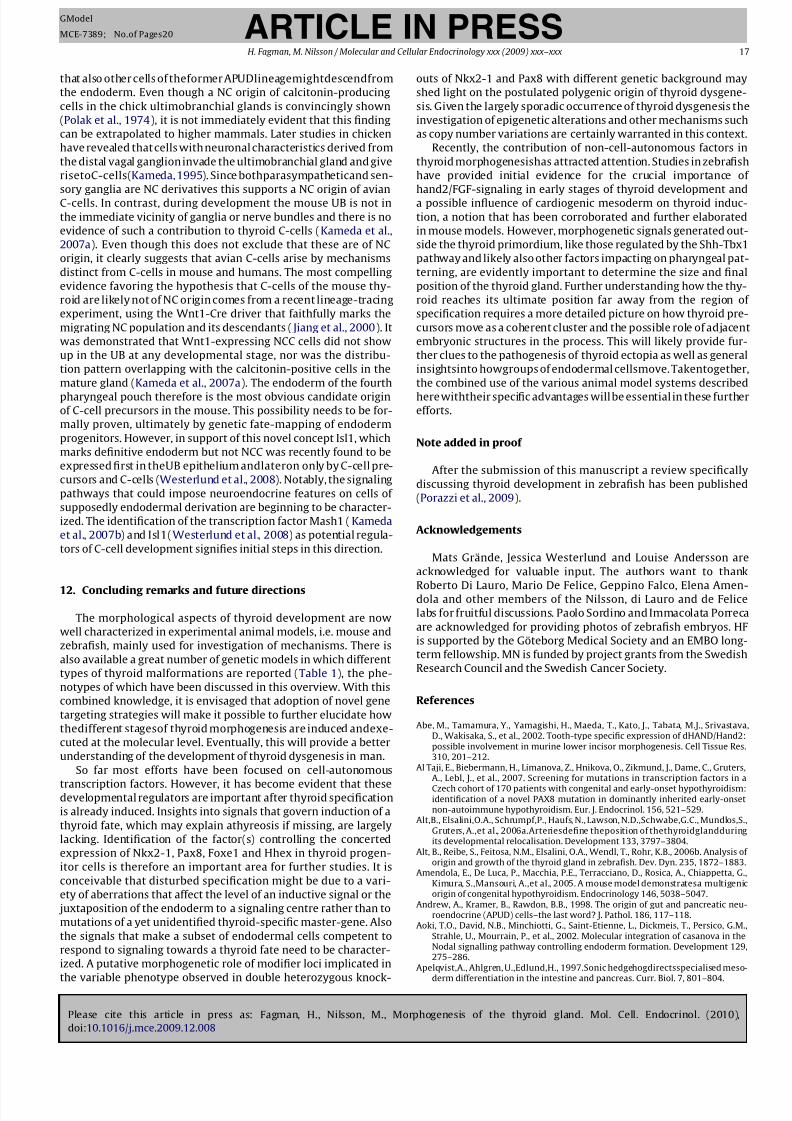
8/10/2019 Molecular Morfgenesis de la glandula Tiroidea.pdf
http://slidepdf.com/reader/full/molecular-morfgenesis-de-la-glandula-tiroideapdf 17/20
Please cite this article in press as: Fagman, H., Nilsson, M., Morphogenesis of the thyroid gland. Mol. Cell. Endocrinol. (2010),
doi:10.1016/j.mce.2009.12.008
ARTICLE IN PRESSGModel
MCE-7389; No.of Pages20
H. Fagman, M. Nilsson / Molecular and Cellular Endocrinology xxx (2009) xxx–xxx 17
that also other cells of theformer APUDlineagemightdescendfrom
the endoderm. Even though a NC origin of calcitonin-producing
cells in the chick ultimobranchial glands is convincingly shown
(Polak et al., 1974), it is not immediately evident that this finding
can be extrapolated to higher mammals. Later studies in chicken
have revealed that cells with neuronal characteristics derived from
the distal vagal ganglion invade the ultimobranchial gland and give
risetoC-cells(Kameda, 1995). Since bothparasympatheticand sen-
sory ganglia are NC derivatives this supports a NC origin of avian
C-cells. In contrast, during development the mouse UB is not in
the immediate vicinity of ganglia or nerve bundles and there is no
evidence of such a contribution to thyroid C-cells (Kameda et al.,
2007a). Even though this does not exclude that these are of NC
origin, it clearly suggests that avian C-cells arise by mechanisms
distinct from C-cells in mouse and humans. The most compelling
evidence favoring the hypothesis that C-cells of the mouse thy-
roid are likely not of NC origin comes from a recent lineage-tracing
experiment, using the Wnt1-Cre driver that faithfully marks the
migrating NC population and its descendants ( Jiang et al., 2000). It
was demonstrated that Wnt1-expressing NCC cells did not show
up in the UB at any developmental stage, nor was the distribu-
tion pattern overlapping with the calcitonin-positive cells in the
mature gland (Kameda et al., 2007a). The endoderm of the fourth
pharyngeal pouch therefore is the most obvious candidate originof C-cell precursors in the mouse. This possibility needs to be for-
mally proven, ultimately by genetic fate-mapping of endoderm
progenitors. However, in support of this novel concept Isl1, which
marks definitive endoderm but not NCC was recently found to be
expressed first in theUB epithelium andlateron only by C-cell pre-
cursors and C-cells (Westerlund et al., 2008). Notably, the signaling
pathways that could impose neuroendocrine features on cells of
supposedly endodermal derivation are beginning to be character-
ized. The identification of the transcription factor Mash1 (Kameda
et al., 2007b) and Isl1(Westerlund et al., 2008) as potential regula-
tors of C-cell development signifies initial steps in this direction.
12. Concluding remarks and future directions
The morphological aspects of thyroid development are now
well characterized in experimental animal models, i.e. mouse and
zebrafish, mainly used for investigation of mechanisms. There is
also available a great number of genetic models in which different
types of thyroid malformations are reported (Table 1), the phe-
notypes of which have been discussed in this overview. With this
combined knowledge, it is envisaged that adoption of novel gene
targeting strategies will make it possible to further elucidate how
thedifferent stagesof thyroid morphogenesis are induced andexe-
cuted at the molecular level. Eventually, this will provide a better
understanding of the development of thyroid dysgenesis in man.
So far most efforts have been focused on cell-autonomous
transcription factors. However, it has become evident that thesedevelopmental regulators are important after thyroid specification
is already induced. Insights into signals that govern induction of a
thyroid fate, which may explain athyreosis if missing, are largely
lacking. Identification of the factor(s) controlling the concerted
expression of Nkx2-1, Pax8, Foxe1 and Hhex in thyroid progen-
itor cells is therefore an important area for further studies. It is
conceivable that disturbed specification might be due to a vari-
ety of aberrations that affect the level of an inductive signal or the
juxtaposition of the endoderm to a signaling centre rather than to
mutations of a yet unidentified thyroid-specific master-gene. Also
the signals that make a subset of endodermal cells competent to
respond to signaling towards a thyroid fate need to be character-
ized. A putative morphogenetic role of modifier loci implicated in
the variable phenotype observed in double heterozygous knock-
outs of Nkx2-1 and Pax8 with different genetic background may
shed light on the postulated polygenic origin of thyroid dysgene-
sis. Given the largely sporadic occurrence of thyroid dysgenesis the
investigation of epigenetic alterations and other mechanisms such
as copy number variations are certainly warranted in this context.
Recently, the contribution of non-cell-autonomous factors in
thyroid morphogenesishas attracted attention. Studies in zebrafish
have provided initial evidence for the crucial importance of
hand2/FGF-signaling in early stages of thyroid development and
a possible influence of cardiogenic mesoderm on thyroid induc-
tion, a notion that has been corroborated and further elaborated
in mouse models. However, morphogenetic signals generated out-
side the thyroid primordium, like those regulated by the Shh-Tbx1
pathway and likely also other factors impacting on pharyngeal pat-
terning, are evidently important to determine the size and final
position of the thyroid gland. Further understanding how the thy-
roid reaches its ultimate position far away from the region of
specification requires a more detailed picture on how thyroid pre-
cursors move as a coherent cluster and the possible role of adjacent
embryonic structures in the process. This will likely provide fur-
ther clues to the pathogenesis of thyroid ectopia as well as general
insightsinto howgroups of endodermal cellsmove. Takentogether,
the combined use of the various animal model systems described
here withtheir specific advantages will be essential in these furtherefforts.
Note added in proof
After the submission of this manuscript a review specifically
discussing thyroid development in zebrafish has been published
(Porazzi et al., 2009).
Acknowledgements
Mats Grände, Jessica Westerlund and Louise Andersson are
acknowledged for valuable input. The authors want to thank
Roberto Di Lauro, Mario De Felice, Geppino Falco, Elena Amen-dola and other members of the Nilsson, di Lauro and de Felice
labs for fruitful discussions. Paolo Sordino and Immacolata Porreca
are acknowledged for providing photos of zebrafish embryos. HF
is supported by the Göteborg Medical Society and an EMBO long-
term fellowship. MN is funded by project grants from the Swedish
Research Council and the Swedish Cancer Society.
References
Abe, M., Tamamura, Y., Yamagishi, H., Maeda, T., Kato, J., Tabata, M.J., Srivastava,D., Wakisaka, S., et al., 2002. Tooth-type specific expression of dHAND/Hand2:possible involvement in murine lower incisor morphogenesis. Cell Tissue Res.310, 201–212.
Al Taji, E., Biebermann, H., Limanova, Z., Hnikova, O., Zikmund, J., Dame, C., Gruters,A., Lebl, J., et al., 2007. Screening for mutations in transcription factors in a
Czech cohort of 170 patients with congenital and early-onset hypothyroidism:identification of a novel PAX8 mutation in dominantly inherited early-onsetnon-autoimmune hypothyroidism. Eur. J. Endocrinol. 156, 521–529.
Alt,B., Elsalini,O.A., Schrumpf,P., Haufs, N., Lawson, N.D.,Schwabe,G.C., Mundlos,S.,Gruters, A.,et al., 2006a.Arteriesdefine theposition of thethyroidglandduringits developmental relocalisation. Development 133, 3797–3804.
Alt, B., Reibe, S., Feitosa, N.M., Elsalini, O.A., Wendl, T., Rohr, K.B., 2006b. Analysis of origin and growth of the thyroid gland in zebrafish. Dev. Dyn. 235, 1872–1883.
Amendola, E., De Luca, P., Macchia, P.E., Terracciano, D., Rosica, A., Chiappetta, G.,Kimura, S.,Mansouri, A.,et al., 2005. A mouse model demonstratesa multigenicorigin of congenital hypothyroidism. Endocrinology 146, 5038–5047.
Andrew, A., Kramer, B., Rawdon, B.B., 1998. The origin of gut and pancreatic neu-roendocrine (APUD) cells–the last word? J. Pathol. 186, 117–118.
Aoki, T.O., David, N.B., Minchiotti, G., Saint-Etienne, L., Dickmeis, T., Persico, G.M.,Strahle, U., Mourrain, P., et al., 2002. Molecular integration of casanova in theNodal signalling pathway controlling endoderm formation. Development 129,275–286.
Apelqvist,A., Ahlgren, U.,Edlund,H., 1997.Sonic hedgehogdirectsspecialised meso-
derm differentiation in the intestine and pancreas. Curr. Biol. 7, 801–804.
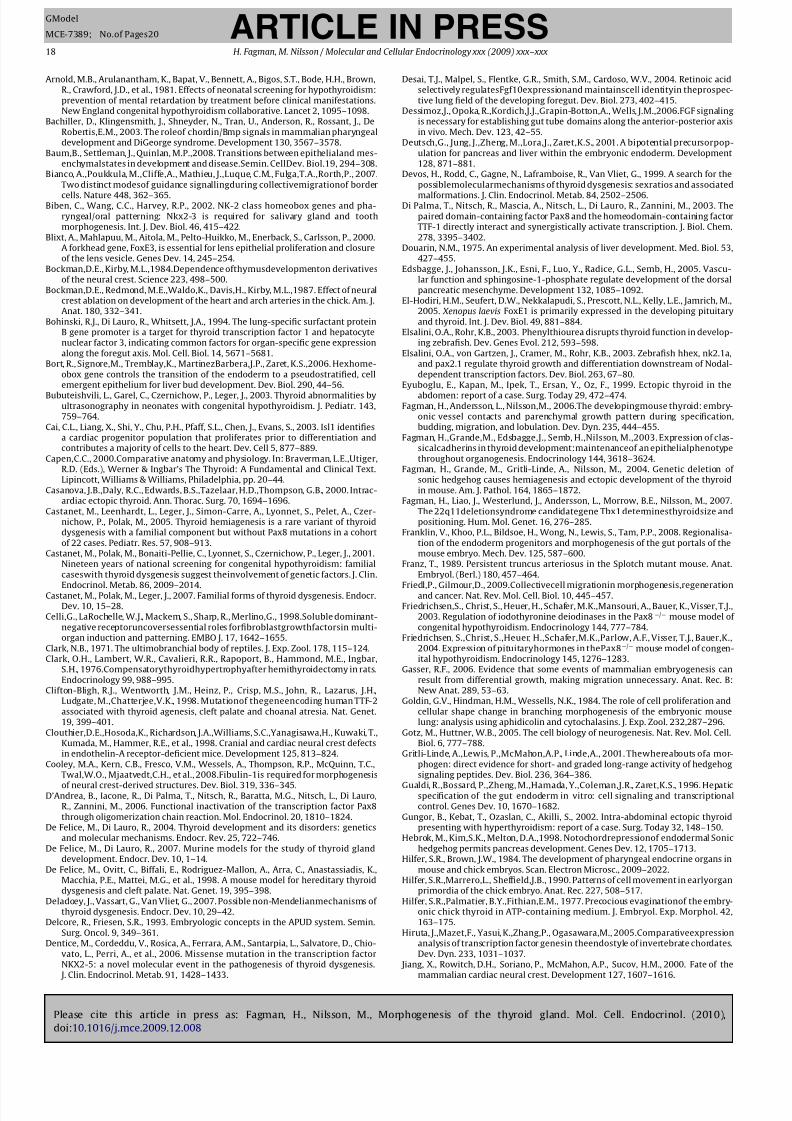
8/10/2019 Molecular Morfgenesis de la glandula Tiroidea.pdf
http://slidepdf.com/reader/full/molecular-morfgenesis-de-la-glandula-tiroideapdf 18/20
Please cite this article in press as: Fagman, H., Nilsson, M., Morphogenesis of the thyroid gland. Mol. Cell. Endocrinol. (2010),
doi:10.1016/j.mce.2009.12.008
ARTICLE IN PRESSGModel
MCE-7389; No.of Pages20
18 H. Fagman, M. Nilsson / Molecular and Cellular Endocrinology xxx (2009) xxx–xxx
Arnold, M.B., Arulanantham, K., Bapat, V., Bennett, A., Bigos, S.T., Bode, H.H., Brown,R., Crawford, J.D., et al., 1981. Effects of neonatal screening for hypothyroidism:prevention of mental retardation by treatment before clinical manifestations.New England congenital hypothyroidism collaborative. Lancet 2, 1095–1098.
Bachiller, D., Klingensmith, J., Shneyder, N., Tran, U., Anderson, R., Rossant, J., DeRobertis,E.M., 2003. The roleof chordin/Bmp signals in mammalian pharyngealdevelopment and DiGeorge syndrome. Development 130, 3567–3578.
Baum,B., Settleman, J., Quinlan, M.P.,2008. Transitions between epithelialand mes-enchymalstates in development and disease.Semin. CellDev. Biol.19, 294–308.
Bianco, A.,Poukkula, M.,Cliffe,A., Mathieu, J.,Luque, C.M., Fulga,T.A.,Rorth,P., 2007.Two distinct modesof guidance signallingduring collectivemigrationof border
cells. Nature 448, 362–365.Biben, C., Wang, C.C., Harvey, R.P., 2002. NK-2 class homeobox genes and pha-
ryngeal/oral patterning: Nkx2-3 is required for salivary gland and toothmorphogenesis. Int. J. Dev. Biol. 46, 415–422.
Blixt, A., Mahlapuu, M., Aitola, M., Pelto-Huikko, M., Enerback, S., Carlsson, P., 2000.A forkhead gene, FoxE3, is essential for lens epithelial proliferation and closureof the lens vesicle. Genes Dev. 14, 245–254.
Bockman,D.E., Kirby, M.L.,1984.Dependence ofthymusdevelopmenton derivativesof the neural crest. Science 223, 498–500.
Bockman,D.E., Redmond, M.E.,Waldo,K., Davis,H., Kirby, M.L.,1987. Effect of neuralcrest ablation on development of the heart and arch arteries in the chick. Am. J.Anat. 180, 332–341.
Bohinski, R.J., Di Lauro, R., Whitsett, J.A., 1994. The lung-specific surfactant proteinB gene promoter is a target for thyroid transcription factor 1 and hepatocytenuclear factor 3, indicating common factors for organ-specific gene expressionalong the foregut axis. Mol. Cell. Biol. 14, 5671–5681.
Bort, R., Signore,M., Tremblay,K., MartinezBarbera,J.P., Zaret, K.S.,2006. Hexhome-obox gene controls the transition of the endoderm to a pseudostratified, cellemergent epithelium for liver bud development. Dev. Biol. 290, 44–56.
Bubuteishvili, L., Garel, C., Czernichow, P., Leger, J., 2003. Thyroid abnormalities byultrasonography in neonates with congenital hypothyroidism. J. Pediatr. 143,759–764.
Cai, C.L., Liang, X., Shi, Y., Chu, P.H., Pfaff, S.L., Chen, J., Evans, S., 2003. Isl1 identifiesa cardiac progenitor population that proliferates prior to differentiation andcontributes a majority of cells to the heart. Dev. Cell 5, 877–889.
Capen,C.C., 2000.Comparative anatomy and physiology. In: Braverman, L.E.,Utiger,R.D. (Eds.), Werner & Ingbar’s The Thyroid: A Fundamental and Clinical Text.Lipincott, Williams & Williams, Philadelphia, pp. 20–44.
Casanova, J.B.,Daly, R.C., Edwards, B.S.,Tazelaar, H.D.,Thompson, G.B., 2000. Intrac-ardiac ectopic thyroid. Ann. Thorac. Surg. 70, 1694–1696.
Castanet, M., Leenhardt, L., Leger, J., Simon-Carre, A., Lyonnet, S., Pelet, A., Czer-nichow, P., Polak, M., 2005. Thyroid hemiagenesis is a rare variant of thyroiddysgenesis with a familial component but without Pax8 mutations in a cohortof 22 cases. Pediatr. Res. 57, 908–913.
Castanet, M., Polak, M., Bonaiti-Pellie, C., Lyonnet, S., Czernichow, P., Leger, J., 2001.Nineteen years of national screening for congenital hypothyroidism: familialcaseswith thyroid dysgenesis suggest theinvolvement of genetic factors. J. Clin.
Endocrinol. Metab. 86, 2009–2014.Castanet, M., Polak, M., Leger, J., 2007. Familial forms of thyroid dysgenesis. Endocr.Dev. 10, 15–28.
Celli,G., LaRochelle, W.J., Mackem, S., Sharp, R., Merlino,G., 1998.Soluble dominant-negative receptoruncoversessential roles forfibroblastgrowthfactorsin multi-organ induction and patterning. EMBO J. 17, 1642–1655.
Clark, N.B., 1971. The ultimobranchial body of reptiles. J. Exp. Zool. 178, 115–124.Clark, O.H., Lambert, W.R., Cavalieri, R.R., Rapoport, B., Hammond, M.E., Ingbar,
S.H., 1976.Compensatorythyroidhypertrophyafter hemithyroidectomy in rats.Endocrinology 99, 988–995.
Clifton-Bligh, R.J., Wentworth, J.M., Heinz, P., Crisp, M.S., John, R., Lazarus, J.H.,Ludgate, M.,Chatterjee,V.K., 1998. Mutationof thegeneencoding human TTF-2associated with thyroid agenesis, cleft palate and choanal atresia. Nat. Genet.19, 399–401.
Clouthier,D.E.,Hosoda,K., Richardson, J.A.,Williams, S.C.,Yanagisawa,H., Kuwaki, T.,Kumada, M., Hammer, R.E., et al., 1998. Cranial and cardiac neural crest defectsin endothelin-A receptor-deficient mice. Development 125, 813–824.
Cooley, M.A., Kern, C.B., Fresco, V.M., Wessels, A., Thompson, R.P., McQuinn, T.C.,Twal,W.O., Mjaatvedt,C.H., et al., 2008.Fibulin-1is required for morphogenesis
of neural crest-derived structures. Dev. Biol. 319, 336–345.D’Andrea, B., Iacone, R., Di Palma, T., Nitsch, R., Baratta, M.G., Nitsch, L., Di Lauro,
R., Zannini, M., 2006. Functional inactivation of the transcription factor Pax8through oligomerization chain reaction. Mol. Endocrinol. 20, 1810–1824.
De Felice, M., Di Lauro, R., 2004. Thyroid development and its disorders: geneticsand molecular mechanisms. Endocr. Rev. 25, 722–746.
De Felice, M., Di Lauro, R., 2007. Murine models for the study of thyroid glanddevelopment. Endocr. Dev. 10, 1–14.
De Felice, M., Ovitt, C., Biffali, E., Rodriguez-Mallon, A., Arra, C., Anastassiadis, K.,Macchia, P.E., Mattei, M.G., et al., 1998. A mouse model for hereditary thyroiddysgenesis and cleft palate. Nat. Genet. 19, 395–398.
Deladoey, J., Vassart, G., Van Vliet, G., 2007. Possible non-Mendelianmechanisms of thyroid dysgenesis. Endocr. Dev. 10, 29–42.
Delcore, R., Friesen, S.R., 1993. Embryologic concepts in the APUD system. Semin.Surg. Oncol. 9, 349–361.
Dentice, M., Cordeddu, V., Rosica, A., Ferrara, A.M., Santarpia, L., Salvatore, D., Chio-vato, L., Perri, A., et al., 2006. Missense mutation in the transcription factorNKX2-5: a novel molecular event in the pathogenesis of thyroid dysgenesis.
J. Clin. Endocrinol. Metab. 91, 1428–1433.
Desai, T.J., Malpel, S., Flentke, G.R., Smith, S.M., Cardoso, W.V., 2004. Retinoic acidselectively regulatesFgf10expressionand maintainscell identityin theprospec-tive lung field of the developing foregut. Dev. Biol. 273, 402–415.
Dessimoz,J., Opoka, R.,Kordich,J.J.,Grapin-Botton,A., Wells, J.M.,2006.FGF signalingis necessary for establishing gut tube domains along the anterior-posterior axisin vivo. Mech. Dev. 123, 42–55.
Deutsch,G., Jung, J.,Zheng, M.,Lora,J., Zaret,K.S., 2001. A bipotential precursorpop-ulation for pancreas and liver within the embryonic endoderm. Development128, 871–881.
Devos, H., Rodd, C., Gagne, N., Laframboise, R., Van Vliet, G., 1999. A search for thepossiblemolecularmechanisms of thyroid dysgenesis: sexratios and associated
malformations. J. Clin. Endocrinol. Metab. 84, 2502–2506.Di Palma, T., Nitsch, R., Mascia, A., Nitsch, L., Di Lauro, R., Zannini, M., 2003. The
paired domain-containing factor Pax8 and the homeodomain-containing factorTTF-1 directly interact and synergistically activate transcription. J. Biol. Chem.278, 3395–3402.
Douarin, N.M., 1975. An experimental analysis of liver development. Med. Biol. 53,427–455.
Edsbagge, J., Johansson, J.K., Esni, F., Luo, Y., Radice, G.L., Semb, H., 2005. Vascu-lar function and sphingosine-1-phosphate regulate development of the dorsalpancreatic mesenchyme. Development 132, 1085–1092.
El-Hodiri, H.M., Seufert, D.W., Nekkalapudi, S., Prescott, N.L., Kelly, L.E., Jamrich, M.,2005. Xenopus laevis FoxE1 is primarily expressed in the developing pituitaryand thyroid. Int. J. Dev. Biol. 49, 881–884.
Elsalini, O.A., Rohr, K.B., 2003. Phenylthiourea disrupts thyroid function in develop-ing zebrafish. Dev. Genes Evol. 212, 593–598.
Elsalini, O.A., von Gartzen, J., Cramer, M., Rohr, K.B., 2003. Zebrafish hhex, nk2.1a,and pax2.1 regulate thyroid growth and differentiation downstream of Nodal-dependent transcription factors. Dev. Biol. 263, 67–80.
Eyuboglu, E., Kapan, M., Ipek, T., Ersan, Y., Oz, F., 1999. Ectopic thyroid in the
abdomen: report of a case. Surg. Today 29, 472–474.Fagman, H., Andersson, L., Nilsson,M., 2006.The developingmouse thyroid: embry-
onic vessel contacts and parenchymal growth pattern during specification,budding, migration, and lobulation. Dev. Dyn. 235, 444–455.
Fagman, H.,Grande,M., Edsbagge,J., Semb, H.,Nilsson, M.,2003. Expression of clas-sicalcadherins in thyroid development:maintenanceof an epithelialphenotypethroughout organogenesis. Endocrinology 144, 3618–3624.
Fagman, H., Grande, M., Gritli-Linde, A., Nilsson, M., 2004. Genetic deletion of sonic hedgehog causes hemiagenesis and ectopic development of the thyroidin mouse. Am. J. Pathol. 164, 1865–1872.
Fagman, H., Liao, J., Westerlund, J., Andersson, L., Morrow, B.E., Nilsson, M., 2007.The 22q11deletionsyndrome candidategene Tbx1 determinesthyroidsize andpositioning. Hum. Mol. Genet. 16, 276–285.
Franklin, V., Khoo, P.L., Bildsoe, H., Wong, N., Lewis, S., Tam, P.P., 2008. Regionalisa-tion of the endoderm progenitors and morphogenesis of the gut portals of themouse embryo. Mech. Dev. 125, 587–600.
Franz, T., 1989. Persistent truncus arteriosus in the Splotch mutant mouse. Anat.Embryol. (Berl.) 180, 457–464.
Friedl,P., Gilmour,D., 2009.Collectivecell migrationin morphogenesis,regenerationand cancer. Nat. Rev. Mol. Cell. Biol. 10, 445–457.Friedrichsen,S., Christ, S., Heuer, H., Schafer, M.K.,Mansouri, A., Bauer, K., Visser, T.J.,
2003. Regulation of iodothyronine deiodinases in the Pax8−/− mouse model of congenital hypothyroidism. Endocrinology 144, 777–784.
Friedrichsen, S.,Christ, S.,Heuer, H.,Schafer,M.K.,Parlow, A.F., Visser, T.J., Bauer,K.,2004. Expression of pituitaryhormones in thePax8−/− mouse model of congen-ital hypothyroidism. Endocrinology 145, 1276–1283.
Gasser, R.F., 2006. Evidence that some events of mammalian embryogenesis canresult from differential growth, making migration unnecessary. Anat. Rec. B:New Anat. 289, 53–63.
Goldin, G.V., Hindman, H.M., Wessells, N.K., 1984. The role of cell proliferation andcellular shape change in branching morphogenesis of the embryonic mouselung: analysis using aphidicolin and cytochalasins. J. Exp. Zool. 232,287–296.
Gotz, M., Huttner, W.B., 2005. The cell biology of neurogenesis. Nat. Rev. Mol. Cell.Biol. 6, 777–788.
Gritli-Linde, A.,Lewis, P.,McMahon,A.P., Linde,A., 2001. Thewhereabouts ofa mor-phogen: direct evidence for short- and graded long-range activity of hedgehogsignaling peptides. Dev. Biol. 236, 364–386.
Gualdi, R.,Bossard, P.,Zheng, M.,Hamada, Y.,Coleman,J.R., Zaret,K.S., 1996. Hepaticspecification of the gut endoderm in vitro: cell signaling and transcriptionalcontrol. Genes Dev. 10, 1670–1682.
Gungor, B., Kebat, T., Ozaslan, C., Akilli, S., 2002. Intra-abdominal ectopic thyroidpresenting with hyperthyroidism: report of a case. Surg. Today 32, 148–150.
Hebrok, M., Kim,S.K., Melton, D.A.,1998. Notochordrepressionof endodermal Sonichedgehog permits pancreas development. Genes Dev. 12, 1705–1713.
Hilfer, S.R., Brown, J.W., 1984. The development of pharyngeal endocrine organs inmouse and chick embryos. Scan. Electron Microsc., 2009–2022.
Hilfer, S.R.,Marrero,L., Sheffield,J.B., 1990. Patterns of cell movement in earlyorganprimordia of the chick embryo. Anat. Rec. 227, 508–517.
Hilfer, S.R.,Palmatier, B.Y.,Fithian,E.M., 1977. Precocious evaginationof the embry-onic chick thyroid in ATP-containing medium. J. Embryol. Exp. Morphol. 42,163–175.
Hiruta, J.,Mazet,F., Yasui, K.,Zhang,P., Ogasawara,M., 2005.Comparativeexpressionanalysis of transcription factor genesin theendostyle of invertebrate chordates.Dev. Dyn. 233, 1031–1037.
Jiang, X., Rowitch, D.H., Soriano, P., McMahon, A.P., Sucov, H.M., 2000. Fate of themammalian cardiac neural crest. Development 127, 1607–1616.
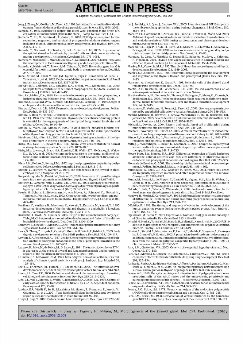
8/10/2019 Molecular Morfgenesis de la glandula Tiroidea.pdf
http://slidepdf.com/reader/full/molecular-morfgenesis-de-la-glandula-tiroideapdf 19/20
Please cite this article in press as: Fagman, H., Nilsson, M., Morphogenesis of the thyroid gland. Mol. Cell. Endocrinol. (2010),
doi:10.1016/j.mce.2009.12.008
ARTICLE IN PRESSGModel
MCE-7389; No.of Pages20
H. Fagman, M. Nilsson / Molecular and Cellular Endocrinology xxx (2009) xxx–xxx 19
Jung, J., Zheng, M., Goldfarb, M., Zaret, K.S., 1999. Initiationof mammalian liver devel-opment from endoderm by fibroblast growth factors. Science 284, 1998–2003.
Kameda, Y., 1995. Evidence to support the distal vagal ganglion as the origin of Ccells of the ultimobranchial gland in the chick. J. Comp. Neurol. 359, 1–14.
Kameda, Y., Ito, M., Nishimaki, T., Gotoh, N., 2009. FRS2alpha is required for theseparation, migration, and survival of pharyngeal-endoderm derived organsincluding thyroid, ultimobranchial body, parathyroid, and thymus. Dev. Dyn.238, 503–513.
Kameda, Y., Nishimaki, T., Chisaka, O., Iseki, S., Sucov, H.M., 2007a. Expression of the epithelial marker E-cadherin by thyroid C cells and their precursors duringmurine development. J. Histochem. Cytochem. 55, 1075–1088.
Kameda,Y., Nishimaki,T., Miura,M., Jiang, S.X., Guillemot,F., 2007b.Mash1regulatesthe development of C cells in mouse thyroid glands. Dev. Dyn. 236, 262–270.
Kameda, Y., Nishimaki, T., Takeichi, M., Chisaka, O., 2002. Homeobox gene hoxa3 isessential for the formation of the carotid body in the mouse embryos. Dev. Biol.247, 197–209.
Kanai-Azuma, M., Kanai, Y., Gad, J.M., Tajima, Y., Taya, C., Kurohmaru, M., Sanai, Y.,Yonekawa, H., et al., 2002. Depletion of definitive gut endoderm in Sox17-nullmutant mice. Development 129, 2367–2379.
Kiehart, D.P., Galbraith, C.G., Edwards, K.A., Rickoll, W.L., Montague, R.A., 2000.Multiple forces contribute to cell sheet morphogenesis for dorsal closure inDrosophila. J. Cell Biol. 149, 471–490.
Kim, S.K., Melton, D.A., 1998. Pancreas development is promoted by cyclopamine, ahedgehog signaling inhibitor. Proc. Natl. Acad. Sci. U.S.A. 95, 13036–13041.
Kimmel, C.B.,Ballard, W.W., Kimmel, S.R.,Ullmann,B., Schilling,T.F., 1995. Stages of embryonic development of the zebrafish. Dev. Dyn. 203, 253–310.
Kimura, J., Deutsch, G.H., 2007. Keymechanisms of earlylung development. Pediatr.Dev. Pathol. 10, 335–347.
Kimura, S., Hara, Y., Pineau, T., Fernandez-Salguero, P., Fox, C.H., Ward, J.M., Gonza-lez, F.J., 1996. The T/ebp null mouse: thyroid-specific enhancer-binding protein
is essential for the organogenesis of the thyroid, lung, ventral forebrain, andpituitary. Genes Dev. 10, 60–69.
Kimura, S., Ward, J.M., Minoo, P., 1999. Thyroid-specific enhancer-binding pro-tein/thyroid transcription factor 1 is not required for the initial specificationof the thyroid and lung primordia. Biochimie 81, 321–327.
Kinebrew, G.M., Hilfer, S.R., 2001. Cellular dynamics during evagination of the thy-roid primordium in the chick embryo. Anat. Rec. 264, 146–156.
Kirby, M.L., Gale, T.F., Stewart, D.E., 1983. Neural crest cells contribute to normalaorticopulmonary septation. Science 220, 1059–1061.
Kirby, M.L., Lawson, A., Stadt, H.A., Kumiski, D.H., Wallis, K.T., McCraney, E., Waldo,K.L., Li, Y.X., et al., 2003. Hensen’s node gives rise to the ventral midline of theforegut:implications fororganizing headand heart development. Dev.Biol. 253,175–188.
Klein, A.H.,Meltzer,S., Kenny,F.M., 1972.Improved prognosisin congenitalhypothy-roidism treated before age three months. J. Pediatr. 81, 912–915.
Knospe, C., Seichert, V., Rychter, Z., 1991. The topogenesis of the thyroid in chickembryos. Eur. J. Morphol. 29, 291–296.
Korpal-Szczyrska, M., Kosiak, W., Swieton, D., 2008. Prevalence of thyroid hemiage-
nesis in an asymptomatic schoolchildren population. Thyroid 18, 637–639.Kreisner, E., Camargo-Neto, E., Maia, C.R., Gross, J.L., 2003. Accuracy of ultrasonog-raphyto establishthe diagnosis and aetiology of permanentprimary congenitalhypothyroidism. Clin. Endocrinol. (Oxf.) 59, 361–365.
Krude, H., Schutz, B., Biebermann, H., von Moers, A., Schnabel, D., Neitzel, H.,Tonnies, H., Weise, D., et al., 2002. Choreoathetosis, hypothyroidism, and pul-monary alterations dueto humanNKX2-1haploinsufficiency.J. Clin.Invest.109,475–480.
Kurihara, Y., Kurihara, H., Maemura, K., Kuwaki, T., Kumada, M., Yazaki, Y., 1995.Impaired development of the thyroid and thymus in endothelin-1 knockoutmice. J. Cardiovasc. Pharmacol. 26 (Suppl. 3), S13–S16.
Kusakabe, T., Hoshi, N., Kimura, S., 2006. Origin of the ultimobranchial body cyst:T/ebp/Nkx2.1 expression is required for development and fusion of the ultimo-branchial body to the thyroid. Dev. Dyn. 235, 1300–1309.
Lammert, E., Cleaver, O., Melton, D., 2001. Induction of pancreaticdifferentiationbysignals from blood vessels. Science 294, 564–567.
Lania,G.,Zhang, Z.,Huynh,T., Caprio, C.,Moon, A.M.,Vitelli,F.,Baldini,A.,2009.Earlythyroid development requires a Tbx1-Fgf8 pathway. Dev. Biol. 328, 109–117.
Lawson, K.A.,Pedersen, R.A., 1987. Cell fate,morphogenetic movement and popula-
tion kinetics of embryonic endoderm at the time of germ layer formation in themouse. Development 101, 627–652.
Lazzaro, D., Price, M., de Felice, M., Di Lauro, R., 1991. The transcription factor TTF-1is expressed at the onset of thyroid and lung morphogenesis and in restrictedregions of the foetal brain. Development 113, 1093–1104.
Le Lievre, C.S., Le Douarin, N.M., 1975. Mesenchymal derivatives of theneural crest:analysis of chimaeric quail and chick embryos. J. Embryol. Exp. Morphol. 34,125–154.
Lee, C.S., Friedman, J.R., Fulmer, J.T., Kaestner, K.H., 2005. The initiation of liverdevelopment is dependent on Foxa transcription factors. Nature 435, 944–947.
Lewis, S.L., Tam, P.P., 2006. Definitive endoderm of the mouse embryo: formation,cell fates, and morphogenetic function. Dev. Dyn. 235, 2315–2329.
Lien, C.L., Wu, C., Mercer, B., Webb, R., Richardson, J.A., Olson, E.N., 1999. Control of early cardiac-specific transcription of Nkx2-5 by a GATA-dependent enhancer.Development 126, 75–84.
Lindsay, E.A., Vitelli, F., Su, H., Morishima, M., Huynh, T., Pramparo, T., Jurecic, V.,Ogunrinu, G., et al., 2001. Tbx1 haploinsufficieny in the DiGeorge syndromeregion causes aortic arch defects in mice. Nature 410, 97–101.
Lough,J., Sugi, Y.,2000. Endodermand heart development.Dev. Dyn. 217, 327–342.
Lu, J., Izvolsky, K.I., Qian, J., Cardoso, W.V., 2005. Identification of FGF10 targets inthe embryonic lung epithelium during bud morphogenesis. J. Biol. Chem. 280,4834–4841.
Macatee,T.L., Hammond,B.P.,Arenkiel,B.R.,Francis, L.,Frank,D.U., Moon, A.M.,2003.Ablation of specific expression domains reveals discrete functions of ectoderm-and endoderm-derived FGF8 during cardiovascular and pharyngeal develop-ment. Development 130, 6361–6374.
Macchia, P.E., Lapi, P., Krude, H., Pirro, M.T., Missero, C., Chiovato, L., Souabni, A.,Baserga, M., et al., 1998. PAX8 mutations associated with congenital hypothy-roidism caused by thyroid dysgenesis. Nat. Genet. 19, 83–86.
Maiorana, R., Carta, A., Floriddia, G., Leonardi, D., Buscema, M., Sava, L., Calaciura,
F., Vigneri, R., 2003. Thyroid hemiagenesis: prevalence in normal children andeffect on thyroid function. J. Clin. Endocrinol. Metab. 88, 1534–1536.
Manley, N.R., Capecchi, M.R., 1995. Theroleof Hoxa-3in mouse thymusand thyroiddevelopment. Development 121, 1989–2003.
Manley, N.R., Capecchi, M.R., 1998. Hox group 3 paralogs regulate the developmentand migration of the thymus, thyroid, and parathyroid glands. Dev. Biol. 195,1–15.
Mansouri, A., Chowdhury, K., Gruss, P., 1998. Follicular cells of the thyroid glandrequire Pax8 gene function. Nat. Genet. 19, 87–90.
Martin, A.C., Kaschube, M., Wieschaus, E.F., 2008. Pulsed contractions of anactin–myosin network drive apical constriction. Nature.
MartinezBarbera,J.P., Clements,M., Thomas, P.,Rodriguez,T., Meloy, D., Kioussis,D.,Beddington, R.S., 2000. The homeobox gene Hex is required in definitive endo-dermal tissues for normal forebrain, liver and thyroid formation. Development127, 2433–2445.
Matsumoto, K., Yoshitomi, H., Rossant, J., Zaret, K.S., 2001. Liver organogenesis pro-moted by endothelial cells prior to vascular function. Science 294, 559–563.
Medina-Martinez, O., Brownell, I., Amaya-Manzanares, F., Hu, Q., Behringer, R.R., Jamrich, M., 2005. Severe defects in proliferation and differentiation of lens cells
in Foxe3 null mice. Mol. Cell. Biol. 25, 8854–8863.Michael, L., Davies, J.A., 2004. Pattern and regulation of cell proliferation during
murine ureteric bud development. J. Anat. 204, 241–255.Michael, L.,Sweeney,D.E., Davies, J.A.,2005. A rolefor microfilament-basedcontrac-
tionin branching morphogenesis of theuretericbud. Kidney Int.68, 2010–2018.Minoo, P., Hamdan, H., Bu, D., Warburton, D., Stepanik, P., deLemos, R., 1995. TTF-1
regulates lung epithelial morphogenesis. Dev. Biol. 172, 694–698.Mittag, J., Winterhager, E., Bauer, K., Grummer, R., 2007. Congenital hypothyroid
female pax8-deficient mice are infertile despite thyroid hormone replacementtherapy. Endocrinology 148, 719–725.
Moore-Scott, B.A., Manley, N.R., 2005. Differential expression of Sonic hedgehogalong the anterior-posterior axis regulates patterning of pharyngeal pouchendoderm and pharyngeal endoderm-derived organs. Dev. Biol. 278, 323–335.
Morreale de Escobar, G., 2001. The role of thyroid hormone in fetal neurodevelop-ment. J. Pediatr. Endocrinol. Metab. 14 (Suppl. 6), 1453–1462.
Muratovska, A., Zhou, C., He, S., Goodyer, P., Eccles, M.R., 2003. Paired-Box genesare frequently expressed in cancer and often required for cancer cell survival.Oncogene 22, 7989–7997.
Muzza, M., Persani, L., de Filippis, T., Gastaldi, R., Vigone, M.C., Sala, D., Weber, G.,Lorini, R., et al., 2008. Absence of sonic hedgehog (Shh) germline mutations inpatients with thyroid dysgenesis. Clin. Endocrinol. (Oxf.) 69, 828–829.
Nakada, C., Iida, A., Tabata, Y., Watanabe, S., 2009. Forkhead transcription factorfoxe1 regulates chondrogenesis in zebrafish. J. Exp. Zool. B: Mol. Dev. Evol..
Nogawa, H., Morita, K.,Cardoso,W.V., 1998.Bud formationprecedesthe appearanceof differential cell proliferation during branching morphogenesis of mouselungepithelium in vitro. Dev. Dyn. 213, 228–235.
O’Rahilly, R., 1983. The timing and sequence of events in the development of thehuman endocrine system during the embryonic period proper. Anat. Embryol.(Berl.) 166, 439–451.
Ogasawara, M., Satou, Y., 2003. Expression of FoxE and FoxQ genes in the endostyleof Ciona intestinalis. Dev. Genes Evol. 213, 416–419.
Ohuchi, H.,Hori,Y., Yamasaki,M.,Harada,H., Sekine, K.,Kato, S.,Itoh,N., 2000.FGF10acts asa majorligand forFGFreceptor 2 IIIb inmousemulti-organ development.Biochem. Biophys. Res. Commun. 277, 643–649.
Olivieri,A., Stazi,M.A., Mastroiacovo, P.,Fazzini,C., Medda, E.,Spagnolo,A., De Ange-lis, S.,Grandolfo,M.E., etal., 2002.A population-based studyon thefrequencyof additionalcongenitalmalformationsin infantswith congenitalhypothyroidism:
data from the Italian Registry for Congenital Hypothyroidism (1991–1998). J.Clin. Endocrinol. Metab. 87, 557–562.
Park, S.M., Chatterjee, V.K., 2005. Genetics of congenital hypothyroidism. J. Med.Genet. 42, 379–389.
Park, W.Y., Miranda, B., Lebeche, D., Hashimoto, G., Cardoso, W.V., 1998. FGF-10 is achemotacticfactor fordistal epithelialbuds during lung development. Dev.Biol.201, 125–134.
Parlato,R., Rosica,A., Rodriguez-Mallon, A.,Affuso,A., Postiglione,M.P., Arra,C., Man-souri, A., Kimura, S., et al., 2004. An integrated regulatory network controllingsurvival and migration in thyroid organogenesis. Dev. Biol. 276, 464–475.
Pearse, A.G., 1969. The cytochemistry and ultrastructure of polypeptide hormone-producing cells of the APUD series and the embryologic, physiologic andpathologic implications of the concept. J. Histochem. Cytochem. 17,303–313.
Pearse, A.G., Carvalheira, A.F., 1967. Cytochemical evidence for an ultimobranchialorigin of rodent thyroid C cells. Nature 214, 929–930.
Pearse, A.G., Polak, J.M., 1971. Neural crest origin of the endocrine polypeptide(APUD) cells of the gastrointestinal tract and pancreas. Gut 12, 783–788.
Pera, E.M., Kessel, M., 1998. Demarcation of ventral territories by the homeoboxgene NKX2.1 during early chick development. Dev. Genes Evol. 208, 168–171.
8/10/2019 Molecular Morfgenesis de la glandula Tiroidea.pdf
http://slidepdf.com/reader/full/molecular-morfgenesis-de-la-glandula-tiroideapdf 20/20
ARTICLE IN PRESSGModel
MCE-7389; No.of Pages20
20 H. Fagman, M. Nilsson / Molecular and Cellular Endocrinology xxx (2009) xxx–xxx
Perl, A.K., Wert, S.E., Nagy, A., Lobe, C.G., Whitsett, J.A., 2002. Early restriction of peripheral and proximal cell lineages during formation of the lung. Proc. Natl.Acad. Sci. U.S.A. 99, 10482–10487.
Peters, H., Neubuser, A., Kratochwil, K., Balling, R., 1998. Pax9-deficient mice lackpharyngealpouchderivatives andteeth andexhibitcraniofacial andlimb abnor-malities. Genes Dev. 12, 2735–2747.
Petryk,A., Anderson,R.M.,Jarcho,M.P.,Leaf,I., Carlson,C.S.,Klingensmith,J.,Shawlot,W., O’Connor, M.B., 2004. The mammalian twisted gastrulation gene functionsin foregut and craniofacial development. Dev. Biol. 267, 374–386.
Plachov, D.,Chowdhury, K.,Walther,C., Simon,D., Guenet, J.L., Gruss,P., 1990. Pax8,a murine paired box gene expressed in the developing excretory system and
thyroid gland. Development 110, 643–651.Pohlenz, J., Dumitrescu, A., Zundel, D., Martine, U., Schonberger, W., Koo, E., Weiss,
R.E., Cohen, R.N., et al., 2002. Partial deficiency of thyroid transcription factor 1producespredominantlyneurologicaldefects inhumans andmice. J. Clin.Invest.109, 469–473.
Polak, J.M.,Pearse,A.G., Le Lievre, C., Fontaine,J., Le Douarin, N.M.,1974. Immunocy-tochemicalconfirmationof the neural crestorigin of aviancalcitonin-producingcells. Histochemistry 40, 209–214.
Porazzi, P., Calebiro, D., Benato, F., Tiso, N., Persani, L., 2009. Thyroid gland develop-ment and function in the zebrafish model. Mol. Cell. Endocrinol. 312, 14–23.
Postiglione, M.P., Parlato, R., Rodriguez-Mallon, A., Rosica, A., Mithbaokar, P.,Maresca, M., Marians, R.C., Davies, T.F., et al., 2002. Role of the thyroid-stimulating hormone receptor signaling in development and differentiation of the thyroid gland. Proc. Natl. Acad. Sci. U.S.A. 99, 15462–15467.
Puppin, C., D’Elia, A.V., Pellizzari, L., Russo, D., Arturi, F., Presta, I., Filetti, S., Bogue,C.W., et al., 2003. Thyroid-specific transcription factors control Hex promoteractivity. Nucleic Acids Res. 31, 1845–1852.
Puppin, C., Presta, I., D’Elia, A.V., Tell, G., Arturi, F., Russo, D., Filetti, S., Damante, G.,2004. Functionalinteraction among thyroid-specific transcription factors: Pax8
regulates the activity of Hex promoter. Mol. Cell. Endocrinol. 214, 117–125.Pyati, U.J., Look, A.T., Hammerschmidt, M., 2007. Zebrafish as a powerful vertebrate
model system for in vivo studies of cell death. Semin. Cancer Biol. 17, 154–165.Reifers, F., Walsh, E.C., Leger, S., Stainier, D.Y., Brand, M., 2000. Induction and
differentiation of the zebrafish heart requires fibroblast growth factor 8(fgf8/acerebellar). Development 127, 225–235.
Reiter, J.F., Kikuchi, Y., Stainier, D.Y., 2001. Multiple roles for Gata5 in zebrafishendoderm formation. Development 128, 125–135.
Rohr,K.B., Concha, M.L.,2000. Expressionof nk2.1a during earlydevelopment of thethyroid gland in zebrafish. Mech. Dev. 95, 267–270.
Romert, P., Gauguin, J., 1973. The early development of the median thyroid glandof the mouse. A light-, electron-microscopic and histochemical study. Z. Anat.Entwicklungsgesch. 139, 319–336.
Rorth, P., 2007. Collective guidance of collective cell migration. Trends Cell Biol. 17,575–579.
Rossi, J.M., Dunn, N.R., Hogan, B.L., Zaret, K.S., 2001. Distinct mesodermal signals,including BMPs from the septum transversum mesenchyme, are required incombination for hepatogenesis from the endoderm.Genes Dev. 15, 1998–2009.
Sakiyama, J., Yamagishi, A., Kuroiwa, A., 2003. Tbx4-Fgf10 system controls lungbud formation during chicken embryonic development. Development 130,1225–1234.
Sala, F.G., Curtis, J.L., Veltmaat, J.M., Del Moral, P.M., Le, L.T., Fairbanks, T.J., Warbur-ton, D., Ford, H., et al., 2006. Fibroblast growth factor 10 is required for survivaland proliferation but not differentiation of intestinal epithelial progenitor cellsduring murine colon development. Dev. Biol. 299, 373–385.
Schultheiss, T.M.,Xydas, S.,Lassar, A.B.,1995. Inductionof aviancardiac myogenesisby anterior endoderm. Development 121, 4203–4214.
Serls, A.E., Doherty, S., Parvatiyar, P., Wells, J.M., Deutsch, G.H., 2005. Differentthresholds of fibroblastgrowth factors pattern the ventral foregut intoliver andlung. Development 132, 35–47.
Shabana, W.,Delange,F., Freson,M., Osteaux, M.,De Schepper,J., 2000.Prevalence of thyroid hemiagenesis: ultrasound screening in normal children. Eur. J. Pediatr.159, 456–458.
Shain, W.G.,Hilfer, S.R.,Fonte, V.G., 1972.Earlyorganogenesisof theembryonic chickthyroid. I. Morphology and biochemistry. Dev. Biol. 28, 202–218.
Shiraishi, T., Imai, H., Fukutome, K., Watanabe, M., Yatani, R., 1999. Ectopic thyroidin the adrenal gland. Hum. Pathol. 30, 105–108.
Sinner, D.,Rankin, S.,Lee,M., Zorn, A.M., 2004. Sox17 andbeta-catenin cooperatetoregulate the transcription of endodermal genes. Development 131, 3069–3080.
Smuts, M.S., Hilfer, S.R., Searls, R.L., 1978. Patterns of cellular proliferation duringthyroid organogenesis. J. Embryol. Exp. Morphol. 48, 269–286.
Spagnoli, F.M., 2007. From endoderm to pancreas: a multistep journey. Cell. Mol.Life Sci. 64, 2378–2390.
Sprinzl, G.M., Koebke, J., Wimmers-Klick, J., Eckel, H.E., Thumfart, W.F., 2000. Mor-phology of the human thyroglossal tract: a histologic and macroscopic study ininfants and children. Ann. Otol. Rhinol. Laryngol. 109, 1135–1139.
Stafford, D., Prince, V.E., 2002. Retinoic acid signaling is required for a critical earlystep in zebrafish pancreatic development. Curr. Biol. 12, 1215–1220.
Stanger, B.Z., Tanaka, A.J., Melton, D.A., 2007. Organ size is limited by the numberof embryonic progenitor cells in the pancreas but not the liver. Nature 445,886–891.
Strohschneider, T., Timm, D., Worbes, C., 1993. Ectopic thyroid gland tissue in theliver. Chirurg 64, 751–753.
Tam, P.P., Kanai-Azuma, M., Kanai, Y., 2003. Early endoderm development in verte-brates: lineage differentiation and morphogenetic function. Curr. Opin. Genet.Dev. 13, 393–400.
Tanaka, H., Yanagisawa, K., Shinjo, K., Taguchi, A., Maeno, K., Tomida, S., Shimada,Y., Osada, H., et al., 2007. Lineage-specific dependencyof lung adenocarcinomason the lung development regulator TTF-1. Cancer Res. 67, 6007–6011.
Teillet, M.A., Ziller, C., Le Douarin, N.M., 2008. Quail-chick chimeras. Methods Mol.Biol. 461, 337–350.
Thomas, P.Q., Brown, A., Beddington, R.S., 1998. Hex: a homeobox gene reveal-ing peri-implantation asymmetry in the mouse embryo and an early transientmarker of endothelial cell precursors. Development 125, 85–94.
Toublanc, J.E., 1992. Comparison of epidemiological data on congenital hypothy-roidismin Europe withthoseof other partsin theworld.Horm. Res.38, 230–235.
Tourneux,J.E.,Verdun,P., 1897. Surles premièrs développements de la thyroide,duthymus et des glandules parathyroidennes chez l’hommes. J. Anat.33, 305–325.
Tremblay, K.D., Zaret, K.S., 2005. Distinct populations of endoderm cells convergeto generate the embryonic liver bud and ventral foregut tissues. Dev. Biol. 280,87–99.
Waldo, K.L., Kumiski, D., Kirby, M.L., 1996. Cardiac neural crest is essential for thepersistence ratherthanthe formationof anarch artery. Dev. Dyn. 205,281–292.
van den Brink, G.R., 2007. Hedgehog signaling in development and homeostasis of the gastrointestinal tract. Physiol. Rev. 87, 1343–1375.
Van Vliet, G., 2003. Development of the thyroid gland: lessons from congenitallyhypothyroid mice and men. Clin. Genet. 63, 445–455.
Warga, R.M., Nusslein-Volhard, C., 1999. Origin and development of the zebrafishendoderm. Development 126, 827–838.
Washington Smoak, I.,Byrd, N.A.,Abu-Issa, R.,Goddeeris, M.M.,Anderson, R.,Morris, J., Yamamura, K., Klingensmith, J., et al., 2005. Sonic hedgehog is required forcardiac outflow tractand neural crest celldevelopment.Dev. Biol.283, 357–372.
Wells, J.M., Melton, D.A., 1999. Vertebrate endoderm development. Annu. Rev. Cell
Dev. Biol. 15, 393–410.Wells, J.M.,Melton, D.A.,2000. Earlymouseendoderm ispatternedby soluble factors
from adjacent germ layers. Development 127, 1563–1572.Venditti, M., Hay, R.W., Kulaga, A., Demetrick, D.J., 2007. Diagnosis of ectopic tissue
versus contamination by genetic fingerprinting in a routine surgical pathologyspecimen. Hum. Pathol. 38, 378–382.
Wendl, T., Adzic, D., Schoenebeck, J.J., Scholpp, S., Brand, M., Yelon, D., Rohr,K.B., 2007. Early developmental specification of the thyroid gland dependson han-expressing surrounding tissue and on FGF signals. Development 134,2871–2879.
Wendl, T.,Lun, K.,Mione,M., Favor, J.,Brand,M., Wilson,S.W.,Rohr, K.B., 2002.Pax2.1is required for the development of thyroid follicles in zebrafish. Development129, 3751–3760.
Wessells, N.K., Sponner, B.S., Ash, J.F., Bradley, M.O., Luduena, M.A., Taylor, E.L.,Wrenn, J.T., Yamada, K.M., 1971. Microfilaments in cellular and developmentalprocesses. Science 171, 135–143.
Westerlund, J., Andersson, L., Carlsson, T., Zoppoli, P., Fagman, H., Nilsson, M., 2008.Expression of Islet1 in thyroid development related to budding, migration, and
fusion of primordia. Dev. Dyn. 237, 3820–3829.Wetzel, B.K., Wollman, S.H., 1969. Fine structure of a second kind of thyroid folliclein the C3H mouse. Endocrinology 84, 563–578.
Wheelock, M.J., Shintani, Y., Maeda, M., Fukumoto, Y., Johnson, K.R., 2008. Cadherinswitching. J. Cell Sci. 121, 727–735.
Williams, R.J., Lindop, G., Butler, J., 2002. Ectopic thyroid tissue on the ascendingaorta: an operative finding. Ann. Thorac. Surg. 73, 1642–1643.
Wistuba,J., Mittag, J.,Luetjens,C.M., Cooper, T.G.,Yeung,C.H., Nieschlag,E., Bauer, K.,2007. Male congenital hypothyroid Pax8−/− mice are infertile despite adequatetreatment with thyroid hormone. J. Endocrinol. 192, 99–109.
Vitelli, F., Taddei, I., Morishima, M., Meyers, E.N., Lindsay, E.A., Baldini, A., 2002. Agenetic link between Tbx1 and fibroblast growthfactor signaling. Development129, 4605–4611.
Wurdak, H., Ittner, L.M., Sommer, L., 2006. DiGeorge syndrome and pharyngealapparatus development. Bioessays 28, 1078–1086.
Xing, Y., Li, C., Hu, L., Tiozzo, C., Li, M., Chai, Y., Bellusci, S., Anderson, S., et al., 2008.Mechanisms of TGFbeta inhibition of LUNG endodermal morphogenesis: therole of TbetaRII, Smads, Nkx2.1 and Pten. Dev. Biol..
Xu, P.X., Zheng, W., Laclef, C., Maire, P., Maas, R.L., Peters, H., Xu, X., 2002. Eya1 is
requiredfor themorphogenesisof mammalianthymus, parathyroidand thyroid.Development 129, 3033–3044.
Yanagisawa,H., Hammer, R.E.,Richardson,J.A.,Williams,S.C.,Clouthier, D.E., Yanagi-sawa,M., 1998.Role of Endothelin-1/Endothelin-A receptor-mediated signalingpathway in the aortic arch patterning in mice. J. Clin. Invest. 102, 22–33.
Yoshitomi, H., Zaret, K.S.,2004. Endothelial cell interactions initiate dorsal pancreasdevelopment by selectively inducing the transcription factor Ptf1a. Develop-ment 131, 807–817.
Yu, J.K., Holland, L.Z., Jamrich, M., Blitz, I.L., Hollan, N.D., 2002. AmphiFoxE4, anamphioxus winged helix/forkhead gene encoding a protein closely related tovertebrate thyroid transcription factor-2: expression during pharyngeal devel-opment. Evol. Dev. 4, 9–15.
Zannini, M., Avantaggiato, V., Biffali, E., Arnone, M.I., Sato, K., Pischetola, M., Taylor,B.A., Phillips, S.J., et al., 1997. TTF-2, a new forkhead protein, shows a tem-poral expression in the developing thyroid which is consistent with a role incontrolling the onset of differentiation. EMBO J. 16, 3185–3197.


![[XLS] de Documentos DPS RIPS... · Web viewPAROTIDECTOMIA TOTAL CONSERVADORA DEL VII PAR CRANEAL 3 SIALOADENECTOMIA DE GLANDULA SUBLINGUAL 2 SIALOADENECTOMIA DE GLANDULA SUBMAXILAR](https://static.fdocuments.us/doc/165x107/5ba3c3b009d3f214538c54fd/xls-de-documentos-dps-rips-web-viewparotidectomia-total-conservadora-del.jpg)