a presynaptic site of action within the mesencephalic reticular ...
17
Transplantation of Foetal Ventral Mesencephalic Grafts in Parkinson’s Disease: A Still Evolving
Concept with New Regulatory Challenges
Sven Möllers, Máté Döbrössy and Guido Nikkhah Department of Stereotactic and Functional Neurosurgery
University Freiburg - Medical Center Germany
1. Introduction
Several chapters of this book describe the physiology and symptoms of Parkinson’s disease (PD) in great detail, nearly 200 years after the typical shaking palsies were first characterised by James Parkinson and further defined by Jean Martin Chacot as “Morbus Parkinson”. More recent investigations, starting in the 1950’s, identified PD as an age related phenomena, mainly effecting patients over 50 years old (Pollock & Hornabrook, 1966). The incidence of the onset of the disease in a patient is relatively high, as approximately 1 out of 1000 people in the western world might develop typical symptoms of the disease characterised by muscular rigidity or tremor, slow movements or slow mental activity (Lees, Hardy & Revesz, 2009). Once the high impact of this disease on the aging population was identified, research studies were initiated to identify a cure or symptomatic treatment of PD. Only a decade later, in the 60’s, the neurotransmitter dopamine, or more precisely the lack of it was pronounced as the main contributory factor in the pathogenesis of PD (Hornykiewicz, 1962, 1966) and initial clinical studies using L-Dopa showed the effectiveness of this drug to reduce the manifestation of classical symptoms of the disease (Boshes et al., 1969; Mones, 1969). As discussed in detail in previous chapters, the main cause of PD is considered to be a slow but continuously progressive reduction of dopamine within the midbrain region, namely the substantia nigra (SN), caused by a continuous loss of dopamine-producing cells within this region and consequently a lack of dopamine in the receptor target areas, i.e. the putamen and caudate nucleus. While many investigators addressed this issue by performing studies on the administration of L-dopa in animal models or in patients in order to directly overcome the lack of this substance, others focussed on different strategies: Replacement of what is lost by transplanting dopamine producing cells into the diseased tissue or into regions depleted of dopamine, and thereby reversing the loss of function and initiating regeneration of function ( Björklund et al., 1982b; Freed, Cannon-Spoor & Wyatt, 1984b). Transplanting organs and tissues in clinical situations was becoming state of the art from the beginning of the last century on, but transplanting dopaminergic cells into the central nervous system (Figure 1) raised technical difficulties and was first performed in Sweden by
www.intechopen.com
Towards New Therapies for Parkinson's Disease
380
Olson and colleagues in the 1970s using animals ( Olson & Seiger, 1972; Perlow, Freed et al., 1979f). In numerous following studies over the next two decades, a wide range of problems, limitations and proof-of-principal concepts were identified and further addressed, leading to the first clinical trial application of dopaminergic cells from autografts in 1982 (Backlund, Granberg et al., 1985a; Olson, Backlund et al., 1985e) up to the first transplantation of allogenic foetal cells in 1987 ( O Lindvall et al. 1989f; Madrazo et al. 1988).
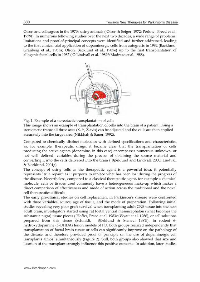
Fig. 1. Example of a stereotactic transplantation of cells This image shows an example of transplantation of cells into the brain of a patient. Using a stereotactic frame all three axes (X, Y, Z axis) can be adjusted and the cells are then applied accurately into the target area (Nikkhah & Sauer, 1992).
Compared to chemically distinct molecules with defined specifications and characteristics as, for example, therapeutic drugs, it became clear that the transplantation of cells producing the active agents (dopamine, in this case) encompasses numerous unknown, or not well defined, variables during the process of obtaining the source material and converting it into the cells delivered into the brain ( Björklund and Lindvall, 2000; Lindvall & Björklund, 2004g). The concept of using cells as the therapeutic agent is a powerful idea: it potentially represents “true repair” as it purports to replace what has been lost during the progress of the disease. Nevertheless, compared to a classical therapeutic agent, for example a chemical molecule, cells or tissues used commonly have a heterogeneous make-up which makes a direct comparison of effectiveness and mode of action across the traditional and the novel cell therapeutics difficult. The early pre-clinical studies on cell replacement in Parkinson’s disease were confronted with three variables: source, age of tissue, and the mode of preparation. Following initial studies revealing very poor graft survival when transplanting adult CNS tissue into the host adult brain, investigators started using rat foetal ventral mesencephalon (what becomes the substantia nigra) tissue pieces ( Hoffer, Freed et al. 1983c; Wyatt et al. 1986), or cell solutions prepared from this tissue (Schmidt, Björklund & Stenevi 1981i), in rodent 6-hydroxydopamine (6-OHDA) lesion models of PD. Both groups realized independently that transplantation of foetal brain tissue or cells can significantly improve on the pathology of the disease, and therefore provided proof of principle on the use of dopaminergic cell transplants almost simultaneously (Figure 2). Still, both groups also showed that size and location of the transplant strongly influence this positive outcome. In addition, later studies
www.intechopen.com
Transplantation of Foetal Ventral Mesencephalic Grafts in Parkinson’s Disease: A Still Evolving Concept with New Regulatory Challenges
381
added a full variety of new information on the variability of cell transplantation in CNS research. Introduction of the 6-hydroxydopamine lesion model provided a stable and highly reproducible animal model of the disease which could be analyzed in detail by also improved animal behaviour assays (Iversen & Dunnett, 1989e; Klein, Metz et al., 2007c; Kloth, Klein et al., 2006d; Kohn Cordeiro, Jiang et al., 2010b; Nikkhah, Rosenthal et al., 1998h). Having these tools at hand for the last 1-2 decades, recently many groups performing cell therapy research on Parkinson disease realized that small or even no methodological changes in the grafting or housing procedure can result in a great variety of the outcome of the transplantation (Döbrössy et al., 2000; Döbrössy et al., 2010;Nikkhah et al., 1993; Nikkhah, Eberhard, Olsson et al., 1995; Nikkhah, Olsson et al., 1994).
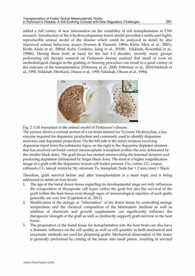
Fig. 2. Cell transplant in the animal model of Parkinson’s disease The picture shows a coronal section of a rat brain stained for Tyrosine Hydroxylase, a key enzyme required for dopamine production and commonly used to identify dopamine neurones and dopamine projections. On the left side is the intact striatum receiving dopamine input from the substantia nigra; on the right is the dopamine depleted striatum that has received rat foetal ventral mesencephalic transplant (within the area delineated by the smaller black dots). The graft tissue has started reinnervating the lesioned striatum and producing dopamine (delineated by larger black dots). The inset is a higher magnification image of a graft with the dopamine neuron cell bodies present. Ctx, cortex; CC, corpus callosum; LV, lateral ventricle; Str, striatum; Tx, transplant. Scale bar = 2 mm; inset = 50µm.
Therefore, graft survival before and after transplantation is a main topic and is being addressed in detail on four levels: 1. The age of the foetal donor tissue regarding its developmental stage not only influences
the composition of therapeutic cell types within the graft but also the survival of the graft within the host tissue even though signs of immunological rejection of such grafts generally are very low (Capetian et al., 2011).
2. Modification of the storage or “hibernation” of the donor tissue by controlling storage temperature and the chemical composition of the hibernation medium as well as addition of chemicals and growth supplements can significantly influence the therapeutic strength of the graft as well as (indirectly support) graft survival in the host tissue.
3. The preparation of the foetal tissue for transplantation into the host brain can also have a dramatic influence on the cell quality as well as cell quantity as both mechanical and enzymatic methods are used for preparing grafts. Mechanical dissociation of the tissue is generally performed by cutting of the tissue into small pieces, resulting in severed
www.intechopen.com

Towards New Therapies for Parkinson's Disease
382
and dead cells at the cut surfaces. The disadvantage of the method are countervailed by the still existing “natural” environment for the cells within each tissue piece, an advantage that usually is lost during enzymatic dissociation of foetal tissue where enzymes are used to actively disintegrate the natural environment (the extracellular matrix) of these cells. Here, the main advantage is that less mechanical cell death is being provoked and that single cell suspensions achieved by such methods usually are reproducibly concentrated.
Both methods also directly influence the last main variable in the full treatment process which is: 4. Stereotactic application of therapeutic foetal tissues or cells deeply within the brain.
Single cell suspension of a very controlled cell concentration and volume can be injected using much thinner cannulae, while tissue pieces require much larger calibre cannulae with higher risks of damaging the host CNS tissue and much less control of amount of the therapeutic tissue, not even regarding the possible high variability of placement of the graft within the individual host (Nikkhah et al., 2009).
Taken together, the relatively simple concept of replacing lost mature dopaminergic neurons with fetal dopaminergic neuroblasts is technically a highly complex and not yet fully understood process. Nevertheless, numerous groups entered the clinical phases of cell therapy in Parkinson’s disease after the strong positive results in the early 80s and the pioneering clinical trials done by Backlund, Madrazo and Lindvall in the mid- and late 80s. These first clinical trials were open-labelled with carefully chosen patients and very low group-sizes. Contrary to most animal data, adrenal autografts containing dopamine producing cells were used in these studies as the need for immunosuppressant after transplantation was eliminated and ethical concerns regarding foetal tissue use were not an issue. The long-term graft survival and functional outcome of adrenal grafts turned out to be poor in the several hundred patients transplanted in numerous trials (Lindvall 1991). In addition, the severity of the side effects of transplanting adrenal medullar tissue into the brain also resulted in high morbidity of patients which was intolerable. Thereby, in the late 80ies of the last century, researchers proceeded with the initial experimental concept of transplanting foetal dopaminergic CNS tissue as the animal results proved robust data on the efficacy of the grafting method (Björklund et al., 1980a; Björklund et al., 1981c; Nikkhah, Bentlage et al., 1994; Nikkhah, Cunningham, Cenci, McKay et al., 1995). In a number of small open-label clinical trials with 2-6 patients each, the benefit of (allogenic) foetal dopaminergic transplants could be shown ranging from moderate to high, stepwise improved by adapting and further developing the methods of application. These early studies showed that foetal dopaminergic neurons can survive long-term for more then 10 years in patients and are capable of improving the classical symptoms of Parkinson’s disease. Most importantly, motivated by these exceptional outcomes, the strong ethical and legal consequences of the use of embryonic and foetal tissue for clinical application were addressed in the creation of clear guidelines restricting the use of these tissues by the applying scientific groups (Boer 1994; Boer 1999). Following these guidelines, more sophisticated double-blinded clinical trials with PD patients were initiated (Freeman et al., 1995d; Olanow et al., 2003). Nevertheless, it also must be stated that during the last century, some of these studies showed a variation of the therapeutic effect of transplanted foetal tissue and cells in patients suffering on PD, including the development of side effects. The variations of the therapeutic
www.intechopen.com
Transplantation of Foetal Ventral Mesencephalic Grafts in Parkinson’s Disease: A Still Evolving Concept with New Regulatory Challenges
383
effects not only differed between study centres, but also within individual study groups of the centres (Brundin et al., 2001; Winkler, Kirik & Björklund, 2005). The side effects of the cell therapy for PD, which occur beside the mentioned variation of the grafts therapeutic potency, mainly consisted of graft induced dyskinesias (GID), motor fluctuations and abnormal involuntary movements similar to those disturbances induced by L-DOPA medication (LID). Both forms of dyskinesias probably accumulated in some patients and can negatively influence the generally moderate improvement of patients studied in clinical trials, and can be associated to either patient related phenomena or transplantation related concurrency and seems to be wide-spread within the highly diverse clinical study designs analyzed (Hagell & Cenci, 2005). As far as safety issues of the grafts are concerned, animal studies have shown that factors influencing the GID-potential of a foetal nigral tissue graft may include age of the foetus, mode of dissection, time and method of storage, culturing of the cells as well as size and translocation of the graft within the brain (Lane & Smith 2010; Lane et al., 2010).
2. Cell therapies in Parkinson’s disease: The new (regulatory) frontier
There are three decades of experience on cell therapy in clinical trials with hundreds of patients having been transplanted with many improving on their disease pattern of PD, yet, many open questions still remain. In particular, since most clinical studies have been conducted independently of one another, there is a marked variability in the individual study protocols that make the general statement about the results highly difficult. This has lead to a desire shared by numerous study centres to generate a single standardized study protocol, based on the experience (positive and negative) of the recent decades that rises to the challenges proposed by clinical cell transplantation into the central nervous system. One of the reasons explaining the high variability in previous study designs may be due to the lack of legislative basis for tissue engineering or cell therapies in several nations. For many years, this fact was recognized in several European nations as such, but many national legislators were waiting for a common, European legislation on the new fields of genetic engineering, cell therapy and biotech manufacturing to adapt such new legislation into their national regulations. After years of discussion a general regulation was finally adopted within the European Union in 2007, which almost generally defines all products from genetically engineered or biotechnologically produced material, tissue or cells as a medicinal drug. This regulation is in force since the 30th December 2008. (Regulation (EC) No 1394/2007 of 13 November 2007).
2.1 What does EC 1394/2007 mean for the field of cell therapies in the central nervous system, particularly for the treatment of PD? The main subject of the regulation EC 1394/2007 is the definition of advanced therapeutic medicinal products (ATMP), their production processes as well as their marketing as an approved drug. The ATMP include gene therapy, somatic cell therapy, tissue engineered products and "combined advanced therapy medicinal products" which are cell and tissue products that contain medical devices or other products as an integral part of it. As a new EU pharmaceutical legislation for Regenerative Medicine, the Regulation 1394/2007/EU (ATMP-Regulation) is valid for all Member States of the EU. The rules set out herein are to be used by all Member States and shall be converted into national law. To meet the special
www.intechopen.com
Towards New Therapies for Parkinson's Disease
384
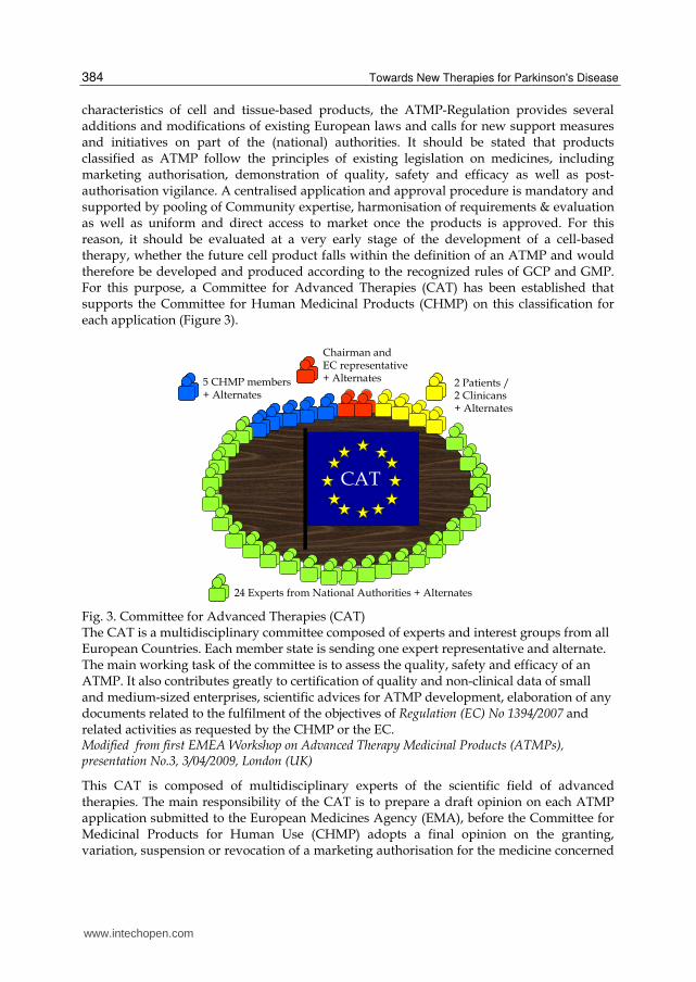
characteristics of cell and tissue-based products, the ATMP-Regulation provides several additions and modifications of existing European laws and calls for new support measures and initiatives on part of the (national) authorities. It should be stated that products classified as ATMP follow the principles of existing legislation on medicines, including marketing authorisation, demonstration of quality, safety and efficacy as well as post-authorisation vigilance. A centralised application and approval procedure is mandatory and supported by pooling of Community expertise, harmonisation of requirements & evaluation as well as uniform and direct access to market once the products is approved. For this reason, it should be evaluated at a very early stage of the development of a cell-based therapy, whether the future cell product falls within the definition of an ATMP and would therefore be developed and produced according to the recognized rules of GCP and GMP. For this purpose, a Committee for Advanced Therapies (CAT) has been established that supports the Committee for Human Medicinal Products (CHMP) on this classification for each application (Figure 3).
CAT CAT
24 Experts from National Authorities + Alternates
5 CHMP members+ Alternates
2 Patients / 2 Clinicans+ Alternates
Chairman and EC representative+ Alternates
Fig. 3. Committee for Advanced Therapies (CAT) The CAT is a multidisciplinary committee composed of experts and interest groups from all European Countries. Each member state is sending one expert representative and alternate. The main working task of the committee is to assess the quality, safety and efficacy of an ATMP. It also contributes greatly to certification of quality and non-clinical data of small and medium-sized enterprises, scientific advices for ATMP development, elaboration of any documents related to the fulfilment of the objectives of Regulation (EC) No 1394/2007 and related activities as requested by the CHMP or the EC. Modified from first EMEA Workshop on Advanced Therapy Medicinal Products (ATMPs), presentation No.3, 3/04/2009, London (UK)
This CAT is composed of multidisciplinary experts of the scientific field of advanced therapies. The main responsibility of the CAT is to prepare a draft opinion on each ATMP application submitted to the European Medicines Agency (EMA), before the Committee for Medicinal Products for Human Use (CHMP) adopts a final opinion on the granting, variation, suspension or revocation of a marketing authorisation for the medicine concerned
www.intechopen.com
Transplantation of Foetal Ventral Mesencephalic Grafts in Parkinson’s Disease: A Still Evolving Concept with New Regulatory Challenges
385
(from EMA webpage). It is also participating in Agency procedures for the certification of quality and non-clinical data for small and medium-sized enterprises developing advanced-therapy medicinal products, and participating in Agency procedures for the provision of scientific recommendations on the classification of advanced-therapy medicinal products in accordance with Article 17 of Regulation (EC) No 1394/2007. Despite several other tasks, this committee is also advising, at the request of the CHMP, on any medicinal product which may require, for the evaluation of its quality, safety or efficacy, expertise in ATMPs.
2.2 How to define the PD cell therapy an ATMP? A PD cell therapy can be considered an ATMP by definition when it is a “tissue engineered product that contains or consists of engineered cells or tissues, and is presented as having properties for, or is used in or administered to human beings with a view to regenerating, repairing or replacing a human tissue. The cells or tissues may be viable or non-viable. It may also contain additional substances, such as cellular products, bio-molecules, biomaterials, chemical substances, scaffolds or matrices.” (Regulation (EC) No 1394/2007) By this definition, cell therapy comes under the ATMP regulations if the cells used are engineered. The term “engineered” is defined for cells or tissues as that which have been subject to substantial manipulation, so that biological characteristics, physiological functions or structural properties relevant for the intended regeneration, repair or replacement are achieved. Also, the cells or tissues are considered ATMP if they are “not intended to be used for the same essential function or functions in the recipient as in the donor”. Any cell or tissue preparation that has been substantially manipulated can thereby be considered an ATMP. “Not substantially manipulated” also has been defined as cutting, grinding, shaping, and centrifugation, soaking in antibiotic or antimicrobial solutions, sterilization, irradiation, cell separation, concentration or purification, filtering, lyophilisation, freezing, cryo-preservation, vitrification. It should be noted that the full process from harvesting of the tissue up to the therapeutic application of the cell therapy needs to be analyzed and considered by the regulatory bodies to decide if a cell therapy becomes an ATMP or not. That decision or definition may have a significant influence on the therapeutic approach and its implementation in clinical practice, as each ATMP is to follow the defined standards of good manufacturing and good clinical practice as within the EU being defined in additional regulations.
2.3 A cell therapy for PD becomes ATMP: What are the implications for the clinical practice? Some cell therapies may be excluded from the ATMP regulation if they fulfil the criteria of the so-called “hospital-exemption”, a paragraph included in most former national health acts. This exemption should allow an application of the therapy which is “prepared on a non-routine basis according to specific quality standards, and used within the same Member State in a hospital under the exclusive professional responsibility of a medical practitioner, in order to comply with an individual medical prescription for a custom-made product for an individual patient.”(Regulation (EC) No 1394/2007). Thereby, some cell therapy approaches might be applied without the full consideration of the EMA, but generally a standard cell therapy approach is too complex to be realized by one medical practitioner only. This is true for some of the cell therapies clinically applied in the late 90’s of the last century. As an example, CNS tissue was dissected from young human foetuses, minced,
www.intechopen.com
Towards New Therapies for Parkinson's Disease
386
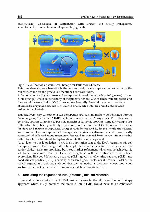
enzymatically dissociated in combination with DNAse and finally transplanted stereotactically into the brain of PD-patients (Figure 4).
Fig. 4. Flow-Sheet of a possible cell therapy for Parkinson’s Disease
This flow sheet shows schematically the conventional process steps for the production of the cell preparation for the previously mentioned clinical studies. A foetus is donated by a woman and transported in medium to the hospital (yellow). In the clinic (orange), under responsibility of the practitioner, the CNS is taken from the foetus and the ventral mesencephalon (VM) dissected mechanically. Foetal dopaminergic cells are obtained by enzymatic dissociation, washed and injected into the brain by stereotactic guided transplantation.
This relatively easy concept of a cell therapeutic approach might now be translated into the “new language” after the ATMP-regulation became active. “Easy concept” in this case is generally spoken compared to possible modern or future approaches using for example iPS cells, which have been genetically engineered, cultured in humid incubators or bioreactors for days and further manipulated using growth factors and hydrogels, while the classical and most applied concept of cell therapy for Parkinson’s disease generally was mostly composed of cells and tissue fragments, dissected from foetal brain tissue without further cell culture but rather direct transplantation into the brain of a patient. As to date - to our knowledge - there is no application sent to the EMA regarding this cell therapy approach. There might likely be applications in the near future as the data of the earlier clinical trials are promising but need further refinement which can be achieved via additional pre-clinical studies. These investigations will be confronted with defined expressions like good laboratory practice (GLP), good manufacturing practice (GMP) and good clinical practice (GCP), generally considered good professional practice (GxP) as the ATMP regulation is defining such cell therapies as medicinal products, whose production are further defined commonly in numerous regulations and directives.
3. Translating the regulations into (practical) clinical research
In general, a new clinical trial in Parkinson’s disease in the EU using the cell therapy approach which likely becomes the status of an ATMP, would have to be conducted
www.intechopen.com

Transplantation of Foetal Ventral Mesencephalic Grafts in Parkinson’s Disease: A Still Evolving Concept with New Regulatory Challenges
387
following the Clinical Trial Directive (Directive 2001/20/EC) as well as the GCP Directive (Directive 2005/28/EC). While these directives are individually to be implemented at a national level for each Member State and can thereby be interpreted variably, the content for EU wide clinical studies is directly applicable. For the example of cell therapy as a clinical study, the manufacture of test material (“the cells”) and the production of scientific data on the safety of the test material are particularly relevant. The GCP-Directive provides, inter alia, that relevant data from preclinical experiments should be planned and conducted by following the standards of good laboratory practice (Directive 2004/10/EC), which means in particular for university facilities a major hurdle as GLP accredited laboratories are rare at such institutes and preclinical data at the university level is usually achieved from via basic research. The logical background to the requirement that certain pre-clinical data shall be generated to the standards of GLP is based on the fact that safety issues of the treatment have been generated by reproducible and fully traceable experiments. GLP also means that the data obtained is reliable and understandable for the authorities; that the data will be archived and regularly monitored; and that audits of the laboratory by the authorities are able to demonstrate sustained and continuous improvement. All aspects of a treatment or manufacturing processes that could result in harm to the patient should therefore be carried out with the best possible method to precisely reduce the risk of the patient. In the field of cell therapy, where the cells are considered an implantable drug, the risks considered are aspects of biocompatibility, toxicity, formulation and use of additives or their removal. Nevertheless, it is our experience that regulatory authorities generally do follow and control upon the principles defined in these GCP and GLP directives. The future clinical trial application on the cell therapy approach for Parkinson’s disease using the mentioned set up of enzymatically dissociated primary foetal cells needs to include experimental data generated according to these principles, for example. In addition, it might also be important for the clinical trial group to include a strategy paper on the requirements for the cell therapy approach once the projects leaves the research phases and is about to enter the therapy phase as at this stage GMP principles are to be followed upon as on the EU level this is directed by Regulation (EC) No 1394/2007.
3.1 PD cell therapy according to GLP and GMP We pointed out that new clinical cell therapy approaches for PD very likely are to be conducted following the international principles of GLP and GMP, and maybe implemented in slightly varied national versions. As both GLP/ GMP were originally developed on an industrial level for decades to ensure product quality (and safety), such industrial standards are likely to be difficult to implement into a historically evolved research institution as most universities hospitals are like. Even though these research institutions generally conduct research in a “good” way, most likely recognized and approved by high ranking, peer reviewed journals and experts in the specific field of science, this “good” way is different to the requirements of GLP and GMP. We highlight the most crucial elements of both GLP and GMP for the example of cell therapy mentioned above and we will try to provide information on the possible implications of these principles for a university hospital set-up. Both GMP/ GLP share a main common feature: A handbook or manual. This collection of written documents should describe all aspects of the quality management and quality assurance of the proposed cell therapy, and ensure that all activities needed to conduct the safety studies, or the manufacture of the test equipment is shown in clear instructions (standard operation procedures or SOPs)
www.intechopen.com
Towards New Therapies for Parkinson's Disease
388
and will be documented in writing. In fact, both manuals have a large amount of common ground as quality is managed similar; but because of their different application areas strong differences stand out. The specific requirements of the manuals are described in the various directives and should be available separately for both the GLP studies as well as GMP manufacturing. It is easily predictable even from this requirement that modern studies of cell therapy now in addition to the clinical study plan, other, more comprehensive documentation are required. As mentioned, both manuals need to define the quality management system. In principle, this management describes who is authorizing the work (Chapter “Management”), where the work is being done (Chapter “Application area”), who is performing the work (Chapter “Personnel”), what equipment and material will be used (Chapter “Equipment”, “Substances”, “Purchase and Storage”) and how the quality is being maintained (Chapter “Document control”, “Quality Assurance”). These possible subdivisions of the term "quality" have been established internationally and this can be found for Europe in the EN ISO 9000 / 9001. Following these requirements, an organization or, as in the example of cell therapy, the clinical facility, can officially be certified to present to the outside the implementation of a quality management system. Especially for the later production according to GMP, the implementation of this process is very important because the regulatory agencies will require an efficient quality management system for the manufacturing license (Regulation (EC) No 1394/2007). A clinical director therefore requires the implementation of a quality system “by the book” (EN ISO 9001) as it is required for GLP and GMP. However, the clinical director must stand behind that decision and also self-identify himself as a model to implement the benefits of an effective quality management system. He must accompany objectives defined by the development of the system, reclaim reports on a regular basis, arrange and identify corrective measures and ensure that agreements are kept up to date. It is not enough here to employ appropriate quality personnel essential to obtain a certification; quality must be understood and experienced as such, otherwise the implementation of the system is condemned to failure from the start. Once the clinical director identified and implemented such quality measures as a crucial part of clinical cell therapy according to the regulations, he now will be confronted with the details of GLP and GMP.
3.2 The good practice in the lab: An introduction to quality assurance As mentioned above, now that it is required to have a quality management system in place, it also is required to produce quality. For example, in both GLP and GMP, it needs to be ensured that cell therapy for the animal safety studies or for the clinical study is being performed under aseptic conditions. While for a specialist in cell culture or the surgeon in the operating room this situation is clearly understood and naturally carried out using corresponding measures consistently, the proof of sterility at medicinal drug manufacturing under GLP and GMP conditions is mandatory. In addition, of course, working environments have to be clean and need to fulfil the prescribed requirements for the manufacturing of ATMP sufficiently. In the given example, all high risk steps of the preparation of the cell product (opening vials, pipetting, etc) must therefore be carried out in a so-called “class A in Class B environment” according to the Directive 2003/94/EC and further defined in EudraLex Volume 4. In general, it means that a conventional safety cabinet (Class A) is installed in a
www.intechopen.com
Transplantation of Foetal Ventral Mesencephalic Grafts in Parkinson’s Disease: A Still Evolving Concept with New Regulatory Challenges
389
category B clean room (EudraLex Volume 4, Annex I). For the class B clean room, strictly defined requirements are in place, which vary significantly from the usual requirements of the operating room. For example, it must be a separated room with restricted access and specifically trained staff. The trained staff of the class B clean room must include a Head of Production and a Head of Quality assurance with separated and defined responsibilities. It should also include a Qualified Person who ensures that each lot was produced and tested in accordance with the guidelines and the specifications set within the QM-Handbook. The responsibilities of and legal requirements for a qualified persons are also defined within national/international directives and regulations and usually include a minimum of 2 years work experience with all processes and within the technical requirements (e.g. class B clean room). Aseptic working conditions of the environment should be ensured by performing online particle counts on volumetric air sampling as well as microbiological monitoring using methods such as settle plates or surface sampling during production. Surface sampling includes the active personnel, which fingers should be tested for microbes after each critical process step and less then 5 microbes / 25cm² in class B environment or >1/25cm² inside the class A workbench should be identified (EudraLex, Volume 4, Annex I). It should also be noted that culture and evaluation of the microbiological sampling plates also need to be performed according to the GLP regulations; for example the incubator should be qualified and monitored constantly. Although these examples cited might be considered as common sense, the actual implementation into a regulatory acceptable level is a very serious and work-intensive task. The authorities’ want these investigations not only being performed and analyzed, but definitely want to have ensured that all tests or production processes can be performed reproducibly within the acceptable specifications. On the industrial side, from where the regulations originate from, these requirements under GMP conditions are usually implemented through activities such as qualification and validation. In consideration of the aseptic work environment mentioned in this paragraph, in the academic research environment one would ensure quality by qualifying the class A work bench, for example. This type of qualification is, according to the GMP regulations, being performed by the implementation steps of design qualification (what kind of class A bench is needed and how to purchase), installation qualification (verification of complete delivery and installation according to the specifications), operational qualification (writing of SOP and test of functionality) and finally performance qualification (is the work bench fully capable of specification delivery for production). This qualification process, using the bench as the example, shows that a large number of documents and tests must be written and performed on a single device, contributing significantly to the manufacturing effort. It is generally not sufficient to order a laboratory work bench from a supplier and use the device directly to produce a cell therapy product. The qualification of the workbench is rather to ensure that optimal aseptic work environment can actually be made possible from a technical point of view and that the equipment is ideal for the production process. The necessary evidence here is carried out as part of the validation, a definition for which there also are continuously evolving industrial standards available (aka ISO norms) that should be followed upon as part of GMP, as well as GLP. For example, the aseptic preparation within the bench it needs to be proven that aseptic conditions actually are achieved before manufacturing begins. This can for example be performed by a cleaning validation, which provides the proof on the
www.intechopen.com
Towards New Therapies for Parkinson's Disease
390
basis of scientific methods that both the optimum cleaning procedure, the best method as well as the most suitable chemicals were used to scientifically exclude risks to the cell product by exposure to microbes or to the cleaning procedure itself. It is conceivable to prepare an aseptic work surface to roughly prepare or cut the tissue for later cell preparation steps. In this case, however, it should be proven by scientific evidence that no risk is transmitted by the surface to the tissue or indirectly to the patient by chemical residues left over after the cleaning process, a simple fact but overlooked easily and actually technically difficult to prove. Using as an example the need for aseptic work environment leading up to clinical treatment, it becomes clear that the preparation for cell therapy under either GLP or GMP purposes is labour intensive, including the generation of several SOPs, performing of several validation tests and filling out forms to collect the data. The validation activities thereby need to be planned very carefully as the clinical director never wants only to focus on single issues of the process but on all aspects of the full manufacturing process. For planning validations in a good practice environment, a so-called validation master plan should thereby be discussed and generated before any validation is actually being performed. This plan is (yet) not mandatory in the regulations, but is one of the first documents a regulator officer will ask for during an audit. It provides information of how the clinical department operates, who is in control of the different validation activities, and how the manufacturing, quality control and personnel are being managed. Main integral part of the validation master plan generally is the previously mentioned qualification procedure, but it also includes activities performed to generate general risk analysis documentation, validation approaches to deal with identified risks and timelines to finish the validation activities. Finally, it is clear that in addition to the head of production, the Quality Assurance Manager and the Qualified Person, a validation expert should also be present in addition to seamlessly link the areas of manufacturing and quality assurance of the product by adapted validation activities. It also becomes clear that once the proposed cell therapy is defined as a medicinal product by the authorities, the principle investigator of the cell therapy - used to research standards - now is confronted with multiple legal requirements that for newcomers might become irritating and frustrating. A high number of needs are to be fulfilled, not only requiring a tremendous piece of work time to be invested to “get the paper work done”, but also technical and personal requirements need to be put in place, most likely coming along with a substantial amount of financial demand. If one considers the usually small numbers of “products” the principle investigator will need to address small clinical trials, these high risk investments will not easily find the internal support needed. Nevertheless, in the long-term, once implemented into a working system, this newly implemented system will lead to less hassles and burdens to carry out additional cell therapy studies and significantly promote the standardisation in multicenter trials.
4. Standardizing new cell therapies for PD: The TRANSEURO project
As discussed in the introduction of this chapter, in recent decades several clinical studies using cell therapy in PD were investigated often producing large differences in the outcome (Winkler, Kirik & Björklund 2005h). In many cases it was found that the protocols needed to conduct the therapy varied significantly in many places and a generalized statement about the efficiency of the studies was difficult to reach. In parallel, the legislation has only
www.intechopen.com
Transplantation of Foetal Ventral Mesencephalic Grafts in Parkinson’s Disease: A Still Evolving Concept with New Regulatory Challenges
391
recently clearly and very significantly changed so that the implementation of studies on cell therapy for small clinical facilities and centres quickly reaches the limits of feasibility, when all the national and international laws and guidelines need to be considered. With the desire of reducing the inconsistencies and variations across centres, a consortium of European based leading clinical and research centres implicated in cell therapy recently have been awarded European Union funding aimed at addressing critical questions in the context of cell therapy in PD, particularly concerning patient selection, tissue preparation, cell delivery, immunosuppression, and off-state dyskinesia. A key objective of the TRANEURO project funded by the FP7 European Commission Health programme is to generate standardized protocols that can serve as a template for all future clinical trials in the cell therapy field including stem cell-based therapies for PD. As part of an international multi-centre clinical study, both preclinical and clinical standards are being defined across the centres in order to generate the best practice approaches investigating the feasibility and clinical efficacy of foetal cell therapy in PD (see also TransEuro homepage http://www.transeuro.org.uk/). Furthermore, the stringent regulatory requirements to implement the standards of GMP and GCP will be addressed by close cooperation between the various centres in particular through joint protocol development and the intensive exchange between the project managers of the different areas to combine synergies to effectively take-on the numerous technical and regulatory hurdles together. By using modern communication media such as web meetings, online training classes, an international project management, but also classic laboratory visits, all elements for manufacturing and the clinical treatment of patients are developed and established, to conserve resources and optimize the expected outcome. The legislature has certainly imposed very high standards of patients’ safety in clinical trials, but as illustrated in the previous examples, such standards hereby reached a high level that can only be met with great difficulty by classical research institutions alone. The declared goal of the TRANSEURO consortium is to achieve successful cell therapy for patients suffering from PD, but at the same time to provide the feasibility of implementing a therapy at regulatory acceptable level. It is the authors’ opinion that the enhancement of the regulations during the last years is a double-edged sword with a potential to become a sharp “weapon”. On one hand it maximizes patient and promotes the standardization of innovative procedures. On the other side, it creates a substantial challenge, both for personal and financial resources, to meet the new regulatory processes and requirements especially for university and other academic institutions.
5. Conclusion
Cell transplantation for PD-patients is a promising therapy, based on the previous pre-clinical and clinical research. The use of foetal tissue as proposed in the TRANSEURO project holds a number of (ethical) concerns and technical handicaps, but is an ideal candidate to merge basic science and industrial standards to achieve an efficient therapy. This may set the basis of future cell therapeutically approaches with considerably more complex processes and thereby noticeable higher regulatory demands. The worldwide effort working with embryonic stem cells and with induced pluripotent stem cells (iPS) might in the near future generate alternative candidate cells for clinical therapy in PD. However, complicated manufacturing processes involving gene manipulation, advanced cell culture techniques and risk of tumorigenicity will benefit greatly from the currently planned
www.intechopen.com
Towards New Therapies for Parkinson's Disease
392
TransEuro clinical trial, as this will set the stage for the potential and risks of clinical cell therapy in PD for the near future.
6. Acknowledgement
The authors are partially funded by the European Commission under the 7th Framework Programme – HEALTH – Collaborative Project Transeuro (Contract n°242003) – www.transeuro.org.uk
7. References
Backlund, E O, P O Granberg, B Hamberger, E Knutsson, A Mårtensson, G Sedvall, A Seiger, and L Olson. 1985a. “Transplantation of adrenal medullary tissue to striatum in parkinsonism. First clinical trials.” Journal of Neurosurgery 62 (2) (February): 169-173. doi:10.3171/jns.1985.62.2.0169.
Björklund, A, SB Dunnett, U Stenevi, M E Lewis, and S D Iversen. 1980a. “Reinnervation of the denervated striatum by substantia nigra transplants: functional consequences as revealed by pharmacological and sensorimotor testing.” Brain Research 199 (2) (October 20): 307-333.
Björklund, A, and O Lindvall. 2000. “Cell replacement therapies for central nervous system disorders.” Nature Neuroscience 3 (6) (June): 537-544. doi:10.1038/75705.
Björklund, A, U Stenevi, SB Dunnett, and F H Gage. 1982b. “Cross-species neural grafting in a rat model of Parkinson’s disease.” Nature 298 (5875) (August 12): 652-654.
Björklund, A, U Stenevi, SB Dunnett, and S D Iversen. 1981c. “Functional reactivation of the deafferented neostriatum by nigral transplants.” Nature 289 (5797) (February 5): 497-499.
Boer, GJ. 1994. “Ethical guidelines for the use of human embryonic or fetal tissue for experimental and clinical neurotransplantation and research. Network of European CNS Transplantation and Restoration (NECTAR).” Journal of Neurology 242 (1) (December): 1-13.
Boer GJ. 1999. “Ethical issues in neurografting of human embryonic cells.” Theoretical
Medicine and Bioethics 20 (5) (September): 461-475. Boshes, B, E R Blonsky, J Arbit, and K Klein. 1969. “Effect of L-dopa on individual
symptoms of parkinsonism.” Transactions of the American Neurological Association 94: 229-231.
Brundin P, S Dunnett, A Björklund, Nikkhah G. 2001. "Transplanted dopaminergic neurons: more or less?" Nature Medicine. (May) 7(5): 512-513.
Capetian, P, M Döbrössy, C Winkler , M Prinz, GNikkhah. 2011. "To be or not to be accepted: the role of immunogenicity of neural stem cells following transplantation into the brain in animal and human studies." Seminars in Immunopathology. [Epub ahead of print]
Cordeiro, Karina Kohn, Wei Jiang, Anna Papazoglou, Sérgio Bernardo Tenório, M Döbrössy, and Guido Nikkhah. 2010b. “Graft-mediated functional recovery on a skilled forelimb use paradigm in a rodent model of Parkinson’s disease is dependent on
www.intechopen.com
Transplantation of Foetal Ventral Mesencephalic Grafts in Parkinson’s Disease: A Still Evolving Concept with New Regulatory Challenges
393
reward contingency.” Behavioural Brain Research 212 (2) (October 15): 187-195. doi:10.1016/j.bbr.2010.04.012.
Directive 2001/20/EC of 4 April 2001, of the European Parliament and of the Council on the approximation of the laws, regulations and administrative provisions of the Member States relating to implementation of good clinical practice in the conduct of clinical trials on medicinal products for human use
Directive 2003/94/EC, of 8 October 2003, of the European Parliament and of the Council laying down the principles of good manufacturing practice in respect of medicinal products for human use and investigational medicinal products for human use
Directive 2004/10/EC, of 11 February 2004, of the European Parliament and of the Council on the harmonisation of laws, regulations and administrative provisions relating to the application of the principles of good laboratory practice and the verification of their applications for tests on chemical substances (codified version).
Directive 2005/28/EC, of 8 April 2005, of the European Parliament and of the Council laying down principles and detailed guidelines for good clinical practice as regards investigational medicinal products for human use, as well as the requirements for authorisation of the manufacturing or importation of such products
Döbrössy, MD, M Le Moal, M F Montaron, and N Abrous. 2000. “Influence of environment on the efficacy of intrastriatal dopaminergic grafts.” Experimental Neurology 165 (1) (September): 172-183. doi:10.1006/exnr.2000.7462.
Döbrössy M, M Busse, T Piroth, A Rosser, S Dunnett, G Nikkhah. 2010. "Neurorehabilitation with neural transplantation." Neurorehabilitation and Neural Repair. (Oct) 24(8): 692-701
EN ISO 9001, 2000, Quality Management Systems - Requirements, International Organization for Standardization
EudraLex, Volume 4, The Rules Governing Medicinal Products in the European Union, EU
Guidelines to Good Manufacturing Practice Medicinal Products for Human and Veterinary
Use, Dec. 2010 Freed, WJ, H E Cannon-Spoor, and R J Wyatt. 1984b. “Embryonic brain grafts in an animal
model of Parkinson’s disease. Criteria for human application.” Applied
Neurophysiology 47 (1-2): 16-22. Freeman, T B, C W Olanow, R A Hauser, G M Nauert, DA Smith, C V Borlongan, P R
Sanberg, D A Holt, J H Kordower, and F J Vingerhoets. 1995d. “Bilateral fetal nigral transplantation into the postcommissural putamen in Parkinson’s disease.” Annals
of Neurology 38 (3) (September): 379-388. doi:10.1002/ana.410380307. Hagell, Peter, and M Angela Cenci. 2005. “Dyskinesias and dopamine cell replacement in
Parkinson’s disease: a clinical perspective.” Brain Research Bulletin 68 (1-2) (December 15): 4-15. doi:10.1016/j.brainresbull.2004.10.013.
Hoffer, B, W Freed, L Olson, and R J Wyatt. 1983c. “Transplantation of dopamine-containing tissues to the central nervous system.” Clinical Neurosurgery 31: 404-416.
Hornykiewicz. 1962. “[Dopamine (3-hydroxytyramine) in the central nervous system and its relation to the Parkinson syndrome in man].” Deutsche Medizinische Wochenschrift
(1946) 87 (September 7): 1807-1810. doi:10.1055/s-0028-1114024.
www.intechopen.com
Towards New Therapies for Parkinson's Disease
394
Hornykiewicz, O. 1966. “Dopamine (3-hydroxytyramine) and brain function.” Pharmacological Reviews 18 (2) (June): 925-964.
Iversen, S D, and SB Dunnett. 1989e. “Functional compensation afforded by grafts of foetal neurones.” Progress in Neuro-Psychopharmacology & Biological Psychiatry 13 (3-4): 453-467.
Klein, A, Gerlinde A Metz, Anna Papazoglou, and Guido Nikkhah. 2007c. “Differential effects on forelimb grasping behavior induced by fetal dopaminergic grafts in hemiparkinsonian rats.” Neurobiology of Disease 27 (1) (July): 24-35. doi:10.1016/j.nbd.2007.03.010.
Kloth, Verena, A Klein, David Loettrich, and Guido Nikkhah. 2006d. “Colour-coded pellets increase the sensitivity of the staircase test to differentiate skilled forelimb performances of control and 6-hydroxydopamine lesioned rats.” Brain Research
Bulletin 70 (1) (June 15): 68-80. doi:10.1016/j.brainresbull.2006.04.006. Lane, EL, and GA Smith. 2010. “Understanding graft-induced dyskinesia.” Regenerative
Medicine 5 (5) (September): 787-797. doi:10.2217/rme.10.42. Lane, E, Anders Björklund, S Dunnett, and Christian Winkler. 2010. “Unravelling the
mechanisms underlying graft-induced dyskinesia.” Progress Brain Research. 184: 295-309.
Lees, Andrew J, John Hardy, and Tamas Revesz. 2009. “Parkinson’s disease.” Lancet 373 (9680) (June 13): 2055-2066. doi:10.1016/S0140-6736(09)60492-X.
Lindvall, O. 1991. “Transplants in Parkinson’s disease.” European Neurology 31 Suppl 1: 17-27.
Lindvall, O, S Rehncrona, P Brundin, B Gustavii, B Astedt, H Widner, T Lindholm, et al. 1989f. “Human fetal dopamine neurons grafted into the striatum in two patients with severe Parkinson’s disease. A detailed account of methodology and a 6-month follow-up.” Archives of Neurology 46 (6) (June): 615-631.
Lindvall, Olle, and Anders Björklund. 2004g. “Cell therapy in Parkinson’s disease.” NeuroRx: The Journal of the American Society for Experimental NeuroTherapeutics 1 (4) (October): 382-393.
Madrazo, I, V León, C Torres, M C Aguilera, G Varela, F Alvarez, A Fraga, R Drucker-Colín, F Ostrosky, and M Skurovich. 1988. “Transplantation of fetal substantia nigra and adrenal medulla to the caudate nucleus in two patients with Parkinson’s disease.” The New England Journal of Medicine 318 (1) (January 7): 51. doi:10.1056/NEJM198801073180115.
Mones, R J. 1969. “An evaluation of L-dopa in Parkinson patients.” Transactions of the
American Neurological Association 94: 307-309. Nikkhah, G, H Sauer. 1992. "Transplantation von Gehirnzellen - Vision oder Realität?"
Deutsches Ärzteblatt. 89 (3A): 106-114 Nikkhah, G, C Bentlage, M G Cunningham, and A Björklund. 1994. “Intranigral fetal
dopamine grafts induce behavioral compensation in the rat Parkinson model.” The
Journal of Neuroscience: The Official Journal of the Society for Neuroscience 14 (6) (June): 3449-3461.
Nikkhah, G, M G Cunningham, M A Cenci, R D McKay, and A Björklund. 1995. “Dopaminergic microtransplants into the substantia nigra of neonatal rats with
www.intechopen.com

Transplantation of Foetal Ventral Mesencephalic Grafts in Parkinson’s Disease: A Still Evolving Concept with New Regulatory Challenges
395
bilateral 6-OHDA lesions. I. Evidence for anatomical reconstruction of the nigrostriatal pathway.” The Journal of Neuroscience: The Official Journal of the Society
for Neuroscience 15 (5 Pt 1) (May): 3548-3561. Nikkhah, G, W M Duan, U Knappe, A Jödicke, and A Björklund. 1993. “Restoration of
complex sensorimotor behavior and skilled forelimb use by a modified nigral cell suspension transplantation approach in the rat Parkinson model.” Neuroscience, 56 (1) (September): 33-43.
Nikkhah, G, M Olsson, J Eberhard, C Bentlage, M G Cunningham, and A Björklund. 1994. “A microtransplantation approach for cell suspension grafting in the rat Parkinson model: a detailed account of the methodology.” Neuroscience, 63 (1) (November): 57-72.
Nikkhah, G, J Eberhard, M Olsson, and A Björklund. 1995. “Preservation of fetal ventral mesencephalic cells by cool storage: in-vitro viability and TH-positive neuron survival after microtransplantation to the striatum.” Brain Research, 687 (1-2) (July 31): 22-34.
Nikkhah, G, C Rosenthal, H J Hedrich, and M Samii. 1998h. “Differences in acquisition and full performance in skilled forelimb use as measured by the ‘staircase test’ in five rat strains.” Behavioural Brain Research 92 (1) (April): 85-95.
Nikkhah G, C Rosenthal, G Falkenstein, A Roedter, A Papazoglou, A Brandis. 2009. "Microtransplantation of dopaminergic cell suspensions: further characterization and optimization of grafting parameters." Cell Transplantation, 18(2): 119-133.
Olanow, C Warren, Christopher G Goetz, Jeffrey H Kordower, A Jon Stoessl, Vesna Sossi, Mitchell F Brin, Kathleen M Shannon, et al. 2003. “A double-blind controlled trial of bilateral fetal nigral transplantation in Parkinson’s disease.” Annals of Neurology 54 (3) (September): 403-414. doi:10.1002/ana.10720.
Olson, L, E O Backlund, W Freed, M Herrera-Marschitz, B Hoffer, A Seiger, and I Strömberg. 1985e. “Transplantation of monoamine-producing cell systems in oculo and intracranially: experiments in search of a treatment for Parkinson’s Disease.” Annals of the New York Academy of Sciences 457: 105-126.
Olson, L, and A Seiger. 1972. “Brain tissue transplanted to the anterior chamber of the eye. 1. Fluorescence histochemistry of immature catecholamine and 5-hydroxytryptamine neurons reinnervating the rat iris.” Zeitschrift Für Zellforschung Und Mikroskopische
Anatomie (Vienna, Austria: 1948) 135 (2): 175-194. Perlow, M J, WJ Freed, BJ Hoffer, A Seiger, L Olson, and R J Wyatt. 1979f. “Brain grafts
reduce motor abnormalities produced by destruction of nigrostriatal dopamine system.” Science (New York, N.Y.) 204 (4393) (May 11): 643-647.
Pollock, M, and RW Hornabrook. 1966. “The prevalence, natural history and dementia of Parkinson’s disease.” Brain 89 (3): 429-448.
Regulation (EC) No 1394/2007 of the European Parliament and of the Council of 13 November 2007 on advanced therapy medicinal products and amending Directive 2001/83/EC and Regulation (EC) No 726/2004,
Schmidt, R H, A Björklund, and U Stenevi. 1981i. “Intracerebral grafting of dissociated CNS tissue suspensions: a new approach for neuronal transplantation to deep brain sites.” Brain Research 218 (1-2) (August 10): 347-356.
www.intechopen.com

Towards New Therapies for Parkinson's Disease
396
Winkler, Christian, Deniz Kirik, and Anders Björklund. 2005h. “Cell transplantation in Parkinson’s disease: how can we make it work?” Trends in Neurosciences 28 (2) (February): 86-92. doi:10.1016/j.tins.2004.12.006.
Wyatt, R J, J M Morihisa, R K Nakamura, and WJ Freed. 1986. “Transplanting tissue into the brain for function: use in a model of Parkinson’s disease.” Research Publications -
Association for Research in Nervous and Mental Disease 64: 199-208.
www.intechopen.com

Towards New Therapies for Parkinson's DiseaseEdited by Prof. David Finkelstein
ISBN 978-953-307-463-4Hard cover, 396 pagesPublisher InTechPublished online 02, November, 2011Published in print edition November, 2011
InTech EuropeUniversity Campus STeP Ri Slavka Krautzeka 83/A 51000 Rijeka, Croatia Phone: +385 (51) 770 447 Fax: +385 (51) 686 166www.intechopen.com
InTech ChinaUnit 405, Office Block, Hotel Equatorial Shanghai No.65, Yan An Road (West), Shanghai, 200040, China
Phone: +86-21-62489820 Fax: +86-21-62489821
Parkinson's disease (PD) is characterised clinically by various non-motor and progressive motor symptoms,pathologically by loss of dopamine producing cells and intraneuronal cytoplasmic inclusions composedprimarily of ?-synuclein. By the time a patient first presents with symptoms of Parkinson's disease at the clinic,a significant proportion of the cells in the substantia nigra have already been destroyed. This degenerationprogresses despite the current therapies until the cell loss is so great that the quality of normal life iscompromised. The dopamine precursor levodopa is the most valuable drug currently available for thetreatment of PD. However for most PD patients, the optimal clinical benefit from levodopa decreases aroundfive to six years of treatment. The aim of the chapters of this book is to work towards an understanding in themechanisms of degeneration and to develop disease modifying therapies.
How to referenceIn order to correctly reference this scholarly work, feel free to copy and paste the following:
Sven Mo ̈llers, Ma ́te ́ Do ̈bro ̈ssy and Guido Nikkhah (2011). Transplantation of Foetal Ventral MesencephalicGrafts in Parkinson’s Disease: A Still Evolving Concept with New Regulatory Challenges, Towards NewTherapies for Parkinson's Disease, Prof. David Finkelstein (Ed.), ISBN: 978-953-307-463-4, InTech, Availablefrom: http://www.intechopen.com/books/towards-new-therapies-for-parkinson-s-disease/transplantation-of-foetal-ventral-mesencephalic-grafts-in-parkinson-s-disease-a-still-evolving-conce

© 2011 The Author(s). Licensee IntechOpen. This is an open access articledistributed under the terms of the Creative Commons Attribution 3.0License, which permits unrestricted use, distribution, and reproduction inany medium, provided the original work is properly cited.