Foetal circulation ppt
38
-
Upload
saadiyah-naeemi -
Category
Science
-
view
247 -
download
12
Transcript of Foetal circulation ppt


Topic Given By:
Dr.Abida Arif.
OBJECTIVE:
• Describe the normal foetal
circulation and mention the
changes that occur in it at placental
stage and after birth.

•The Foetus:It is the term used to refer to a prenatal mammal between it’s
embryonic state and it’s birth.
•The Placenta:The organ in human mother responsible for the supplying of
oxygen and nutritive material to the fetus and for the elimination of CO2
and nitrogenous waste out of the fetus.

•The Umiblical Cord:A flexible cord-like structure containing blood vessels and
attaching a human to the placenta responsible for the supplying and
elimination of CO2 and O2.

•Foetal Circulation:
The circulation of oxygenated
blood, de-oxygenated blood,
nutritive material etc in the foetus
is termed as foetal circulation.

•Placental Role in Foetal Circulation:The circulatory system of the mother is not directly connected to that
of the fetus, so the placenta functions as the respiratory center for the
fetus as well as a site of filtration for plasma nutrients and wastes.
•Foetal Lungs:Pulmonary vascular resistance is the resistance offered to blood
through lungs. The resistance is very high in fetus because of the
non-functioning of fetal lungs. Because of this high pressure, the
blood is diverted from pulmonary artery into aorta.

•Blood Vessels In
Foetus:The blood vessels responsible for foetal circulation are:
1. Umblical Vein.
2. Umblical Artery.
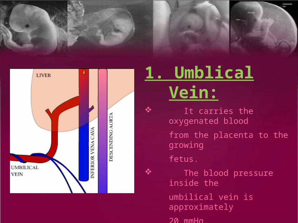
1. Umblical Vein: It carries the oxygenated
blood
from the placenta to the growing
fetus.
The blood pressure inside the
umbilical vein is approximately
20 mmHg.
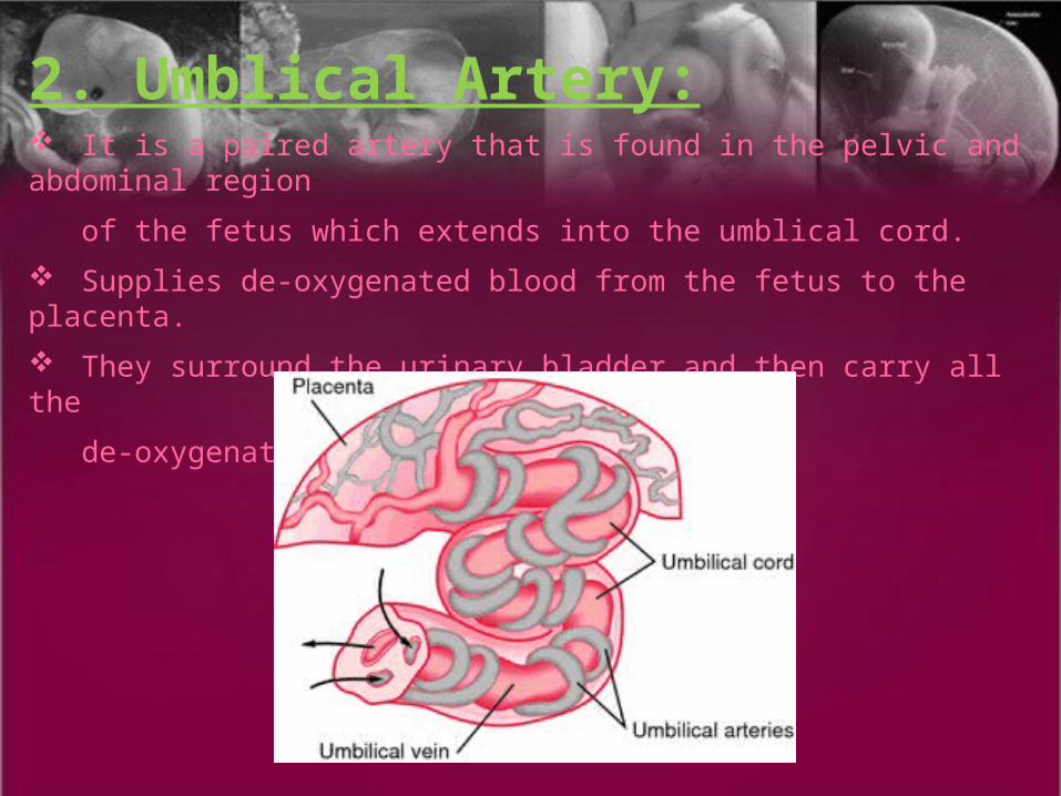
2. Umblical Artery: It is a paired artery that is found in the pelvic and abdominal region
of the fetus which extends into the umblical cord.
Supplies de-oxygenated blood from the fetus to the placenta.
They surround the urinary bladder and then carry all the
de-oxygenated blood out of the fetus.
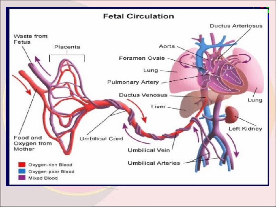
•Shunts Involved In Foetal Circulation:
There are three shunts present in a fetus, they are:
1. Ductus Venosus.
2. Ductus Arteriosus.
3. Foramen Ovale.

1. Ductus Venosus:
The Ductus Venosus shunts the portion of left umblical vein blood
flow directly to the inferior vena cava.
Allows oxygenated blood from the placenta to bypass the liver.
2. Ductus Arteriosus:
Also called Ductus Botalli.
Connects the pulmonary artery to the proximal descending aorta.
It allows most of the blood from the right-ventricle to bypass the
fetus’ fluid-filled non-functioning lungs.

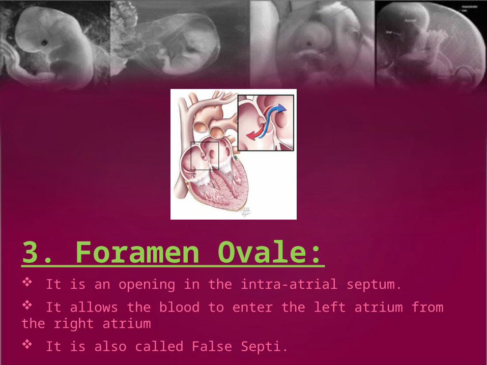
3. Foramen Ovale: It is an opening in the intra-atrial septum.
It allows the blood to enter the left atrium from the right atrium
It is also called False Septi.

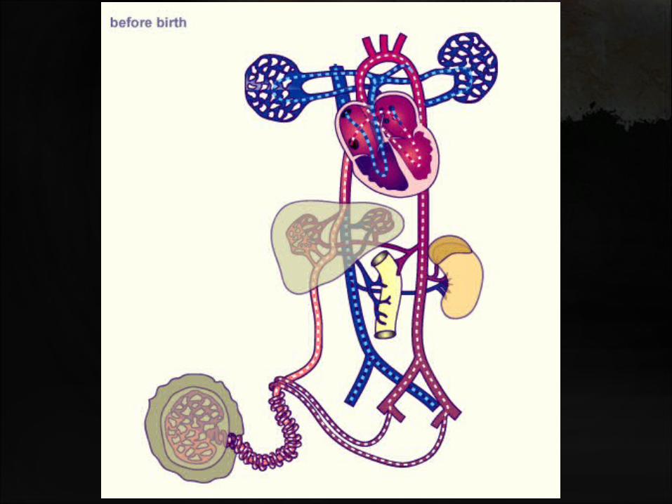
Steps Of Foetal Circulation:
Step.1:The placenta accepts the bluest blood (blood without oxygen) from
the fetus through blood vessels that leave the fetus through the Umbilical
Cord (Umblical Arteries).
Step.2:When blood goes through the placenta it picks up oxygen and
becomes Red.


Step.3:The red blood then returns to the fetus via the umbilical cord
(umbilical vein).
Step.4:The red blood that enters the fetus passes through the fetal liver
and enters the right side of the heart.
Step.5:
Foramen Ovale allows the reddest blood to go from the right
atrium to left atrium and then to the left ventricle and out the aorta. As a
result the blood with the most oxygen gets to the brain.

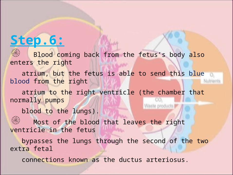
Step.6:۞ Blood coming back from the fetus’s body also enters the right
atrium, but the fetus is able to send this blue blood from the right
atrium to the right ventricle (the chamber that normally pumps
blood to the lungs).
۞ Most of the blood that leaves the right ventricle in the fetus
bypasses the lungs through the second of the two extra fetal
connections known as the ductus arteriosus.

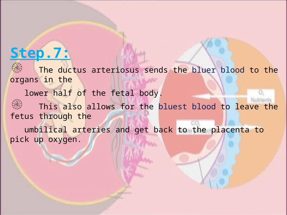
Step.7:۞ The ductus arteriosus sends the bluer blood to the organs in the
lower half of the fetal body.
۞ This also allows for the bluest blood to leave the fetus through the
umbilical arteries and get back to the placenta to pick up oxygen.

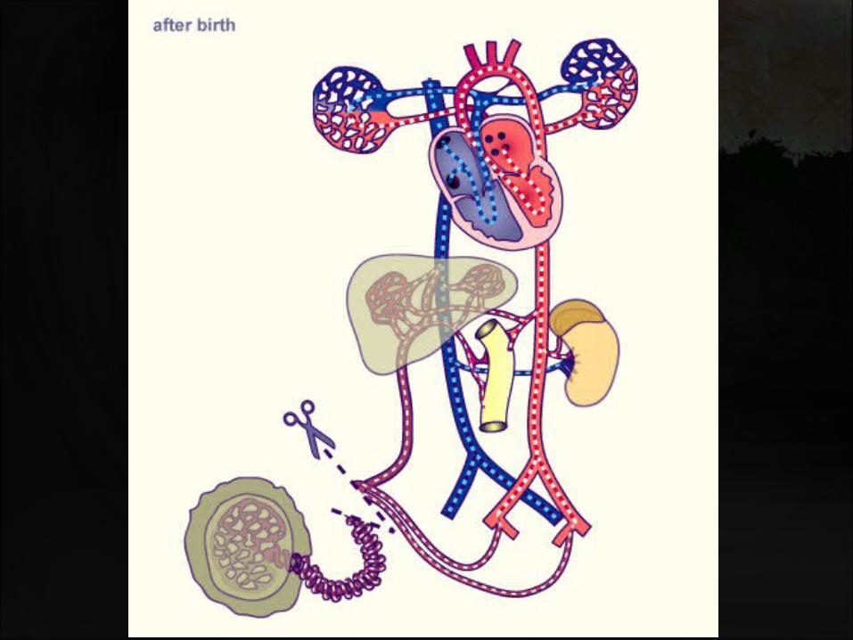
•The Circulatory Changes
After Birth:
Ѡ The Placenta is replaced by the Lungs as the organ of respiratory exchange.
Ѡ The lungs and pulmonary vessels expand thereby significantly lowering the resistance to blood flow.
Ѡ Subsequently the pressure in the pulmonary artery and the right side of the heart is decreased.
Ѡ The pressure of the left side of the heart increases
Ѡ The increasing pressure of blood in the left side of the heart decreases the vascular resistance of the lungs, therefore, the blood now enters the lungs as a respiratory exchange.

• What happens to the shunts
at birth?
Ѡ Closure of the Ductus Venosus.
Ѡ Closure of the Foramen Ovale.
Ѡ Closure of the Ductus Arteriosus.

Ѡ Closure of the Ductus Venosus:
1. Functional closure occurs within minutes of birth.
2. Structural closure occurs within 3 to 7 days.
3. After it closes, the remnant is known as ligamentum venosum.
4. Closure of ductus venosus is caused by strong contraction of muscle wall of ductus venosus, but the cause of this contraction is not revealed yet.
Ѡ Closure of the Ductus Arteriosus:1. Closure of ductus arteriosus is by smooth muscle contraction.
2. It is further replaced by fibrous tissue, called ligamentum arteriosum.
3. At birth, opposite direction of blood flow from aorta to pulmonary artery supplies more oxyginated blood than before.
4. This contraction of smooth muscle occurs becuase of the increase in availability of oxygen.
5. The degree of smooth muscle contraction is highly dependant on more availability of oxygen.
Ѡ Closure of the Foramen Ovale:
1. Before birth the foramen ovale allows most of the oxygenated blood entering the right atrium from the Inferior Vena Cava to pass into the left atrium.
2. Closes at birth due to decreased flow from placenta and Inferior Vena Cava to hold open foramen.
3. More importantly because of increased pulmonary blood flow and pulmonary venous return to left heart causing the pressure in the left atrium to be higher than in the right atrium.
4. The increased left atrial pressure then closes the foramen ovale against the septum secundum (between right and left atrium).
5. The output from the right ventricle now flows entirely into the pulmonary circulation.


‽ The Umblical arteries carry Deoxygenated blood.‽ The Umblical veins carry Oxygenated Blood.‽ When placental blood flow is cut off, there is sudden hypoxia(deficiency of oxygen) and hypercapnia(elevation of CO2).

‽ Oxygen in fetal circulation is
15-20 mmHg.‽ Oxygen after birth increases to
100 mmHg.‽ The right Ventricular wall is thicker in
foetal circulation.‽ The Left Ventricular wall gets thicker by the end of the first month after birth.


![[PPT]Slide 1 AND EMBRYOLOGY... · Web viewFetal circulation Fetal circulation : Physiological and morphological aspects Post natal and transitional circulation: Changes at birth and](https://static.fdocuments.us/doc/165x107/5b5d0cb07f8b9a9c398d539f/pptslide-1-and-embryology-web-viewfetal-circulation-fetal-circulation-.jpg)