Poultry Diseases CVM
34
Rob Porter Small Flock Diseases 1 Diseases of Small Poultry Flocks Rob Porter, D.V.M., Ph.D., DACVP, DACPV Minnesota Veterinary Diagnostic Laboratory 1333 Gortner Avenue St. Paul, MN 55109 (612) 624-7400 and Department of Veterinary Population Medicine University of Minnesota College of Veterinary Medicine [email protected] References: Disease of Poultry: M. Saif (ed.); Iowa S tate University Press, 2003 Poultry Production (13 th Edition); R. Austic and C. Neshem, Lea & Febiger Publishers, 1990 Avian Disease Manual (4 th Edition): C. Whiteman and A. Bickford (eds.), Kendall Hunt Publishing, 1990 Avian Histopathology (2 nd Edition), C. Riddell (ed); AAAP, 1996. The following list is the most common diseases I have diagnosed in backyard or small poultry flocks (mostly chickens). 1. Staphylococcus aureus infection 2. Escherichia coli infection (colibacillosis) 3. Mycoplasma gallisepticum infection 4. Marek’s disease 5. Infectious laryngotracheitis (ILT) 6. Visceral gout 7. Fatty liver 8. Cloacal prolapse 9. Osteomalacia 10. Ascite syndrome 11. Coccidiosis 12. Ascarids 13. Ectoparasites- mites and lice
-
Upload
parashuram-shanigaram -
Category
Documents
-
view
244 -
download
1
Transcript of Poultry Diseases CVM
8/17/2019 Poultry Diseases CVM
http://slidepdf.com/reader/full/poultry-diseases-cvm 1/34
Rob Porter Small Flock Diseases
1
Diseases of Small Poultry Flocks
Rob Porter, D.V.M., Ph.D., DACVP, DACPV
Minnesota Veterinary Diagnostic Laboratory
1333 Gortner AvenueSt. Paul, MN 55109
(612) 624-7400
and
Department of Veterinary Population Medicine
University of Minnesota College of Veterinary Medicine
References:
Disease of Poultry: M. Saif (ed.); Iowa State University Press, 2003
Poultry Production (13th Edition); R. Austic and C. Neshem, Lea & Febiger Publishers, 1990
Avian Disease Manual (4th Edition): C. Whiteman and A. Bickford (eds.), Kendall Hunt
Publishing, 1990
Avian Histopathology (2nd
Edition), C. Riddell (ed); AAAP, 1996.
The following list is the most common diseases I have diagnosed in backyard or small poultry
flocks (mostly chickens).
1. Staphylococcus aureus infection
2. Escherichia coli infection (colibacillosis)
3. Mycoplasma gallisepticum infection
4. Marek’s disease
5. Infectious laryngotracheitis (ILT)
6. Visceral gout
7. Fatty liver
8. Cloacal prolapse
9. Osteomalacia
10. Ascite syndrome
11. Coccidiosis
12. Ascarids
13. Ectoparasites- mites and lice
8/17/2019 Poultry Diseases CVM
http://slidepdf.com/reader/full/poultry-diseases-cvm 2/34
Rob Porter Small Flock Diseases
2
Staphylococcus aureus Infection
Definition: Septicemic infection of many birds characterized by arthritis and tenosynovitis
(inflammation of tendon sheath)
Synonyms: Staphylococcosis- often associated with bumblefoot or omphalitis (navel ill)
Cause: Staphylococcus aureus- Gram-positive coccus
Epidemiology: S. aureus is an opportunist that must penetrate skin barrier. Normal inhabitant
on skin surfaceskin injuryorganism penetrates skin to invade blood. Skin injury may be
scratches, excessively moist skin, needle or claw punctures
Transmission: Ubiquitous on skin and in environment and must penetrate skin. Not directlytransmitted from bird to bird
Clinical signs: Large variety of diseases: 1. Omphalitis (yolk sac infection), 2. Necrotic
dermatitis. 3. Necrotic skin lesions/abscesses 4. Arthritis and tenosynovitis, 5. Osteomyelitis
(infection and inflammation of bone)
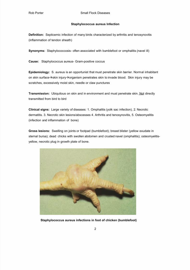
Gross lesions: Swelling on joints or footpad (bumblefoot); breast blister (yellow exudate in
sternal bursa); dead chicks with swollen abdomen and crusted navel (omphalitis); osteomyelitis-
yellow, necrotic plug in growth plate of bone.
Staphylococcus aureus infections in foot of chicken (bumblefoot)
8/17/2019 Poultry Diseases CVM
http://slidepdf.com/reader/full/poultry-diseases-cvm 3/34
Rob Porter Small Flock Diseases
3
Diagnosis: Bacterial culture of joints, bone, or yolk sac is definitive. Differential diagnosis:
Pasteurella multocida (fowl cholera) and Erysipelothrix (erysipelas)
Prevention/Treatment: Reduce trauma in house- eliminate sharp objects, beak and toe trim,
avoid wet litter or leaking drinkers; antibiotics (gram-positive spectrum such as erythromycin) may
be useful during outbreak. Prevention is much more important than treatment.
Escherichia coli Infection (Colibacillosis)
Definition: Infectious disease in which at Escherichia coli is the primary pathogen or a
secondary invader causing septicemia, peritonitis, cellulitis, omphalitis, salpingitis and
airsacculitis.
Synonyms: Escherichia coli infection; coligranuloma, colisepticemia
Cause: Escherichia coli, Gram-negative rod; serotypes 01, 02, or 078, but often untypeable
Epidemiology: Ubiquitous, present in intestine of birds and mammals and is disseminated in
feces. E. coli infections often result from management failures; often a secondary infection. Fecal
contamination is most important.
Transmission: Birds infected by direct contact with dirty litter and hatchers or contaminated egg
shells. This is an environmental disease. Not transmitted from bird to bird.
Clinical signs: Nonspecific and include ill-thrift, ruffled feathers, enlarged and swollen navel,
decreased appetite, depression, diarrhea, pasting of feathers around vent
Gross lesions: A variety of lesions depending on system that is affected:
1. Airsacculitis, perihepatitis and pericarditis- secondary invader as part of chronic
respiratory disease results in white, friable material covering air sacs, liver and pericardial
sac. Infection often preceded by Mycoplasma, Newcastle disease, or infectious
bronchitis.
2. Omphalitis- swollen, red, crusted navels can be caused by contamination of egg shells
through dirty setter, fecal covered eggs, excessive moisture during storage of eggs
3. Septicemia- hepatosplenomegaly (hepatitis), hemorrhages and necrosis in tissues
4. Salpingitis/peritonitis- especially laying hens; oviduct filled with yellow, cheesy exudate
8/17/2019 Poultry Diseases CVM
http://slidepdf.com/reader/full/poultry-diseases-cvm 4/34
Rob Porter Small Flock Diseases
4
5. Cellulitis (“scabby hip”) of broiler chickens: yellow exudate underneath skin of hip, leg
and breast
6. Synovitis/arthritis
7. Hypopyon- pus in eye following E. coli septicemia
Diagnosis: Histopathology; bacterial culture of affected organs is required for diagnosis.
Treatment: E. coli isolates from commercial poultry are often resistant to a variety of antibiotics.
This is compounded by the fact that few antibiotics are being put on the market for poultry.
Enrofloxacin was effective against this organism, but the antibiotic was removed from food
production use in 2006. Regardless, culture and sensitivity may assist treatment if more than one
bird is affected.
Prevention: Vigorous sanitation program in breeder house, at hatchery and at grow-out facility;Egg fumigation; dust control in house; routinely remove dead birds from house, avoid stress and
immunosuppression (IBD).
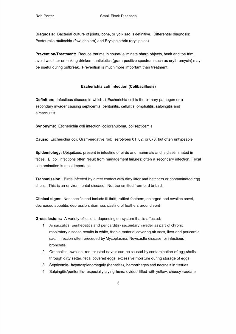
Colibacillosis: One-day-old White leghorn chicks with omphalitis (left photo) and yolksacculitis (right photo).
8/17/2019 Poultry Diseases CVM
http://slidepdf.com/reader/full/poultry-diseases-cvm 5/34
Rob Porter Small Flock Diseases
5
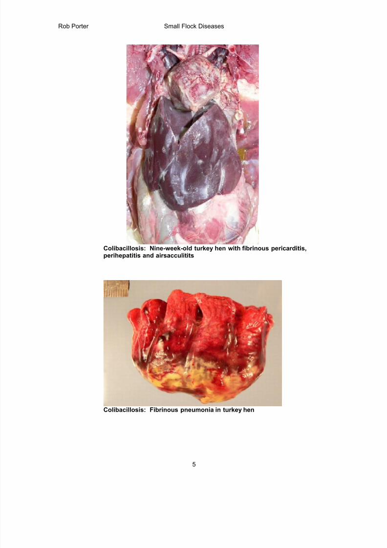
Colibacillosis: Nine-week-old turkey hen with fibrinous pericarditis,perihepatitis and airsacculitits
Colibacillosis: Fibrinous pneumonia in turkey hen
8/17/2019 Poultry Diseases CVM
http://slidepdf.com/reader/full/poultry-diseases-cvm 6/34
Rob Porter Small Flock Diseases
6
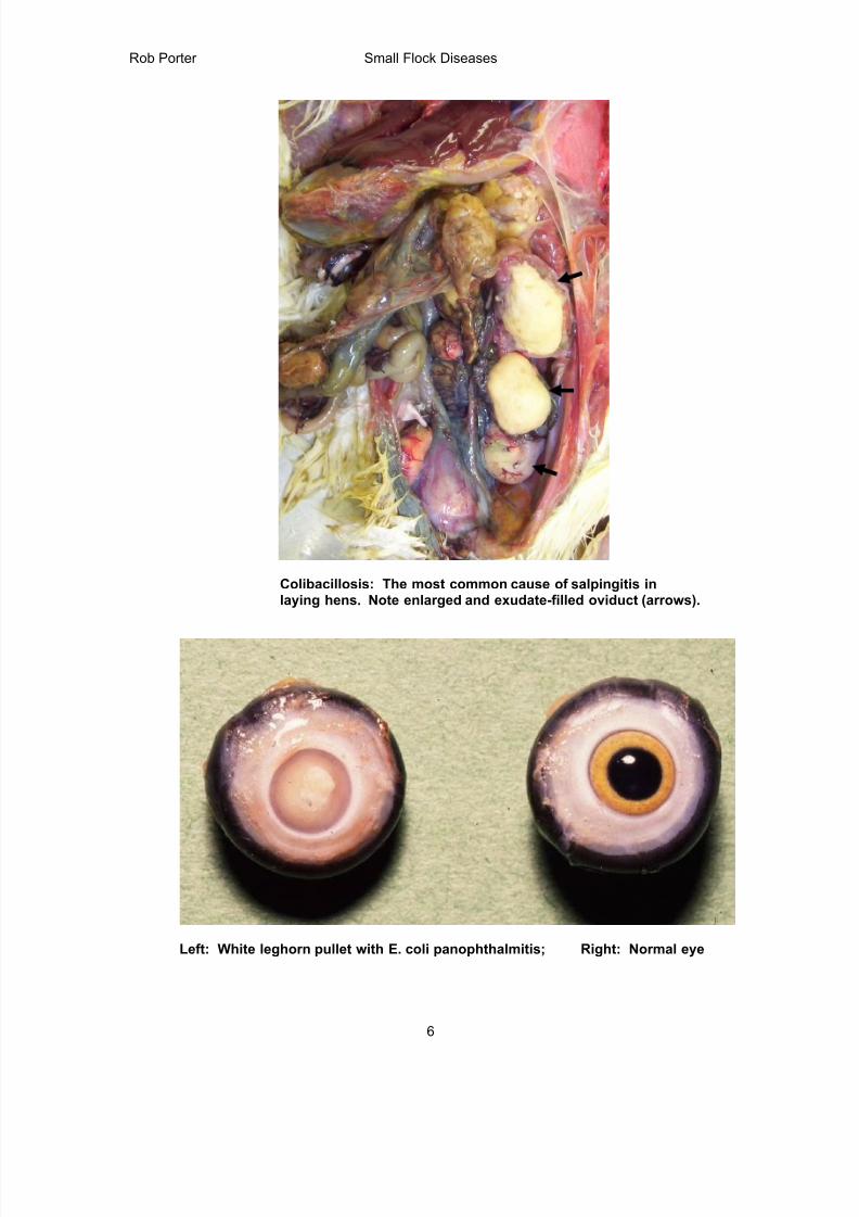
Colibacillosis: The most common cause of salpingitis inlaying hens. Note enlarged and exudate-filled oviduct (arrows).
Left: White leghorn pullet with E. coli panophthalmitis; Right: Normal eye
8/17/2019 Poultry Diseases CVM
http://slidepdf.com/reader/full/poultry-diseases-cvm 7/34
Rob Porter Small Flock Diseases
7
Mycoplasma gallisepticum Infection
Definition: A chronic disease of a variety of birds, especially chickens and turkeys,
characterized by nasal exudate, coughing and debilitation. Major problem in poultry industry for
over 60 years. Monitored by NPIP.
Synonyms: “Chronic respiratory disease” of chickens; “Infectious sinusitis” of turkeys; MG, CRD
Cause: Mycoplasma gallisepticum; Mycoplasmas are small prokaryotes that lack a cell wall and
are covered only by a plasma membrane. These organisms form fried-egg colonies in culture
plates and generally require a protein-rich medium that contains 10-15% animal serum. M.
gallisepticum, M. synoviae and M. meleagridis have the ability to agglutinate turkey or chicken
erythrocytes, a feature that is utilized in the hemagglutination-inhibition assay to detect antibodies
to these agents.
Epidemiology/Transmission: Transovarian- transmitted from breeder birds to offspring through
egg; Horizontal infection via infected aerosols; Chronic respiratory disease of chickens is usually
complicated by Escherichia coli infection, Newcastle disease virus or infectious bronchitis virus.
MG alone produces mild lesions in chickens. MG can be common inhabitant of upper respiratory
tract of clinically healthy birds.
Clinical signs: Adult laying hens- signs are rare; decreased egg production; decreased feed
consumption and increased medication costs
Broiler chickens- Chronic respiratory disease with coughing, sneezing (snicks), oculnasal
discharge, poor feed conversion and air sac condemnations at processing
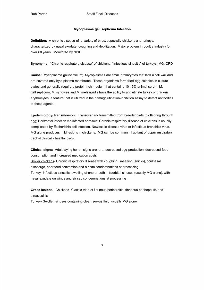
Turkey- Infectious sinusitis- swelling of one or both infraorbital sinuses (usually MG alone), with
nasal exudate on wings and air sac condemnations at processing
Gross lesions: Chickens- Classic triad of fibrinous pericarditis, fibrinous perihepatitis and
airsacculitis
Turkey- Swollen sinuses containing clear, serous fluid, usually MG alone
8/17/2019 Poultry Diseases CVM
http://slidepdf.com/reader/full/poultry-diseases-cvm 8/34
Rob Porter Small Flock Diseases
8
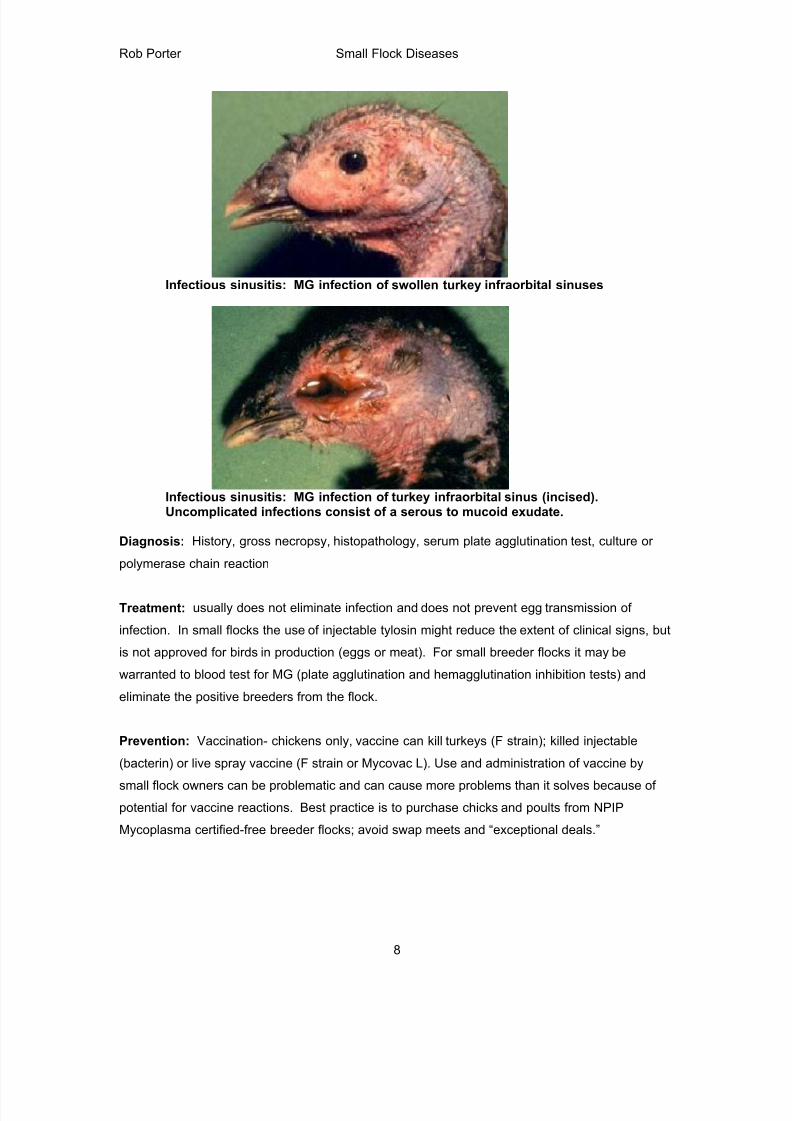
Infectious sinusitis: MG infection of swollen turkey infraorbital sinuses
Infectious sinusitis: MG infection of turkey infraorbital sinus (incised).Uncomplicated infections consist of a serous to mucoid exudate.
Diagnosis: History, gross necropsy, histopathology, serum plate agglutination test, culture or
polymerase chain reaction
Treatment: usually does not eliminate infection and does not prevent egg transmission of
infection. In small flocks the use of injectable tylosin might reduce the extent of clinical signs, but
is not approved for birds in production (eggs or meat). For small breeder flocks it may be
warranted to blood test for MG (plate agglutination and hemagglutination inhibition tests) and
eliminate the positive breeders from the flock.
Prevention: Vaccination- chickens only, vaccine can kill turkeys (F strain); killed injectable
(bacterin) or live spray vaccine (F strain or Mycovac L). Use and administration of vaccine by
small flock owners can be problematic and can cause more problems than it solves because of
potential for vaccine reactions. Best practice is to purchase chicks and poults from NPIP
Mycoplasma certified-free breeder flocks; avoid swap meets and “exceptional deals.”
8/17/2019 Poultry Diseases CVM
http://slidepdf.com/reader/full/poultry-diseases-cvm 9/34
Rob Porter Small Flock Diseases
9
Marek's Disease (MD)
Definition: Marek's disease is herpesvirus infection that causes lymphoma of T lymphocytes.
Tumors may occur in the nerves, ovaries, testes, viscera, eye, muscle, and skin. MD is ubiquitous
throughout the world. The leg paralysis was often referred to as range paralysis. This is a classicexample of vaccines being used to prevent cancer in chickens.
Cause: The disease is caused by a cell-associated herpesvirus (double-stranded DNA virus,
hexagonal enveloped virus 150-160 nm diameter). There are three serotypes of MD virus: The
oncoviruses (tumor-causing) are serotype 1; non-oncogenic viruses are serotype 2; and the
herpes virus turkey (HVT) is serotype 3. The virus is intranuclear (cell-associated) and normally
cannot live outside the host cell being protected from the environment by host epithelium.
Viral replication occurs in three phases of virus-cell interactions: Productive, latent and
transformation.
1. Productive infection: Occurs mainly in nonlymphocytic cells with replication of viral DNA ,
antigen synthesized and viral particles sometimes produced. Full productive infection
occurs in feather follicle epithelium that develops large numbers of enveloped and fully
infectious virus. Productive restrictive infection occurs in some lymphoid and epithelial
cells with viral antigen being produced, but most viruses formed have no envelope and
therefore are noninfectious.
2. Latent infection: Occurs in lymphocytes, predominantly T cells, but occasionally B cells.
The viral genome is not expressed and no virus or tumor-associated antigen is produced.
3. Transforming infection: Occurs in only T lymphocytes and is caused by only the virulent
serotype 1 Marek’s disease viruses. This transformation in T lymphocytes results in
formation of neoplastic lymphocytes with viral antigen expressed, but not virus. MD
tumor-associated antigen is expressed on the cell surface. MD viral DNA is not
integrated into the host genome.
Transmission: Infectious virus is produced only in feather follicle epithelium and spreads by
direct or indirect contact between birds. The infectious virus contaminates the premises through
infected molted feathers and dander. Birds become infected when they inhale dust containing the
virus. Contaminated dust may remain infectious for several months.
8/17/2019 Poultry Diseases CVM
http://slidepdf.com/reader/full/poultry-diseases-cvm 10/34
Rob Porter Small Flock Diseases
10
Many apparently normal birds are carriers that can transmit the infection. Some birds have been
found to shed virus from skin as long as eighteen months. Darkling beetles may also act as a
mechanical vector to carry the virus between houses.
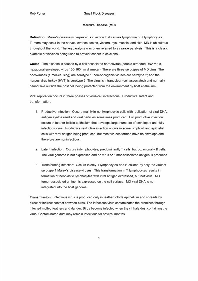
Clinical Signs: Under field conditions, it is difficult to determine the incubation period.
Experimentally, clinical signs and gross lesions generally do not appear until between 3 and 4
weeks post-infection. In acute outbreaks, birds become severely depressed, anorectic and
uncoordinated followed by unilateral or bilateral paralysis of legs and wings. Many birds become
dehydrated, emaciated, and eventually die. The extremities affected include the legs, wings, and
neck. In an infected flock, mortality gradually builds and generally persists for 4-10 weeks. Ocular
Marek’s disease is characterized by decreased pupil size and irregular diameter (“grey eye”). A
number of factors influence the extent of losses in affected flocks, such as virus strain, dosage,
route of exposure, and genetic resistance of the host. Immunosuppression can be a long-term
effect.
Marek’s disease: Splay-leg posture of pullet with sciatic neuropathy.
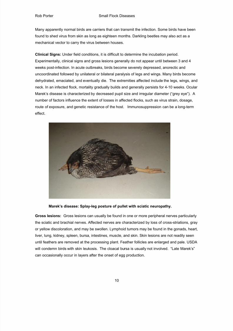
Gross lesions: Gross lesions can usually be found in one or more peripheral nerves particularly
the sciatic and brachial nerves. Affected nerves are characterized by loss of cross-striations, gray
or yellow discoloration, and may be swollen. Lymphoid tumors may be found in the gonads, heart,
liver, lung, kidney, spleen, bursa, intestines, muscle, and skin. Skin lesions are not readily seen
until feathers are removed at the processing plant. Feather follicles are enlarged and pale. USDA
will condemn birds with skin leukosis. The cloacal bursa is usually not involved. “Late Marek’s”
can occasionally occur in layers after the onset of egg production.
8/17/2019 Poultry Diseases CVM
http://slidepdf.com/reader/full/poultry-diseases-cvm 11/34
Rob Porter Small Flock Diseases
11
Marek’s disease: Hepatic lymphoma (tumors)
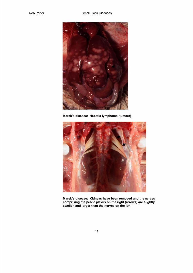
Marek’s disease: Kidneys have been removed and the nervescomprising the pelvic plexus on the right (arrows) are slightlyswollen and larger than the nerves on the left.
8/17/2019 Poultry Diseases CVM
http://slidepdf.com/reader/full/poultry-diseases-cvm 12/34
Rob Porter Small Flock Diseases
12
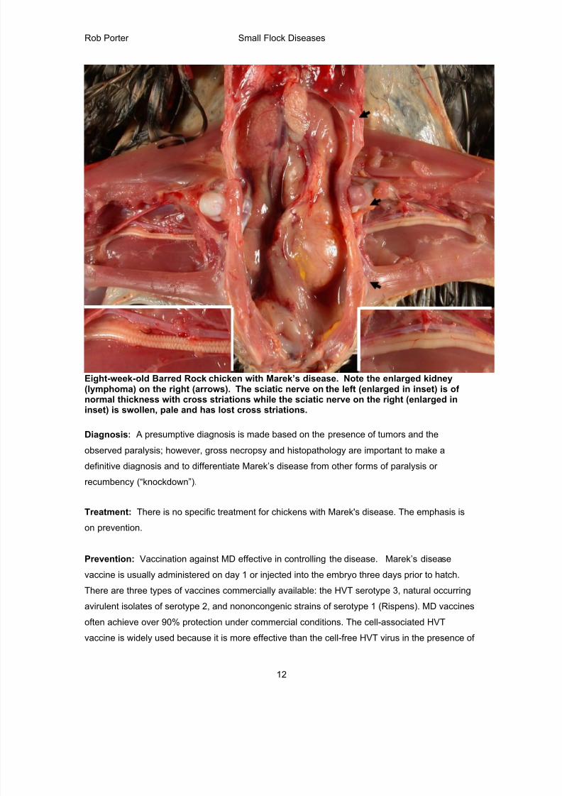
Eight-week-old Barred Rock chicken with Marek’s disease. Note the enlarged kidney(lymphoma) on the right (arrows). The sciatic nerve on the left (enlarged in inset) is ofnormal thickness with cross striations while the sciatic nerve on the right (enlarged ininset) is swollen, pale and has lost cross striations.
Diagnosis: A presumptive diagnosis is made based on the presence of tumors and the
observed paralysis; however, gross necropsy and histopathology are important to make a
definitive diagnosis and to differentiate Marek’s disease from other forms of paralysis or
recumbency (“knockdown”).
Treatment: There is no specific treatment for chickens with Marek's disease. The emphasis is
on prevention.
Prevention: Vaccination against MD effective in controlling the disease. Marek’s disease
vaccine is usually administered on day 1 or injected into the embryo three days prior to hatch.
There are three types of vaccines commercially available: the HVT serotype 3, natural occurring
avirulent isolates of serotype 2, and nononcongenic strains of serotype 1 (Rispens). MD vaccines
often achieve over 90% protection under commercial conditions. The cell-associated HVT
vaccine is widely used because it is more effective than the cell-free HVT virus in the presence of
8/17/2019 Poultry Diseases CVM
http://slidepdf.com/reader/full/poultry-diseases-cvm 13/34
Rob Porter Small Flock Diseases
13
maternal antibodies. At least 1000-1500 plaque-forming units of virus are needed per dose. The
HVT dose is often diluted or cut in hatcheries to save money, which may not protect against
highly pathogenic MD. In areas where highly pathogenic field strains are prevalent, polyvalent
vaccines containing serotype 2 and 3 viruses are commonly used. Chicks are normally
vaccinated at the hatchery because they are exposed to the field virus at an early age. Cell-
associated vaccines require extreme care in handling and application. Condemnations due to
Marek's Disease can often be traced to errors in vaccination. In problem regions all three
serotypes of MD virus can be included in vaccines.
Infectious Laryngotracheitis (ILT)
Definition: Infectious laryngotracheitis is an acute, highly infectious viral disease, characterized
by conjunctivitis, loud gasping, tracheal plugs and coughing of blood-filled exudate.
Cause: Family Herpesviridae, Subfamily Alphaherpesvirnae, Genus Iltovirus, Species Gallid
herpesvirus 1, (infectious laryngotracheitis virus), dsDNA virus. ILT is a herpesvirus, a hexagonal
shaped, enveloped, ether-sensitive virus that contains DNA. Four major envelope glycoproteins
are the major immunogens. The virus replicates in the tracheal and bronchiolar epithelium;
viremia does not occur. Field strains have been isolated that vary in their pathogenicity and
virulence. Virus is destroyed by most commonly used disinfectants. In some instances infection
is caused by vaccine virus increasing in virulence (“heating up”) as it spreads from vaccinated to
non-vaccinated birds.
Transmission: This is primarily a human traffic-associated problem. Birds are infected by
inhaling virus or via the intraocular route. Rapid virus replication occurs after the virus infects the
upper respiratory tract. Transmission is rapid. Mechanical transmission can occur by use of
contaminated equipment, clothing, shoes and litter. Some birds that recover from an infection can
act as carriers (for up to 16 months virus is latent in dorsal root ganglia) and intermittently shed
the virus. Because the virus spreads slowly birds can be vaccinated during an outbreak.
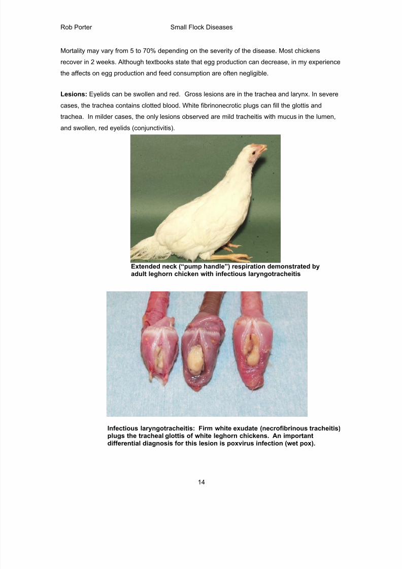
Clinical Signs: In acute infections, there may be nasal discharge, moist rales, coughing and
gasping. The neck may be extended (“pump handle respiration”). The eyelids can be red with
increased ocular drainage. In severe cases there is marked labored breathing and coughing of
blood-stained mucus, which covers the wing and breast feathers as a result of the bird shaking
the head with violent coughing. Blood and yellow exudate in the trachea cause death by
suffocation. In mild cases, birds are unthrifty with red, swollen eyelids, and nasal discharge.
8/17/2019 Poultry Diseases CVM
http://slidepdf.com/reader/full/poultry-diseases-cvm 14/34
Rob Porter Small Flock Diseases
14
Mortality may vary from 5 to 70% depending on the severity of the disease. Most chickens
recover in 2 weeks. Although textbooks state that egg production can decrease, in my experience
the affects on egg production and feed consumption are often negligible.
Lesions: Eyelids can be swollen and red. Gross lesions are in the trachea and larynx. In severe
cases, the trachea contains clotted blood. White fibrinonecrotic plugs can fill the glottis and
trachea. In milder cases, the only lesions observed are mild tracheitis with mucus in the lumen,
and swollen, red eyelids (conjunctivitis).
Extended neck (“pump handle”) respiration demonstrated byadult leghorn chicken with infectious laryngotracheitis
Infectious laryngotracheitis: Firm white exudate (necrofibrinous tracheitis)plugs the tracheal glottis of white leghorn chickens. An importantdifferential diagnosis for this lesion is poxvirus infection (wet pox).
8/17/2019 Poultry Diseases CVM
http://slidepdf.com/reader/full/poultry-diseases-cvm 15/34
Rob Porter Small Flock Diseases
15
Diagnosis: A presumptive diagnosis can be made on the history and the presence of blood and
caseous/fibrinous plugs in the trachea. Since the clinical signs and lesions of the milder form
resemble other respiratory diseases, such as wet pox, infectious bronchitis, Newcastle disease
and avian inlfuenza, a positive diagnosis must be made by histopathology of the trachea or virus
isolation. The virus is cultivated on the chorioallantoic membrane of 10-day-old chicken
embryos.
Treatment: If the disease is diagnosed early in an outbreak, vaccinating the entire flock and
adjacent flocks may reduce the overall mortality. The administration of broad-spectrum antibiotics
may help reduce secondary infections.
Prevention: In endemic areas, keep unauthorized personnel out of the poultry house, in addition
vaccinating all breeder/layer birds at the proper time. Two types of ILT vaccines are most
commonly used: Tissue culture-origin is administered by eyedrop at 1-day of age and a chick
embryo origin vaccine, which is administered by drinking water or spray. Layers should be
vaccinated prior to beginning egg production. Commercial layers are usually vaccinated by
eyedrop or spray at 7-8 weeks of age, again by spray or drinking water at 12-14 weeks. Extra
care must be taken to assure good vaccination technique avoid problems with vaccination
reaction to ILT vaccines. Be sure to get vaccine to all birds in the flock because the vaccine virus
tends to increase in pathogenicity (“heat up”) as it passes from bird to bird, and can eventually
cause disease in nonvaccinated birds. For this reason, I rarely recommend ILT vaccination of
small flocks.
Miscellaneous/Metabolic Diseases
Visceral gout/urolithiasis: Gout/urolithiasis is a condition very commonly seen in older layer
flocks and is related to kidney failure. On occasion, gout can be a very significant part of flock
mortality, sometimes as high as 0.5 % per week, but is often an insignificant cause of low-grade
mortality. Kidneys can be damaged by low phosphorus diets at any age, water deprivation at
housing, high vitamin D3 in the ration, or excessive calcium before sexual maturity (15 to 16
weeks). A nephrotropic infectious bronchitis (e.g., Australian T or Italian strain) can cause similar
lesions. Losses due to gout tend to be chronic with the number of affected birds depending on
the severity of the kidney insult. The strain of bird can also affect the severity and incidence of
gout. Birds with gout usually show no clinical signs before death or may be extremely thin. The
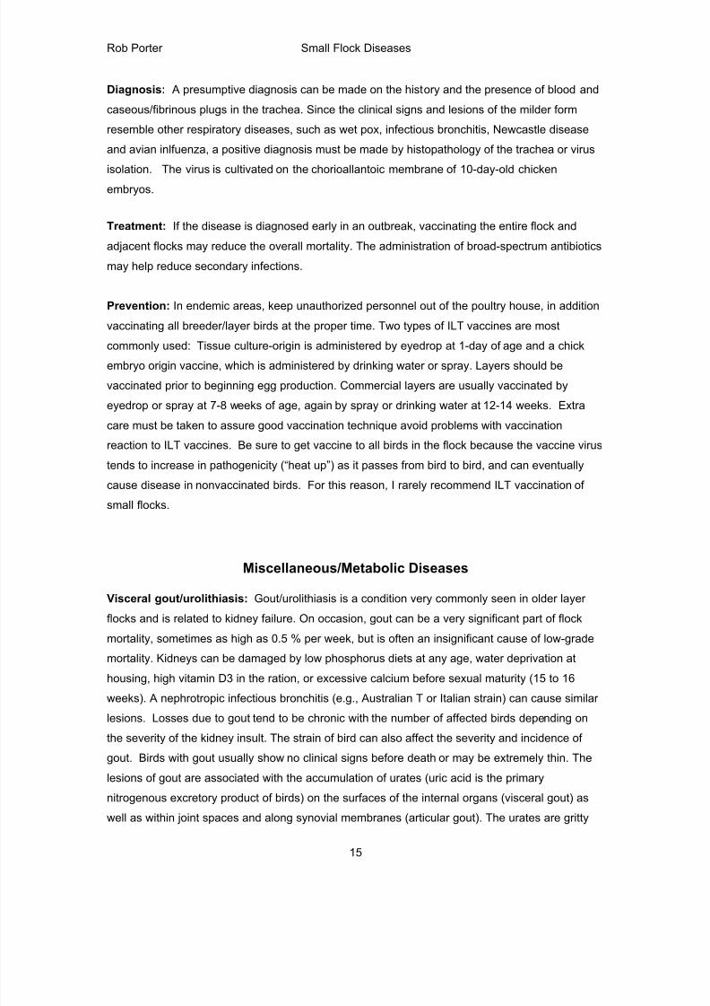
lesions of gout are associated with the accumulation of urates (uric acid is the primary
nitrogenous excretory product of birds) on the surfaces of the internal organs (visceral gout) as
well as within joint spaces and along synovial membranes (articular gout). The urates are gritty
8/17/2019 Poultry Diseases CVM
http://slidepdf.com/reader/full/poultry-diseases-cvm 16/34
Rob Porter Small Flock Diseases
16
and white as opposed to inflammatory exudate from bacterial infections, such as colibacillosis,
which are yellow and friable. Portions of kidney are atrophic or absent and contralateral portions
are often swollen (compensatory hypertrophy). Birds can be treated with varying success by
adding ammonium sulfate or ammonium chloride to the ration but these treatments may cause
wet droppings and deterioration of shell quality. Gout can be prevented or minimized by providing
the proper nutrition of calcium and phosphorus throughout growing (1 % calcium and 0.50 to
0.45% available phosphorus), starting layer levels of calcium feeding at the proper time (one
week prior to first egg), and avoiding water deprivation at housing.
Visceral gout: Chaulk-like urate deposits on pericardial sac and liver capsule
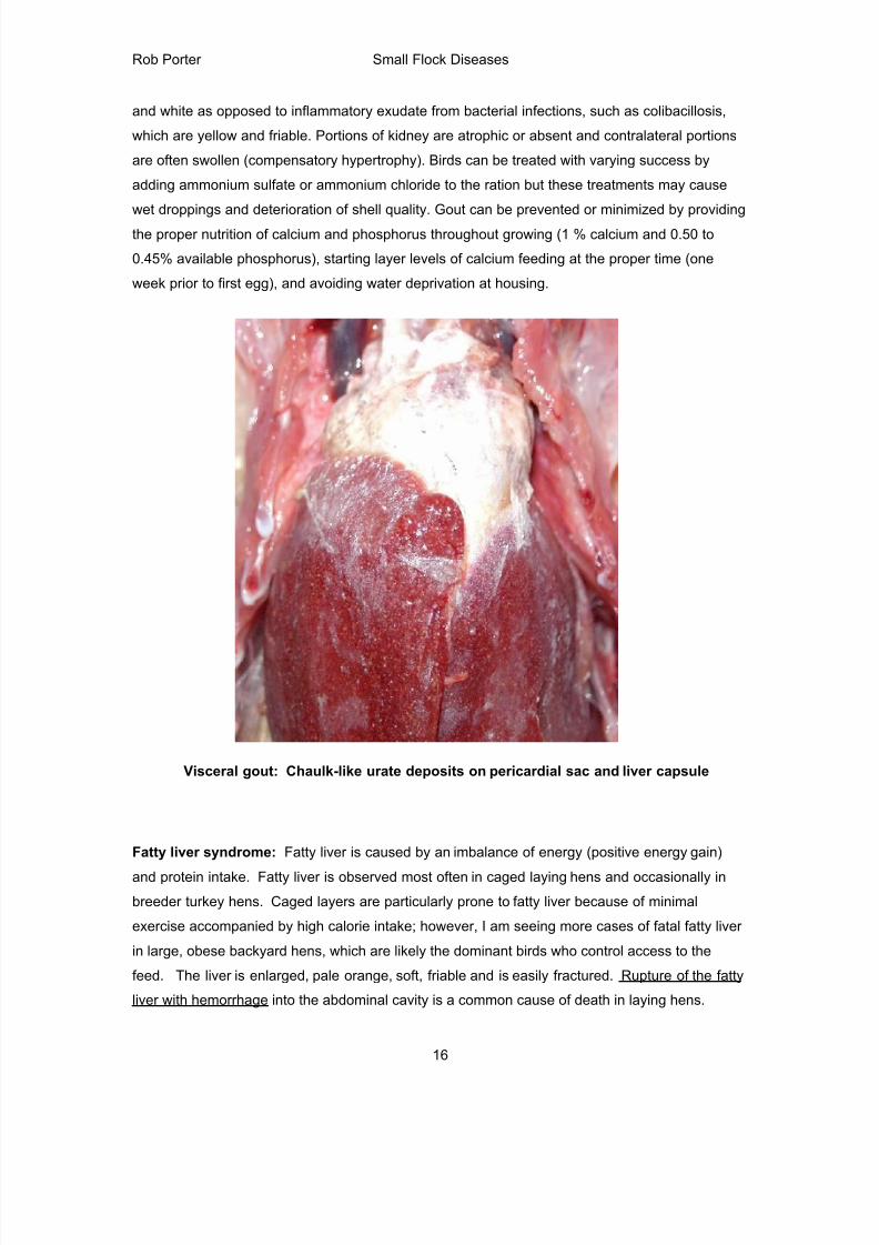
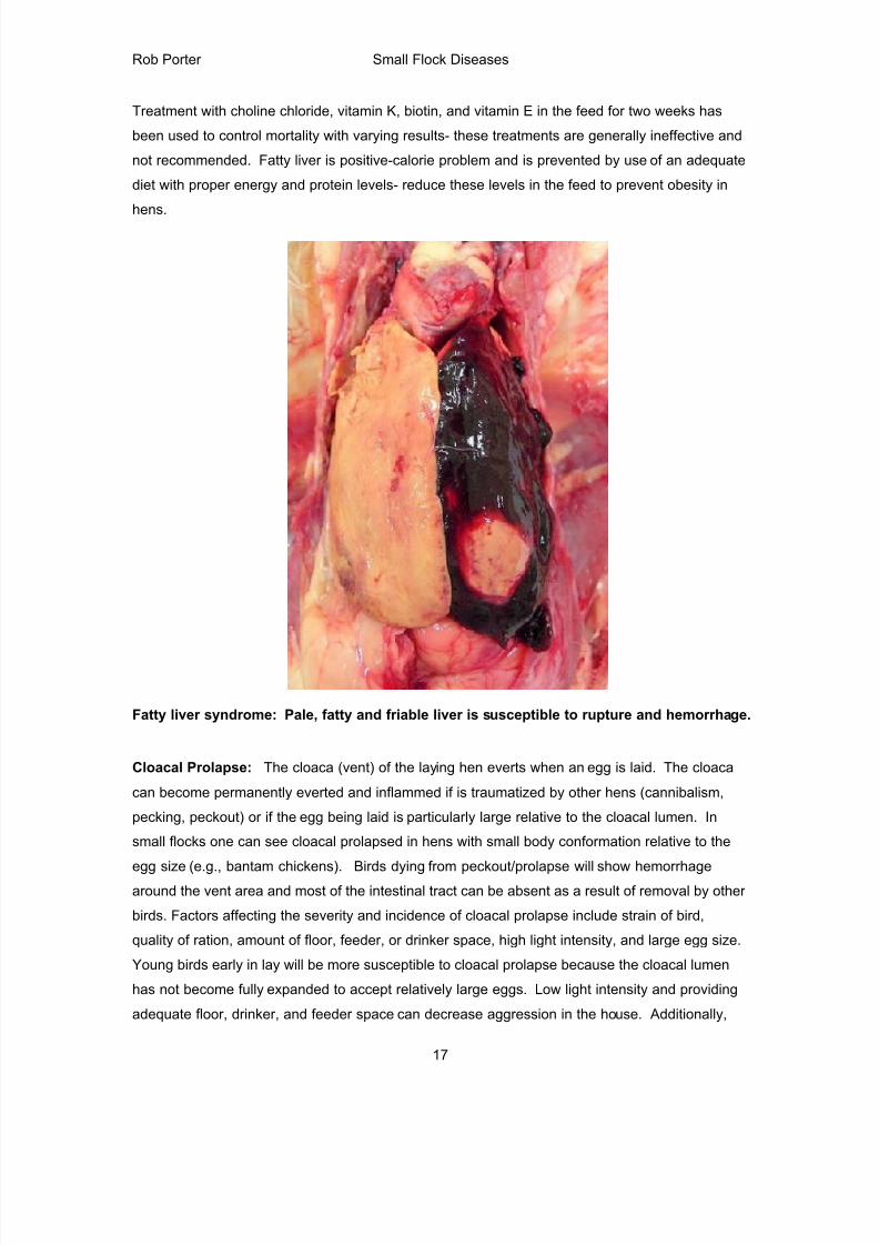
Fatty liver syndrome: Fatty liver is caused by an imbalance of energy (positive energy gain)
and protein intake. Fatty liver is observed most often in caged laying hens and occasionally in
breeder turkey hens. Caged layers are particularly prone to fatty liver because of minimal
exercise accompanied by high calorie intake; however, I am seeing more cases of fatal fatty liver
in large, obese backyard hens, which are likely the dominant birds who control access to the
feed. The liver is enlarged, pale orange, soft, friable and is easily fractured. Rupture of the fatty
liver with hemorrhage into the abdominal cavity is a common cause of death in laying hens.
8/17/2019 Poultry Diseases CVM
http://slidepdf.com/reader/full/poultry-diseases-cvm 17/34
Rob Porter Small Flock Diseases
17
Treatment with choline chloride, vitamin K, biotin, and vitamin E in the feed for two weeks has
been used to control mortality with varying results- these treatments are generally ineffective and
not recommended. Fatty liver is positive-calorie problem and is prevented by use of an adequate
diet with proper energy and protein levels- reduce these levels in the feed to prevent obesity in
hens.
Fatty liver syndrome: Pale, fatty and friable liver is susceptible to rupture and hemorrhage.
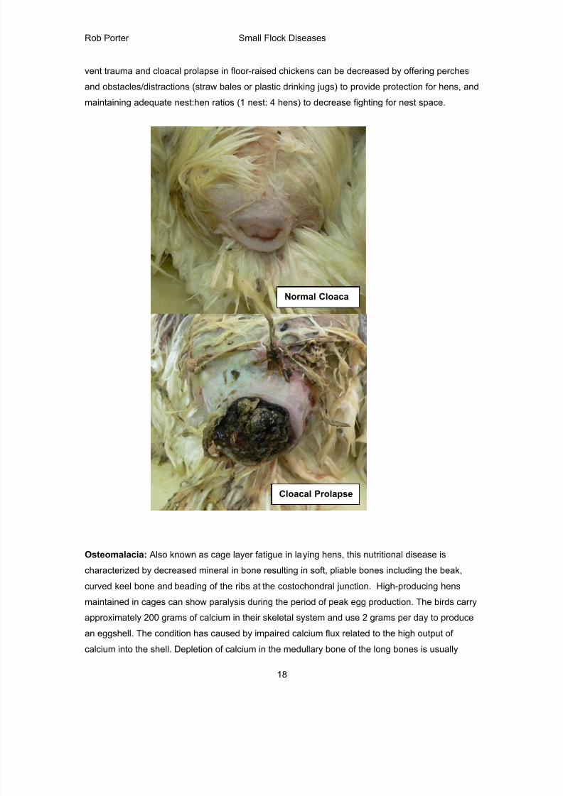
Cloacal Prolapse: The cloaca (vent) of the laying hen everts when an egg is laid. The cloaca
can become permanently everted and inflammed if is traumatized by other hens (cannibalism,
pecking, peckout) or if the egg being laid is particularly large relative to the cloacal lumen. In
small flocks one can see cloacal prolapsed in hens with small body conformation relative to the
egg size (e.g., bantam chickens). Birds dying from peckout/prolapse will show hemorrhagearound the vent area and most of the intestinal tract can be absent as a result of removal by other
birds. Factors affecting the severity and incidence of cloacal prolapse include strain of bird,
quality of ration, amount of floor, feeder, or drinker space, high light intensity, and large egg size.
Young birds early in lay will be more susceptible to cloacal prolapse because the cloacal lumen
has not become fully expanded to accept relatively large eggs. Low light intensity and providing
adequate floor, drinker, and feeder space can decrease aggression in the house. Additionally,
8/17/2019 Poultry Diseases CVM
http://slidepdf.com/reader/full/poultry-diseases-cvm 18/34
Rob Porter Small Flock Diseases
18
vent trauma and cloacal prolapse in floor-raised chickens can be decreased by offering perches
and obstacles/distractions (straw bales or plastic drinking jugs) to provide protection for hens, and
maintaining adequate nest:hen ratios (1 nest: 4 hens) to decrease fighting for nest space.
Osteomalacia: Also known as cage layer fatigue in laying hens, this nutritional disease ischaracterized by decreased mineral in bone resulting in soft, pliable bones including the beak,
curved keel bone and beading of the ribs at the costochondral junction. High-producing hens
maintained in cages can show paralysis during the period of peak egg production. The birds carry
approximately 200 grams of calcium in their skeletal system and use 2 grams per day to produce
an eggshell. The condition has caused by impaired calcium flux related to the high output of
calcium into the shell. Depletion of calcium in the medullary bone of the long bones is usually
Normal Cloaca
Cloacal Prolapse
8/17/2019 Poultry Diseases CVM
http://slidepdf.com/reader/full/poultry-diseases-cvm 19/34
Rob Porter Small Flock Diseases
19
seen in caged birds, suggesting that reduced activity/exercise is a predisposing factor. Affected
birds can be found on their sides in the back of the cage. At the time of initial paralysis, birds can
appear healthy and will have a shelled egg in the oviduct. Death occurs from starvation or
dehydration, a failure of the birds to reach the feet or water. A high incidence of cage layer fatigue
can be prevented if pullets receive a high calcium diet (minimum of 3.5% calcium) at least two
weeks prior to the first oviposition. Older caged layers are also quite susceptible to bone
breakage, particularly during transport to processing.
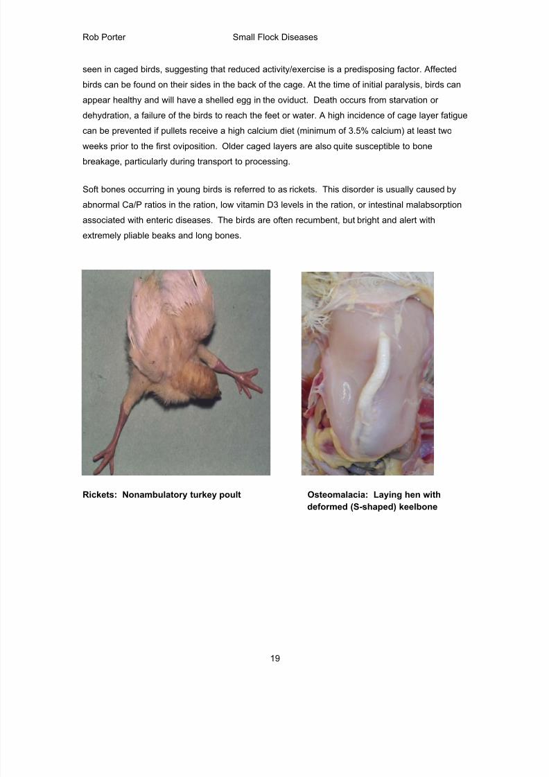
Soft bones occurring in young birds is referred to as rickets. This disorder is usually caused by
abnormal Ca/P ratios in the ration, low vitamin D3 levels in the ration, or intestinal malabsorption
associated with enteric diseases. The birds are often recumbent, but bright and alert with
extremely pliable beaks and long bones.
Rickets: Nonambulatory turkey poult Osteomalacia: Laying hen with
deformed (S-shaped) keelbone
8/17/2019 Poultry Diseases CVM
http://slidepdf.com/reader/full/poultry-diseases-cvm 20/34
Rob Porter Small Flock Diseases
20
Ascites Syndrome and Right Ventricular Failure of Broiler Chickens
Definition: Ascites = accumulation of clear (serous transudate) fluid in the abdomen. Ascites syndrome
is a noninfectious, metabolic disease of broiler chickens and ducklings characterized by pulmonary
hypertension, right-sided heart failure and accumulation of excessive fluid (transudate) in the abdomen.
This is a form of congestive heart failure in chickens. Birds are predisposed to right-sided heart failure
because of a muscular flap that forms part of the right atrioventricular valve
Epidemiology: Originally described in broiler chickens raised at high altitudes (decreased PO2).
Ascites is ommonly observed in a small percentage of broilers at processing and mortality can approach
1-2% or greater in some flocks. Respiratory infection or poor ventilation can promote this problem
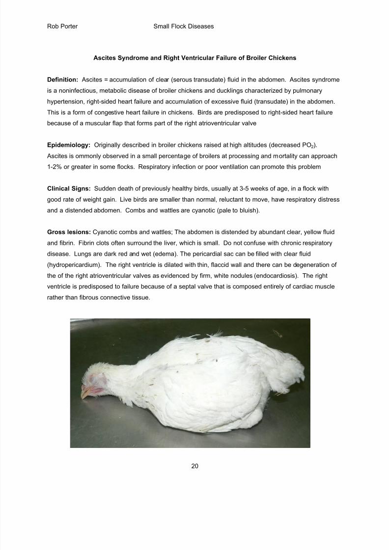
Clinical Signs: Sudden death of previously healthy birds, usually at 3-5 weeks of age, in a flock with
good rate of weight gain. Live birds are smaller than normal, reluctant to move, have respiratory distress
and a distended abdomen. Combs and wattles are cyanotic (pale to bluish).
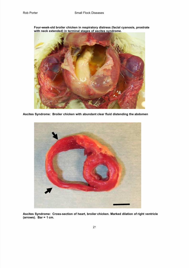
Gross lesions: Cyanotic combs and wattles; The abdomen is distended by abundant clear, yellow fluid
and fibrin. Fibrin clots often surround the liver, which is small. Do not confuse with chronic respiratory
disease. Lungs are dark red and wet (edema). The pericardial sac can be filled with clear fluid
(hydropericardium). The right ventricle is dilated with thin, flaccid wall and there can be degeneration of
the of the right atrioventricular valves as evidenced by firm, white nodules (endocardiosis). The right
ventricle is predisposed to failure because of a septal valve that is composed entirely of cardiac musclerather than fibrous connective tissue.
8/17/2019 Poultry Diseases CVM
http://slidepdf.com/reader/full/poultry-diseases-cvm 21/34
Rob Porter Small Flock Diseases
21
Four-week-old broiler chicken in respiratory distress (facial cyanosis, prostratewith neck extended) in terminal stages of ascites syndrome.
Ascites Syndrome: Broiler chicken with abundant clear fluid distending the abdomen
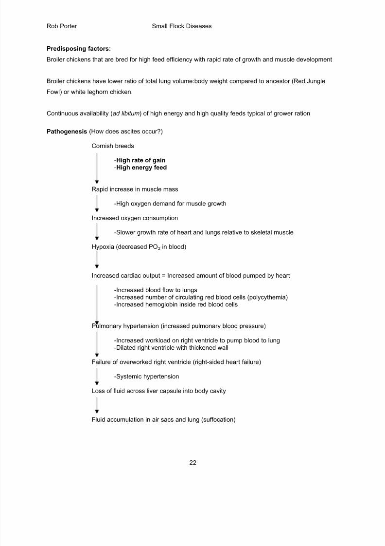
Ascites Syndrome: Cross-section of heart, broiler chicken. Marked dilation of right ventricle(arrows). Bar = 1 cm.
8/17/2019 Poultry Diseases CVM
http://slidepdf.com/reader/full/poultry-diseases-cvm 22/34
Rob Porter Small Flock Diseases
22
Predisposing factors:
Broiler chickens that are bred for high feed efficiency with rapid rate of growth and muscle development
Broiler chickens have lower ratio of total lung volume:body weight compared to ancestor (Red Jungle
Fowl) or white leghorn chicken.
Continuous availability (ad libitum) of high energy and high quality feeds typical of grower ration
Pathogenesis (How does ascites occur?)
Cornish breeds
-High rate of gain -High energy feed
Rapid increase in muscle mass
-High oxygen demand for muscle growth
Increased oxygen consumption
-Slower growth rate of heart and lungs relative to skeletal muscle
Hypoxia (decreased PO2 in blood)
Increased cardiac output = Increased amount of blood pumped by heart
-Increased blood flow to lungs-Increased number of circulating red blood cells (polycythemia)-Increased hemoglobin inside red blood cells
Pulmonary hypertension (increased pulmonary blood pressure)
-Increased workload on right ventricle to pump blood to lung-Dilated right ventricle with thickened wall
Failure of overworked right ventricle (right-sided heart failure)
-Systemic hypertension
Loss of fluid across liver capsule into body cavity
Fluid accumulation in air sacs and lung (suffocation)
8/17/2019 Poultry Diseases CVM
http://slidepdf.com/reader/full/poultry-diseases-cvm 23/34
Rob Porter Small Flock Diseases
23
Prevention:
Decrease energy level in feed to slow down growth rate. Try limiting feed during the grow-out or turning
off house lights at night. The birds should be acclimated to dark-out periods at an early age to minimize
panic.
If mortality is >1-2% from ascites look for other complicating factors such as high sodium levels in feed or
water, vitamin E or selenium deficiency, respiratory infection, furazolidone or coal tar toxicity
Maintain good air quality with adequate ventilation, especially during winter
Coccidiosis (Eimeria spp.)
Cause: Protozoan parasite of the intestinal tract of poultry
Remains on of the most common and expensive diseases of poultry despite improved
chemotherapy, management and genetics
Most coccidia affecting chickens are in genus Eimeria (up to 9 species)
Species-specificity: The species of coccidia that infect chickens do not infect turkeys and vice
versa.
Most field outbreaks involve infection with two or more species of Eimeria.
Infections more common in birds raised on a floor environment, but infections can occur in caged
birds (e.g., pullets and layers) to produce substantial mortality
Life cycle
Initiated by ingestion of sporulated oocysts (eggs) and usually takes 4-6 days to complete life
cycle
Involves both asexual (schizogony or merogony) and sexual (microgametes fertilizemacrogametes) stages
Asexual and sexual stages occur in different sites of intestinal tract depending thespecies of Eimeria.
8/17/2019 Poultry Diseases CVM
http://slidepdf.com/reader/full/poultry-diseases-cvm 24/34
Rob Porter Small Flock Diseases
24
Eimeria: A single, mature oocyst (egg) contains four sporocysts, and eachsporocyst contains two sporozoites (eight sporozoites in each oocyst)
Sporulated oocysts in poultry house
Oocysts consumed by bird
Gizzard: oocysts crushed to release sporozoites DAY 1
Intestine and cecum epithelium (species dependent) DAY 2-5
Asexual Stage: Sporozoites invade epithelial cells and divide toproduce numerous schizonts (cysts) filled with merozoites
At least two generations of asexual development(first and second generation schizonts containing first andsecond generation merozoites)
Merozoites are final product of asexual stage
Intestine and cecum epithelium DAY 5-7
Sexual Stage: Macrogametes and microgametes producedinside epithelial cells
Microgametes (small) are released to seek out and fertilize thelarger macrogametes to form zygote
Zygote matures into oocysts
Oocysts rupture from epithelial cell to enter intestinal lumen
Feces Oocysts undergo further maturation (sporulation) in environmentto become infectious. Sporulation takes less than 48 hours andoccurs more rapidly in warm, moist litter
Clinical signs
Usually turkeys and broilers
Range-reared, cage-free layers are at increased risk for coccidiosis
Pale combs and wattles from blood loss in gut
Ruffled feathers, depression, blood in droppings, shivering
Mortality rate may increase, particularly in caged pullets
Decreased egg production in birds at reproductive age
8/17/2019 Poultry Diseases CVM
http://slidepdf.com/reader/full/poultry-diseases-cvm 25/34
Rob Porter Small Flock Diseases
25
Gross lesions
Chickens: Lesions vary depending on what species of Eimeria are involved
Eimeria acervulina: white “tiger -striping” of upper small intestine (duodenum)
Eimeria necatrix: Mid and distal small intestines (jejunum and ileum): White spots (large
schizonts) in mucosa with mucus and blood in lumen
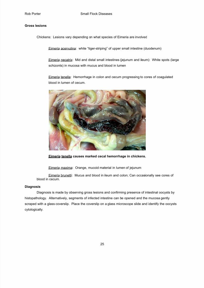
Eimeria tenella: Hemorrhage in colon and cecum progressing to cores of coagulated
blood in lumen of cecum.
Eimeria tenella causes marked cecal hemorrhage in chickens.
Eimeria maxima: Orange, mucoid material in lumen of jejunum
Eimeria brunetti: Mucus and blood in ileum and colon; Can occasionally see cores ofblood in cecum.
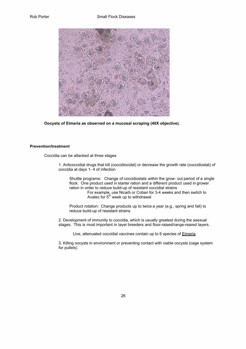
Diagnosis
Diagnosis is made by observing gross lesions and confirming presence of intestinal oocysts by
histopathology. Alternatively, segments of infected intestine can be opened and the mucosa gently
scraped with a glass coverslip. Place the coverslip on a glass microscope slide and identify the oocysts
cytologically.
8/17/2019 Poultry Diseases CVM
http://slidepdf.com/reader/full/poultry-diseases-cvm 26/34
Rob Porter Small Flock Diseases
26
Oocysts of Eimeria as observed on a mucosal scraping (40X objective).
Prevention/treatment
Coccidia can be attacked at three stages
1. Anticoccidial drugs that kill (coccidiocidal) or decrease the growth rate (coccidiostat) ofcoccidia at days 1- 4 of infection
Shuttle programs: Change of coccidiostats within the grow- out period of a single
flock. One product used in starter ration and a different product used in growerration in order to reduce build-up of resistant coccidial strains
For example, use Nicarb or Coban for 3-4 weeks and then switch to Avatec for 5
th week up to withdrawal
Product rotation: Change products up to twice a year (e.g., spring and fall) toreduce build-up of resistant strains
2. Development of immunity to coccidia, which is usually greatest during the asexualstages. This is most important in layer breeders and floor-raised/range-reared layers.
Live, attenuated coccidial vaccines contain up to 6 species of Eimeria
3. Killing oocysts in environment or preventing contact with viable oocysts (cage systemfor pullets)
8/17/2019 Poultry Diseases CVM
http://slidepdf.com/reader/full/poultry-diseases-cvm 27/34
Rob Porter Small Flock Diseases
27
Roundworms = Ascariasis
Cause: Ascaridia galli; a nematode residing in the upper small intestine of chickens and turkeys
Young birds (<3 months) are most susceptible to intestinal damage and light-weight breeds(e.g., White Leghorns) are more susceptible to infection than heavy meat-type breeds (e.g.,White Rock)
Malcom Reid (University of Georgia): Layers harboring 10-14 adult worms in intestinal tracthad 5-7% reduction in egg production
Infection can occasionally occur in caged layers that have been exposed to contaminatedflies.
Direct Life Cycle (no intermediate host): About 30 days to complete cycle
Embryonated egg (takes 10-12 days in environment; faster if warm and moist litter)
Lumen of proventriculus or duodenum
Larvae hatch and live in lumen of duodenum for 9-10 days
Wall of duodenum
Larvae migrate in wall of small intestine for 8-9 daysto cause tissue damage and hemorrhage
Lumen of duodenum
Young adult (male and female) re-enters lumen of intestine
Lumen of small intestine
Feces Mature adult produces eggs by 30 days after infection
Eggs contaminate environment and can remain infective forup to 160 weeks on the ground. Resistant to low temperatures
Clinical Signs
Depression, weight loss, diarrhea
Decreased growth
Lowered egg production in heavy infection
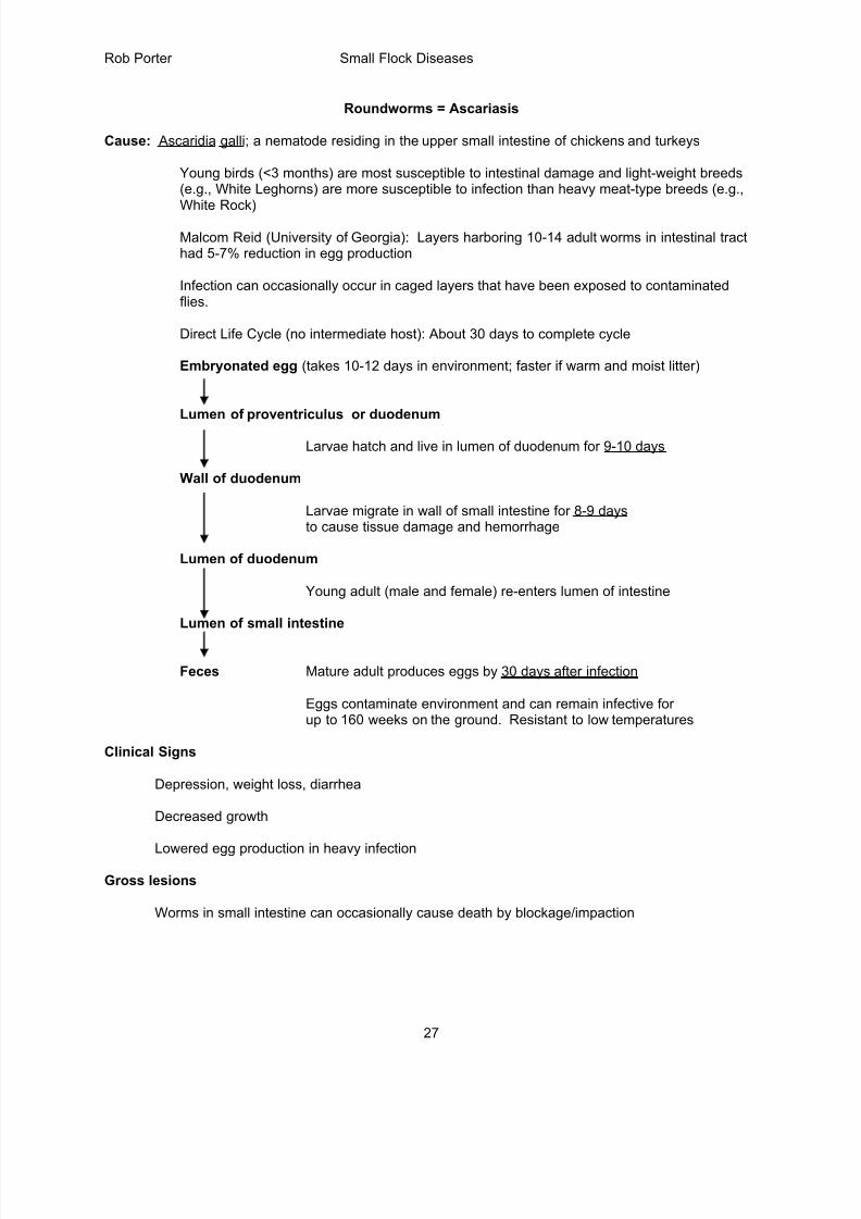
Gross lesions
Worms in small intestine can occasionally cause death by blockage/impaction
8/17/2019 Poultry Diseases CVM
http://slidepdf.com/reader/full/poultry-diseases-cvm 28/34
Rob Porter Small Flock Diseases
28
Intestinal Roundworms in a floor-raised pullet
Reddened intestinal mucosa (enteritis)
Thin bird; atrophy of breast muscle, decreased body fat
Ascarid can migrate to oviduct and penetrate egg prior to shell formation
Diagnosis
Necropsy: identification of worms that are large, yellow-white, and 5-11 cm long
Fecal flotation (eggs float in zinc sulfate or sodium nitrate solution)
Prevention/Treatment
Confinement and cage rearing has reduced problems with most intestinal parasites
Deep litter (4-6 inches of wood shavings) to reduce exposure to parasite eggs
Proper clean up between flocks
Treat pullets before they are transferred to laying house
Treatment: Piperazine
Ectoparasites
Mites and lice are the most common ectoparasites in poultry. Ectoparasites can cause decreased
production and uniformity and also provide opportunity for transmission of other diseases.
Feather mites
Synonyms: Acariasis; Dermanyssus gallinae = the red roost mite or red chicken mite
Causes: The two common mites of poultry are the Northern fowl mite (Ornithonyssus sylvarium) and the
red chicken mite (Dermanyssus gallinae).
Northern fowl mite (Ornithonyssus sylviarum): This mite can cause economic loss as a result of
8/17/2019 Poultry Diseases CVM
http://slidepdf.com/reader/full/poultry-diseases-cvm 29/34
Rob Porter Small Flock Diseases
29
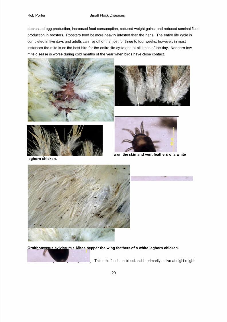
decreased egg production, increased feed consumption, reduced weight gains, and reduced seminal fluid
production in roosters. Roosters tend be more heavily infested than the hens. The entire life cycle is
completed in five days and adults can live off of the host for three to four weeks; however, in most
instances the mite is on the host bird for the entire life cycle and at all times of the day. Northern fowl
mite disease is worse during cold months of the year when birds have close contact.
Ornithonyssus sylviarum : Mites and mite excreta on the skin and vent feathers of a whiteleghorn chicken.
Ornithonyssus sylviarum : Mites pepper the wing feathers of a white leghorn chicken.Left = adult mite (10x).
Red roost mite (Dermanyssus gallinae): This mite feeds on blood and is primarily active at night (night
8/17/2019 Poultry Diseases CVM
http://slidepdf.com/reader/full/poultry-diseases-cvm 30/34
Rob Porter Small Flock Diseases
30
feeder), and during the day will leave birds to reside in nest boxes and cracks and crevices of perches
and slats in the poultry house.
Clinical signs: Mites are most often observed in young laying birds and appear as rough, scaley skin,
dark, stained feathers around the vent as a result of accumulation of mite eggs, scabs and fecal material.
Mites feed on blood and heavy infestations can cause anemia. Decreased egg production can occur.
Gross lesions: Mite can be observed on the feathers of the vent, particularly in hens, and over the entire
body in roosters. Check more than one bird for the presence of mites because not all birds are infested.
Check birds at night if you are looking for the red roost mite (mites are off of the bird during the day).
Treatment: In heavy infestations chemical insecticide sprays can be used. The insecticides are
available as wettable powders, emusifiable concentrates, soluble concentrates, granules and
microencapsulated products. The spray must be at high enough pressure (100-125 PSI with one gallon ofwater per 100 birds) to penetrate the feathers of the vent region. Commercial acaricidal insecticides(e.g.,
pyrethrins and permethrins such as Ectobon, Rabon and Ravap) often contain wetting agents to help the
active chemical penetrate to the feathers. Instructions on the commercial insecticide sprays should be
carefully followed. Caged layers should receive the spray from beneath the cage to increase access to
the vent region. Floor raised breeders should be sprayed at night when they are resting on slats.
Because the red roost mite commonly spends time off of the bird at night, these infestations require that,
in addition to bird treatment, the nests and cracks/crevices of the poultry house should be sprayed or
dusted with insecticide.
In small flocks it may be economical to use a carbamate product, commercial rose dust (Sevin Dust).
Place 2.5 pounds of 5% Sevin dust in one dust box per fifty birds. Carefully dust the product on the vent
region and also sprinkle the dust around nest boxes and along cracks and crevices in the hen house.
Prevention: Make sure the house is cleaned, disinfected and free of mites and lice before introducing
new birds. New birds introduced to a house should be carefully examined to determine that they are free
of ectoparasites. Examine equipment to prevent transmission of mites on clothing, egg flats, crates, etc.
Use washable egg flats and steam clean or power wash equipment that is moved into a new house.
Maintain biosecurity to keep rodents and wild birds out of houses.
Scaley-leg mites
Knemidocoptes mutans. This is one of several species of scaley-leg mite that exist on a variety of birds
including chickens, turkeys, pheasants, partridges, and many passerine birds. The mite found most often
8/17/2019 Poultry Diseases CVM
http://slidepdf.com/reader/full/poultry-diseases-cvm 31/34
Rob Porter Small Flock Diseases
31
on older birds and spends the entire life cycle in the skin. A similar mite infection (Knemidocoptes pilae)
in psittacine birds causes scaling on the beak and legs.
Transmission occurs through direct contact with other birds. Female scaley-leg mites burrow into
nonfeathered portions of the body, particularly the scales of the legs, but also the comb and wattles,
resulting in marked proliferation of the skin with heavy and irregular keratin crusts. The females deposit
eggs for two months after burrowing. Both larval and nymph stages follow after the eggs hatch. The
heavy crusting on the leg can interfere with joint flexion and can cause lameness. Sever infections can
result in loss of toes, decreased feed consumption and decreased egg production. The disease can be
treated by dipping the legs in dilute insecticide (acaricidal) solutions. Birds that are not used for egg or
meat production can be effectively treated with a drop of ivermectin solution (Ivomec, off-label treatment)
on the comb or wattle. Repeat this treatment two weeks later.
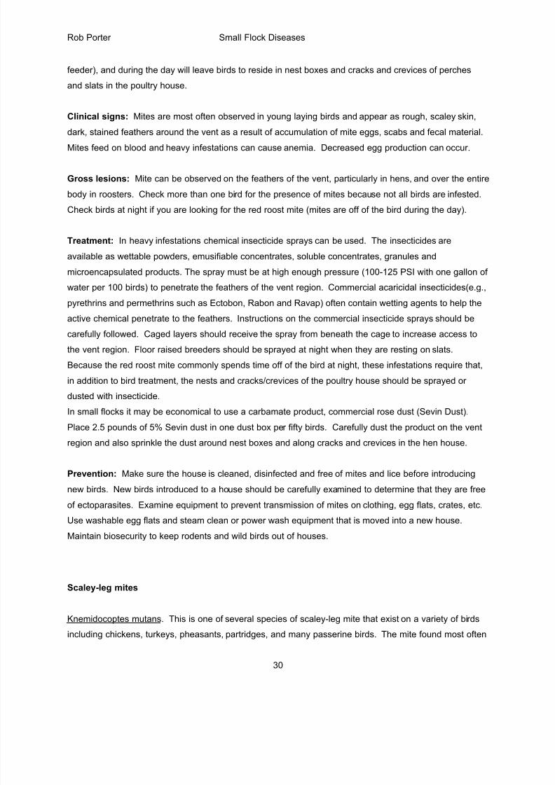
Adult bantam roosters (A and B) with scaley-let mites: Note the enlarged and deformed shanksand feet with excess accumulation of keratin caused by Knemidocoptes mutans.
A B
8/17/2019 Poultry Diseases CVM
http://slidepdf.com/reader/full/poultry-diseases-cvm 32/34
Rob Porter Small Flock Diseases
32
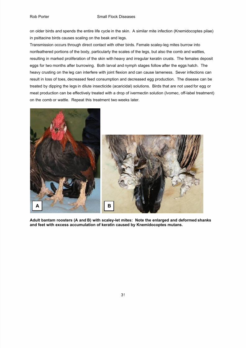
Microphotograph of skin from leg of chicken with round scaley-leg mite (blackarrows) embedded in thick keratin layer (white arrows; H&E, bar = 20 microns).
Lice (Pediculosis)
Body lice are larger than mites and can build up in large numbers on affected birds, especially caged
layers. Lice spend the entire life cycle on the bird and can live for several months. The lice that infest
birds are chewing lice (Mallophaga) rather than blood feeders, feeding on dander and feathers. Laying
hens that are infested can have significant decreases in egg production and weight loss.
Causes:
Chicken: Cuclogaster (head louse), Goniocotes (fluff louse), Menacanthus (body louse)
Turkey: Menacanthus, Chelopistes, Oxylipeurus
Duck and goose: Anaticola, Trinoton
Pigeon: Columbicola, Campanulotes
8/17/2019 Poultry Diseases CVM
http://slidepdf.com/reader/full/poultry-diseases-cvm 33/34
Rob Porter Small Flock Diseases
33
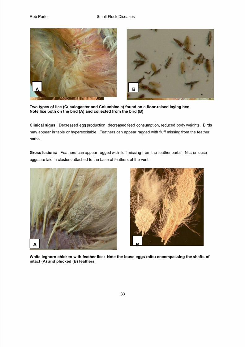
Two types of lice (Cuculogaster and Columbicola) found on a floor-raised laying hen. Note lice both on the bird (A) and collected from the bird (B)
Clinical signs: Decreased egg production, decreased feed consumption, reduced body weights. Birds
may appear irritable or hyperexcitable. Feathers can appear ragged with fluff missing from the feather
barbs.
Gross lesions: Feathers can appear ragged with fluff missing from the feather barbs. Nits or louse
eggs are laid in clusters attached to the base of feathers of the vent.
White leghorn chicken with feather lice: Note the louse eggs (nits) encompassing the shafts ofintact (A) and plucked (B) feathers.
BA
A B

8/17/2019 Poultry Diseases CVM
http://slidepdf.com/reader/full/poultry-diseases-cvm 34/34
Rob Porter Small Flock Diseases
Treatment and Prevention: This is similar to recommendations made for the Northern Fowl mite, but the
louse eggs are resistant to insecticides and a second treatment (spray or dust) is required at two weeks
after the first to kill the lice that have recently hatched. Be sure to treat the entire body of the birds
because in heavy infestations the lice tend to spread from the vent to the neck region.