Surface modification of polyamide composite membranes by ...

Instructions for use
Title Modification of spectrin in red cell membranes by the lipid peroxidation product 4-hydroxy-2-nonenal associated withthe changes in red cell membrane properties
Author(s) Arashiki, Nobuto
Citation 北海道大学. 博士(獣医学) 甲第9655号
Issue Date 2010-06-30
DOI 10.14943/doctoral.k9655
Doc URL http://hdl.handle.net/2115/43145
Type theses (doctoral)
File Information arashiki_thesis.pdf
Hokkaido University Collection of Scholarly and Academic Papers : HUSCAP

Modification of Spectrin in Red Cell Membranes by the Lipid
Peroxidation Product 4-Hydroxy-2-nonenal Associated with
the Changes in Red Cell Membrane Properties
(赤血球膜スペクトリンの脂質過酸化産物 4-ヒドロキシ-2-ノネナールによる 分子修飾と赤血球膜物性の変化)
Nobuto Arashiki
Laboratory of Molecular Medicine, Department of Veterinary Clinical Sciences, Graduate School of Veterinary Medicine, Hokkaido University

Contents
Abbreviations
Preface ……………………………………………………………………………….. 1
Introduction ……………………………………………………………………………. 3
Materials and Methods …………………………………………………………….. 7
Results 11
I. Characterization of HNE modification of RBC α- and β-spectrin ……………… 11
II. The effects of HNE-spectrin adduct formation on the membrane properties of human RBCs ……………………………………………………………………
16
Discussion 30
Physiological and pathological roles of HNE modification of spectrin …………… 30 Characteristics of HNE modification of spectrin and its effects on the protein
function ………………………………………………………………………….
33
References …………………………………………………………………………… 37
Acknowledgements …………………………………………………………………. 42
Abstract ……………………………………………………………………………… 43
Abstract in Japanese ………………………………………………………………... 44
i

Abbreviations
Abbreviations used in this study are as follows:
HNE ........................ 4-hydroxy-2-nonenal
IOV ......................... inside-out vesicle
MALDI-TOF MS.... matrix-assisted laser desorption/ionization time-of-flight mass spectrometry
MgATP .................... adenosine 5'-triphosphate, magnesium salt
PBS ......................... phosphate-buffered saline
PVP ......................... polyvinylpyrrolidone
RBC ........................ red blood cell
SDS-PAGE .............. sodium dodecylsulfate-polyacrylamide gel electrophoresis
t-BOOH ................... tert-butylhydroperoxide
In the present study, one-letter and three-letter abbreviations for amino acid residues are
employed.
ii

1
Preface
Oxidative stress is associated with various pathological conditions, including
inflammation, diabetes, neurodegenerative diseases, and hemolytic anemia with various
degrees. The potential for red blood cells (RBCs) to undergo autooxidative destruction is great
because the cell is loaded with about 20 mM hemoglobin, most of which is bound to oxygen
and can release a highly reactive superoxide ion, leaving behind it methemoglobin. Continued
oxidation of methemoglobin leads to generation of irreversibly oxidized hemichromes, to
precipitation, and eventually to the formation of Heinz bodies. Hemichromes and Heinz
bodies can destroy functions of RBC membranes directly or can cause oxidation of membrane
components. These may include lipid peroxidation and protein cross-linking, resulting in
formation of RBCs that are rigid and are susceptible to trapping in sinusoidal structures.
Consequently, the extravascular and intravascular destructions of such RBCs occur.
The mechanical properties of RBC membranes, i.e., membrane stability and
deformability, are primarily attributed to the functions of membrane skeletal proteins that
underlie the cytoplasmic surface of the lipid bilayer. Therefore, these membrane proteins are
likely to be the targets of the oxidative attacks. In addition, the local generation and
accumulation of peroxidation products in the membrane, by affecting membrane protein
function, might be involved in senescence of RBCs under physiological conditions. However,
no substantial study has been reported in RBCs except for a few studies on the adduct
formation between spectrin and denatured globin and the association of band 3 with Heinz
bodies.

2
The purpose of the present study was to examine the presence and significance of the
covalent modification of membrane skeletal proteins by lipid peroxidation products in RBCs.
The present study demonstrates that protein modification with a lipid peroxidation end
product, 4-hydroxy-2-nonenal occurs preferentially in spectrin at the interface between the
skeletal proteins and lipid bilayer. The data also show that this reaction is associated with
reduction in membrane mechanical properties of RBCs.
This study has been published in part as follows:
1) Arashiki, N., Otsuka, Y., Ito, D., Yang, M., Komatsu, T., Sato, K., and Inaba, M. (2010)
The covalent modification of spectrin in red cell membranes by the lipid peroxidation
product 4-hydroxy-2-nonenal. Biochem. Biophys. Res. Commun. 391, 1543-1547.
3
Introduction
Spectrin, a major constituent of the human RBC membrane skeleton, acts as a
scaffolding protein and mediates the organization and assembly of a diverse set of proteins in
specialized membrane domains (Bennett and Healy, 2008). Spectrin is a flexible, rod-like
protein formed by two end-to-end associated heterodimers composed of α and β subunits,
with sizes of 280 kDa and 246 kDa, respectively. In RBCs, the spectrin-actin network is
linked to the lipid bilayer through protein-protein interactions, specifically spectrin-protein
4.1R-glycophorin C and spectrin-ankyrin-band 3 associations, and through direct association
of spectrin with phosphatidylserine (PS) in the inner bilayer leaflet (Cohen et al., 1986;
Manno et al., 2002; Mohandas and Gallagher, 2008). Such vertical interactions between the
spectrin-actin skeleton and lipid bilayer play pivotal roles in maintaining RBC membrane
stability (Manno et al., 2002; Low et al., 1991; Inaba et al., 1996). Spectrin loss reduces the
membrane mechanical resilience, leading to spherocyte formation in various types of
hereditary spherocytosis (Tse and Lux, 2001; Mohandas and Gallagher, 2008; Perrotta et al.,
2008). In contrast with its role in congenital disorders, less is known about acquired spectrin
damage. Previous studies have focused on the oxidation of spectrin sulfhydryl groups,
revealing that RBC exposure to oxidative conditions causes morphological and mechanistic
changes, principally through spectrin oxidation (Snyder et al., 1998; Wagner et al., 1987).
Furthermore, mild oxidation reportedly causes functional and structural changes, leading to
the reduced interaction of spectrin with protein 4.1 (Becker et al., 1986).
In contrast, membrane phospholipid peroxidation, that is proceeded by a free radical

4
chain reaction mechanism (Yang et al., 2003), generates various cytotoxic aldehydes and
alkenals that can damage proteins and other cellular constituents (Esterbauer et al., 1991;
Schneider et al., 2001; Uchida, 2003). 4-Hydroxy-2-nonenal (HNE) is the most abundant and
toxic aldehyde generated by the oxidation of plasma membrane polyunsaturated fatty acids,
such as arachidonic acid. HNE is fairly stable and present in relatively higher amounts in
biological membranes under the conditions of oxidative stress (Esterbauer et al., 1991). HNE
is a highly reactive electrophile that reacts with the side chains of various amino acid residues,
including Cys, His, and Lys, to form Michael-type adducts and Schiff-base adducts of
HNE-Lys (Fig. 1A) (Esterbauer et al., 1991; Liu et al., 2003; Peterson and Doorn, 2004).
Protein modification by HNE causes serious detrimental effects in the cell, due to the
resulting functional defects and cross-linking of proteins (Fig. 1B) (Uchida et al., 1993;
Montine et al., 1996; Lauderback et al., 2001; Murray et al., 2007; Stewart et al., 2007;
Kokubo et al., 2008). HNE also accumulates in RBCs (Ando et al., 1995), leading to the
covalent modification of membrane proteins (Uchida et al., 1997). However, the target
proteins for HNE modification remain unknown.
These findings have suggested that oxidative environments under physiological or
disease conditions may cause spectrin modifications by HNE, leading to deleterious changes
in the plasma membrane mechanical properties. The purpose of the present study was to
examine the presence and significance of covalent spectrin modifications by HNE in RBCs.
The present study was focused on membrane proteins in human RBCs since their structures
and the roles in the mechanical properties of the RBC membrane have been well documented
(Tse and Lux, 2001; Mohandas and Gallagher, 2008). We used anti-HNE antibodies and
5
matrix-assisted laser desorption/ionization time-of-flight mass spectrometry (MALDI-TOF
MS) to identify HNE-modified proteins in RBC membranes, and found that spectrin was the
primary target of HNE. Then, the effects of HNE modification on the RBC membrane
mechanical properties were characterized by some biophysical and biochemical approaches.
6
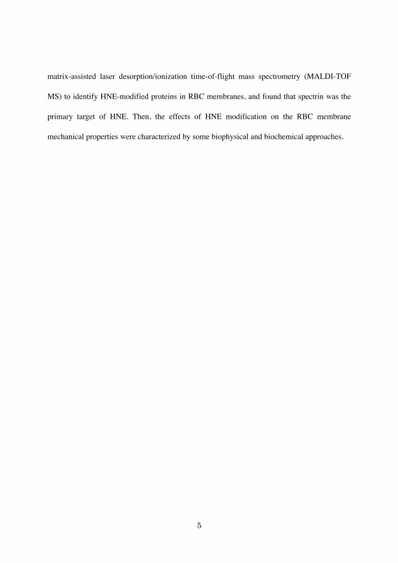
Figure 1. Lipid peroxidation, formation of HNE-amino acid adducts, and protein crosslinking. A. A chain reaction of lipid (L) peroxidation generates lipid peroxides. Lipid peroxides undergo spontaneous decomposition yielding a number of aldehydes including HNE. HNE can react with Cys, His, and Lys in proteins, forming Michael adducts or Schiff-base adducts (only with Lys). B. The reaction of HNE with proteins is frequently associated with their covalent crosslinking leading to the formation of fluorophores such as a Lys-derived dihydro-pyrrole derivative. Reproduced from Yang et al. (2003) and Liu et al. (2003) with several changes.
OOH
Michael-typeadducts
HN-R
OOH
S-R
OOH
HNE-Cys
HNE-His
N-ROH
HNE-Lys
HNE-Lys
OOH
NN
R
4-Hydroxynonenal(HNE)
L (Lipid)
LOO
LH
LOOH
Fe3+
Fe2+
LO!-Scission
Schiff-baseadducts
A
+NHLys
NH-LysO
O2
Lysine
NHLys
ON-Lys
NHLys
NH-LysOH
O2
N-LysOH
NH-LysOH
ON-Lys
HO
+NH
NLys
Lys
Schiff-base Lys adduct
Michael Lys adduct
Dihydro-pyrrolecrosslink
B
Fig. 1
Formation of lysine-derived protein crosslink

7
Materials and Methods
Preparation of RBC membrane ghosts and spectrin-actin extracts
After obtaining informed consent, blood was obtained from healthy volunteers.
Blood was also obtained from dogs, cats, and cattle housed at the Graduate School of
Veterinary Medicine, Hokkaido University. All experimental procedures met with the
approval of the Laboratory Animal Experimentation Committee, Graduate School of
Veterinary Medicine, Hokkaido University.
Preparation of RBC membrane ghosts and extraction of spectrin heterodimers (αβ)
with a hypotonic buffer were performed as described previously (Inaba et al., 1996). In some
experiments, RBC ghosts were prepared by hemolysis in hypotonic buffer (5 mM Tris/Cl [pH
7.4], 5 mM KCl, and 1 mM MgCl2) with or without 0.6 mM MgATP (Sigma), as described
previously (Manno et al., 2002). We also obtained ghosts from RBCs separated into different
fractions using a discontinuous arabinogalactan gradient (Tiffert et al., 2005).
SDS-PAGE and immunoblotting
Membrane proteins were separated by SDS-PAGE on 6.5% or 10% SDS-gels
followed by staining with Coomassie brilliant blue or immunoblotting. Immunoblotting was
performed as described previously (Ito et al., 2006) using antibodies against HNE-Michael
adducts (Calbiochem), HNE-His (monoclonal antibody HNEJ-2; Nikken SEIL Co., Fukuroi,
Japan), and canine spectrin (Inaba et al., 1996). One-dimensional spectrin peptide mapping
was performed as described previously (Inaba and Maede, 1989; Inaba et al., 1992) using V8

8
protease (Sigma).
MALDI-TOF MS analysis of spectrins.
After separation by SDS-PAGE, proteins in the gel slices were reduced with NaBH4
for 30 minutes at an ambient temperature unless otherwise indicated. This was done to detect
unstable reversibly formed adducts, such as HNE-Lys Michael adducts (Fenaille et al., 2003).
The proteins were subsequently stained with Coomassie brilliant blue and digested in gel
slices with 10 µg/ml of trypsin Gold (Promega) for 16 hours at 37ºC. Prior to protease
digestion, the Cys residues were reduced with 10 mM dithiothreitol and alkylated with 55
mM iodoacetamide. Peptides were eluted from gels, desalted using C18 ZipTips (Millipore),
and crystallized using saturated α-cyano-4-hydroxy-cinnamic acid (Bruker Daltonics) as a
matrix. Full-scan mass spectra of the tryptic peptides from 800-3,000 m/z were collected in
positive mode by averaging 100-250 spectra using a Bruker autoflex MALDI-TOF mass
spectrometer. Measured peptide masses were used to search the NCBI and Swiss-Prot
sequence database for protein identification using MASCOT software (Matrix Science).
In vitro formation of HNE-protein adducts in red cells and red cell ghosts
A packed 10% cellular volume of RBCs was suspended in phosphate-buffered saline
(PBS) containing 5 mM glucose. After incubation for various times (0-6 hours) at 37ºC in the
presence or absence of 0.1-1.0 mM HNE (Calbiochem), the cells were washed with PBS.
Membrane ghosts were then prepared as described above. Ghost proteins were labeled with
HNE by incubating PBS-suspended ghosts (1 mg protein/ml) with 0.01-1.0 mM HNE at 37ºC
9
for appropriate time periods (0-60 minutes), followed by SDS-PAGE and immunoblotting. In
some experiments, incubation was performed in hypotonic buffer with 0.1 mM MgATP.
In vitro oxidation of RBCs with tert-butylhydroperoxide and analysis of spectrin
RBCs suspended in PBS containing 5 mM glucose were incubated at 37ºC for the
time periods indicated in the presence of 0.3 or 3.0 mM tert-butylhydroperoxide (t-BOOH,
Wako Pure Chemical Industries, Tokyo, Japan). After incubation, RBCs were collected by
centrifugation and directly dissolved in SDS-PAGE sample buffer. An SDS-PAGE was
performed, followed by immunoblotting to detect the HNE-spectrin adducts.
Measurement of red cell membrane mechanical properties
After incubation in the presence or absence of HNE, as described above, RBCs were
washed with PBS, suspended in 3.5% polyvinylpyrrolidone (PVP, Sigma) at 30 µl of packed
red cells/4 ml of PVP solution, and exposed to an increasing shear stress (0-150 dynes/cm2) in
the ektacytometer as described previously (Chasis and Mohandas, 1986). Deformability index
(DI) versus applied shear stress curve was analyzed to quantitate membrane deformability.
Measurement of binding of spectrin to the inside-out vesicles
The RBC ghosts were extracted for spectrin as described above. The resultant
inside-out vesicles (IOVs) were resealed by incubating in PBS at 37ºC for 40 minutes.
Extracted spectrin heterodimers (0-150 µg) were incubated in PBS in the presence or in the
absence of 0.1 mM HNE at 37ºC for the appropriate time. To this were added IOVs
10
containing 10 µg of proteins and incubated on ice for 90 minutes. The reactions were applied
on 8% sucrose in PBS followed by centrifugation at 33,500 x g for 50 minutes at 4ºC to
obtain spectrin bound to the IOVs. The contents of α- and β-spectrin were analyzed by
SDS-PAGE and normalized with the amount of band 3 in the IOVs by densitometric scanning
of the Coomassie blue-stained gels.
Statistical analysis
Paired Student's t-test was used to assess statistical significance.

11
Results
I. Characterization of HNE modification of RBC α- and β-spectrin
HNE modification of α- and β-spectrin in human RBC membranes
Immunoblotting analysis revealed the presence of polypeptides that had reacted with
the anti-HNE antibody. These polypeptides were located at positions corresponding to α- and
β-spectrin in the freshly prepared human RBC membranes and in the crude spectrin-actin
preparation extracted from the ghosts (Fig. 2A). These signals were weak and required > 15
seconds for detection when 5 ~ 10 µg of membrane proteins were loaded. Spectrin, especially
β-spectrin, also reacted with the anti-HNE-His adducts, suggestive of a predominance of
HNE-His adducts in β-spectrin (Fig. 2A). When limited digestion of β-spectrin with V8
protease was performed, more than 20 polypeptides with sizes of < 50 kDa were detected by
Coomassie blue staining. Among these, only the 45- and 40-kDa fragments strongly reacted
with the anti-HNE antibodies (Fig. 2B).
The spectrin polypeptides were analyzed to determine the positions where these
spontaneous modification with HNE occurred in spectrins from freshly prepared RBC ghosts.
MALDI-TOF MS detected 50 ~ 70 tryptic peptides that covered ~ 25-35% of the total amino
acid residues of α- and β-spectrin. Several independent analyses consistently showed that a
dozen or more distinct peptides derived from either α- and β-spectrin contained one or more
Michael-type HNE-amino acid adduct and Schiff base conjugate (Table 1 and Fig. 3). For
example, in β-spectrin reduced with NaBH4, the MALDI-TOF MS consistently detected an

12
HNE-modified species at 2,622.4 m/z for the peptides IHCLENVDKALQFLKEQR (amino
acid residues 110-127) derived from the N-terminal actin-binding domain of this subunit. The
observed mass shift of 438 m/z (= 158 + 2 × 140 m/z) was indicative of a Michael addition
reaction with mass shifts of 158 m/z at either the 2nd or 3rd underlined His or Cys residue,
respectively. The mass shift of 438 m/z was also indicative of Schiff base adducts with mass
shifts of 280 m/z (= 2 × 140 m/z) at the two underlined Lys residues. This is because reduction
with NaBH4 increases the characteristic mass shifts of 156 and 138 m/z for Michael and
Schiff base adducts, respectively, by 2 atomic mass units (Fenaille et al., 2003). When
β-spectrin was analyzed without reduction, peaks at 1,382.6 and 2,239.2 m/z, shifted by 312
(= 2 × 156) and 468 (= 3 × 156) atomic mass units, respectively, were detected for the
corresponding peptides IHCLENVDK and IHCLENVDKALQFLK, suggesting that all
underlined residues could form Michael-type HNE adducts. Together with the data for the
mass shifts of several other peptides (Table 1), these results allow us to predict that all Cys-,
His-, and Lys-Michael adducts and Schiff base Lys adducts are present in both α- and
β-spectrin, consistent with our immunoblot results with anti-HNE antibodies.
Fig. 3 illustrates the positions and types of HNE adduct formation obtained by
MALDI-TOF MS for α- and β-spectrin. The presence of many HNE-amino acid adducts in
the β-spectrin N-terminal region is compatible with the reactivity of the 45- and 40-kDa
proteolytic fragments in immunoblotting shown in Fig. 2B. Subsequent MALDI-TOF MS
analysis revealed that these proteolytic fragments were derived from the N-terminal domain
and the first triple helical structural repeat, and that they contained peptides such as
IHCLENVDK displaying [M + H]+ with an m/z of 1,382.6, as described above.

13
Spectrins are the major targets of the lipid peroxidation product HNE in human
RBC membranes
To further characterize HNE-spectrin adduct formation, we analyzed the covalent
modification of spectrin in RBCs incubated with extrinsic HNE. Membrane ghosts treated
with 0.1 mM HNE exhibited a marked increase in HNE-spectrin adduct contents within 10
minutes of incubation (Fig. 4A). Incubation also caused HNE labeling of several other
membrane proteins with apparent sizes of 115, 110, 100, 80, and 43 kDa (Fig. 4A). These
polypeptides were likely α- and β-adducin, band 3, protein 4.1R, and actin, respectively,
judging from their migrating positions and antibody reactivities (data not shown). In contrast,
when intact RBCs were incubated with HNE for several hours, a remarkable increase in the
signals of HNE-protein adducts was found for spectrin, especially β-spectrin, but not for other
membrane proteins (Fig. 4B).
Next, HNE-spectrin adduct formation was examined in RBCs exposed to the
well-known oxidant t-BOOH. Incubation with 0.3 mM t-BOOH for 1 hour increased the HNE
modification of spectrin, while decreasing the spectrin content of the human RBC membranes
(Fig. 4C). As observed in RBCs incubated with extrinsic HNE (Fig. 4B), HNE signals were
predominantly found in spectrins and were more prominent in β- than in α-spectrin. HNE
adduct signals were also detected at the position corresponding to globins (~ 15 kDa), since
RBCs were directly subjected to SDS-PAGE after incubation. While no obvious increase in
HNE-spectrin adducts was demonstrated during incubation, the signal intensity of
HNE-spectrin relative to that of spectrin (relative abundance of HNE-spectrin) increased

14
significantly due to the reduction in spectrin contents. Increasing the t-BOOH concentration
to 3 mM caused striking decreases in α- and β-spectrin contents and the formation of high
molecular weight species reactive to both the anti-HNE adducts and anti-spectrin antibodies
within 2 hours of incubation (Fig. 4C). This indicates that the HNE-modified spectrin
accumulated and formed irreversible aggregates in RBCs exposed to t-BOOH.
These results demonstrate that both extrinsic circulating HNE and intrinsic HNE,
generated presumably through membrane lipid peroxidation, selectively react with spectrin at
the protein-lipid bilayer interface. The data also suggest that the association status of spectrin
with membrane lipids was different in intact RBCs and in the isolated membrane ghosts, so as
to cause distinct patterns in HNE adduct formation for α- and β-spectrin.
Spectrin modification with HNE in RBC membranes from several animal
species
To assess if HNE modification occurs, in general, in spectrins from various animals,
membrane proteins in bovine, canine, and feline RBC membranes were analyzed for their
HNE-protein adducts by immunoblotting. Membranes from these animals showed protein
compositions that were principally the same with protein constituents in human RBCs so far
judged by Coomassie brilliant blue staining (Fig. 5A) as reported previously (Inaba and
Maede, 1988). However, there was a remarkable difference in HNE-modified proteins and
their contents. In bovine and feline RBC membranes, HNE-spectrin adducts (α and β) was
observed, but the their signal intensities were much less than that in human RBCs (Fig. 5B).
Moreover, canine RBC spectrin showed no signals of HNE-protein adducts, indicating no
15
substantial and spontaneous formation of HNE-spectrin under physiological conditions.
Instead, strong signals of HNE adducts were found in other polypeptides including a 95-kDa
unknown polypeptide and protein 4.2. The abundant signals in protein 4.2 were also observed
in feline RBC ghosts.
On the other hand, exposure to 0.1 mM HNE caused generation of HNE-protein
adducts with profound signals in membranes from these animals; α- and β-spectrin were
modified with HNE in all species, while HNE adducts appeared in the positions
corresponding to band 3 and protein 4.1 in bovine RBC membranes, and protein 4.2 in canine
and feline RBC membranes, respectively (Fig. 5C). Unidentified polypeptides of 200 kDa in
bovine and 95 kDa in canine membranes also represented abundant HNE-protein adduct
signals. These results demonstrated that the RBC membrane proteins accessible to HNE
modification differ among species. Nonetheless, spectin appeared to be one of the major
targets for HNE-adduct formation as in human RBC membranes as described above.
Effect of spectrin-lipid association on HNE modification of α- and β-spectrin
Therefore, we next compared spectrin labeling with HNE in membrane ghosts
possessing different association status of spectrin with membrane lipids, i.e., ghosts prepared
in the presence (MgATP ghost) or absence (control ghost) of MgATP (Manno et al., 2002).
When ghosts were incubated with increasing HNE concentrations, we observed marked
increases in the intensities of HNE-spectrin adducts in both MgATP and control ghosts (Fig.
6). In principle, when membrane proteins containing equivalent amounts of spectrins were
exposed to HNE, spectrin labeling with HNE in the MgATP ghost was less abundant than in

16
the control ghost, and was much less than that in the crude spectrin-actin extract (Fig. 6A).
Remarkable differences between the MgATP and control ghosts were observed at
lower HNE concentrations (0.05-0.3 mM). Immunoblotting revealed that the abundance of
HNE signals relative to spectrin contents was significantly higher in the control than in the
MgATP ghost (Figs. 6B and 6C), demonstrating that HNE spectrin labeling preferentially
occurred in the control ghosts. In contrast, the MgATP ghost showed reductions in the spectrin
contents and corresponding increases in high molecular weight aggregate formation, with
prominent signals for both the anti-HNE adducts and anti-spectrin antibodies (Fig. 6B). These
data suggest that HNE-modified spectrins in the MgATP ghosts tended to be cross-linked and
aggregated, although the adduction levels were lower than those in control ghosts. Moreover,
the HNE labeling of β-spectrin was nearly equivalent to that of α-spectrin in the MgATP
ghost, whereas HNE labeling was predominantly found in α-spectrin in the control ghost, as
observed in the crude spectrin-actin extract (Figs. 6A-6C).
II. The effects of HNE-spectrin adduct formation on the membrane properties
of human RBCs
HNE-spectrin adducts in density-separated RBCs
Then, the contents of HNE-modified spectrin were analyzed for density-separated
RBCs to examine if these adducts accumulate during RBC aging (Fig. 7). The
lightest/youngest cell fraction showed HNE/spectrin ratios equivalent to those in fractions 3
and 4, which comprised ~90% of total cell counts (Fig. 7). No significant differences in the
HNE levels of α- and β-spectrin were observed among fractions 1 through 4. The densest

17
fractions (fractions 5 and 6) had reduced HNE/spectrin ratios; an increase would have been
expected if there was accumulation of HNE-spectrin adducts during RBC senescence. Similar
results were obtained in three independent experiments, suggesting that human RBCs possess
HNE-modified spectrin throughout their life-span with a slight reduction, rather than
accumulation, in the levels of HNE-modified spectrins at senescence.
Effect of HNE modification of spectrin on RBC membrane mechanical
properties
To determine the effect of HNE-spectrin adduct formation on mechanical properties
of RBC membranes, we analyzed deformability, by ektacytometry, of RBCs in which
spectrins were adducted with HNE by incubating the intact RBCs with HNE as described
above (Fig. 4). RBCs exposed to 0.1 mM HNE exhibited reduction in the DI values to about
80% that of the control cells incubated in the absence of HNE, judging from the maximum DI
values (DImax) under shear stress at 150 dynes/cm2 (Fig. 8). Difference in exposure periods,
i.e., 1 hour or 4 hours, appeared to affect the initial phase of increasing the shear stress (0 ~ 40
dynes/cm2), with less DI values in RBCs treated with 0.1 mM HNE for 4 hours. RBCs
incubated for 1 hour with 1.0 mM HNE showed remarkably attenuated increase in DI in
response to shear stress, and had a decreased DImax value that was 60% that of the control
cells. The DImax value of HNE-treated cells exposed for 4 hours showed further reduction to
only 40% of the control value. Since spectrin was the major target of HNE modification in the
membrane under the condition employed (Fig. 4), the data obtained by ektacytometry analysis
demonstrated that HNE modification of spectrin affected the mechanical properties, resulting

18
in reduced RBC membrane deformability.
Effect of HNE modification on binding of spectrin to the IOV
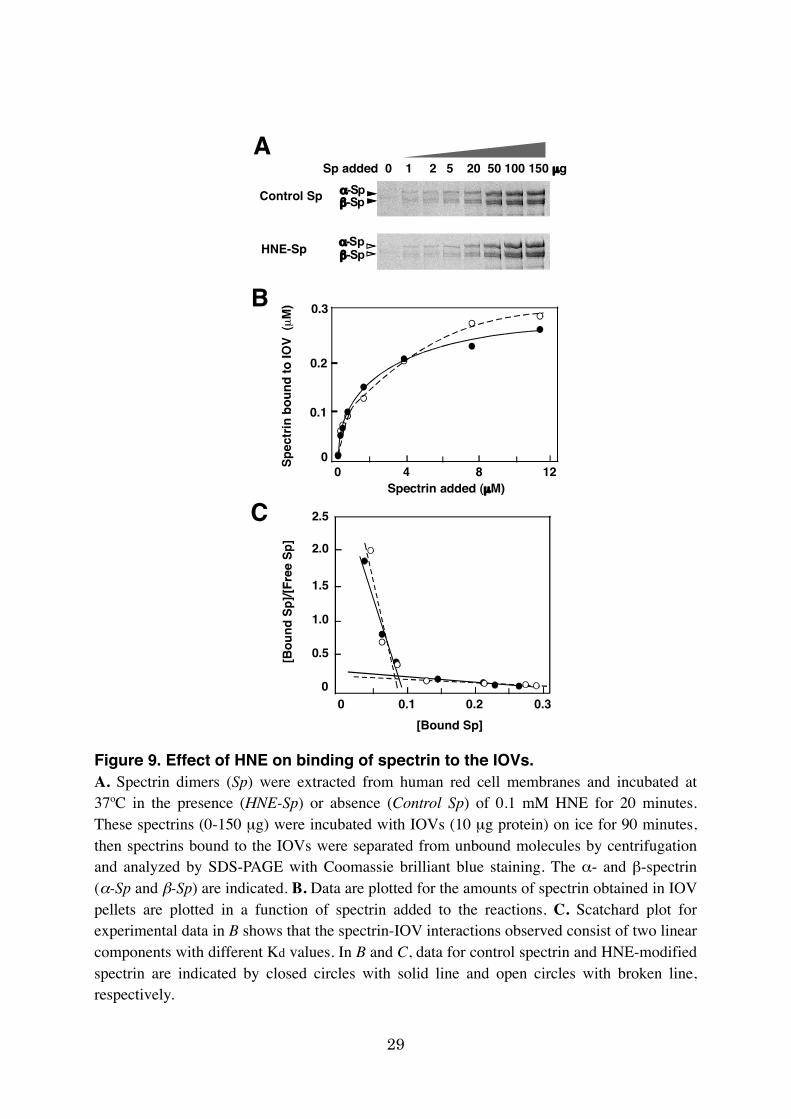
Spectrin bound to the IOVs in a concentration-dependent manner (Figs. 9A and 9B).
The binding appeared to contain two phases with distinct dissociation constants (Kd) of 0.79
µM and 32 µM (Fig. 9C). Binding of HNE-spectrin to the IOVs exhibited a profile similar to
that of the control spectrin, while it possessed Kd values of 0.41 µM and 40 µM, for high and
low affinity phases, respectively (Figs. 9B and 9C), suggesting a slightly increased affinity of
HNE-modified spectrin in binding to the IOVs in the high affinity component (Figs. 9B and
9C).

19
Figure 2. HNE-spectrin adducts in human RBC membranes. A. Ghost membrane proteins (G, 8 µg/lane) and the crude spectrin/actin extracted from ghosts (S, 2-3 µg/lane) were separated on SDS-PAGE gels. Protein components were analyzed by Coomassie brilliant blue staining (CBB) and HNE-protein adduct formation was assessed by immunoblotting using anti-HNE (Anti-HNE) or anti-HNE-His (Anti-HNE-His) adduct antibodies. In immunoblotting, the membranes were exposed to the films for 1 minute. The migrating positions of the major membrane proteins, including α- and β-spectrin (α-Sp and β-Sp, respectively), are indicated. B. β-Spectrin separated on SDS-PAGE gels was sliced out and analyzed by one-dimensional peptide mapping in the presence (β-Sp + V8) or absence (β-Sp) of V8 protease, followed by CBB staining and immunoblotting with Anti-HNE. The 45- and 40-kDa proteolytic polypeptides exhibiting strong signals for HNE-protein adducts are indicated. The migrating positions of β-spectrin and of marker proteins (kDa) are also presented. Asterisks indicate polypeptides from V8 protease preparation.
!-Sp"-Sp
GAPDH
Band 34.1
Actin
G S G S G S
4.2
CBB Anti-HNE Anti-HNE-His
A B
-"-Sp
**
250
-
-25
50 4540
CBB Anti-HNE
Fig. 2

20
Table 1. Analysis of HNE-modified peptides derived from α- and β-spectrin by MALDI-TOF MS. Tryptic peptides derived from α- and β-spectrin were analyzed for Michael-type (Michael) and Schiff base (Schiff) HNE adducts by MALDI-TOF MS. The amino acid residues, sequences, and theoretical and observed masses are shown for peptides deduced to contain HNE adduct(s) by at least two independent analyses (peptides 1-16 and 1-18 for α- and β-spectrin, respectively). Localization of each peptide in the spectrin repeats is also indicated.
Spectrin Peptide number
Spectrin repeats
Amino acid residues
Sequence Mass Numbers of HNE adducts†
Theoretical Observed Michael Schiff α-Spectrin 1* 3-4 257-271 QKALSNAANLQRFKR 1743.99 2057.18 2 0 2 4 295-307 DLVASEGLFHSHK 1438.72 1597.85 1 0 3* 4 312-326 NLAVMSDKVKELCAK 1647.87 2117.21 3 0 4 4 322-329 ELCAKAEK 890.45 1207.65 2 0 5* 5 412-416 HQQHK 676.34 989.56 2 0 6 7 569-575 EKAATRR 830.47 989.56 1 0 7* 7 596-601 NWINKK 801.45 958.54 1 0 8* 13 1225-1230 HEGFER 773.35 930.49 1 0 9* 13-14 1282-1290 KESLNEAQK 1045.54 1202.70 1 0 10 13-14 1283-1299 ESLNEAQKFYLFLSKAR 2043.08 2202.15 1 0 11* 16 1586-1594 EHVVDHLLER 1233.59 1546.89 2 0 12 17 1648-1659 KHQLLEREMLAR 1522.84 1663.83 0 1 13 18 1744-1756 DLQGVQNLLKKHK 1519.88 1818.94 1 1 14* 20 2016-2027 WEQLLEASAVHR 1437.74 1594.95 1 0 15 21 2133-2141 HLSDIIEER 1110.57 1269.71 1 0 16* EF1 2281-2293 HFDENLTGRLTHK 1566.79 1723.98 1 0 β-Spectrin 1 Nt 45-52 IKALADER 914.52 1055.56 0 1 2* Nt 75-86 ITDLYKDLRDGR 1463.77 1620.88 1 0 3* Nt 110-118 IHCLENVDK 1069.52 1382.65 2 0 4* Nt 110-124 IHCLENVDKALQFLK 1769.95 2239.24 3 0 5 Nt 110-127 IHCLENVDKALQFLKEQR 2183.15 2622.46 1 2 6 Nt 227-243 DSNARHNLEHAFNVAER 1978.94 2138.08 1 0 7* 1 293-302 VIDHAIETEK 1153.60 1310.70 1 0 8* 2 491-498 ENYHDQKR 1088.50 1401.74 2 0

21
*Obtained for α- and β-spectrin without reduction. Other HNE-peptides were detected for polypeptides reduced with NaBH4 prior to
tryptic digestion. †Numbers of deduced HNE-amino acid adducts in each peptide.
9 3 570-593 HKLMEADIAIQGDKVKAITAATLK 2564.44 2705.48 0 1 10* 4 631-641 TQLEQSKRLWK 1415.79 1572.79 1 0 11* 7 956-975 VHTLCVDCEETSKWITDKTK 2335.12 2705.48 2 0 12 7 974-996 TKVVESTKDLGRDLAGIIAIQRK 2510.45 2827.62 2 0 13 9 1161-1175 SHTLAQCLGFQEFQK 1735.84 1894.98 1 0 14 11 1404-1420 SDDPRKDLTSVNRMLAK 1945.00 2104.03 1 0 15 11 1417-1431 MLAKLKRVEDQVNVR 1798.02 1938.91 0 1 16 14 1671-1684 HYAGLKDVAEERKR 1670.89 1830.01 1 0 17 14 1677-1696 DVAEERKRKLENMYHLFQLK 2546.34 2705.48 1 0 18* 17 1991-2004 RKEMNEKWEARWER 1946.95 2260.14 2 0

22
Figure 3. Schematic illustration of HNE-amino acid adduct positions in α- and β-spectrin deduced from MALD-TOF MS analysis. Positions of HNE-amino acid adducts found in α- and β-spectrin by MALDI-TOF MS analysis (Table 1) are summarized and schematically presented. Closed and open circles indicate Michael-type and Schiff base adducts, respectively, for spectrins reduced with NaBH4 prior to MS analysis. Closed triangles indicate Michael adducts deduced for spectrins without reduction. Illustrations for spectrin and phosphatidylserine-binding repeats are according to previous studies (Mohandas and Gallagher, 2008; An et al., 2004). Interactions between spectrin and membrane proteins through glycophorin C-protein 4.1-spectrin and band 3-ankyrin-spectrin linkages are also shown.
Fig. 3

23
Figure 4. HNE-spectrin adduct formation in RBC ghosts and intact red cells exposed to HNE. A. Human RBC ghosts were suspended in PBS containing 0.1 mM HNE and placed on ice for 10 minutes then incubated at 37ºC for the indicated periods and analyzed for HNE-protein adduct formation by immunoblotting (5 µg/lane) with the anti-HNE antibody. B. Human RBCs were incubated at 37ºC in the presence of 0.1 or 1.0 mM HNE for 0, 1 or 4 hours. RBC membrane ghosts were prepared and analyzed by immunoblotting (5 µg/lane). Immunoblotting membranes were exposed to films for 3-5 seconds. C. RBCs were incubated at 37ºC for 0-12 hours with 0.3 or 3.0 mM t-BOOH. After a brief wash, packed RBCs were directly solubilized in an equivalent volume of 2× SDS-PAGE sample buffer, and were subjected to immunoblotting to detect HNE-protein adducts (Anti-HNE) and spectrin (Anti-Sp). Closed arrowheads indicate α- and β-spectrin (α-Sp and β-Sp) and some major proteins in the red cell membranes, including α- and β-adducin (α-Add and β-Add), band 3, protein 4.1, actin, and hemoglobin (Hb). Open arrowheads indicate high molecular weight aggregates (HMW).
Fig. 4
C
!-Sp"-Sp
HMW0 1 2 4 12 1 2 0 1 2 4 12 1 2 (h)
t-BOOH0.3 3.0 0.3 3.0
t-BOOH
Hb
A
!-Sp"-Sp
0.1 1.00 1 4 1 4 (h)
Actin
HNE
Anti-HNE Anti-HNE Anti-HNE Anti-Sp
B
!-Sp"-Sp
!-Ad"-AddBand 34.1
Actin
0 5 10 20 (min)0.1 mM HNE

24
Figure 5. HNE adduct formation in red cell membranes from several animal species. A. Membrane proteins (8 µg/lane) from human (Human), bovine (Cattle), canine (Dog), and feline (Cat) red cells were separated by SDS-PAGE followed by Coomassie brilliant blue staining (CBB). B and C. HNE-protein adduct formation was analyzed by immunoblotting using the anti-HNE adduct (Anti-HNE) antibody. In immunoblotting, the membranes were exposed to the films for 1 minute. In C, red cell ghosts were suspended in PBS containing 0.1 mM HNE and placed on ice for 10 minutes then incubated at 37ºC for 1 hour and analyzed for HNE-protein adduct formation by immunoblotting (5 µg/lane). Immunoblotting membranes were exposed to films for 3-5 seconds. The migrating positions of the major membrane proteins, including α- and β-spectrin (α-Sp and β-Sp, respectively), are indicated. Arrowheads with asterisks indicate unidentified polypeptides of 200 kDa and 95 kDa in bovine and canine membranes, respectively.
Band 34.1
Actin
4.2
!-Sp"-Sp
A CB
GAPDH
!-Sp"-Sp
4.2
!-Sp"-Sp
!-Sp"-Sp
Band 34.1
Actin
4.2
GAPDH
4.2
!-Sp"-Sp
CBB Anti-HNE Anti-HNE
*
*
*
Fig. 5

25
Figure 6. HNE-spectrin adduct formation in MgATP ghosts and control ghosts exposed to HNE. (continued on the following page)
ATP ghost Control ghost Sp/Act ext
HNE 0 0.1 0.3 0.5 1 0 0.1 0.3 0.5 1 0 0.1 0.3 0.5 1 (mM)* *
!-Sp"-Sp
!-Sp"-Sp
HMW*HMW
HMW * * **
ATP ghost Control ghost
!-Sp"-Sp
!-Sp"-Sp
Anti-HNE Anti-Sp Anti-HNE Anti-Sp
HNE 0.1 0.2 0.3 0.1 0.2 0.3 0.1 0.2 0.3 0.1 0.2 0.3 (mM)
**
5
4
3
2
1
0
HNE (mM) HNE (mM) HNE (mM)0 0.1 0.2 0.3 0 0.1 0.2 0.3 0 0.1 0.2 0.3
5
4
3
2
1
0
ATP ghost Control ghost Sp/Act ext25
20
15
10
5
0
B
A
C
Fig. 6

26
Figure 6. HNE-spectrin adduct formation in MgATP ghosts and control ghosts exposed to HNE. A. Human red cell ghosts were prepared in hypotonic buffer (5 mM Tris/Cl. pH 7.4, 5 mM KCl, 1 mM MgCl2) with or without 0.6 mM MgATP (MgATP ghost and Control ghost, respectively) and were diluted in the same buffer to contain 0.1 mM MgATP when present. These ghosts were incubated at 37ºC and 1 mg protein/ml for 1 hour with 0, 0.05, 0.1, 0.2, 0.3, 0.4, 0.5, or 1.0 mM HNE. After rapid centrifugation, the membrane proteins (1 µg/lane) were analyzed for HNE-protein adducts by immunoblotting. Crude extracts containing spectrin and actin (Sp/Act ext, 0.3 mg protein/ml) were also incubated with HNE and processed to detect HNE-protein adducts (0.3 µg/lane). B. Typical immunoblots similar to those shown in A, with longer exposure time for HNE-protein adducts (Anti-HNE), and the corresponding immunoblots for spectrin (Anti-Sp). In A and B, α- and β-spectrin (α-Sp and β-Sp) and high molecular weight species (HMW) are indicated. Asterisks mark the top of the separating gel. Immunoblot membranes were exposed to films for 3 seconds and 10 seconds in A and B, respectively. C. Contents of HNE-modified α-spectrin (open circles) and β-spectrin (closed circles) shown in B were quantified by densitometric scanning, normalized by the corresponding spectrin, and shown as the relative abundance of the HNE-spectrin (HNE/Spectrin). Data are the means ± S.D. (n = 3). **P < 0.01. The third panel displays data for the crude extracts of spectrin/actin obtained from immunoblots shown in A, and the corresponding immunoblots for spectrin (Sp/Act ext, n = 2).

27
Figure 7. HNE-spectrin adduct contents in density-separated human red cells. A. Human red cells were separated into fractions 1 to 6 by centrifugation on an arabinogalactan (AG) density gradient (Tiffert et al., 2005). B. Fractions were examined for their HNE-spectrin and whole spectrin contents as described above by immunoblotting (Anti-HNE, 8 µg/lane, exposed for 1 minute) and Coomassie brilliant blue staining (CBB, 8 µg/lane), respectively. C. Contents of HNE-modified α-spectrin (hatched columns) and β-spectrin (unhatched columns) shown in B were quantified by densitometric scanning, normalized by the corresponding spectrin, and shown as the relative abundance of the HNE-spectrin (HNE/Spectrin). Data represent a typical result from several independent experiments.
!-Sp"-Sp
!-Sp"-Sp
Top BottomGravity
Fraction No. 1 2 3 4 5 6
Anti-HNE
CBB
1 2 3 4 5 6
8
4
6
20
Fraction No.
A B
Fig. 7
C
1
2
3
456
AG (%)Fraction

28
Figure 8. Reduction in deformability of human red cells incubated with HNE. Human red cells were incubated at 37ºC in the presence (0.1 mM or 1.0 mM) or absence (Control) of HNE for 1 or 4 hours (1 h or 4 h), suspended in the PVP solution, and examined for their deformability under an increasing shear stress (0-150 dynes/cm2) in the ektacytometer. Symbols indicate as follows: closed square, control red cells; closed and open circles, red cells incubated with 0.1 mM HNE for 1 and 4 hours, respectively; closed and open triangles, red cells incubated with 1.0 mM HNE for 1 and 4 hours, respectively.
Fig. 8
0 30 60 90 120 150
0.2
0.4
0.6
0.8
1.0
Shear stress (dyn/cm2)
0
Control
0.1 mM, 1 h
0.1 mM, 4 h
1.0 M, 1 h1.0 mM, 4 h
29
Figure 9. Effect of HNE on binding of spectrin to the IOVs. A. Spectrin dimers (Sp) were extracted from human red cell membranes and incubated at 37ºC in the presence (HNE-Sp) or absence (Control Sp) of 0.1 mM HNE for 20 minutes. These spectrins (0-150 µg) were incubated with IOVs (10 µg protein) on ice for 90 minutes, then spectrins bound to the IOVs were separated from unbound molecules by centrifugation and analyzed by SDS-PAGE with Coomassie brilliant blue staining. The α- and β-spectrin (α-Sp and β-Sp) are indicated. B. Data are plotted for the amounts of spectrin obtained in IOV pellets are plotted in a function of spectrin added to the reactions. C. Scatchard plot for experimental data in B shows that the spectrin-IOV interactions observed consist of two linear components with different Kd values. In B and C, data for control spectrin and HNE-modified spectrin are indicated by closed circles with solid line and open circles with broken line, respectively.
Fig. 9
00
2.5
2.0
1.5
1.0
0.5
[Bound Sp]0.30.20.1
00
0.3
0.2
0.1
Spectrin added (µM)1284
!-Sp"-Sp
!-Sp"-Sp
Control Sp
HNE-Sp
Sp added 0 1 2 5 20 50 100 150 µgA
B
C
30
Discussion
Physiological and pathological roles of HNE modification of spectrin
Various diseases involve proteins modified by HNE and protein cross-linking
through HNE adducts contributes to disease pathogenecities (Montine et al., 1996; Murray et
al., 2007; Stewart et al., 2007; Kokubo et al., 2008). Here, we showed that spectrins are the
primary targets of HNE, and that HNE-modified spectrins generate irreversibly cross-linked
aggregates in human RBCs. Since HNE accumulates in RBCs in circulation (Ando et al.,
1995), the extensive modification of spectrins by HNE may occur in peripheral blood RBCs
under physiological and pathological conditions, leading to RBC senescence or accelerated
destruction of RBCs (Fig. 10).
The relative abundance of HNE-spectrin adducts was higher in control ghosts than in
MgATP ghosts. MgATP helps maintain the asymmetric distribution of PS through the action
of MgATP-dependent phospholipid translocase. Interactions between aminophospholipids and
skeletal proteins modulate the human RBC membrane mechanical stability (Manno et al.,
2002). The spectrin-actin network is thus tightly associated with the lipid bilayer through the
direct binding of spectrin with PS in the presence of MgATP (Manno et al., 2002) as well as
through protein-protein interactions (Mohandas and Gallagher, 2008). Therefore, in the
absence or under reduced concentrations of MgATP, greater HNE adduct formation may
occur in spectrins that are dissociated from the lipid bilayer and have increased susceptibility
to the amphiphilic HNE. Then, local aggregation of HNE-modified spectrins is inevitable,
since these spectrins contain Cys-, His-, and Lys-Michael adducts and Schiff bases on the Lys

31
residues, that are required for intra- or intermolecular cross-linking (Esterbauer et al., 1991;
Uchida et al., 1993; Petersen and Doorn, 2004; Murray et al, 2007; Stewart et al., 2007).
Local spectrin aggregation may free the lipid bilayer from underlying spectrin-actin skeletons,
possibly leading to membrane surface area loss by extrusion, which occurs in RBCs under
oxidative conditions (Snyder et al., 1998; Wagner et al., 1987) and various RBC membrane
disorders (Tse and Lux, 2001; Mohandas and Gallagher, 2008; Perotta et al., 2008) (Fig. 10).
Thus, in addition to modulating RBC membrane mechanical properties, the
spectrin-membrane lipid interactions may have an obligatory role in preventing skeletal
proteins from excessive HNE adduct formation.
In contrast to the observations under ATP-free conditions described above, in which
HNE modifications preferentially occurred in α-spectrin (Figs. 4 and 6), the intensities of
β-spectrin HNE adducts were comparable to those of α-spectrin in HNE-exposed MgATP
ghosts (Fig. 6). Furthermore, β-spectrin was the principal target of HNE when intact RBCs
were exposed to HNE or t-BOOH (Fig. 4). Spectrin reportedly associates directly with PS
(Cohen et al., 1986; Manno et al., 2002) in several distinct segments involving the N-terminal
actin-binding domain, triple helical structural repeats 2-4 and 12-14 of β-spectrin, and repeats
8-10 of α-spectrin (An et al., 2004). Thus, under physiological conditions, HNE adduction
occurs preferentially in membrane-associated β-spectrin at the interface of the skeletal protein
and the inner leaflet of the membrane lipid bilayer. Intrinsic HNE is generated in the lipid
bilayer by fatty acid peroxidation; extrinsic HNE reaches the inner leaflet presumably by
diffusion through the lipid bilayer (Uchida, 2003). These assumptions are consistent with the
presence of HNE adducts in the N-terminal regions of β-spectrin, including repeats 2-4 (Fig.
32
3). Moreover, selective HNE-spectrin formation and protection of other membrane proteins
from HNE addition probably involve HNE elimination by cellular glutathione. Glutathione
readily reacts with HNE via Michael addition spontaneously or enzymatically to form
glutathione-HNE conjugate and much of aqueous HNE in a cell is bound to glutathione
(Petersen and Doorn, 2004).
Interestingly, in the presence of MgATP, the HNE-modified spectrins tended to be
cross-linked and aggregated (Fig. 6). HNE reportedly modifies the three His residues of
amyloid β protein (Aβ). These HNE-modified Aβ molecules have an increased affinity for
membrane lipids and possess a conformation similar to that of mature amyloid fibrils (Murray
et al., 2007; Liu et al., 2008). The observed enrichment of HNE-His adducts (Fig. 2 and Table
1) suggests that the propensity of HNE-modified spectrin to form aggregates may involve
mechanisms similar to those found for Aβ. Thus, the closer to the physiological environment
the RBC membrane is, the more readily membrane damage through spectrin aggregation can
occur. Consequently, restricted membrane surface area loss would occur with much less
severity compared to pathological conditions described above. This mechanism, involving
preferential β-spectrin HNE-modification and aggregate formation, may provide significant
protection against excessive lipid peroxidation product accumulation during RBC aging. The
slightly decreased contents of HNE-spectrin adducts in the most dense population (Fig. 7),
which displayed reduced membrane surface area/volume ratios possibly represent the gradual
accumulation followed by the loss of aggregated HNE-modified proteins and membrane
lipids during RBC aging (Fig. 10). This mechanism also gives an explanation for the
difference between ghosts from intact RBCs (Fig. 2) and ghosts from HNE-exposed RBCs

33
(Fig. 4) in HNE modification patterns of α- and β-spectrin. HNE modification and consequent
aggregate formation would be expected for spectrin family members expressed at the
cytoplasmic plasma membrane surface of most metazoan cells (Bennett and Healy, 2008).
HNE-spectrin aggregation in the presence of ATP may also involve interactions with
hemoglobin. The MgATP ghost contained more residual hemoglobin (~12% of the total
membrane proteins) than the control ghost (< 3% of the total membrane proteins). Retained
hemoglobin can augment irreversible aggregate formation, possibly by cross-linking spectrin,
as previously shown in human red blood exposed to hydrogen peroxide (Snyder et al., 1985).
Characteristics of HNE modification of spectrin and its effects on the protein
function
One of the most notable findings of the present study is that human RBCs exposed to
HNE exhibited marked reduction in their deformability (Fig. 8). Since spectrin was the major
target of HNE in the RBC membranes under the condition employed (Figs. 4 and 8), this
implies that HNE modification caused structural and/or functional changes of spectrin,
leading to reduced mechanical properties of the RBC membrane. The possible changes in
spectrin skeleton in RBCs treated with HNE appear to differ from those reported for the
RBCs exposed to oxidants to generate spectrin-hemoglobin cross-linking (Snyder et al., 1985;
Wagner et al., 1987). In these RBCs, lipid peroxidation, though present, was not necessary for
the formation of spectrin-hemoglobin aggregates and the various cellular changes (Snyder et
al., 1985). As discussed above, therefore, surface area loss due to aggregate formation of
HNE-spectrin might be a cause for the decreased deformability of RBCs incubated with HNE.

34
The other possibility involves a slight increase in interactions between spectrin and
its binding proteins in the membrane such as protein 4.1R and/or ankyrin-band 3 complex
(Tse and Lux, 2001; Mohandas and Gallagher, 2008). The stoichiometry of binding of control
spectrin heterodimers to the IOVs with Kd of 0.8 µM observed (Fig. 9) is comparable with
that reported for binding of protein 4.1R and ankyrin to spectrin in the 10-7 M range (Tyler et
al., 1980). The increased affinity of HNE-modified spectrin with Kd of 0.4 µM likely
represent that structural changes caused by HNE adduction can modulate the membrane
binding stoichiometry of spectrin through the protein-protein association. On the other hand,
there was no significant change in the low affinity component of the spectrin-IOV binding
that appears to represent a direct association of spectrin to the membrane lipids (PS) with Kd
= 10-5 ~ 10-6 M range (An et al., 2004). This is contrary to the assumption discussed above,
i.e., an increased affinity of HNE-spectrin to the membrane lipids. This discrepancy may be
attributed to the lack of asymmetric distribution of PS in the IOVs due to the absence of
MgATP in the reaction. Measurement of deformability of resealed ghosts would provide some
clues to these issues.
HNE adduct formation was shown to be a posttranslational modification of spectrin
polypeptides, with several major, distinct target sites susceptible to HNE (Fig. 3). This
posttranslational event seemingly can occur soon after spectrin synthesis, since HNE-spectrin
adducts were found in all fractions, including the lightest/youngest fraction, of
density-separated red cells (Fig. 7). We also observed that an anti-HNE antibody reacted with
bacterial recombinant polypeptides of the N-terminal domain and the adjacent triple helical
structural repeats 2-4 of β-spectrin (An et al., 2004), and that some tryptic peptides of these

35
recombinants showed mass shifts indicative of Michael-type adducts (data not shown). For
example, the peptide IHCLENVDK (amino acid residues 110-118, [M + H]+ with an m/z of
1,382.6) exhibited a mass shift of 312 m/z due to two Michael adducts at the His and Cys
residues, as demonstrated for the native RBC spectrins. There should be some structural
changes subsequent to HNE adductions, particularly in the N-terminal β-spectrin region. It
should be ascertained whether such structural changes affect or modulate spectrin function,
since this region and the α-spectrin C-terminus play key roles in αβ heterodimer formation
(Speicher et al., 1992; Begg et al., 2000) and actin and protein 4.1 binding (Tse and Lux,
2001; An et al., 2005; Zhu et al., 2007).
In conclusion, this study demonstrates that HNE-spectrin adducts spontaneously
form at the interface of membrane skeletal proteins and the inner lipid bilayer leaflet in
human RBCs. Furthermore, our data suggests that HNE-spectrin aggregates may accumulate
in the RBC, followed by their extrusion with membrane lipids from RBCs. This process may
be involved in the physiological and pathological destruction of RBCs.

36
Figure 10. Schematic illustration for HNE-spectrin adduct formation and its implications in physiological and pathological changes of RBCs. Both intrinsic HNE generated in the membrane through lipid peroxidation in RBCs and extrinsic HNE penetrated through the lipid bilayer can react with spectrin to form HNE-spectrin adducts. Accumulated HNE-modified spectrin, depending on the extent to which levels accumulation occurs, may generate local or large and dispersed aggregates. Vesicular extrusion of the local aggregates consequently cause membrane surface area loss in RBCs under physiological condition, resulting in production of aged and sphere RBCs. By contrast, under some pathological conditions generating HNE at profound levels, aggregates formed aberrantly and extensively at the cytoplasmic surface of RBC membranes would cause fragmentation of RBCs, resulting in hemolysis.
Profound anddispersed aggregates
Physiological changes:RBC senescence
Pathological changes:RBC lesions and hemolysis
HNE-spectrin accumulation
Ankyrin !-Spectrin
Fig. 10

37
References
An, X., Debnath, G., Guo, X., Liu, S., Lux, S. E., Baines, A., Gratzer, W. and Mohandas, N. (2005) Identification and functional characterization of protein 4.1R and actin-binding sites in erythrocyte spectrin: regulation of the interactions by phosphatidylinositol-4,5-bisphosphate. Biochemistry 44: 10681-10688.
An, X., Guo, X., Sum, H., Morrow, J., Gratzer, W. and Mohandas, N. (2004) Phosphatidylserine binding sites in erythroid spectrin: location and implications for membrane stability. Biochemistry 43: 310-315.
Ando, K., Beppu, M. and Kitagawa, K. (1995) Evidence for accumulation of lipid hydroperoxides during the aging of human red blood cells in the circulation. Biol
Pharmacol. Bull. 18: 659-663.
Becker, P. S., Cohen, C. M, and Lux, S. E. (1986) The effect of mild diamide oxidation on the structure and function of human erythrocyte spectrin. J. Biol. Chem. 261: 4620-4628.
Begg, G. E., Harper, S. L., Morris, M. B. and Speicher, D. W. (2000) Initiation of spectrin dimerization involves complementary electrostatic interactions between paired triple-helical bundles. J. Biol. Chem. 275: 3279-3287.
Bennett, V. and Healy, J. (2008) Organizing the fluid membrane bilayer: diseases linked to spectrin and ankyrin. Trends Mol. Med. 14: 28-36.
Chasis, J. A. and Mohandas, N. (1986) Erythrocyte membrane deformability and stability: two distinct membrane properties that are independently regulated by skeletal protein associations. J. Cell Biol. 103: 343-350.
Cohen, A. M., Liu, S.-C., Derick, L. H. and Palek, J. (1986) Ultrastructural studies of the interaction of spectrin with phosphatidylserine liposomes. Blood 68: 920-926.
Esterbauer, H., Schauer, R. J. and Zollner, H. (1991) Chemistry and biochemistry of 4-hydroxynonenal, malondialdehyde and related aldehydes. Free Rad. Biol. Med. 11: 81-128.

38
Fenaille, F., Guy, P. A. and Tabet, J. C. (2003) Study of protein modified by 4-hydroxy-2-nonenal and other short chain aldehydes analyzed by electrospray ionization tandem mass spectrometry. J. Am. Soc. Mass Spectrom. 14: 215-226.
Ito, D., Koshino, I., Arashiki, N., Adachi, H., Tomihari, M., Tamahara, S., Kurogi, K., Amano, T., Ono, K. and Inaba, M. (2006) Ubiquitylation-independent ER-associated degradation of an AE1 mutant associated with dominant hereditary spherocytosis in cattle. J. Cell Sci. 119: 3602-3612.
Inaba, M. and Maede, Y. (1988) A new major transmembrane glycoprotein, gp155, in goat erythrocytes. Isolation and characterization of its association to cytoskeleton through binding with band 3-ankyrin complex. J. Biol. Chem. 263: 17763-17771.
Inaba, M. and Maede, Y. (1989) O-N-Acetyl-D-glucosamine moiety on discrete peptide of multiple protein 4.1 isoforms regulated by alternative pathways. J. Biol. Chem. 264: 18149-18155.
Inaba, M., Gupta, K. C., Kuwabara, M., Takahashi, T., Benz, E. J. Jr, and Maede, Y. (1992) Deamidation of human erythrocyte protein 4.1: possible role in aging. Blood 79: 3355-3361.
Inaba, M., Yawata, A., Koshino, I., Sato, K., Takeuchi, M., Takakuwa, Y., Manno, S.. Kanzaki, A., Sakai, J., Ban, A., Ono, K. and Maede, Y. (1996) Defective anion transport and marked spherocytosis with membrane instability caused by hereditary total deficiency of red cell band 3 in cattle due to a nonsense mutation. J. Clin. Invest. 97: 1804-1817.
Jain, S. K. (1988) Evidence for membrane lipid peroxidation during the in vivo aging of human erythrocytes. Biochim. Biophys. Acta 937: 205-210.
Kokubo, J., Nagatani, N., Hiroki, K., Kuroiwa, K., Watanabe, N. and Arai, T. (2008) Mechanism of destruction of microtubule structures by 4-hydroxy-2-nonenal. Cell Struct. Funct. 33: 51-59.
Lauderback, C. M., Hackett, J. M., Huang, F. F., Keller, J. N., Szweda, L. I., Markesbery, W. R. and Butterfield, D. A. (2001) The glial glutamate transporter, GLT-1, is oxidatively modified by 4-hydroxy-2-nonenal in the Alzheimer’s disease brain: the role of Aβ1-42. J. Neurosci. 78: 413-416.

39
Liu, L., Komatsu, H., Murray, I. V. J. and Axelsen, P. H. (2008) Promotion of amyloid β protein misfolding and fibrillogenesis by a lipid oxidation product. J. Mol. Biol. 377, 1236-1250.
Liu, Z., Minkler, P. E. and Sayre, L. M. (2003) Mass spectroscopic characterization of protein modification by 4-hydroxy-2-(E)-nonenal and 4-oxo-2-(E)-nonenal. Chem. Res. Toxicol. 16, 901-911.
Low, P. S., Willardson, B. M., Mohandas, N., Rossi, M. and Shohet, S. (1991) Contribution of the band 3-ankyrin interaction to erythrocyte membrane stability. Blood 77: 1581-1586.
Manno, S., Takakuwa, Y. and Mohandas, N. (2002) Identification of a functional role for lipid asymmetry in biological membranes: phosphatidylserine-skeletal protein interactions modulate membrane stability. Proc. Natl. Acad. Sci. U.S.A. 99: 1943-1948.
Mohandas, N. and Gallagher, P. G. (2008) Red cell membrane: past, present, and future. Blood 112: 3939-3948.
Montine, T. J., Amarnath, V., Martin, M. E., Strittmatter, W. J. and Graham, D. G. (1996) E-4-Hydroxy-2-nonenal is cytotoxic and cross-links cytoskeletal proteins in P19 neuroglial cultures. Am. J. Pathol. 148: 89-93
Murray, I. V. J., Liu, L., Komatsu, H., Uryu, K., Xiao, G., Lawson, J. A. and Axelsen, P. H. (2007) Membrane-mediated amyloidogenesis and the promotion of oxidative lipid damage by amyloid β proteins. J. Biol. Chem. 282: 9335-0345.
Perrotta, S,, Gallagher, P. G. and Mohandas, N. (2008) Hereditary spherocytosis. Lancet 372: 1411-1426.
Petersen, D. R. and Doorn, J. A. (2004) Reactions of 4-hydroxynonenal with proteins and cellular targets. Free Rad. Biol. Med. 37: 937-945.
Schneider, C., Tallman, K. A., Porter, N. A. and Brash, A. R. (2001) Two distinct pathways of formation of 4-hydroxynonenal. Mechanisms of nonenzymatic transformation of the 9- and 13-hydroperoxides of linoleic acid to 4-hydroxyalkenals. J. Biol. Chem. 276: 20831-20838.
Speicher, D. W., Weglarz, L. and SeSilva, T. M. (1992) Properties of human red cell spectrin

40
heterodimer (side-to-side) assembly and identification of an essential nucleation site. J. Biol. Chem. 267: 14775-14782.
Stewart, B. J., Doorn, J. A. and Petersen, D. R. (2007) Residue-specific adduction of tubulin by 4-hydroxynonenal and 4-oxononenal causes cross-linking and inhibits polymerization. Chem. Res. Toxicol. 20: 1111-1119.
Snyder, L. M., Fortier, N. L., Trainer, J., Jacobs, J., Leb, L., Lubin, B., Chiu, D., Shohet, S. and Mohandas, N. (1985) Effect of hydrogen peroxide exposure on normal human erythrocyte deformability, morphology, surface characteristics, and spectrin-hemoglobin cross-linking. J. Clin. Invest. 76: 1971-1977.
Tiffert, T., Lew, V. L., Ginsburg, H., Krugliak, M., Croisille, L. and Mohandas, N. (2005) The hydration state of human red blood cells and their susceptibility to invasion by Plasmodium falciparum. Blood 105: 4853-4860.
Tse, W. T. and Lux, S. E. (2001) Hereditary spherocytosis and hereditary elliptocytosis. In: Scriver CR, Beaudet AL, Sly WS, Valle DV, eds. The Metabolic & Molecular Bases of Inherited Disease (8th edition). New York, NY: McGraw-Hill: 4665-4727.
Tyler, J. M., Reinhardt, B. N. and Branton, D. (1980) Association of erythrocyte membrane proteins. Binding of purified bands 2.1 and 4.1 to spectrin. J. Biol. Chem. 255: 7034-7039.
Uchida, K. (2003) Review. 4-Hydroxy-2-nonenal: a product and mediator of oxidative stress. Prog. Lipid Res. 42: 318-343.
Uchida, K., Hasui, Y. and Osawa, T. (1997) Covalent attachment of 4-hydroxy-2-nonenal to erythrocyte proteins. J. Biochem. 122: 1246-1251.
Uchida, K., and Stadtman, E. R. (1993) Covalent attachment of 4-hydroxynonenal to glyceraldehyde-3-phosphate dehydrogenase. A possible involvement of intra- and intermolecular cross-linking reaction. J. Biol. Chem. 268: 6388-6393.
Wagner, G. M., Chiu, D. T.-Y., Qju, J. H., Heath, R. H. and Lubin, B. H. (1987) Spectrin oxidation correlates with membrane vesiculation in stored RBCs. Blood 69: 1777-1781.
Yang, Y., Sharma, R., Sharma, A., Awasthi, S and Awasthi, Y. C. (2003) Lipid peroxidation

41
and cell cycle signaling: 4-hydroxynonenal, a key molecule in stress mediated signaling. Acta Biochim. Polon. 50: 319-336.
Zhu, Q., Vera, C., Asaro, R. J., Sche, P. and Sung, L. A. (2007) A hybrid model for erythrocyte membrane: a single unit of protein network coupled with lipid bilayer. Biophys. J. 93: 386-400.

42
Acknowledgments
The author express sincere gratitude to Professor Mutsumi Inaba (Laboratory of
Molecular Medicine, Graduate School of Veterinary Medicine, Hokkaido University) for his
excellent supervision and encouragements throughout this work.
I would like to express my great thanks to Professor Yuichi Takakuwa and
Associate Professor Sumie Manno (Department of Biochemistry, Tokyo Women’s Medical
University) for helpful discussions and technical support, Professor Osamu Inanami
(Laboratory of Radiation Biology), Associate Professor Akira Terao (Laboratory of
Biochemistry), and Associate Professor Kota Sato (Laboratory of Molecular Medicine), all
from Graduate School of Veterinary Medicine, Hokkaido University, for their helpful
discussion and critical reading of the manuscript. I also express my great thanks to Drs. Yayoi
Otsuka, Daisuke Ito, Hiroshi Ohta, Mizuki Tomihari, Tomohiko Komatsu, Hirokazu Adachi
and all members of the Laboratory of Molecular Medicine for discussion and technical
assistance.

43
Abstract
Spectrin strengthens the red cell membrane through its direct association with membrane
lipids and through protein-protein interactions. Spectrin loss reduces the membrane stability
and results in various types of hereditary spherocytosis. However, less is known about
acquired spectrin damage. The present study showed that α- and β-spectrin in human red cells
are the primary targets of the lipid peroxidation product 4-hydroxy-2-nonenal (HNE) by
immunoblotting and mass spectrometry analyses. The level of HNE adducts in spectrin
(particularly α-spectrin) and several other membrane proteins was increased following the
HNE treatment of red cell membrane ghosts prepared in the absence of MgATP. In contrast,
ghost preparation in the presence of MgATP reduced HNE adduct formation, with preferential
β-spectrin modification and increased cross-linking of the HNE-modified spectrins. Exposure
of intact red cells to HNE resulted in selective HNE-spectrin adduct formation with a similar
preponderance of HNE–β-spectrin modifications and marked reduction in the membrane
mechanical properties. These findings indicate that HNE adduction occurs preferentially in
spectrin at the interface between the skeletal proteins and lipid bilayer in red cells and suggest
that HNE-spectrin adduct aggregation results in the extrusion of damaged spectrin and
membrane lipids under physiological and disease conditions.

44
Abstract in Japanese(要 旨)
Modification of Spectrin in Red Cell Membranes by the Lipid Peroxidation Product
4-Hydroxy-2-nonenal Associated with the Changes in Red Cell Membrane Properties
(赤血球膜スペクトリンの脂質過酸化産物 4-ヒドロキシ-2-ノネナールによる分子修飾
と赤血球膜物性の変化)
膜脂質の過酸化は、4-hydroxyl-2-nonenal (HNE)をはじめとする種々のアルデヒドやア
ルケナールを産生する。これらの脂質過酸化産物は、核酸やタンパク質と共有結合して、そ
の構造と機能の障害を生じる。赤血球は、こうした脂質過酸化産物による障害を最も受け得
る細胞であり、その老化にともなう HNE の蓄積が知られるが、実際にいかなる分子修飾が
どのような影響をもたらすのかは不明である。本研究は、その解明を目的に、主にヒト赤血
球膜における HNE 付加タンパク質と修飾部位の同定、ならびに膜物性に対する影響の解析
を行った。
まず、抗 HNE 抗体を用いたイムノブロッティングの結果、膜骨格網状構造の主体をな
すタンパク質、α-、ならびにβ-スペクトリンに HNE 付加体のシグナルが検出された。赤血球
膜ゴーストを HNE と孵置すると、アクチンやバンド3をはじめとする他の主要膜タンパク
質に HNE 付加が生じたが、無傷赤血球に HNE を作用させた場合にはスペクトリンにのみ、
特にβ-スペクトリンに HNE 付加体の著しい増加が認められ、α-、ならびにβ-スペクトリンが
HNE の主たる標的分子であることが明らかになった。飛行時間型質量分析装置を用いた解析
から、その修飾部位は、β-スペクトリン分子 N 末端ドメイン(βN)中の Ile110-…-Arg127配列な
ど、膜脂質と直接に相互作用する領域を中心に複数存在し、Cys、His、ならびに Lys 残基の
Michael 付加体として、また Lys 残基では加えて Schiff 塩基付加体として存在すると推定さ
れた。一方、ヒト以外の赤血球膜でも、HNE との孵置により HNE-スペクトリンの著増がみ
られたが、自然付加体の量はヒト赤血球に比べて少なく、また動物種により差異が認められ
た。これらの知見は、スペクトリン分子と膜脂質との相互作用が HNE 修飾に影響すること
を示唆するものである。

45
そこで、赤血球におけるスペクトリン-膜脂質間結合を保つMgATP の存在/非存在下に
膜ゴーストを調製し、これらの HNE 付加を検討した。その結果、MgATP 存在下では、HNE
との孵置でスペクトリンの不可溶性凝集体形成が生じ、α鎖よりもβ-スペクトリンの修飾が優
位であった。対照的に、MgATP 非存在下では、HNE-α-スペクトリンの生成がβ鎖のそれを
上回り、明瞭な凝集はみられなかった。また、比重遠心で分画した赤血球では、予想に反し
て全ての分画の赤血球でスペクトリンの HNE 修飾がみられ、最下層の老化赤血球では軽度
の HNE-スペクトリン減少が認められた。これらの知見は、スペクトリンの膜への組み込み
後早期に一定量の HNE 付加が生じることを示すとともに、赤血球の末梢循環過程で限定的
な HNE-スペクトリン凝集体の形成と膜小胞としての除去が生じることを示唆している。
さらに、HNE 付加による赤血球膜の機械的特性の変化を知るために、エクタサイトメー
ターで赤血球の変形能を解析したところ、変形能指数は、HNE の濃度と孵置時間に応じて対
照赤血球の値の 40%~80%に低下した。また、HNE-スペクトリンの赤血球反転小胞への結
合定数は対照スペクトリンの約 1/2 とわずかではあるが低下を示した。したがって、一時的
な HNE 付加体の増加は、スペクトリン-アクチン膜骨格の変形能を低下させて局所的な膜の
断片化を生じ得ると推測され、その一因として赤血球膜との相互作用の強まりが考えられた。
以上のように、本研究の成績は、酸化ストレス下、赤血球内外で生成する HNE が赤血
球膜に作用してスペクトリンと共有結合付加体を形成し、スペクトリンの性状と機能を変化
させることを通して赤血球膜の変形能を低下させることを明らかにしたものである。これら
の知見は赤血球の老化や様々な疾患病態にともなう赤血球膜物性変化に新しい視点を与える
とともに、酸化ストレスが関わる疾患の細胞病態に、膜インターフェースにおける脂質過酸
化産物による骨格タンパク質の構造・機能修飾が関わる可能性を示すものである。