![CYTOSKELETON NEWS - fnkprddata.blob.core.windows.net · Dynamic remodeling of the actin cytoskeleton [i.e., rapid cycling between filamentous actin (F-actin) and monomer actin (G-actin)]](https://static.fdocuments.us/doc/165x107/609edd2b88630103265d18ee/cytoskeleton-news-dynamic-remodeling-of-the-actin-cytoskeleton-ie-rapid-cycling.jpg)
Spectrin promotes the association of F-actin with the cytoplasmic
10
University of Massachuses Medical School eScholarship@UMMS Women’s Health Research Faculty Publications Women's Faculty Commiee 2-1-1981 Spectrin promotes the association of F-actin with the cytoplasmic surface of the human erythrocyte membrane V. M. Fowler Elizabeth J. Luna University of Massachuses Medical School W. R. Hargreaves See next page for additional authors Follow this and additional works at: hps://escholarship.umassmed.edu/wfc_pp Part of the Cell Biology Commons , and the Medicine and Health Sciences Commons is material is brought to you by eScholarship@UMMS. It has been accepted for inclusion in Women’s Health Research Faculty Publications by an authorized administrator of eScholarship@UMMS. For more information, please contact [email protected]. Repository Citation Fowler, V. M.; Luna, Elizabeth J.; Hargreaves, W. R.; Taylor, D. L.; and Branton, D., "Spectrin promotes the association of F-actin with the cytoplasmic surface of the human erythrocyte membrane" (1981). Women’s Health Research Faculty Publications. 287. hps://escholarship.umassmed.edu/wfc_pp/287
Transcript of Spectrin promotes the association of F-actin with the cytoplasmic

University of Massachusetts Medical SchooleScholarship@UMMS
Women’s Health Research Faculty Publications Women's Faculty Committee
2-1-1981
Spectrin promotes the association of F-actin withthe cytoplasmic surface of the human erythrocytemembraneV. M. Fowler
Elizabeth J. LunaUniversity of Massachusetts Medical School
W. R. Hargreaves
See next page for additional authors
Follow this and additional works at: https://escholarship.umassmed.edu/wfc_pp
Part of the Cell Biology Commons, and the Medicine and Health Sciences Commons
This material is brought to you by eScholarship@UMMS. It has been accepted for inclusion in Women’s Health Research Faculty Publications by anauthorized administrator of eScholarship@UMMS. For more information, please contact [email protected].
Repository CitationFowler, V. M.; Luna, Elizabeth J.; Hargreaves, W. R.; Taylor, D. L.; and Branton, D., "Spectrin promotes the association of F-actin withthe cytoplasmic surface of the human erythrocyte membrane" (1981). Women’s Health Research Faculty Publications. 287.https://escholarship.umassmed.edu/wfc_pp/287

Spectrin promotes the association of F-actin with the cytoplasmic surfaceof the human erythrocyte membrane
AuthorsV. M. Fowler, Elizabeth J. Luna, W. R. Hargreaves, D. L. Taylor, and D. Branton
Creative Commons License
This work is licensed under a Creative Commons Attribution-Noncommercial-Share Alike 4.0 License.
Rights and PermissionsPublisher PDF posted as allowed by the publisher's terms of use policy at: http://www.rupress.org/termsAfter the Initial Publication Period, RUP will grant to the public the non-exclusive right to copy, distribute, ordisplay the Article under a Creative Commons Attribution-Noncommercial-Share Alike 4.0 Internationallicense, as described at https://creativecommons.org/licenses/by-nc-sa/4.0/legalcode, or updates thereof.
This article is available at eScholarship@UMMS: https://escholarship.umassmed.edu/wfc_pp/287

Spectrin Promotes the Association of F-Actin with the
Cytoplasm is Surface of the Human Erythrocyte Membrane
VELIA M. FOWLER, ELIZABETH J . LUNA, WILLIAM R. HARGREAVES, D. LANSINGTAYLOR, and DANIEL BRANTONCell and Developmental Biology, The Biological Laboratories, Harvard University, Cambridge,Massachusetts 02138. Dr. Fowler's present address is the Clinical Hematology Branch, National Instituteof Arthritis, Metabolism and Digestive Diseases, National Institutes of Health, Bethesda, Maryland20205. Dr. Hargreaves's present address is the Clinical Systems, Photoproducts Division, Wilmington,Delaware 19898 .
ABSTRACT We have studied the binding of actin to the erythrocyte membrane by a novelapplication of falling ball viscometry . Our approach is based on the notion that if membraneshave multiple binding sites for F-actin they will be able to cross-link and increase the viscosityof actin . Spectrin- and actin-depleted inside-out vesicles reconstituted with purified Spectrindimer or tetramer induce large increases in the viscosity of actin. Comparable concentrationsof Spectrin alone, inside-out vesicles alone, inside-out vesicles plus heat-denatured Spectrin,ghosts, or ghosts plus Spectrin have no effect on the viscosity of actin. Centrifugation experi-ments show that the amount of actin bound to the inside-out vesicles is enhanced in thepresence of Spectrin . The interactions detected by low-shear viscometry reflect actin interactionwith membrane-bound Spectrin because (a) prior removal of band 4.1 and ankyrin (band 2.1,the high-affinity membrane attachment site for Spectrin) reduces both Spectrin binding to theinside-out vesicles and their capacity to stimulate increases in viscosity of actin in the presenceof Spectrin, and (b) the increases in viscosity observed with mixtures of inside-out vesicles +Spectrin + actin are inhibited by the addition of the water-soluble 72,000-dalton fragment ofankyrin, which is known to inhibit Spectrin reassociation to the membrane .The increases in viscosity of actin induced by inside-out vesicles reconstituted with purified
Spectrin dimer or tetramer are not observed when samples are incubated at 0° C. This temper-ature dependence may be related to temperature-dependent associations we observe insolution studies with purified proteins : addition of ankyrin inhibits actin cross-linking bySpectrin tetramer plus band 4.1 at 0°C, and enhances it at 32°C.We conclude (a) that falling ball viscometry can be used to assay actin binding to membranes
and (b) that Spectrin is involved in attaching actin filaments or oligomers to the cytoplasmicsurface of the erythrocyte membrane .
The shape and deformability of the human erythrocyte (27,32), the distribution of membrane surface markers (14, 30), andmembrane protein mobility (17) are believed to be modulatedby a Spectrin-actin network that underlies the membrane (6,18, 19, 27, 32, 34, 36, 37, 41, 44, 48). Although the molecularfeatures of the interaction of Spectrin with the cytoplasmicsurface of the membrane (in the absence of actin) have beencharacterized in some detail (1-5, 25, 45, 46, 49), less is knownconcerning the attachment of actin to the membrane .
Reassociation of monomeric (G) actin with spectrin-actin-
388
depleted vesicles has been measured directly (10, 11). G-actindoes not reassociate with these vesicles unless the vesicles arefirst reconstituted with a high molecular weight complex con-taining Spectrin, actin, band 4.1 and band 4.9 .' Actin reassocia-tion appears to occur by polymerization of the actin fromnucleating sites associated with the reconstituted membranes(11) . Thus, the high molecular weight complex itself probably
' Nomenclature of erythrocyte membrane proteins is according to Steck(39, 41) .
THE JOURNAL Of CELL BIOLOGY - VOUumi 88 FEBRUARY 1981 388-395
©The Rockefeller University Press - 0021-9525/81/02/0388/08 $1 .00
on October 18, 2007
ww
w.jcb.org
Dow
nloaded from

contains preexisting filamentous (F) actin seeds which serve asnucleating sites for the exogenous actin (7, 16, 23, 33) .The ability of purified spectrin (6, 18, 19), 2_
or a spectrin-band 4.1 complex (18, 19, 48), 2 to interact with and cross-linkactin in the absence ofmembranes has led to the idea that actinis associated with the cytoplasmic surface of the membrane asshort oligomers cross-linked either directly by spectrin or,alternatively, by a spectrin-band 4 .1 complex (7, 18, 19, 28, 48) .However, it is also possible that F-actin interacts directly withcomponents on the cytoplasmic surface of the membrane,independent of spectrin or band 4.1 .We decided to study the interaction of actin with the cyto-
plasmic surface ofthe erythrocyte membrane by measuring theability ofmembrane vesicles to cross-link' actin . We reasonedthat if membranes have multiple F-actin-binding sites, theyshould cross-link F-actin just as multivalent actin-binding pro-teins isolated from a variety of nonmuscle cells (8, 9, 22, 42,43), including erythrocytes (6, 18, 19, 48), 2 cross-link actin.Because many of the protein associations in the erythrocytemembrane have been studied extensively by other techniques,we have used the erythrocyte to verify this reasoning . Theresults presented here and in the accompanying paper (26)demonstrate that the low-shear viscometric assay (20, 29),previously used to monitor actin cross-linking (18, 19, 20, 29),can provide useful information about membrane-associatedactin-binding sites. We find that reconstitution of spectrin-actin-depleted inside-out membrane vesicles with purified spec-trin confers on them the ability to cross-link F-actin . Theseresults show that spectrin promotes the attachment of actinfilaments or oligomers to the inner surface of the membrane .
MATERIALS AND METHODS
PreparationsMEMBRANES :
Fresh whole human blood drawn into acid-citrate-dextrosewas obtained through the Northeastern Regional Red Cross, and wasused within1 wk of drawing . White erythrocyte ghost membranes (depleted of band 6),spectrin-actin-depletedinside-out vesicles, and inside-out vesicles furtherdepletedof ankyrin and band 4.1 by high-salt extraction (ankyrin-band 4.l-depletedinside-out vesicles) were prepared as described by Hargreaves et al . (21) (Fig . l,lanes a-c) . Sodium hydroxide-stripped vesicles were prepared essentially asdescribed by Steck and Yu (40). All of the membrane preparations were given afinal wash in 20 mM KCI, 1 .0 mM EDTA, 0.2 mM dithiothreitol (DTT), 3.0 mMNaN,, 1.0mM sodium phosphate, pH 7.6, and then resuspended in this buffer toa volume equivalent to that of the ghost membranes from which they werederived . Electrophoresis on 5% polyacrylamide gels was performed as describedby Fairbanks et al. (15) .
SPECTRIN :
Spectrin dimers or tetramers were extracted from ghosts into 1.0mM N-tris(hydroxymethyl)methyl-2-aminoethane sulfonic acid, pH 8.0 (mea-sured at 0°C), 0.1 mM EDTA, 0.4 mM diisopropyl fuorophosphate, at 37 ° or0°C, respectively, and purified by gel filtration over Sepharose 4B (35, 47), in 20mM KCI, 1 .0 mM EDTA, 3.0 mM NaN3, 1 .0mM sodium phosphate, pH 7.6 (18,46). Peak fractions containing pure spectrin (Fig. l, lane d) were pooled andstored in this buffer at 0°C without further manipulation .
ACTIN :
G-actin was prepared from an acetone powder of rabbit skeletalmuscle with a single cycle of polymerization and sedimentation from 0.8 MKCI(38) . After subsequent depolymerization and clarification of the G-actin at100,000 g for 3 h at 4°C, the G-actin either was used directly in the viscositymeasurements or was stored as a lyophilized powder at -20°C for later resus-pension and preparation of G-actin (l8). In either case, the G-actin was dialyzedup to 10 d at 0°C against 2.0 mM Tris, pH 8.0, 0.2 mM CaC12 , 0.2 mM ATP, 0.5
2 Cohen, C. M., and C. Korsgren . Personal communication.3 In this paper, the term "cross-link" refers to interactions of filamen-tous actin with polypeptides or membranes that contain a multiplicityof actin-binding sites and are therefore multivalent for actin. Theseinteractions give rise to anastomosing networks or gel-like structures inwhich actin filaments are bound to one another, or "cross-linked" (fora more extended discussion see references 8, 9, 22, 42, and 43).
FIGURE 1
SDS polyacrylamide gels of (a) human erythrocyte mem-branes depleted of band 6 (white ghosts), (b) spectrin-actin-de-pleted inside-out vesicles, (c) ankyrin(band 2.1)-band 4.1-depletedinside-out vesicles, (d) purified spectrin dimer, and (e) purifiedrabbit skeletal muscle actin . Gels a, b, and c were loaded with equalvolumes of membrane samples, gel dwith 10 Ag of spectrin, and gele with 11 lag of actin . The spectrin and actin samples were over-loaded to demonstrate the purity of the preparations, and themembrane samples were overloaded to show minor bands.
mM DTT, 3.0 mM NaN3. The low-shear viscosity of F-actin varied from batchto batch (18) but remained constant with time within a batch. Protein concentra-tions of actin and membrane preparations were determined by the method ofLowry et al. (24) .
Viscosity MeasurementsViscosity was measured using a low-shear falling ball viscometer (20, 29) as
modified by Fowler (l9) and Fowler and Taylor (I8), in an assay bufferpreviously found to be optimal for gelation of actin by extracts from erythrocytemembranes (18,19). The assay buffer contained 50 mM KCI, 20 mM PIPES, pH7.0, 0.5 mM DTT, 2.0 mM EGTA, 0.1 mM CaCl2, and 1 .0 mM MgATP([Ca"]ape, -10-8 M). (Less than 1.0 mM each ofsodium phosphate, EDTA, andTris was contributed by the addition of membranes, spectrin, and actin to theassay mixture .)
Increasing the free calcium ion concentration from ^ 1 x 10 -8 M(free calciumion concentrations calculated as in reference 18) to -2 x l0-s M inhibited theincreases in viscosity of inside-out vesicle-spectrin-actin mixtures in some exper-
FOWLER ET At . Actin-Spectrin-Membrane Cross-Linking
389
on October 18, 2007
ww
w.jcb.org
Dow
nloaded from

iments but not in others, in contrast to the consistent inhibitory effect of calciumon the interaction of actin with spectrin- and band 4.1-containing extracts fromerythrocyte membranes (18, 19). Interestingly, the inhibitory effect of -2 x 10⁻⁶M free calcium on inside-out vesicle-spectrin-actin interactions was never ob-served when preformed F-actin rather than G-actin was added to the assaymixture and incubated with inside-out vesicles and spectrin (under polymerizingconditions). Similarly, the inhibitory effect of micromolar free calcium on theincreases in viscosity of actin induced by purified spectrin plus band 4.1 (nomembranes) was observed only when G-actin and not preformed F-actin wasused . The difference in calcium sensitivity between G- and preformed F-actinwas not further investigated.
Unless otherwise indicated, rabbit skeletal muscle actin was added to the assaymixture immediately after addition of the purified spectrin to the inside-outvesicles at 0°C. After mixing of all components, samples were vortexed briefly,drawn up into the 100-pl micropipettes, and incubated in a horizontal position(so as to prevent destruction of the gel by rising bubbles) at 32 °Cfor 1 h beforethe viscosities were measured (18, 19) . The viscosities presented are averages oftriplicate determinations for each sample ; thus, a 0.3-ml sample volume wasusedto fill three 100-,ul micropipettes . Viscosities above -500 cp (indicated by anasterisk in the figures, where the sample begins to gel) were not measured becauseour calibration curves do not extend beyond this point (18, 19) .
RESULTS
Increases in Viscosity of Actin Induced byInside-Out Vesicles plus Spectrin
When erythrocyte membranes (ghosts) (Fig . 1, lane a) areextracted at 37°C with 0.3 mM sodium phosphate, pH 7.6,spectrin and actin are eluted from the membrane (Fig. 1, laneb) and the membranes vesiculate into inside-out vesicles (1,41). Low concentrations ofthese spectrin-actin-depleted inside-out vesicles reconstituted with purified spectrin dimer inducelarge increases in the viscosity ofpurified rabbit skeletal muscleG-actin under ionic conditions that promote actin polymeri-zation (50 mM KCI, 1 h, 32°C) (Fig . 2) . In the absence ofmembranes, these low concentrations of purified spectrin haveno detectable effect on the viscosity of actin (Fig . 2, and seereferences 18 and 19). The viscosity of comparable concentra-
FIGURE 2
Effect of purified spectrin dimer on increases in viscosityof actin induced by spectrin-actin-depleted inside-out vesicles : ef-fect of varying the inside-out vesicle concentration in the presenceof (O) 0 Rg/ml spectrin, (") 2 Rg/ml spectrin, (/) 5 Ftg/ml spectrin,(") 30 ttg/ml spectrin, or (0) 5 ttg/ml heat-treated spectrin (5 min,60°G). (A) White ghosts plus actin in the presence or absence of 30wg/ml spectrin . Rabbit muscle G-actin (final concentration 0.8 mg/ml) was incubated with the inside-out vesicles (in the presence orabsence of spectrin) under polymerizing conditions as described inMaterials and Methods. (Inset) Increases in viscosity of actin in-duced by higher concentrations of inside-out vesicles .
390
THE JOURNAL OF CELL BIOLOGY " VOLUME 88, 1981
tions ofinside-out vesicles plus actin in the absence ofspectrin,or of the inside-out vesicles plus heat-treated spectrin (5 min,60°C) plus actin, is not appreciably different from that ofactinalone (Fig. 2). Neither leaky (Fig . 2) nor resealed (not shown)ghosts cause increases in the viscosity of actin, in the presenceor absence of spectrin . (In the absence of actin, all sampleshave viscosities equivalent to that of the buffer : 1-2 cp).The increases in viscosity of the inside-out vesicle-spectrin-
actin mixtures depend on both the vesicle concentration andthe spectrin concentration (Fig . 2) . In the absence of addedspectrin, greater than 1 mg/ml of inside-out vesicles (whichcontain some residual spectrin) is required to induce extensiveincreases in the viscosity of actin (Fig . 2, inset) .
Viscosity Increases are Mediated Only bySpecifically Bound SpectrinFour lines ofevidence demonstrate that the viscosity changes
we measure are caused by bound rather than unbound spectrin.First, inside-out vesicles were preincubated with spectrin butwithout actin, and then centrifuged to separate the vesicles withbound spectrin from any free spectrin in the supernate. Mostof the spectrin binds to the inside-out vesicles (Fig . 3 a) andthus, as expected, these preincubated and washed vesiclesinduce increases in the viscosity of actin comparable to anequivalent volume of inside-out vesicles preincubated withspectrin but not washed (Fig . 36). (Washed vesicles are notused routinely because the added steps are cumbersome .)
Second, we extracted the inside-out vesicles with salt orsodium hydroxide to deplete them of the spectrin-bindingproteins, ankyrin and band 4.1, and thus reduce spectrin reas-sociation with the cytoplasmic surface of the membrane (45) .The capacity of such ankyrin-band 4.1-depleted inside-outvesicles (Fig . 1, lane c) to stimulate increases in the viscosity ofactin in the presence of spectrin is reduced in comparison tountreated inside-out vesicles (Fig . 4) . In the absence of spectrin,the viscosity of the salt- or NaOH-treated inside-out vesiclesplus actin is not appreciably different from that of actin alone,even at inside-out vesicle concentrations >1 mg/ml (notshown) .
Third, the increases in viscosity of actin induced by bothnative and ankyrin-band 4.1-depleted inside-out vesicles ap-pear to be saturable for spectrin (Fig . 4b), just as the bindingof spectrin to inside-out vesicles is saturable, both at 0°C (l,45) and also under our assay conditions (50 mM KCI, pH 7.0,32°C, 1 h; not shown) . Although we have observed a significantamount of nonsaturable binding of í25I-labeled spectrin tosodium hydroxide-stripped inside-out vesicles (not shown), thisapparently nonspecific association of spectrin with the mem-brane does not result in increases in viscosity of actin (Fig . 4 b) .Similarly, increased nonspecific binding ofheat-denatured 1251_
labeled spectrin to inside-out vesicles observed at 32 °C (notshown) in comparison to 0°C (1, 45) does not confer actincross-linking activity on the inside-out vesicles (Fig . 2, opensquares) .
Fourth, the increases in viscosity observed with mixtures ofinside-out vesicles, spectrin, and actin are inhibited by theaddition of the 72,000-dalton fragment of ankyrin which in-hibits the reassociation of spectrin with spectrin-actin-depletedinside-out vesicles (2) (Fig. 5) . The inhibition cannot be theresult ofproteolysis because SDSpolyacrylamide gels ofinside-out vesicles plus spectrin appear identical by Coomassie Bluestaining in the presence or absence of the 72,000-dalton poly-
on October 18, 2007
ww
w.jcb.org
Dow
nloaded from

FIGURE 3 (a) SIDS polyacrylamide gels of (i) inside-out vesicleswithout added spectrin, (ii) inside-out vesicles preincubated withspectrin, (iii) inside-out vesicles preincubated with spectrin andwashed, (iv) supernateobtained after pelleting of inside-out vesiclespreincubated with spectrin . Equal volumes of all samples wereelectrophoresed . (b) Effect of removing unbound spectrin on theability of spectrin-actin-depleted inside-out vesicles reconstitutedwith purified spectrin dimer to induce increases in the viscosity ofactin . (Blank bars) inside-out vesicles (IOVs) alone, spectrin alone,or inside-out vesicles plus spectrin preincubated but not washedbefore mixing with actin. (Dotted bar) inside-out vesicles preincu-bated with spectrin, then washed before mixing with actin. Purifiedspectrin dimer and inside-out vesicles (final concentrations 130 and500 ttg/ml, respectively), spectrin alone, or inside-out vesicles alonewere preincubated without actin under standard assay conditionsfor 1 h at 32°C. Samples were then left on ice (blank bars), orcentrifuged to separate membrane-bound from free spectrin (1)(dotted bar) before mixing with actin. The membrane pellet wasresuspended to the initial volume in the assay buffer and parallelsamples (30 pl) of the resuspended pellet and the preincubated butnot centrifuged samples were then mixed with G-actin and incu-bated as specified in Materials and Methods.
peptide (except for a reduction in the amount of spectrinpelleting with the membranes) .
Inside-Out Vesicles Reconstituted with PurifiedSpectrin Cross-link Preformed F-Actin as well asG-Actin under Polymerizing ConditionsUnder the conditions used in the experiments presented in
Figs . 2-5, the G-actin added to the assay mixture would beexpected to polymerize . Inside-out vesicles plus spectrin canalso induce increases in the viscosity of preformed F-actin (Fig .6) . The different viscosities achieved in experiments that startwith preformed F-actin and those that start with G-actin mayreflect the fact that the viscous actin-containing solutions areonly partially thixotropic, that is, the final viscosity is notcompletely recovered after the solution is subjected to mechan-ical disruption and reincubated (Fig. 6, triangles) . It should beremembered that the assay requires us to vortex and pipette,and thus disrupt (8)4 the preformed F-actin to place it in themicropipettes used to measure viscosity.
'Taylor, D. L., J. Reidler, J. A. Spudich, and L. Stryer. The detectionand measurement of actin assembly by fluorescence energy transfer.Manuscript submitted for publication .
ca)manmn
0
160 260-300 400 500
0~ 20 40 60 8o
100
80
cm
40
500
400
á 300mn
200
IOVs (Ng/ml)
Spectrin (Ng/ml)
FIGURE 4 (a) Comparison of the increases in viscosity of actininduced by spectrin-actin-depleted inside-out vesicles (IOVs) in thepresence (0) or absence (O) of 5 Mg/ml purified spectrin dimer withthe increases in viscosity of actin induced by ankyrin-band 4.1-depleted inside-out vesicles in the presence (A) or absence (0) of5 pg/ml purified spectrin dimer: effect of varying the inside-outvesicle concentration . Increasing concentrations of sodium hydrox-ide-stripped vesicles had very little effect on the viscosity of actin inthe presence or absence of spectrin (not shown) . (b) Influence ofincreasing concentrations of purified spectrin dimer on increases inviscosity of actin induced by (0) spectrin-actin-depleted inside-outvesicles, (A) ankyrin-band 4.1-depleted inside-out vesicles, (0) so-dium hydroxide-stripped inside-out vesicles, or (O) no membranes.Rabbit muscle G-actin (final concentration 0.8 mg/ml) was incu-bated under polymerizing conditions with equivalent volumes ofinside-out vesicles, ankyrin-band 4.1-depleted inside-out vesicles,or sodium hydroxide-stripped inside-out vesicles (final concentra-tions of 113, 102, and 33lAg/ml, respectively) in the presence of theindicated concentrations of purified spectrin dimer.
72,000 Dalton Polypeptide (pg/ml)
FIGURE 5
Effect of increasing concentrations of the 72,000-daltonpolypeptide on the increases in viscosity of actin induced by spec-trin-actin-depleted inside-out vesicles in the presence of purifiedspectrin dimer. (") 72,000-dalton polypeptide, inside-out vesicles,spectrin, and actin. (O) 72,000-dalton polypeptide, inside-out vesi-cles, and actin. (0) 72,000-dalton polypeptide, spectrin and actin.(,L) 72,000-dalton polypeptide and actin . The 72,000-dalton poly-peptide was prepared by chymotryptic digestion of spectrin-actin-depleted inside-out vesicles and purified by ion-exchange chroma-tography over DEAE cellulose (2) . Components were added to theassay mixture in the following order at the indicated final concen-trations : inside-out vesicles (100,ug/ml), 72,000-dalton polypeptide(see figure), spectrin (5lAg/ml), G-actin (0.8 mg/ml), and incubatedas specified in Materials and Methods.
FOWEER ET AE. Actin-Spectrin-Membrane Cross-Linking
391
on October 18, 2007
ww
w.jcb.org
Dow
nloaded from
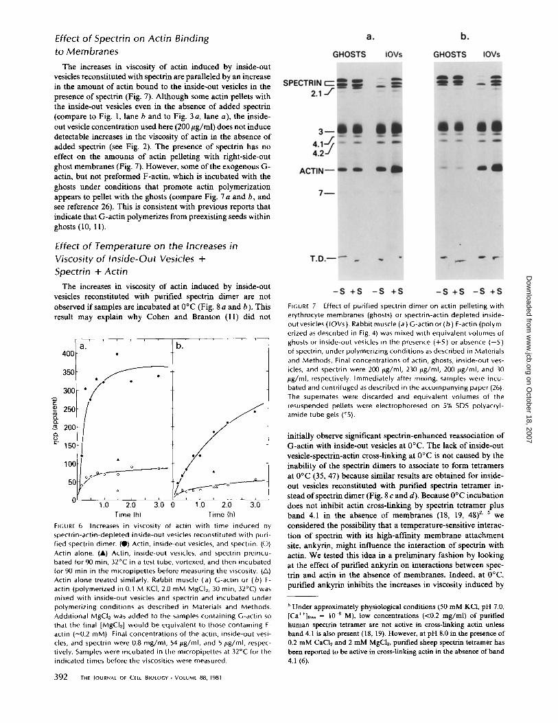
Effect of Spectrin on Actin Binding
to Membranes
The increases in viscosity of actin induced by inside-outvesicles reconstituted with spectrin are paralleled by an increasein the amount of actin bound to the inside-out vesicles in thepresence of spectrin (Fig. 7) . Although some actin pellets withthe inside-out vesicles even in the absence of added spectrin(compare to Fig. 1, lane b and to Fig. 3 a, lane a), the inside-out vesicle concentration used here (200 tLg/ml) does not inducedetectable increases in the viscosity of actin in the absence ofadded spectrin (see Fig. 2) . The presence of spectrin has noeffect on the amounts of actin pelleting with right-side-outghost membranes (Fig. 7) . However, some ofthe exogenous G-actin, but not preformed F-actin, which is incubated with theghosts under conditions that promote actin polymerizationappears to pellet with the ghosts (compare Fig. 7 a and b, andsee reference 26). This is consistent with previous reports thatindicate that G-actin polymerizes from preexisting seeds withinghosts (10, 11) .
Effect of Temperature on the Increases in
Viscosity of Inside-Out Vesicles +
Spectrin + Actin
The increases in viscosity of actin induced by inside-outvesicles reconstituted with purified spectrin dimer are notobserved if samples are incubated at 0°C (Fig . 8 a and b) . Thisresult may explain why Cohen and Branton (11) did not
'-3 .0 0
1 .0 2.0
3.0Time (h)
FIGURE 6 Increases in viscosity of actin with time induced byspectrin-actin-depleted inside-out vesicles reconstituted with puri-fied spectrin dimer. (") Actin, inside-out vesicles, and spectrin . (O)Actin alone. (A) Actin, inside-out vesicles, and spectrin preincu-bated for 90 min, 32°C in a test tube, vortexed, and then incubatedfor 90 min in the micropipettes before measuring the viscosity . (A)Actin alone treated similarly . Rabbit muscle (a) G-actin or (b) F-actin (polymerized in 0.1 M KCI, 2.0 mM M902,30 min, 32°C) wasmixed with inside-out vesicles and spectrin and incubated underpolymerizing conditions as described in Materials and Methods.Additional MgCl2 was added to the samples containing G-actin sothat the final [MgCl21 would be equivalent to those containing F-actin (---0 .2 mM) . Final concentrations of the actin, inside-out vesi-cles, and spectrin were 0.8 mg/ml, 54,ug/ml, and 5 tug/ml, respec-tively. Samples were incubated in the micropipettes at 32°C for theindicated times before the viscosities were measured .
392
THE JOURNAL OF CELL BIOLOGY " VOLUME 88, 1981
FIGURE 7
Effect of purified spectrin dimer on actin pelleting witherythrocyte membranes (ghosts) or spectrin-actin depleted inside-outvesicles (IOVs ) . Rabbit muscle (a) G-actin or (b) F-actin (polym-erized as described in Fig . 4) was mixed with equivalent volumes ofghosts or inside-out vesicles in the presence (+S ) or absence (-S )of spectrin, under polymerizing conditions as described in Materialsand Methods . Final concentrations of actin, ghosts, inside-out ves-icles, and spectrin were 200 I+g/ml, 230 jLg/ml, 200 flg/ml, and 30Wg/ml, respectively . Immediately after mixing, samples were incu-bated and centrifuged as described in the accompanying paper (26) .The supernates were discarded and equivalent volumes of theresuspended pellets were electrophoresed on 5% SDS polyacryl-amide tube gels (15) .
initially observe significant spectrin-enhanced reassociation ofG-actin with inside-out vesicles at 0°C. The lack of inside-outvesicle-spectrin-actin cross-linking at 0°C is not caused by theinability of the spectrin dimers to associate to form tetramersat 0°C (35, 47) because similar results are obtained for inside-out vesicles reconstituted with purified spectrin tetramer in-stead ofspectrin dimer (Fig . 8c and d) . Because 0°C incubationdoes not inhibit actin cross-linking by spectrin tetramer plusband 4.1 in the absence of membranes (18, 19, 48)2, 5 weconsidered the possibility that a temperature-sensitive interac-tion of spectrin with its high-affinity membrane attachmentsite, ankyrin, might influence the interaction of spectrin withactin . We tested this idea in a preliminary fashion by lookingat the effect of purified ankyrin on interactions between spec-trin and actin in the absence of membranes. Indeed, at 0°C,purified ankyrin inhibits the increases in viscosity induced by
'Under approximately physiological conditions (50 mM KCI, pH 7.0,[Ca"]r*ee = 10-8 M), low concentrations (<0.2 mg/ml) of purifiedhuman spectrin tetramer are not active in cross-linking actin unlessband 4.1 is also present (18, 19) . However, at pH 8.0 in the presence of0.2 mM CaC12 and 2 MM MgC12, purified sheep spectrin tetramer hasbeen reported to be active in cross-linking actin in the absence ofband4.1(6).
a .4001
3501
300c
250an
200a
150
100 0
500
0'1 .0 2.0
Time (h)
on October 18, 2007
ww
w.jcb.org
Dow
nloaded from

250
a 200dá 150
Ç 100
50
0
0
0.z, 2d
á 15n
0250
002, 1
oS
Actin Spectrin INS INS
Actin Spectrin INS INS
Actin Actin Spectrin
Actin Actin Spectrin
Actin
Actin
FIGURE 8
Inhibition at 0°C of increases in viscosity of actin inducedby spectrin-actin-depleted inside-out vesicles (IOVs) reconstitutedwith a and b) purified spectrin dimer or (c and d) purified spectrintetramer . Rabbit muscle (a and c) G-actin or (b and d) F-actin(polymerized as in Fig . 4) was mixed with inside-out vesicles in thepresence of spectrin dimer or tetramer as described in Materials andMethods, at final concentrations of 0 .8 mg/ml actin, 75 ttg/ml inside-out vesicles, and 30 pg/ml spectrin dimer or tetramer . Samples wereincubated under ionic conditions favoring polymerization (blankbars) for 4 h at 0°C or (dotted bars) for 4 h at 32°C before theviscosities were measured .
spectrin tetramer plus band 4.1 (Fig . 9 a) . In contrast, at 32°C,ankyrin markedly enhances the increases in viscosity inducedby spectrin tetramer plus band 4.1 (Fig. 9 b).
DISCUSSION
Our results show that the ability of inside-out vesicles to cross-link and increase the viscosity of actin preparations is depend-ent on the specific reassociation ofspectrin with the membrane .We conclude that F-actin can attach to the erythrocyte mem-brane via an interaction with spectrin. This conclusion extendsprevious observations of spectrin-actin interactions in solution(6, 18, 19, 48)2 and agrees with recent direct measurements ofactin binding to membranes using radiolabeled F-actin (12) .
Actin Associates with Membrane-bound Spectrin
In all of our experiments the increases in viscosity of actin-containing solutions are attributable to spectrin (either spectrinheterodimers or tetramers) bound to the inside-out vesicles .Conditions known to reduce spectrin reassociation with themembrane inhibit the increases in viscosity; conditions thatmaximize specific spectrin binding maximize the increases inviscosity. Although it is known that, in the absence of mem-branes, spectrin alone or in the presence of band 4.1 can cross-link actin, such cross-linking requires spectrin tetramer andrequires spectrin concentrations higher than those used in our
assays.' It is unlikely that in our experiments membranessimply provide a source of soluble band 4.1 . Band 4.1 does notelute from the membrane under our conditions ofionic strengthand pH (40, 41, 45). Furthermore, the maximal amount ofband 4.1 that could conceivably be eluted from the concentra-tions of inside-out vesicles used in our assays is <5 ttg/ml . Thisamount of band 4.1 has no effect on the actin cross-linkingactivity of low concentrations of spectrin in the absence ofmembranes (Fig . 9 and see references 18 and 19) .
Further evidence that the role of the inside-out vesicles isnot simply to furnish band 4.1 for spectrin-band 4.1-actin cross-linking in solution is the fact that the 72,000-dalton polypeptide
Spectrin 4.1
2.1 4.1+2 .1
Actin Spectrin Spectrin Spectrin
Actin Actin Actin
FIGURE 9
Effect of purified ankyrin (2 .1) on the increases in viscos-ity of actin at (a) 0°C or (b) 32°C induced by purified spectrintetramer plus purified band 4.1 . Ankyrin and band 4.1 were purifiedas previously described (21, 45) . Rabbit muscle G-actin was used ata final concentration of 0 .8 mg/ml, spectrin tetramer at (a ) 301ag/mlor (b) 50 lag/ml, band 4.1 at 8 ttg/ml, and ankyrin at 20 ttg/ml .Components were added to the assay mixture in the followingorder: spectrin, band 4.1, ankyrin, actin. Samples were drawn upinto the micropipettes immediately after all components wereadded to the assay mixture, and incubated under ionic conditionsfavoring polymerization at (a ) 0°C or (b) 32°C for 4 h before theviscosities were measured . Ankyrin also enhances actin cross-linkingby spectrin dimer plus band 4.1 at 32°C (not shown), a temperaturewhich favors dimer to tetramer conversion (47) .
FowLER ET At . Actin-Spectrin-Membrane Cross-Linking
393
a.O 0° CO32' C
®a /, al z®
b.
,L tr=... 0w o® mom
on October 18, 2007
ww
w.jcb.org
Dow
nloaded from

does not inhibit the increases in viscosity observed at 32 °Cwhen spectrin, band 4.1, and actin are mixed in appropriateconcentrations in the absence of membranes (not shown) .However, it is possible that interactions of spectrin with band4 .1 in situ on the membrane mayenhance the spectrin-mediatedinteraction of actin with the cytoplasmic surface of the mem-brane. Our data do not directly address this question becausewe were not able to selectively extract all of the band 4 .1 fromthe membrane without also removing some of the ankyrin (seeFig. 1, lane c) .
Does Actin Interact with Components OtherThan Spectrin on the Cytoplasmic Surface ofthe Membrane?
In contrast to the increases in viscosity of actin induced bylow concentrations of inside-out vesicles reconstituted withpurified spectrin, comparable concentrations of inside-out ves-icles alone have relatively little effect on the viscosity of actin .The increases in viscosity of actin observed at very high con-centrations of inside-out vesicles in the absence of addedspectrin (Fig. 2, inset) may reflect incomplete extraction of theendogenous spectrin from the membrane (see Fig. 1, lane b),or perhaps a lower affinity interaction ofactin with a nonspec-trin component (e .g ., band 4.1) (12) . Under conditions whereinside-out vesicles alone have no effect on the viscosity of theactin, some actin does pellet with the spectrin-actin-depletedinside-out vesicles, even in the absence of added spectrin (Fig .7, and reference 12) . However, it is difficult to evaluate F-actinbinding in the absence of independent measures of polymeri-zation and filament length . For example, the number of actinfilaments associated with the membrane could change (asreflected in differences in viscosity), with no correspondingchange in the absolute amount of actin pelleting with thevesicles .
Mode of Attachment of Actin to the Inside-OutVesicles : Lateral vs . End-on AssociationAn actin oligomer or filament could form either end-on or
lateral attachments to components on the surface of an inside-out vesicle . Assuming that spectrin does not bind equally toboth the sides and ends of actin filaments, the followingconsiderations lead us to favor lateral attachments for actin-spectrin-inside-out vesicle interactions. First, because the twoends ofa single actin filament are not identical (31), both endscannot bind with the same specificity . On the other hand, actinfilaments could associate laterally with identical componentson two or more vesicles, thus linking them to one another.Second, if end-on attachment of actin to the inside-out vesiclesoccurred, then as the number of sites available for end-onattachment increased with increasing inside-out vesicle andspectrin concentration, the length of the actin filaments at-tached to the inside-out vesicles would become shorter andshorter . Shortening of filaments would probably tend to de-crease, not increase, the viscosity of actin as the inside-outvesicle concentration was increased. Third, electron microscopeimages of actin-spectrin-band 4.1 interactions show that spec-trin associates laterally ratherthan at the ends ofactin filaments(13, 48): actin filaments are bridged by spectrin to neighboringfilaments along the entire length of the actin filaments.A combination of end-on attachment of actin filaments to a
component on one inside-out vesicle and lateral associations
394
THE JOURNAL OF CELL BIOLOGY " VOLUME 88, 1981
with a different component on another inside-out vesicle couldalso lead to the increases in viscosity and gelation observed.This may account for actin binding to low concentrations ofinside-out vesicles in the absence ofspectrin as well as spectrin-stimulated increases in actin binding (this paper and see ref-erence 12) .
Interaction of Actin with the CytoplasmicSurface of the Erythrocyte Membrane In Vivo
It is important to note that we have no independent criteriafor assessing whether the actin-spectrin-membrane associationswe observe reconstitute the native cytoskeletal spectrin-actinnetwork. Some indication of the complex interrelations thatmay exist among cytoskeletal components in the erythrocyte isprovided by our observations of temperature effects on actin-membrane interactions . Inside-out vesicles reconstituted withpurified spectrin do not cross-link actin at 0°C and, in theabsence of membranes, purified ankyrin inhibits spectrin tet-ramer-band 4.1-actin cross-linking at 0°C and enhances it at32°C (Figs . 8 and 9) . This may be because of an ankyrin-induced temperature-dependent conformational change in thespectrin (or in a spectrin-band 4.1 complex) or, alternatively,it may be because of a temperature-dependent formation ofadditional cross-links between ankyrin, actin, spectrin, and/orband 4.1 .
Application of Falling Ball Viscometry to theStudy of Cytoskeleton-Membrane InteractionsThe excellent agreement between our results using visco-
metric assays and a recent study using direct binding measure-ments (12) is evidence that the falling ball viscometer canprovide a valid indication of how membranes and actin mayinteract . But, while actin cross-linking by membranes requiresactin to be bound to the membrane, actin binding to themembrane may not necessarily result in cross-linking of actinby membranes. The technique of low-shear viscometry maybeparticularly sensitive to associations of actin with membraneswhich lead to extensive cross-linking and gelation of actinfilaments but may be insensitive to other modes of association.
Because low-shear viscometry is rapidly and easily per-formed, this assay in combination with selective strippingtechniques could be useful in the preliminary identification ofactin-binding components of intracellular organelles such asendocytic vesicles and secretory vesicles, as well as invertedvesicles from the plasma membrane itself. We have recentlyapplied this technique to study actin-binding components inmembranes from the ameboid stage ofDictyostelium discoideum(26) .
The authors would like to acknowledge the technical assistance of K.Giedd, B. N. Reinhardt, and N. Buklad, as well as numerous helpfuldiscussions with J. M. Tyler, A. Verkleij, E. Ungewickell, J. M. Heiple,L. Tanasugarn, Y.-L . Wang, and G. Ralston while these experimentswere in progress . V. Fowler would particularly like to thank H. B .Pollard and the members of his laboratory for providing the time andspace necessary for completing this manuscript. This work was sup-ported by a grant from the National Institute of Arthritis, Metabolismand Digestive Diseases (AM 18111) to D. L. Taylor, and by NationalScience Foundation (PCM-80-07669) and National Institutes ofHealth(NIH) (HL 17411) grants to D. Branton. W. R. Hargreaves (HL 05823)and E. J. Luna (HL 05555) were postdoctoral fellows ofNIH.
Received for publication 12 June 1980, and in revisedform 14 October1980.
on October 18, 2007
ww
w.jcb.org
Dow
nloaded from

REFERENCES
I . Bennett, V., and D. Branlon . 1977 . Selective association of speclrin with the cytoplasmicsurface of human erythrocyte plasma membranes . J. Biol. Chem. 252 :2753-2763 .
2 . Bennett, V . 1978. Purification of an active proleolytic fragment of the membrane attach-ment site for human erythrocyte speclrin . J. Biol. Chem. 253:2292-2299 .
3 . Bennett, V ., and P . Stenbuck . 1979 . Identification and partial purification of ankyrin, thehigh affinity membrane attachment site for human erythrocyte speclrin . J. Biol. Chem.254:2533-2541 .
4. Bennett, V., and P . J . Stenbuck. 1979. The membrane attachment protein for speclrin isassociated with band 3 in human erythrocyte membranes. Nature (Land.) . 280:468-473.
5 . Bennett, V ., and P. J. Stenbuck . 1980. Human erythrocyte ankyrin. Purification andproperties . J. Biol. Chem. 255:2540-2548.
6. Brenner, S . L ., and E . D . Korn . 1979 . Spectrin-actin interaction . Phosphorylated anddephosphorylated speclrin tetramer cross-link F-actin. J. Biol. Chem. 254:8620-8627 .
7. Brenner, S. L ., and E. D . Korn. 1980. Spectrin/actin complex isolated from sheeperythrocytes accelerates actin polymerization by simple nucleation. Evidence for oligo-meric actin in the erythrocyte cytoskeleton. J Biol. Chem. 255 :1670-1676 .
8. Brotschi, E. A ., J. H . Hartwig, and T. P . Stossel . 1978 . Th e gelation of actin by actin-binding protein . J. Biol. Chem. 253:8988-8993.
9. Clarke, M., and J . A . Spudich. 1977 . Non-muscle contractile proteins . The role of actinand myosin in cell motility and shape determination. Annu. Rev. Biochem . 46:797-822.
10. Cohen, C . M ., P. L . Jackson, and D. Branton. 1978 . Actin-membrane interactions :association of G-actin with the red cell membrane. J. Supramol. Stmct. 9:113-124 .
It . Cohen, C. M., and D . Branton. 1979. Erythrocyte membrane stimulated actin polymeri-zation : the role of speclrin. Nature (Land. ) . 279 :163-165 .
12. Cohen, C . M., and S . F . Foley . 1980. Spectrin dependent and independent association ofF-actin with red cell membranes. J. Cell Biol. 86 :694-698.
13. Cohen, C. M., l. M . Tyler, and D . Branton. 1980 . Spectrin-actin associations studied byelectron microscopy of shadowed preparations . Cell 21 :875-883 .
14. Elgsaeter, A., and D . Branton. 1974 . Intramembrane particle aggregation in erythrocyteghosts . I. The effects of protein removal. J. Cell Biol. 63 :1018-1030.
15 . Fairbanks, G ., T. L . Steck, and D. F . H . Wallach. 1971 . Electrophoreti c analysis of themajor polypeplides of the human erythrocyte membrane . Biochemistry. 10:2606-2617 .
16. Flanagan, M. D ., and S . Lin. 1980. Cytochalasins block actin filament elongation bybinding to high affinity sites associated with F-actin . J. Biol. Chem. 255 :835-838 .
17 . Fowler, V., and V. Bennett. 1978 . Association of speclrin with its membrane attachmentsite restricts lateral mobility of human erythrocyte membrane proteins. J. Supramol. Siruct.8:215-221 .
18. Fowler, V., and D . L . Taylor. 1980 . Spectri n plus band 4.1 cross-link actin. Regulation bymicromolar calcium. J. Cell Biol. 85 :361-376 .
19. Fowler, V . 1980. Cytoskeletal structures and membrane protein mobility in the humanerythrocyte . Ph.D. Thesis. Th e Biology Department . Harvard University, Cambridge,Mass.
20. Griffith, L . M., and T. D . Pollard . 1978 . Evidence for actin filament-microtubule inter-action mediated by microtubule-associated proteins. J. Cell Biol. 78 :958-965 .
21 . Hargreaves, W. R ., K. N . Giedd, A . Verkleij, and D . Branton . 1980 . Reassociation ofankyrin with band 3 in erythrocyte membranes and in lipid vesicles. J. Biol. Chem. 255 .In press.
22. Kom, E . D . 1978 . Biochemistry of actomyosin-dependent cell motility . Proc. Nail A cad.Sci. U. S. A . 75:588-599 .
23 . Lin, D . C., and S . Lin . 1979 . Actin polymerization induced by a motility-related high-affinity cytochalasin binding complex from human erythrocyte membrane . Proc. Nail.Acad. Sci. U. S. A . 76 :2345-2349 .
24. Lowry, O . H ., N . Rosebrough, A . L . Farr, and R . l . Randall. 1951 . Protein measurementwith the Folin phenol reagent. J. Biol. Chem. 193 :265-275 .
25 . Luna, E. J., G. H . Kidd, and D. Branlon. 1979 . Identification by peptide analysis of the
speclrin-binding protein in human erythrocytes. J. Biol. Chem . 254:2526-2532 .26 . Luna, E . J ., V . M . Fowler, 1. Swanson, D . Branton, and D . L . Taylor. 1980. A membrane
cytoskeleton from Dictvostelium discoideum . I . Identification and partial characterizationof an actin-binding activity . J. Cell Biol. 88 :396-409 .
27 . Lux, S . E . 1979. Spectrin-actin membrane skeleton of normal and abnormal red bloodcells . Semin. Hemalol. 16 :21-51 .
28 . Lux, S . E. 1979. Dissecting the red cell membrane skeleton . Nature (Loud). 281 :426-429 .29 . MacLean-Fletcher, S . D ., and T . D . Pollard . 1980 . Viscometric analysis of the gelation of
Acamhamoeba extracts and purification of two gelation factors . L Cell Biol. 85:414-428 .30 . Nicolson, G . L ., andR. G . Painter . 1973 . Anionic sites of human erythrocyte membranes.
II. Antispectrin induced transmembrane aggregation of the binding sites for positivelycharged collodial particles. J. Cell Biol. 59 :395-406.
31 . Oosawa, F., andM . Kasai . 1979. Actin. In Subunits in Biological Systems . S . N . Timasheffand G . D . Fasman, editors . Marcel Dekker, New York . 261-322 .
32 . Palek, J., and S-.C. Liu. 1979. Dependence of speclrin organization in red blood cellmembranes on cell metabolism : implication for control of red cell shape, deformability,and surface area. Semin . Hemalol. 16:75-93 .
33 . Pinder, J . C ., E. Ungewickell, R. Calvert, E . Morris, and W. B . Gratzer . 1979 . Polymeri-zatio n of G-actin by speclrin preparations : identification of the active constituent . FEBS(Fed . Ent. Biochem . Soc.) Lett. 104 :396-400.
34 . Pinder, J. C., D. Bray, and W . B . Gratzer. 1977 . Control of interaction of speclrin andactin by phosphorylation . Nature (Loud).270:752-754.
35 . Ralston, G . B., J . Dunbar, and M . White . 1977 . The temperature-dependent dissociationof speclrin. Biochim. Biophys. Acta. 491 :345-348 .
36 . Sheetz, M. P., and D . Sawyer. 1978. Triton shells of intact erythrocytes . J. Supramol.Strict. 8:399-412 .
37 . Sheetz, M . P. 1979 . Integral membrane protein interaction with Triton cytoskeletons oferythrocytes. Biochim . Biophys. Acta. 577 :122-134 .
38 . Spudich, 1 . A ., and S . Watt . 1971 . The regulation of rabbit skeletal muscle contraction . I .Biochemical studies of the interaction of the Tm-Tn complex with actin and the proleolyticfragments of myosin . J. Biol. Chem . 246:4866-4871 .
39 . Steck, T. L. 1972. Cross-linking the major proteins of the isolated erythrocyte membrane .J. Mol. Biol. 66 :295-305 .
40 . Steck, T. L ., and l . Yu . 1973. Selective solubilization of proteins from red blood cellmembranes by protein perturbants . J. Supramol. Struct. 1 :220-232.
41 . Steck, T. L. 1974. The organization of proteins in the human red blood cell membrane . J.Cell Biol. 62 :1-19 .
42 . Stossel, T. P . 1978 . Contractile proteins in cell structure and function . Annu. Rev. Med. 29 :427-457.
43 . Taylor, D . L., and J . S . Condeelis. 1979 . Cytoplasmic structure and contractility inamoeboid cells. Int. Rev. Cyiol. 56 :57-144 .
44 . Tilney, L . G., and P. Detmers. 1975 . Actin in erythrocyte ghosts and its association withspeclrin . Evidence for a non-filamentous form of these two molecules in situ . J. Cell Biol.66 :508-520 .
45 . Tyler, J. M ., W. R . Hargreaves, and D . Branton. 1979. Purification of two speclrin-bindingproteins: biochemical and electron microscopical evidence for site-specific reassociationbetween speclrin and bands 2 .1 and 4.1 . Proc . Nadl. A cad. Sci. U. S. A. 76 :5192-5196.
46 . Tyler, J . M ., B . N. Reinhardt, and D . Branlon . 1980. Associations of erythrocyte cytoske-letal proteins : binding of purified bands 2.1 and 4 .1 to speclrin. J Biol. Chem. 255 :7034-7039.
47 . Ungewickell, E ., and W. Gratzer. 1978. Self-association of human speclrin. A thermody-namic and kinetic study . Eur. J. Biochem. 88:379-385 .
48 . Ungewickell, E ., P. M . Bennett, R . Calvert, V. Ohanian, and W. B . Gratzer. 1979 . In vitroformation of a complex between cytoskeletal proteins of the human erythrocyte. Nature(Land.) 280:811-814 .
49. Yu, J ., and S . R . Goodman . 1979. Syndeins : the speclrin-binding protein(s) of the humanerythrocyte membrane. Proc . Natl. Acad. Sci. U. S. A . 76 :2340-2344.
FOWLER IT At . Actin-Spectrin-Membrane Cross-linking
395
on October 18, 2007
ww
w.jcb.org
Dow
nloaded from


![Research Cytoplasmic PCNA is located in the actin belt and ... · [3]. Opposite to the promotive role of osteoblasts on bone formation, osteoclasts are mainly responsible for bone](https://static.fdocuments.us/doc/165x107/5fd1259682ee337ab638e5df/research-cytoplasmic-pcna-is-located-in-the-actin-belt-and-3-opposite-to.jpg)