Expression of Functional Domains of Beta -Spectrin Disrupts ...
12
Expression of Functional Domains of Beta -Spectrin Disrupts Epithelial Morphology in Cultured Cells Ren-Ju Hu,* Suraj Moorthy,* and Vann Bennett* Howard Hughes Medical Institute and Departments of* Biochemistry and ¢Cell Biology, Duke University School of Medicine, Durham, North Carolina 27710 Abstract. Spectrin is a major structural protein as- sociated with the cytoplasmic surface of plasma mem- branes of many types of cells. To study the functions of spectrin, we transfected Caco-2 intestinal epithelial cells with a plasmid conferring neomycin resistance and encoding either actin-binding or ankyrin-binding domains of beta~-spectrin fused with beta-galactosi- dase. These polypeptides, in principle, could interfere with the interaction of spectrin with actin or ankyrin, as well as block normal assembly of alpha- and beta- spectrin subunits. Cells expressing the fusion proteins represented only a small fraction of neomycin-resistant cells, but they could be detected based on expression of beta-galactosidase. Cells expressing spectrin do- mains exhibited a progressive decrease in amounts of endogenous betac-spectrin, although alpha-spectrin was still present. Betac-spectrin-deficient cells lost epithelial cell morphology, became multinucleated, and eventually disappeared after 10-14 d in culture. Spectrin-associated membrane proteins, ankyrin and adducin, as well as the Na÷,K÷-ATPase, which binds to ankyrin, exhibited altered distributions in cells transfected with betac-spectrin domains. E-cadherin and F-actin, in contrast to ankyrin, adducin, and the Na÷,K÷-ATPase, were expressed, and they exhibited unaltered distribution in betao-spectrin-deficient cells. Cells transfected with the same plasmid encoding beta- galactosidase alone survived in culture as the major population of neomycin-resistant cells, and they exhib- ited no change in morphology or in the distribution of spectrin-associated membrane proteins. These results establish that betac-spectrin is essential for the normal morphology of epithelial cells, as well as for their maintenance in monolayer culture. S PV.CTRIN is an elongated actin-binding protein that is the principal component of a system of structural proteins associated with the cytoplasmic surface of plasma membranes of most metazoan cells (reviewed by Bennett and Gilligan, 1993). Spectrin is comprised of two subunits, termed alpha and beta, that are aligned side-to-side to form heterodimers, and the dimers are linked head-to-head to form tetramers. Beta subunits contain most of the recogni- tion sites of spectrin for other proteins including ankyrin, protein 4.1 actin, as well as the site for ankyrin-independent association of spectrin with membranes. Beta~-spectrin is the most common type of beta subunit, and it is expressed in most vertebrate tissues (Hu et al., 1992). The structure and function of spectrin has been best characterized in mam- malian erythrocytes from both in vivo and in vitro studies (Palek and Lambert, 1990; Delaunay and Dhermy, 1993; Gallagher and Forget, 1993; Bennett and Gilligan, 1993). Erythrocyte spectrin forms a membrane-associated polygo- nal network by the associations of spectrin with actin illa- ments and with integral membrane proteins through linkages Address all correspondence to Vann Bennett, Howard Hughes Medical In- stitute, Department of Cell Biology, Duke University Medical School, Durham, NC 27710. Tel.: (919) 684-3538. Fax: (919) 684-3590. with ankyrin and protein 4.1. Defects and deficiencies in components of the spectrin-actin network result in abnor- mally fragile erythrocytes in humans and mice, suggesting that one function of spectrin is to physically stabilize the phospholipid bilayer. Structures similar to the spectrin-based membrane skele- ton of erythrocytes are likely to exist in other tissues. Genes encoding proteins closely related to erythrocyte spectrin and spectrin-associated proteins are expressed in brain, as well as most other tissues (reviewed by Bennett and Gilligan, 1993). In vitro studies also show that tissue spectrins can in- teract with isoforms of spectrin-associated proteins includ- ing ankyrin (Davis and Bennett, 1984) and adducin (Bennett et al., 1988). However, the organization of spectrin-based membrane structures and their cellular roles are likely to be more complex in nonerythroid tissues. Major differences be- tween the spectrin-based membrane skeleton of erythrocytes and that of more complex cells include the presence of multi- ple isoforms of spectrin and associated proteins caused by al- ternate exon usage and distinct genes, additional proteins as- sociating with spectrin that do not occur in erythrocytes, and localization of spectrin in areas in addition to the plasma membrane. A candidate function of spectrin and associated proteins, © The Rockefeller University Press, 0021-9525/95/03/1069/12 $2.00 The Journal of Cell Biology, Volume 128, Number 6, March 1995 1069- 1080 1069 on March 15, 2018 jcb.rupress.org Downloaded from
Transcript of Expression of Functional Domains of Beta -Spectrin Disrupts ...

Expression of Functional Domains of Beta -Spectrin Disrupts Epithelial Morphology in Cultured Cells Ren-Ju Hu,* Suraj Moorthy,* and Vann Bennett*
Howard Hughes Medical Institute and Departments of* Biochemistry and ¢Cell Biology, Duke University School of Medicine, Durham, North Carolina 27710
Abstract. Spectrin is a major structural protein as- sociated with the cytoplasmic surface of plasma mem- branes of many types of cells. To study the functions of spectrin, we transfected Caco-2 intestinal epithelial cells with a plasmid conferring neomycin resistance and encoding either actin-binding or ankyrin-binding domains of beta~-spectrin fused with beta-galactosi- dase. These polypeptides, in principle, could interfere with the interaction of spectrin with actin or ankyrin, as well as block normal assembly of alpha- and beta- spectrin subunits. Cells expressing the fusion proteins represented only a small fraction of neomycin-resistant cells, but they could be detected based on expression of beta-galactosidase. Cells expressing spectrin do- mains exhibited a progressive decrease in amounts of endogenous betac-spectrin, although alpha-spectrin was still present. Betac-spectrin-deficient cells lost
epithelial cell morphology, became multinucleated, and eventually disappeared after 10-14 d in culture. Spectrin-associated membrane proteins, ankyrin and adducin, as well as the Na÷,K÷-ATPase, which binds to ankyrin, exhibited altered distributions in cells transfected with betac-spectrin domains. E-cadherin and F-actin, in contrast to ankyrin, adducin, and the Na÷,K÷-ATPase, were expressed, and they exhibited unaltered distribution in betao-spectrin-deficient cells. Cells transfected with the same plasmid encoding beta- galactosidase alone survived in culture as the major population of neomycin-resistant cells, and they exhib- ited no change in morphology or in the distribution of spectrin-associated membrane proteins. These results establish that betac-spectrin is essential for the normal morphology of epithelial cells, as well as for their maintenance in monolayer culture.
S PV.CTRIN is an elongated actin-binding protein that is the principal component of a system of structural proteins associated with the cytoplasmic surface of plasma
membranes of most metazoan cells (reviewed by Bennett and Gilligan, 1993). Spectrin is comprised of two subunits, termed alpha and beta, that are aligned side-to-side to form heterodimers, and the dimers are linked head-to-head to form tetramers. Beta subunits contain most of the recogni- tion sites of spectrin for other proteins including ankyrin, protein 4.1 actin, as well as the site for ankyrin-independent association of spectrin with membranes. Beta~-spectrin is the most common type of beta subunit, and it is expressed in most vertebrate tissues (Hu et al., 1992). The structure and function of spectrin has been best characterized in mam- malian erythrocytes from both in vivo and in vitro studies (Palek and Lambert, 1990; Delaunay and Dhermy, 1993; Gallagher and Forget, 1993; Bennett and Gilligan, 1993). Erythrocyte spectrin forms a membrane-associated polygo- nal network by the associations of spectrin with actin illa- ments and with integral membrane proteins through linkages
Address all correspondence to Vann Bennett, Howard Hughes Medical In- stitute, Department of Cell Biology, Duke University Medical School, Durham, NC 27710. Tel.: (919) 684-3538. Fax: (919) 684-3590.
with ankyrin and protein 4.1. Defects and deficiencies in components of the spectrin-actin network result in abnor- mally fragile erythrocytes in humans and mice, suggesting that one function of spectrin is to physically stabilize the phospholipid bilayer.
Structures similar to the spectrin-based membrane skele- ton of erythrocytes are likely to exist in other tissues. Genes encoding proteins closely related to erythrocyte spectrin and spectrin-associated proteins are expressed in brain, as well as most other tissues (reviewed by Bennett and Gilligan, 1993). In vitro studies also show that tissue spectrins can in- teract with isoforms of spectrin-associated proteins includ- ing ankyrin (Davis and Bennett, 1984) and adducin (Bennett et al., 1988). However, the organization of spectrin-based membrane structures and their cellular roles are likely to be more complex in nonerythroid tissues. Major differences be- tween the spectrin-based membrane skeleton of erythrocytes and that of more complex cells include the presence of multi- ple isoforms of spectrin and associated proteins caused by al- ternate exon usage and distinct genes, additional proteins as- sociating with spectrin that do not occur in erythrocytes, and localization of spectrin in areas in addition to the plasma membrane.
A candidate function of spectrin and associated proteins,
© The Rockefeller University Press, 0021-9525/95/03/1069/12 $2.00 The Journal of Cell Biology, Volume 128, Number 6, March 1995 1069- 1080 1069
on March 15, 2018
jcb.rupress.orgD
ownloaded from

inferred from their localization in tissues and association with proteins in in vitro assays, is in assembly and/or main- tenance of specialized domains on the cell surface. Sites of cell-cell contact in epithelial tissues are the best-charac- terized example of a role of membrane skeletal proteins in a membrane domain (reviewed in Nelson, 1992; Nelson et al., 1991). Direct evaluation of functions of spectrin in non- erythroid cells has been approached by several methods. Mi- croinjection of fibroblasts with antibodies against spectrin induced the formation of spectrin aggregates as well as the condensation of intermediate filaments, but had little effect on cell shape, actin filaments, or microtubules (Mangeat and Burridge, 1984). In contrast, properties of alpha-spectrin mutants in Drosophila demonstrate that alpha-spectrin is es- sential for larval survival and development, and further- more, that spectrin plays essential roles in maintenance of cell shape and cell-cell interactions (Lee et al., 1993).
To further understand the functions of spectrin, we have expressed functional domains of betao-spectrin in cultured epithelial cells with the expectation that these domains would interfere with the activity of endogenous beta-spec- trin. Results of these experiments demonstrate that spectrin plays an essential role in maintenance of epithelial cell mor- phology in culture.
Materials and Methods
Reagents Affinity-purified antibodies against brain adducin, Na+,K+-ATPase, eryth- rocyte ankyrin, and betaG spectrin (repeats 4-9) were prepared from rabbit antisera as described (Davis and Bennett, 1984). Antibody against alpha- spectrin was kindly provided by Dr. Velia Fowler (Scripps Research Insti- tute, La Julia, CA). Monoclonal antibody against beta-galactosidase was from Promega Corp. (Madison, WI). Polyclonal antibody against beta- galactosidase was from 5 Prime-3 Prime, Inc. (Boulder, CO). Monoclonal antibody against E-cadherin was from Sigma Immunochemicals (St. Louis, MO). Fluorescein- and rhodamine-conjugated secondary antibodies were from Pierce Chemical Co. (Rockford, IL). Fluorescein-labeled phalloidin was obtained from Molecular Probes (Eugene, OR). Restriction enzymes were from New England Biolabs Ltd. (Mississiauga, Ontario). Reagents for lipofectin transfection were from Gibco BRL (Gaithersburg, MD), and those for immunoblot staining were from Bio Pad Laboratories (Her- cules, CA).
Plasmid Construction DNA encoding actin- and ankyrin-binding domains of betaG-spectrin was inserted into a mammalian expression vector containing a beta-actin pro- moter, and conferring neomycin resistance (pHbetaApr-I neo-CB-17). The pHbetaApr-1 neo~CB-17 vector was kindly provided by Dr. EIwood Linney (Duke University) (Espeseth et al., 1989). DNA fragments encoding the actin-binding domain (amino acids 1-396) and ankyrin-binding domain (amino acids 1661-1990) of betao-spectrin were made from betaG spectrin eDNA (Hu et al., 1992) by a PCR reaction using primers to introduce a HindIII restriction site and an AT(; initiation site at the 5' end of the DNA sequence, and BamHI site at the 3' end. HindIII- and BamHI-restricted DNA fragments were inserted into the pHbetaApr-I neo-CB 17 mammalian expression vector. The BamHI site was made in-frame with the beta- galactosidase gene to produce a COOH-terminai fusion protein. For a con- trol plasmid, an AT(3 initiation site was inserted in the pHbetaApr-1 neo- CB-17 vector such that beta-golactosidase was expressed by itself in cells. Beta-galactosidase fusion proteins were detected either by immunofluores- cence of fixed and permeabilized cells or by enzymatic activity of living cells. The DNA sequence of various constructs was confirmed by direct se- quence analysis.
Mammalian Cell Culture Human Caco-2 intestinal epithelial cells were grown in MEM medium, sup- plemented with 20% fetal bovine serum, MEM nonessential amino acids, and penicillin/streptomycin. Medium was changed every 3 d, and the cells were replated when they grew to confluency. G418 (0.5 mg/ml) was added to select transfected cells 48 h after transfection, and fresh G418 medium was added every 3-4 d.
Immunofluorescence Staining and Detection of Beta-Galactosidase Activity Caco-2 cells were grown on 1-ml glass slide chamber coated with 1 mg/ml of poly-D-lysine. The cells were transfected by lipofectin as described in the Gibco protocol, washed three times with PBS, and fixed with 3 % parafor- maldehyde/PBS at 4°C for 30 rain. In some experiments, cells were stained for beta-galactosidase activity before fixation, and transfected cells were de- tected as described (Espeseth et al., 1989).
After 3% paraformaldehyde/PBS fixation, cells were extracted with 0.25% Triton X-100 in PBS for 5 min at room temperature. The cells were then incubated in 1% BSA, 10% normal goat serum, and 0.1% Tween-20 in PBS at room temperature for 15 rain to block nonspecific binding. 1 #g/ ml of monoclunal anti-beta-galactosidase antibody was added to the block- ing buffer and further incubated overnight at 4°C. After washing thoroughly with PBS, fluorescein- or rhodamine-labeled goat anti-monse secondary antibodies in blocking buffer were added to label the primary antibody. Cells were then washed five times with PBS and mounted with 5 mg/ml n-propyl gallate in 30% glycerol. Double immunofluorescence was per- formed to determine the localization in cells producing beta-gaiactosidase fusion proteins, endogenous spectrin, and spcctrin-nssociated proteins, such as ankyrin, adducin, and Na+,K+-ATPase. The betao spectrin anti- body was directed against repeats 4--9, and they did not recognize either the actin or ankyrin-binding domains expressed by transfected cells. The same protocol was used as described above, but antibody incubations were per- formed by mixing antibodies from rabbit with antibodies from mouse. A mixture of rhodamine-labeled goat anti-rabbit antibody and fluorescein- labeled goat anti-mouse antibody was used to visualize the antigens. Dou- ble immunofluorescence for colocalizing beta-gaiactosidase expression and E-cadherin was performed stepwise to decrease the cross-reaction of the goat anti-mouse secondary antibody with rat monoclonal anti-E-cadherin antibody. Cells were first incubated with mouse monoclonal anti-beta- gaiactosidase antibody, followed by rhndamine-labeled goat anti-mouse an- tibody. After washing thoroughly with PBS buffer, the cells were incubated with rat monoclonai anti-E-cadherin antibody, followed by the incubation with fluorescein-labeled goat anti-rat antibody. The stained slides were then examined by epifluorescence using a Bio Rad confocal microscope.
4',6'-diaminophenylindole (DAPI) 1 was used to visualize cell nuclei by fluorescence microscopy. Cells were stained with rhodamine-conjugated anti-beta-galactosidase antibody and goat anti-mouse antibody, as de- scribed above. DAPI in PBS buffer was then added into the glass chamber and incubated for 5 rain at room temperature.
Immunoblotting Studies Cell extracts of transfected Caco-2 cells were obtained by solubilizing cells in SDS-electrophoresis buffer. The cell extracts were passed through a 23- gauge needle several times to shear DNA, heated for 15 rain at 65°C, elec- trophoresed on SDS-polyacrylamide gels, and transferred by electrophore- sis onto nitrocellulose paper (Davis and Bennett, 1983). After blotting, the paper was incubated in TTBS blocking buffer: 20 mM Tris HCI, pH 7.5, 500 mM NaCI, 0.05% Tween 20, and 3% nonfat dry milk for 20 rain at room temperature. Monoclonal anti-beta-galactosidase antibody was added to the buffer and incubated with the paper for overnight at 4°C. After wash- ing with TTBS buffer, goat anti-mouse antibody conjugated with alkaline phosphatase was used to label the anti-beta-galactosidase antibody on the nitrocellulose paper. The color on the paper was developed as described in the Bio Pad protocol.
1. Abbreviation used in this paper: DAPI, 4',6'-diaminophenylindole.
The Journal of Cell Biology, Volume 128, 1995 1070
on March 15, 2018
jcb.rupress.orgD
ownloaded from

Results
Strategies for Evaluation of Beta~-Spectrin Function in Cultured Cells
Experiments were designed to disrupt endogenous spectrin function by either using antisense DNA or expression of spectrin domains. A mammalian expression vector contain- ing antisense sequence of betaG-spectrin under control of a cytomegalovirus promoter and conferring neomycin resis- tance was transfected into epithelial cell lines. After (3418 selection, permanent clones of transfected cells were ob- tained. The cells lost their normal epithelial morphology, and they eventually disappeared from culture (data not shown). Unfortunately, because of loss of cells, it was difficult to study the specific function of spectrin by the method of antisense expression. Therefore, we chose the ap- proach of interfering with endogenous spectrin by expression of truncated forms of beta~-spectrin as fusion proteins with beta-galactosidase. It was possible to follow the transfected cells using beta-galactosidase as a marker at early stages and to characterize individual cells by immunofluorescence and morphology.
Constructs encompassing two important protein-binding domains of beta~-spectrin, the actin- and ankyrin-binding domains, were expressed in Caco-2 epithelial cells. The binding site of betac-spectrin for ankyrin is localized to re- peat 15 (Kennedy et al., 1991), and the site for actin is lo- cated in the NH2-terminal domain from Ala47 to Lys186 (Karinch et al., 190). Constructs used in this study extend from residues 1-396 and 1661-1990, and they are referred to as the actin- and ankyrin-binding domains of betaG-spec- trin, respectively. The expression vector conferred neomycin resistance, and it contained a beta-actin promoter (Fig. 1 A). Betao-spectrin domains were linked to the 5' end of trun- cated beta-galactosidase in the vector (see Materials and Methods). For control experiments, an ATG initiation site was introduced at the 5' end of the beta-galactosidase gene (Materials and Methods) (Fig. 1 B, a).
Growth of cells expressing beta-galactosidase was deter- mined during (3418 selection, using X-gal as a chromogenic substrate (Fig. 2 A). For the control plasmid, >95 % of the surviving cells were neomycin resistant and beta-galactosi- dase positive (Fig. 2 A). However, <5 % of cells transfected with betaa-spectrin actin-binding domain, and 40% of cells transfected with ankyrin-binding domain expressed beta- galactosidase after 15 d of G418 selection (Fig. 2 A). These data suggested that expression of actin- and ankyrin-binding domains of beta~ spectrin resulted in selective advantage for those neomycin-resistant cells that did not express the fu- sion proteins. Cells expressing betaG-spectrin domains may die during culture, have a reduced rate of cell division, and/or detach from the epitheli~ monolayer as a result of loss of cell-cell contacts (see below). Cells transfected with ankyrin-binding domain had a higher percentage of blue- stained cells than cells transfected with actin-binding do- main, suggesting that expression of actin-binding domain was more disruptive than expression of the ankyrin-binding domain. The effects of beta~-spectrin actin-binding domain were not caused by a generalized competition for other actin- binding proteins since the ankyrin-binding domain yielded
A
POLY A pGAL
B Expressed fusion protelem
I}-galactosidase
b I . . . . . . . . . . . . . . . . . . . . . . . . . . . . .
~¢ ~oectrin actin-binding doma in I}-galactosidase a.a. # 1-396
. . . . . . . . . . . . 1 . . . . . . . . . . . . . . .
spectrin ~galactosidase ankyrin-binding domain a~. #1661-1990
Figure 1. Schematic model of mammalian expression plasmid used for transfection experiments. (.4) Mammalian expression vector, pHbetaApr-l-neo coil bGAL (pHbetaAPr-l-neo-CB-17), contains unique restriction sites for HindlH and BamI-II, which are used as insertion sites for betaG-spectrin-domains. (B) Expressed fusion proteins in mammalian cells after insertion of initiation site or betao-spectrin-domains, a, ATG initiation codon; b, actin-binding domain of betaG-spectrin; c, ankyrin-binding domain of betao- spectrin.
qualitatively similar results, and since F-actin morphology was unperturbed (see the next section). Moreover, perturba- tion of the cells was not caused by the transfection procedure or to beta-galactosidase itself because the cells transfected with control plasmid were still normal and >95 % of the cells still expressed beta-galactosidase.
To determine if the constructs expressed the correct pro- teins, immunoblot analysis of transfected cells was per- formed using antibody against beta-galactosidase to detect the fusion proteins. Proteins of 100, 143, and 136 kD corre- sponding to predicted sizes of beta-galactosidase alone, beta-galactosidase plus beta~-spectrin actin-binding domain (amino acids 1-396), and beta-galactosidase plus betao- spectrin ankyrin-binding domain (amino acids 1661-1990) were detected in the extracts of cells transfected with plas- mids encoding control, actin-binding domain, and ankyrin- binding domain, respectively (Fig. 2 B, lanes 1-3). These results indicate that the fusion proteins were expressed, at least in a subpopulation of cells. The relative amount of ex- pressed fusion proteins in transfected cells correlated well with the percentage of blue-stained cells (Fig. 2 B). Expres-
Hu et al. Beta~-Spectrin Function in Epithelial Cells 1071
on March 15, 2018
jcb.rupress.orgD
ownloaded from
100
8o
6O
¢) >
5 10 15
Days of G418 selection
0 "
0 20
Figure 2. Caco-2 cells expressing the actin- or ankyrin-binding do- main of betao-spectrin disappear from monolayer culture during G418 selection. Caco-2 cells were transfected with vectors encod- ing beta-galactosidase, beta-galactosidase fused with actin-binding domain, or ankyrin-binding domain of beta~-spectrin. (A) Trans- fected Caco-2 cells were fixed and evaluated for beta-galactosidase enzyme activity using X-gal as a chromogenic substrate. The per- centage of blue-stained cells is plotted as a function of time of G418 selection. Caco-2 ceils transfected with beta-galactosidase alone (D), actin-binding domain of beta~-spectrin-beta-gaiactosidase fusion protein (o), and ankyrin-binding domain of beta~-spec- trin-beta-galactosidase fusion protein (e) were compared. (B) Im- munoblot analysis of transfected cell extracts after 3, 6, 9, 12, and 15 d of G418 selection. A monoclonal antibody against beta gaiac- tosidase was used to stain the expressed fusion proteins. Lane 1, Caco-2 cells expressing beta-galactosidase alone; lane 2, cells ex- pressing actin-binding domain of beta~-spectrin-beta-galactosi-
sion of beta-galactosidase reached a plateau after 12 and 15 d in cells transfected with the control plasmid (Fig. 2 B, lanes 1 ). In contrast, the amount of fusion proteins contain- ing spectrin domains reached maximal levels at ~ 6 d of (3418 selection and declined after 9 d of G418 selection. Neomycin-resistant cells, therefore, started to lose the ex- pressed fusion protein after the 6 d of G418 selection, either because of degradation of the fusion proteins or loss of the cells expressing fusion proteins. Immunofluorescence ex- periments were typically performed within 5 d of (3418 se- lection (see below).
Expression of Domains of Beta~-Spectrin in Caco-2 Cells Results in Decreased Levels of Endogenous Betc~ Spectrin
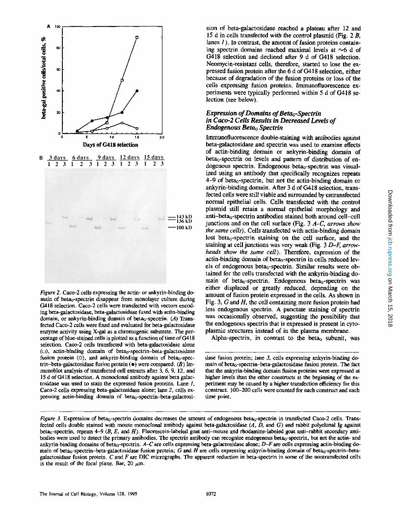
Immunofluorescence double-staining with antibodies against beta-galactosidase and spectrin was used to examine effects of actin-binding domain or ankyrin-binding domain of betaG-spectrin on levels and pattern of distribution of en- dogenous spectrin. Endogenous beta~-spectrin was visual- ized using an antibody that specifically recognizes repeats 4-9 of betao-spectrin, but not the actin-binding domain or ankyrin-binding domain. After 3 d of G418 selection, trans- fected cells were still viable and surrounded by untransfected normal epithelial cells. Cells transfected with the control plasmid still retain a normal epithelial morphology and anti-betaG-spectrin antibodies stained both around cell-cell junctions and on the cell surface (Fig. 3 A-C, arrows show the same cells). Cells transfected with actin-binding domain lost betao-spectrin staining on the cell surface, and the staining at cell junctions was very weak (Fig. 3 D-F, arrow- heads show the same cell). Therefore, expression of the actin-binding domain of beta~-spectrin in cells reduced lev- els of endogenous beta~-spectrin. Similar results were ob- tained for the cells transfected with the ankyrin-binding do- main of beta~-spectrin. Endogenous betaG-spectrin was either displaced or greatly reduced, depending on the amount of fusion protein expressed in the cells. As shown in Fig. 3, G and H, the cell containing more fusion protein had less endogenous spectrin. A punctate staining of spectrin was occasionally observed, suggesting the possibility that the endogenous spectrin that is expressed is present in cyto- plasmic structures instead of in the plasma membrane.
Alpha-spectrin, in contrast to the beta~ subunit, was
dase fusion protein; lane 3; cells expressing ankyrin-binding do- main of betao-spectrin-beta-galactosidase fusion protein. The fact that the ankyrin-binding domain fusion proteins were expressed at higher levels than the other constructs at the beginning of the ex- periment may be caused by a higher transfection efficiency for this construct. 100-200 cells were counted for each construct and each time point.
Figure 3. Expression of betaG-spectrin domains decreases the amount of endogenous beta6-spectrin in transfected Caco-2 cells. Tram- fected cells double stained with mouse monoclonal antibody against beta-galactosidase (A, D, and G) and rabbit polyclonal Ig against betao-spectrin, repeats 4-9 (B, E, and H). Fluorescein-labeled goat anti-mouse and rhodamine-labeled goat anti-rabbit secondary anti- bodies were used to detect the primary antibodies. The spectrin antibody can recognize endogenous betao-spectrin, but not the actin- and ankyrin-binding domains of betaG-spectrin. A-C are cells expressing beta-galactosidase alone; D-F are cells expressing actin-binding do- main of betaG-spectrin-beta-galactosidase fusion protein; G and H are cells expressing ankyrin-binding domain of beta6-spectrin-beta- galactosidase fusion protein. C and F are DIC micrographs. The apparent reduction in beta-spectrin in some of the nontransfected cells is the result of the focal plane. Bar, 20 #m.
The Journal of Cell Biology, Volume 128, 1995 1072
on March 15, 2018
jcb.rupress.orgD
ownloaded from

Hu et al. Beta6~Spectrin Function in Epithelial Cells 1073
on March 15, 2018
jcb.rupress.orgD
ownloaded from

present at the same levels of intensity in untransfected cells, and in ceils transfected with beta-galactosidase alone or with the ankyrin-binding domain-beta-galactosidase fusion pro- tein, as visualized by immunofluorescence (Fig. 4). Alpha- spectrin was evident in the cytoplasm as well as in associa- tion with the plasma membrane of transfected cells (Fig. 4).
Beta~-Spectrin Is Essential for Normal Epithelial Cell Morphology Caco-2 epithelial cell morphology was disrupted after ex- pression of actin-binding or ankyrin-binding domains of betaG-spectrin (Fig. 5, C-F). Caco-2 cells still retained the normal morphology as epithelial cells after the cells were transfected with the control plasmid (Fig. 5, A and B). Cells expressing either beta~-spectrin actin-binding domain or ankyrin-binding domain became enlarged 5-10 times the size of normal epithelial cells, and they contained numerous vacuoles. Cells observed by confocal microscopy frequently overlayed other epithelial cells, and they apparently grew without contact inhibition (data not shown).
Another effect of expression of spectrin domains was that cells became multinucleated. On different days of G418 selec- tion, transfected cells were double stained with anti-beta- galactosidase antibody and a nuclear staining dye, DAPI. The percentage of multinucleated cells in the total cells that expressed fusion protein was counted (Fig. 6). More than 50% of the cells transfected with actin-binding domain or ankyrin-binding domain were multinucleated after 10 d of G418 selection, while <25 % of cells transfected with control plasmid contained more than one nucleus. However, there appeared to be no significant difference in the number of pyknotic nuclei in these cells, indicating that the cells were still viable at the time of observation. Therefore, loss of betao-spectrin may cause either malfunction of some step in cell division such as cytokinesis, or it may result in cell-cell fusion. Further experiments are required to distin- guish these two possibilities.
Localization of Actin, Ankyrin, Adducin, Na+,K+-ATPase, and E-Cadherin in Cells Expressing the Ankyrin-binding Domain of Beta~-Spectrin Spectrin associates with actin in in vitro assays through con- tact with the actin-binding domain of the beta subunit, and spectrin-actin complexes are the basic structural unit of the spectrin-skeleton in erythrocytes (Bennett and Gilligan, 1993). It was, therefore, of interest to determine the effect of betao-spectrin deficiency on F-actin localization. The lo- calization of F-actin was evaluated in cells expressing the ankyrin- and actin-binding domains of beta~-spectrin using fluorescein-labeled phalloidin to identify actin filaments and polyclonal antibody against beta-galactosidase to identify the cells expressing foreign proteins (Fig. 7). F-actin was present in stress fibers (Fig. 7, C and F) and in close associa- tion with the plasma membrane (Fig. 7, B and E) in cells expressing beta-galactosidase alone or either of the beta~- spectrin domain fusion proteins. The apparently normal dis- tribution of F-actin in transfected cells is in contrast to their bizarre morphology and lack of betaG-spectrin. These re- suits suggest that proteins other than beta~-spectrin are re- sponsible for targeting of F-actin to the plasma membrane, and that the altered cell morphology in betac-spectrin-
deficient cells is not a consequence of perturbation of the population of actin molecules detected using phalloidin as a probe.
Cells were double stained with anti-beta-galactosidase antibody (Fig. 8, a2-e2) and antibodies against E-cadherin (Fig. 8, a/-d/) . E-cadherin colocalizes with spectrin, and it coisolates with spectrin and ankyrin in extracts of epithelial cells (Nelson and Hammerton, 1989). Moreover, expression of E-cadherin induces a polarized distribution of spectfin and ankyrin to sites of cell-cell contact (McNeill et al., 1990). E-cadherin was localized at cell-cell junctions of cells expressing the ankyrin-binding domain of beta,rspec- trin, even though these cells had lost other aspects of epithe- lial morphology (Fig. 8 d/). Similar results were obtained for the cells expressing actin-binding domain fusion protein (data not shown). The normal targeting of E-cadherin to cell-cell junctions suggests that E-cadherin sorts to the plasma membrane by a pathway independent from betao- spectrin, and it is consistent with observations that E-cad- herin is concentrated at cell-cell contact before appearance of spectrin (McNeill et al., 1993).
Ankyrin and adducin associate with spectrin and are colocalized with spectrin at sites of cell-cell contact in epi- thelial cells (Nelson and Veshnock, 1987a; Nelson and Ham- merton, 1989; Kaiser et al., 1989). Na÷,K+-ATPase inter- acts with ankyrin in in vitro assays, and it is colocalized with spectrin and ankyrin in epithelial cells (Nelson and Vesh- nock, 1987b; Koob et al., 1988; Morrow et al., 1989). All three antibodies stain normal epithelial cells at sites of cell-cell contact. The cells transfected with control plasmid showed normal morphology of epithelial cells, and a normal distribution of adducin at cell-cell junctions (Fig. 8 e), as well as a normal distribution of ankyrin, Na+,K+,-ATPase, or E-cadherin (data not shown). Cells transfected with ankyrin-binding domain of betao-spectrin were stained with antibodies against ankyrin (Fig. 8 a/) , Na+,K+,- ATPase (Fig. 8 bl), or adducin (Fig. 8 cl). The distribution of ankyrin, Na+,K +,-ATpase, and adducin was diffuse in the cytoplasm, and was not restricted to cell-cell junctions. Na+,K+,-ATPase and adducin also exhibited punctate stain- ing in the cytoplasm of some of these cells. The altered dis- tribution of ankyrin, adducin, and the Na+,K÷-ATPase is in contrast to relatively normal patterns for alpha-spectrin, F-actin, and E-cadherin.
Discussion
Expression of actin- and ankyrin-binding domains of betaG- spectrin in cultured epithelial cells was used in this study as an initial step in resolving functions of spectrin in vivo. Cells expressing beta~-spectrin domains exhibited a progressive reduction in levels of endogenous betaa-spectrin, based on results of immunofluorescence, and they eventually disap- peared after 10-14 d in culture. BetaG-spectrin-deticient cells exhibited dramatic changes in cell morphology, includ- ing increased numbers of multinucleated cells, 5-10-fold en- largement of cell size, formation of many vacuoles, and growth on top of the monolayer of normal epithelial cells. The cells continued to express cadherin, F-actin, and alpha- spectrin normally, but they failed to target adducin, ankyrin, and the Na÷,K+-ATPase to their correct subcellular loca-
The Journal of Cell Biology, Volume 128, 1995 1074
on March 15, 2018
jcb.rupress.orgD
ownloaded from

F.
l~gu
re 4.
Alp
ha-s
pect
rin
expr
essi
on p
ersi
sts
in C
aco-
2 ce
lls t
ran
~'~
l w
ith b
etaG
-spe
ctri
n an
kyri
n-bi
ndin
s do
mai
n. C
ells
wer
e tr
an.~
ecte
d w
ith b
eta-
gala
ctos
idas
e al
one
(,4 a
nd D
) or
with
th
e an
kyri
n-bi
ndin
g do
mai
n of
bet
aa-s
pect
rin
fusi
on t
o be
ta-g
alac
tosi
dase
(B
, C
, E a
nd F
) T
ran~
ecte
d ce
lls w
ere
dete
cted
usi
ng a
n an
ti-b
eta-
gala
ctos
idas
e m
onoc
lona
l an
tibo
dy f
ollo
wed
by
fluo
resc
ein-
conj
ugat
ed s
econ
dary
ant
ibod
y. A
lpha
-spe
ctri
n w
as s
tain
ed u
sing
a s
ubun
it-sp
ecif
ic a
ntib
ody
follo
wed
by
rhod
amin
e-co
njug
ated
sec
onda
ry a
ntib
ody.
Not
e th
at a
lpha
-spe
ctri
n co
ntin
ues
to s
tain
the
regi
ons
of c
ell-
cell
con
tact
, ev
en in
cel
ls d
emon
stra
ting
exte
nsiv
e ch
an~g
es in
cel
l m
orph
olog
y (C
and
F).
Raw
con
foca
l im
ages
wer
e pr
oces
sed
in C
OM
OS,
fol
low
ed
by A
dobe
Pho
tosh
op 2
.5,
and
prin
ted
usin
g a
Tek
tron
ix P
hase
r H
SD
X P
rint
er.
Bar
s, 2
5/~m
.
on March 15, 2018jcb.rupress.orgDownloaded from

Figure 5. Expression of betaG-spectrin actin-binding domain and ankyrin-binding domain in Cacoo2 cells results in loss of normal epithe- lial morphology. Transfected cells were stained with monoclonal antibody against beta-galactosidase. (.4 and B) Cells expressing beta- galactosidase alone. (C and D) Cells expressing actin-binding domain of betaG-spectfin-beta-galactosidase fusion protein. (E and F) Cells expressing ankyrin-binding domain of beta6-spectrin-beta-galactosidase fusion protein. Bar, 20/~m.
The Journal of Cell Biology, Volume 128, 1995 1076
on March 15, 2018
jcb.rupress.orgD
ownloaded from

Figure 6. Expression of betaG-spectdn actin- and ankyrin-binding domains results in an increased percentage of multinucleated Caco-2 ceils. Caco-2 ceils were cultured in the presence of G418 after transfection, and ceils expressing beta-galactosidase and beta- galactosidase fusion proteins were stained with anti-beta-galac- tosidase monoclonal antibody and detected by rhodamine-labeled goat anti-mouse secondary antibody. Nuclei of ceils were visual- ized by DAPI staining. The percentages of multinucleated cells versus total transfected ceils were calculated after 3, 6, 10, and 16 d of G418 selection. Clear bars, ceils expressing beta-galactosidase alone; cross-hatched bars, cells expressing actin-binding domain of betaG-spectrin-beta-galactosidase fusion protein; stippled bars, cells expressing ankyrin-binding domain of betaG-spectrin-beta- galactosidase fusion protein. 100-180 ceils were counted for each time point.
tion. These experiments provide strong evidence that spec- trin plays essential roles in maintaining cell morphology in cultured epithelial cells. These conclusions are in contrast to those of an earlier study of spectrin function based on microinjection of antibody against spectrin into fibroblasts (Mangeat and Burridge, 1984), and they could result from the following differences in experimental systems: (a) the present study involved epithelial ceils that have a well- defined differentiated phenotype in culture; (b) the time of the perturbation in transfection studies was prolonged over a period of days compared to shorter term observations per- formed after microinjection; (c) the present study focused on the beta subunit, which may be the more important of spec- trin subunits in terms of protein interactions.
The decrease in endogenous betao-spectrin in cells ex- pressing domains of beta~-spectrin was striking, although the molecular basis for the decrease is not known. Conjec- tures regarding mechanisms for beta-spectrin deficiency in- clude the following possibilities. High levels of betaG-SpeC- trin domains could activate a normal cellular pathway for downregulating levels of spectrin expression. Alternatively, beta~-spectrin domains could associate with alpha-spectrin subunits and compete with endogenous beta~-spectrin for assembly of normal spectrin dimers and tetramers. These possibilities could be resolved by determining the levels of
beta~-spectrin mRNA and the half-life of beta~-spectrin polypeptides in transfected cells.
F-acfin and E-cadherin were normally distributed in beta~- spectrin-deficient cells (Figs. 7 and 8), suggesting that these proteins do not require beta~-spectrin for their targeting. Alpha-spectrin is still expressed, and it exhibits localization at the plasma membrane (Fig. 4). It is not likely that alpha- spectrin would interact with F-actin based on results of in vitro assays (Davis and Bennett, 1983), although an interac- tion with E-cadherin is conceivable. The independent be- havior of E-cadherin and beta-spectrin is consistent with observations that E-cadherin becomes localized at sites of cell-ceU contact before spectrin (McNeil et al., 1993). Moreover, in the choroid plexus, cadherins are localized at distinct membrane domains from the Na+,K~,-ATPase, as well as a subpopulation of spectrin and ankyrin (Marrs et al., 1993).
Ankyrin, adducin, and the Na÷,K+-ATPase were local- ized primarily in the cytoplasm in spectrin-deficient cells in- stead of the plasma membrane at sites of cell-cell contact, as in normal epithelial cells (Fig. 8). A straightforward in- terpretation of this observation is that lack of spectrin results directly in the absence of these proteins at their normal location on the plasma membrane. In this case, the obser- vation of lack of targeting provides evidence for in vivo in- teractions of spectrin with ankyrin and adducin, and for a re- quirement of a spectrin skeleton for maintenance of the Na÷,K÷-ATPase in a polarized distribution in epithelial cells, These conclusions are consistent with earlier observa- tions of association of ankyrin and adducin with spectrin, and of ankyrin with the Na÷,K÷-ATPase in in vitro assays, and colocalization of these proteins at sites of cell-cell con- tacts in epithelial cells (reviewed by Bennett and GiUigan, 1993; Nelson, 1992). However, an important caveat is that betaQ-spectrin--deficient ceils were so altered in overall mor- phology that missorting conceivably also could have resulted from indirect consequences of a major perturbation of the cells. It will be important in the future to design experiments to selectively interfere with individual spectrin-protein in- teractions without total disruption of the spectrin skeleton.
Increased numbers of multinucleated cells noted in cells expressing beta~-spectrin domains could result from a de- fect at some stage of cell division or from cell fusion. A pos- sible role of spectrin in dividing cells has been suggested pre- viously by the observation that spectrin is phosphorylated and that it redistributes to the cytosol during mitosis (Fowler and Adam, 1992). It will be of interest in future experiments to follow the progression of spectrin-deficient cells through mitosis and to evaluate whether these ceils are capable of forming cleavage furrows and undergoing cytokinesis. Spec- trin could also be required for correct delivery of ion chan- nels to precise locations on the plasma membrane to prevent altered levels of intracellular ions.
The transient transfection system used in this study per- mits conclusions that spectrin is essential for maintaining cell shape and possibly establishing polarized domains in the plasma membrane. A major limitation imposed by this sys- tem was that it was not possible to obtain a homogeneous population of cells lacking spectrin. In the future, it will be important to develop methods to disrupt spectrin expression synchronously in a population of cells and to selectively in- terfere with individual spectrin-protein interactions. Issues that can then be addressed include the role of spectrin in con-
Hu et al. Betat--Spectrin Function in Epithelial Cells 1077
on March 15, 2018
jcb.rupress.orgD
ownloaded from

o o o
F/gu
re 7
. F-a
ctin
is
dist
ribu
ted
in s
tres
s fi
bers
and
cor
tical
x~i
ons
of C
aco-
2 ce
lls e
xpre
ssin
g ei
ther
the
act
in-
or a
nkyr
in-b
indi
ng d
omai
ns o
f be
tao-
spec
trin
. C
ells
wer
e tr
ansf
ecte
d w
ith
the
anky
rin-
bim
tin~
(A-C
) or
act
in-b
imtin
~ (D
-F)
dom
ain
of b
etao
-spe
ctri
n fu
sed
to b
eta-
gala
ctus
idas
e. T
rans
fect
ed c
ells
wer
e vi
sual
ized
usi
ng a
n an
ti-b
eta-
gala
ctos
idas
e po
lycl
onal
ant
i-
body
, fol
low
ed b
y rh
odam
ine-
labe
led
seco
ndar
y an
tibod
y (A
and
D).
Act
in w
as d
etec
ted
usin
g fl
uore
scei
nqab
eled
pha
lloid
in (
B, C
, E, a
nd F
). N
ote
that
bot
h co
rtic
al a
ctin
(B
and
E, a
rrow
s)
and
stre
ss f
iber
act
in (
C a
nd F
, arr
owhe
ads)
app
ear
norm
ally
dis
trib
uted
. R
aw c
onfo
cal i
mag
es w
ere
proc
esse
d in
CO
MO
S, f
ollo
wed
by
Ado
be P
hoto
shop
2.5
, an
d pr
inte
d us
ing
a T
ektr
onix
Ph
aser
H S
DX
Pri
nter
. B
ars,
25
~m.
on March 15, 2018jcb.rupress.orgDownloaded from

Figure 8. Localization of ankyrin, adducin, Na+,K+-ATPase, or E-cadherin in Caco-2 cells transfected with the ankyrin-binding domain of beta~-spectrin. Caco-2 cells transfected with ankyrin-binding domain of beta~-spectrin were double stained with anti-beta- galactosidase monoclonai antibody and with affinity-purified antibody against one of the following proteins: ankyrin, addu¢in, Na+,K +- ATPase, or E-cadherin. The stained cells were visualized by epifluorescence using a confocal microscope. (d-d/) Cells transfected with plasmids encoding the ankyrin-binding domain of beta6-spectrin-beta-gaiactosidase fusion protein and stained with antibody against ankyrin (a/), Na+,K÷-ATPase (bl), adducin (cl), and E-cadherin (d/). (a2-d2) Cells stained with anti-beta-galactosidase monoelonal antibody. (eJ and e2) Cells transfected with plasmid encoding beta-galactosidase alone and stained with adducin antibody (el) and anti-beta-galactosidase monoclonal antibody (e2). Bar, 20 #m.
trolling dynamic behavior of specific proteins in the plasma membrane, and design of experiments to distinguish be- tween targeting and stabilization of proteins in specialized membrane domains.
Dr. E. Linney is gratefully acknowledged for providing the pHbetaApr-1 neo-CB-17 vector, and Lydia Davis for providing the antibody against betaG spectrin. We thank Susan Hester for help with the confocal microscopy, and Brenda Sampson for help in preparing the manuscript.
Hu et al. Betaa-Spectrin Function in Epithelial Cells 1079
on March 15, 2018
jcb.rupress.orgD
ownloaded from

This research was supported in part by a grunt from the National Insti- tutes of Health (537DK29808).
Received for publication 15 April 1994 and in revised form 20 December 1994.
References
Bennett, V., and D. Gilligan. 1993. The spectrin-based membrane skeleton and micron-scale organization of the plasma membrane. Annu. Rev. Cell Biol. 9:27-66.
Bennett, V., K. Gardner, and J. Steiner. 1988. Brain addncin: a protein kinase C substrate that may mediate site-directed assembly at the spectrin-actin junction. J. Biol. Chem. 263:5860-5869.
Davis, J. Q., and V. Bennett. 1983. Brain spectrin. Isolation of subunits and formation of hybrids with erythrocyte spectrin subunits. J. Biol. Chem. 258:7757-7766.
Davis, J. Q., and V. Bennett. 1984. Brain ankyrin: a membrane-associated pro- tein with binding sites for spoctrin, tubulin, and the cytoplasmic domain of the erythrocyte anion channel. J. Biol. Chem. 259:13550-13559.
Delaunay, J., and D. Dhermy. 1993. Mutations involving the spectrin beterodi- mer contact site: clinical expression and alterations in specific function. Sere. Hematol. 30:21-33.
Espeseth, A. S., S. P. Murphy, and E. Linney. 1989. Retinoie acid receptor expression vector inhibits differentiation of F9 embryonal carcinoma cells. Genes &Dev. 3:1647-1656.
Fowler, V. M., and E. J. H, Adam. 1992. Spectrin redistributes to the cytosol and is phosphorylated during mitosis in cultured cells. J. Cell Biol. 119: 1559-1572.
Gallagher, P. G., and B. G. Forget. 1993. Spectrin genes in health and disease. Sere. Hematol. 30:4-20.
Hu, R.-J., M. Watanabe, and V. Bennett. 1992. Characterization of human brain cDNA encoding the general isoform of beta spectrin. J. Biol. Chem. 267:18715-18722.
Kaiser, H. W., E. O'Keefe, and V. Bennett. 1989. Addncin: Ca2+-dependent association with sites of cell-cell contact. J. Cell Biol. 109:557-569.
Karinch, A. M., W. E. Zimmer, and S. R. Goodman. 1990. The identification and sequence of the actin-binding domain of human red blood cell beta- spectrin. J. Biol. Chem. 265:11833-11840.
Kennedy, S. P., S. L. Warren, B. G. Forget, andJ. S. Morrow. 1991. Ankyrin binds to the 15th repetitive unit of erythroid and nonerythroid beta-spectrin.
J. Cell Biol. 115:267-277. Koob, R., M. Zimmerman, W. Schoner, and D. Drenckhahn. 1988. Co-
localization and coprecipitation of ankyrin and Na÷,K ÷,-ATPase in kidney epithelial cells. Fur. J. Cell Biol. 45:230-237,
Lee, J. K., R. S. Coyne, R. R. Dubreuil, L. S. B. Goldstein, and D. Branton. 1993, Cell shape and interaction defects in alpha-spectrin mutants of Dro- sophila melanoga~ter. J. Cell Biol. 123:1797-1809.
Mange, at, P. H., and K. Burridge. 1984. Immunoprecipitation of nonerythro- cyte spectrin within live cells following microinjection of specific antibodies: relation to cytoskeletal structures. J. Cell Biol. 98:1363-1377.
Marts, J. A., E. W. Napolitano, C. Murphy-Erdosh, R. W. Mays, L. F. Reichardt, and W. J. Nelson. 1993. Distinguishing roles of the membrane- cytoskeleton and eadberin mediated cell-cell adhesion in generating different Na÷,K+-ATPase distributions in polarized epithelia. J. Cell Biol. 123: 149-164,
McNeill, H., T. A. Ryan, S. J. Smith, and W. J. Nelson. 1993. Spatial and temporal dissection of immediate and early events following cadherin- mediated epithelial cell adhesion. J. Cell Biol. 120:1217-1226.
McNeill, H., M. Ozawa, R. Kemier, and W. J. Nelson. 1990, Novel function of the cell adhesion molecule uvomornlin as an inducer of cell surface polar- ity. Cell. 62:309-316.
Morrow, J. S., C. D. Cianci, T. Ardito, A. S. Mann, and M. Kashgarian. 1989. Ankyrin links fodrin to the alpha subunit of Na÷,K'-ATPase in Madin- Darby canine kidney cells and in intact renal tubule cells. J. Cell Biol. 108:455--465.
Nelson, W. J., and P. J. Veshnock. 1987a. Modulation of fodrin (membrane skeleton) stability by cell-cell contact in Madin-Darby canine kidney epithe- lial cells. J. Cell Biol. 104:1527-1537.
Nelson, W. J., and P, J. Veshnock. 1987b. Ankyrin binding to Na+,K +- ATPase and implications for the organization of membrane domains in polar- ized cells. Nature (Lond.). 328:533-536.
Nelson, W. J., and R. W. Hammerton. 1989. A membrane-cytoskeletal com- plex containing Na÷,K÷-ATPase, ankyrin and fodrin in Madin Darby ca- nine kidney cells: implications for the biogenesis of epithelial cell polarity. J. Cell Biol. 108:893-902.
Nelson, W. J., R. W. Hammerton, and H. McNeill. 1991. Role of the membrane-cytoskeleton in the spatial organization of the Na÷,K÷-ATPase in polarized epithelial cells. Soc. Gen. Physiol. Ser. 46:77-87.
Nelson, W. J. 1992. Regulation of ceil surface polarity from bacteria to mam- mals. Science (Wash. DC). 258:948-955.
Palek, J., and S. Larnben. 1990. Genetics of the red cell membrane skeleton. Sere. Hematol. 27:290-332.
The Journal of Cell Biology, Volume 128, 1995 1080
on March 15, 2018
jcb.rupress.orgD
ownloaded from