Laboratory diagnosis of meningitis - kimsmedicalcollege.org fluid analysis-07-10-2015.pdf ·...
33
Laboratory diagnosis of meningitis Dr. B i n du pav ani .C h Ass o c ia t e pro f e sso r Depart m e nt of b io c he m i s t 1
Transcript of Laboratory diagnosis of meningitis - kimsmedicalcollege.org fluid analysis-07-10-2015.pdf ·...
Laboratory diagnosisof meningitis
Dr. Bindu pavani .Ch
Associate professor
Department of biochemist
1
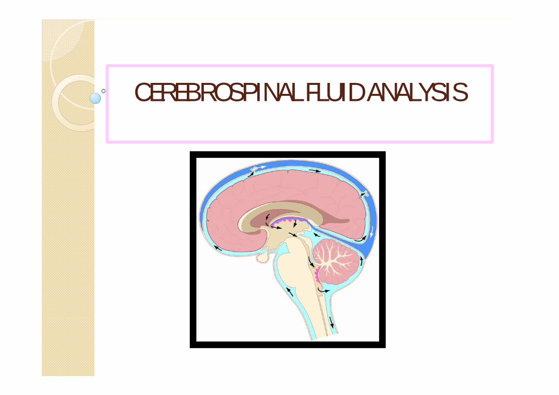
CEREBROSPINAL FLUID ANALYSIS
2016-1-19
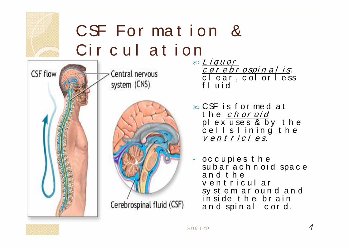
Liquor cerebrospinalis: clear, colorless fluid
CSF is formed at the choroid plexuses & by the cells lining the ventricles.
• occupies the subarachnoid space and the ventricular system around and inside the brain and spinal cord.
4
CSF Formation & Circulation

5
Method of CSF Sampling
Traumatic tap (damage to blood vessel during specimen collection) blood in CSF
Obtained by lumbar puncture (At the interspace L3-4, or lower)
Using aseptic technique
6
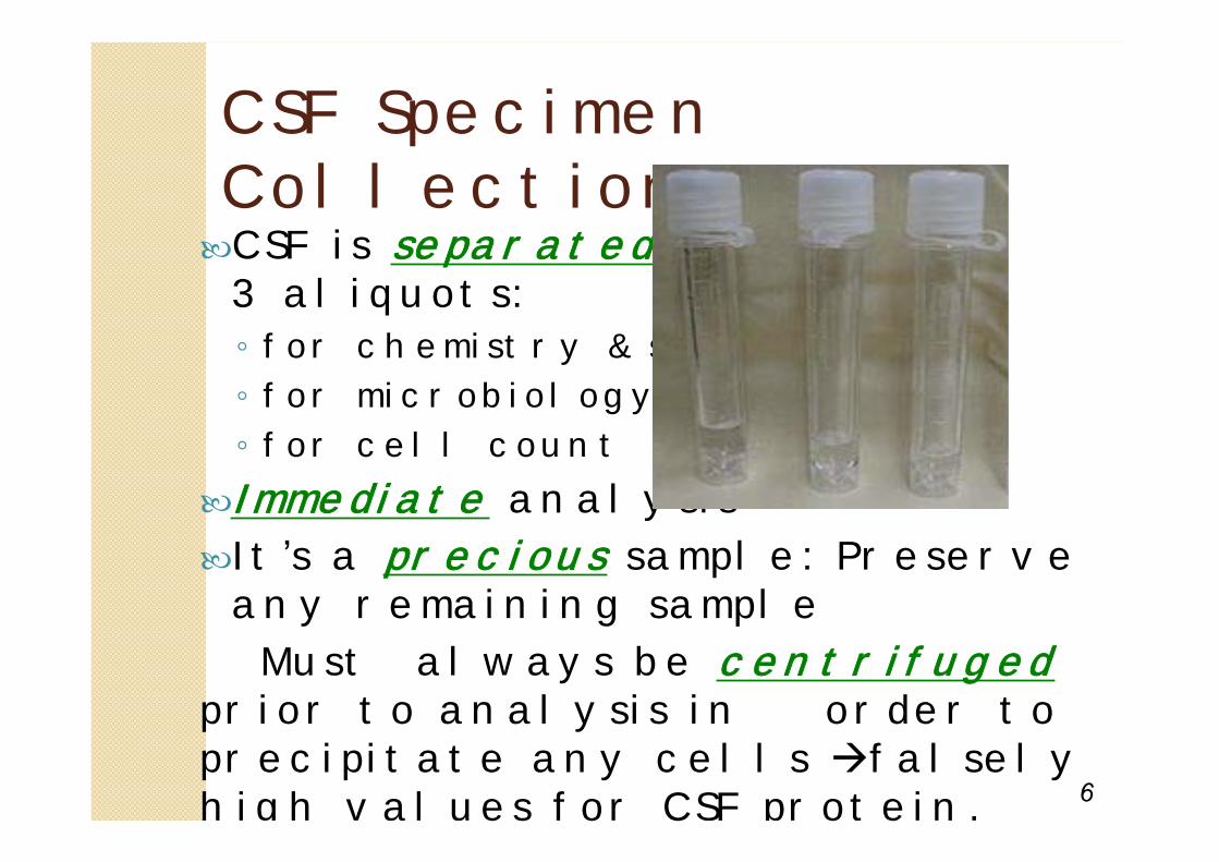
CSF Specimen CollectionCSF is separated into 3 aliquots:◦ for chemistry & serology
◦ for microbiology
◦ for cell count
Immediate analysis
It’s a precious sample: Preserve any remaining sample
Must always be centrifuged prior to analysis in order to precipitate any cells falsely high values for CSF protein.
7
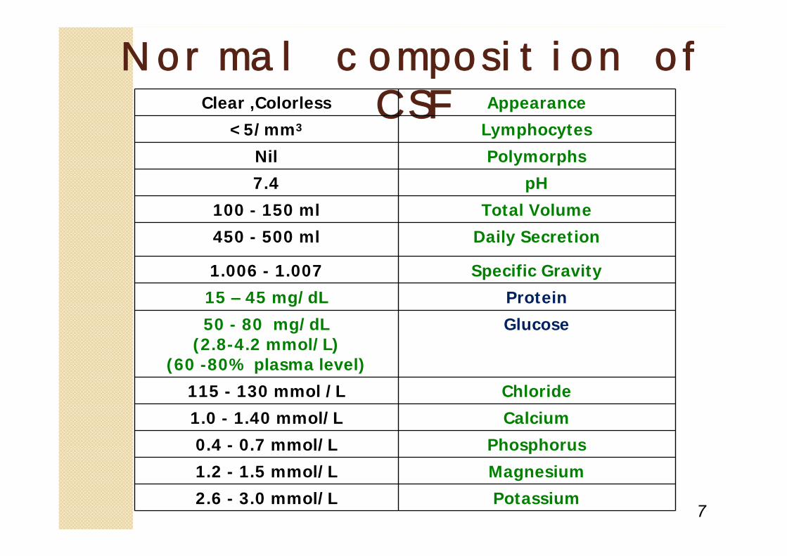
Normal composition of CSFClear ,Colorless Appearance
<5/mm3 Lymphocytes
Nil Polymorphs
7.4 pH
100 - 150 ml Total Volume
450 - 500 ml Daily Secretion
1.006 - 1.007 Specific Gravity15 – 45 mg/dL Protein
50 - 80 mg/dL(2.8-4.2 mmol/L)
(60 -80% plasma level)
Glucose
115 - 130 mmol /L Chloride
1.0 - 1.40 mmol/L Calcium
0.4 - 0.7 mmol/L Phosphorus
1.2 - 1.5 mmol/L Magnesium
2.6 - 3.0 mmol/L Potassium
8
Examination of CSF:1- Physical examinationoNormal CSF is:
oColorlessoClearoFree of clotsoFree of blood
9
Blood & Hemoglobin pigments in CSF
Subarachnoid hemorrhage (SAH)
Xanthochromia (hemoglobin breakdown pigments) = RBCs lysis & metabolism previously occurred (at least 2 hr earlier)

10
When would Xanthochromia indicate hemorrhage?
If you exclude:
1. Prior traumatic tap
2.Hyperbilirubinemia (bilirubin > 20 mg/dL)
11
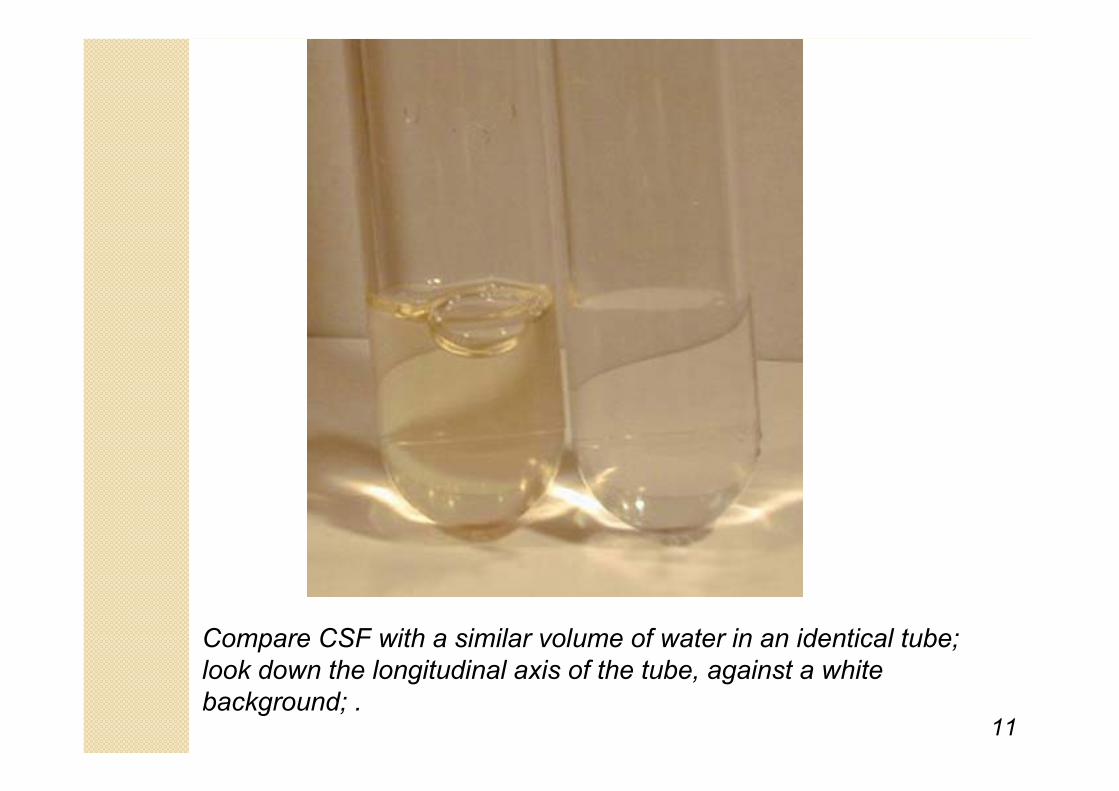
Compare CSF with a similar volume of water in an identical tube; look down the longitudinal axis of the tube, against a white background; .
2016-1-19
Turbidity
CSF is cloudy (turbid) ◦ is usually due to leucocytes◦ may be due to micro-organisms
Meningitis – coccal forms
400-500 polymorphs per cu.mm
12
2016-1-19
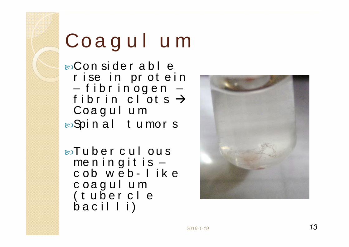
Coagulum Considerable rise in protein – fibrinogen – fibrin clots Coagulum
Spinal tumors
Tuberculous meningitis – cob web- like coagulum (tubercle bacilli)
13
14
Examination of CSF:2- Biochemical analysis of CSFTests of interest:◦ Glucose
◦ Protein
Total
Specific:Albumin
Immunoglobulin
Others (e.g. myelin basic protein; MBP)
◦ Chlorides
◦ Lactate
◦ Enzymes
√√ The most reliable parameters
diagnostically &accessible analytically
15
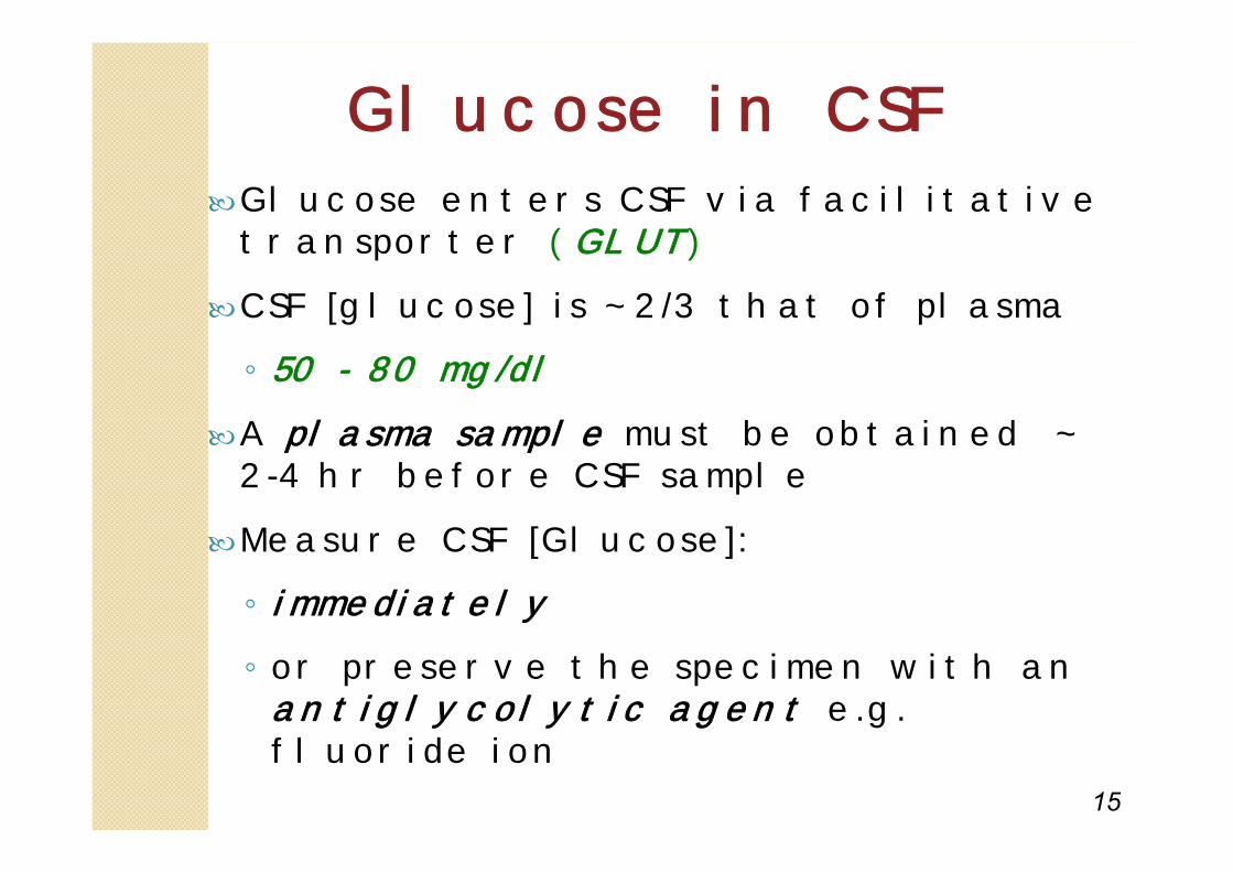
Glucose in CSFGlucose enters CSF via facilitative transporter (GLUT)
CSF [glucose] is ~ 2/3 that of plasma
◦ 50 - 80 mg/dl
A plasma sample must be obtained ~ 2-4 hr before CSF sample
Measure CSF [Glucose]:
◦ immediately
◦ or preserve the specimen with an antiglycolytic agent e.g. fluoride ion
16
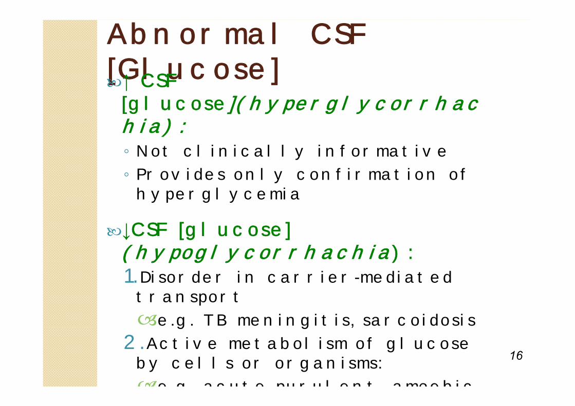
Abnormal CSF [Glucose]↑ CSF [glucose](hyperglycorrhachia):◦ Not clinically informative
◦ Provides only confirmation of hyperglycemia
↓CSF [glucose] (hypoglycorrhachia):1.Disorder in carrier-mediated transport
e.g. TB meningitis, sarcoidosis
2.Active metabolism of glucose by cells or organisms:
e g acute purulent amoebic
2016-1-19
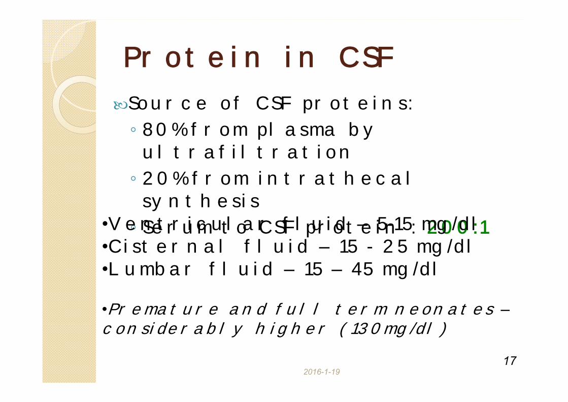
Protein in CSF
Source of CSF proteins:
◦ 80% from plasma by ultrafiltration
◦ 20% from intrathecal synthesis
◦ Serum to CSF protein : 200:1
17
•Ventricular fluid – 5-15 mg/dl•Cisternal fluid – 15 - 25 mg/dl•Lumbar fluid – 15 – 45 mg/dl
•Premature and full term neonates – considerably higher (130mg/dl)
2016-1-19
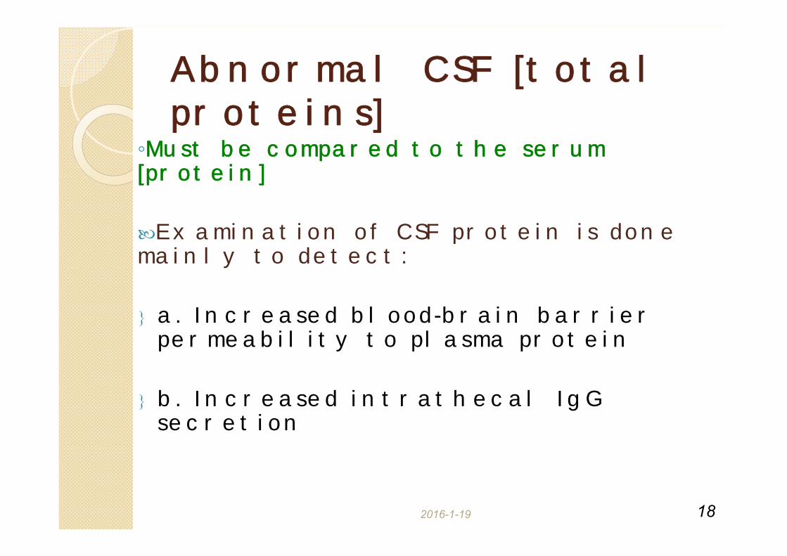
◦Must be compared to the serum [protein]
Examination of CSF protein is done mainly to detect:
a. Increased blood-brain barrier permeability to plasma protein
b. Increased intrathecal IgG secretion
18
Abnormal CSF [total proteins]
19
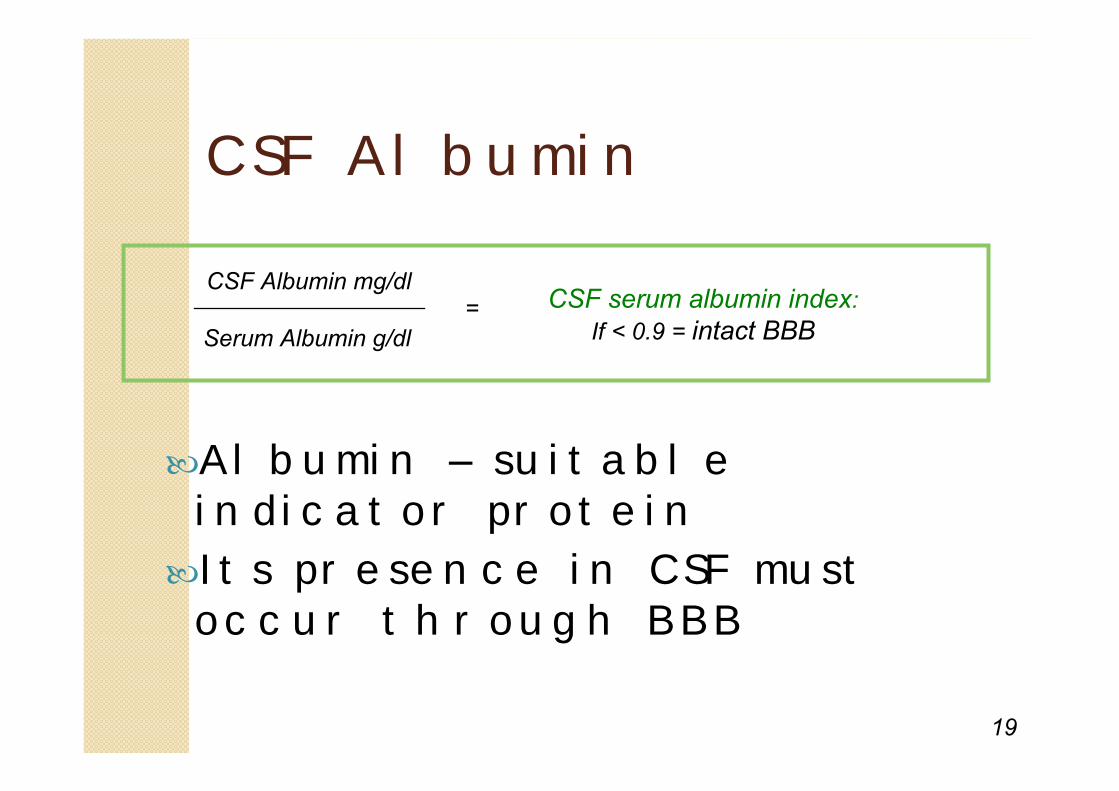
CSF Albumin
Albumin – suitable indicator protein
Its presence in CSF must occur through BBB
CSF Albumin mg/dl
Serum Albumin g/dlCSF serum albumin index:
If < 0.9 = intact BBB=
2016-1-19
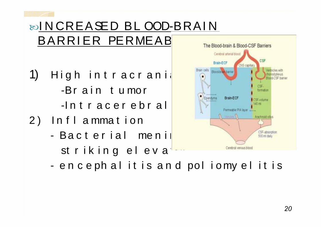
INCREASED BLOOD-BRAIN BARRIER PERMEABILITY
1) High intracranial pressure
-Brain tumor
-Intracerebral haemorrhage
2) Inflammation
- Bacterial meningitis – striking elevation
- encephalitis and poliomyelitis
20
2016-1-19
INCREASED INTRATHECAL SYNTHESIS OF IMMUNOGLOBULINS
IgG – Demyelinating diseasesMultiple sclerosis (MS)Subacute Sclerosing Panencephalitis (SSPE)
B lymphocytes infiltrating the leisions synthesize IgG
21
22
What to do if ↑ CSF [protein] was detected?
Perform electrophoretic separation
If multiple banding of the IgG band is detected (oligoclonal bands): ◦ MS
◦ SSPE◦ Inflammatory diseases
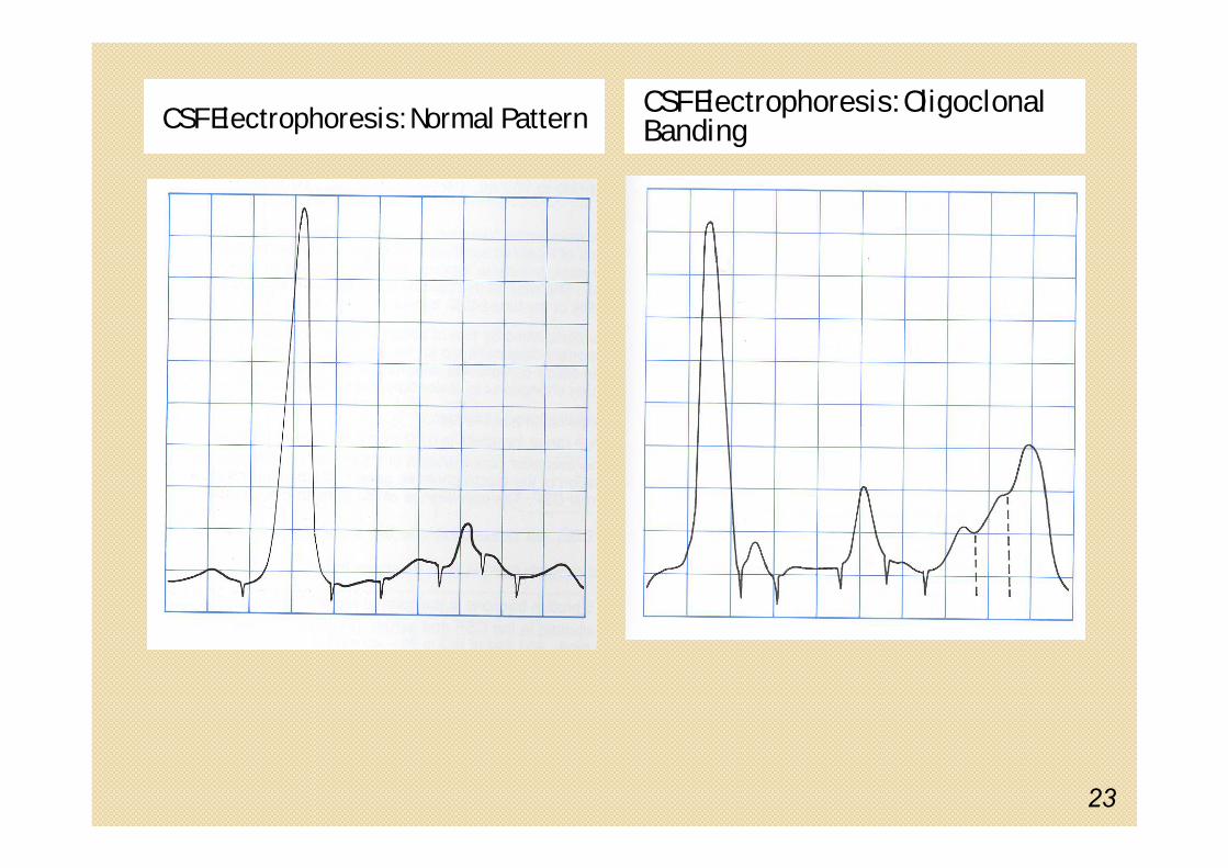
CSF Electrophoresis: Normal Pattern
23
CSF Electrophoresis: Oligoclonal Banding
2016-1-19
Abnormal CSF Chloride
◦ 120 – 130 meq per litre
◦ Higher than the plasma chloride
marked in acute bacterial meningitis
slight in viral meningitis & brain tumors
24
25
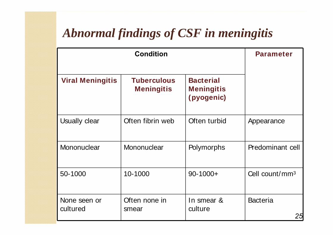
Abnormal findings of CSF in meningitisCondition Parameter
Viral Meningitis Tuberculous Meningitis
Bacterial Meningitis(pyogenic)
Usually clear Often fibrin web Often turbid Appearance
Mononuclear Mononuclear Polymorphs Predominant cell
50-1000 10-1000 90-1000+ Cell count/mm3
None seen or cultured
Often none in smear
In smear & culture
Bacteria
26
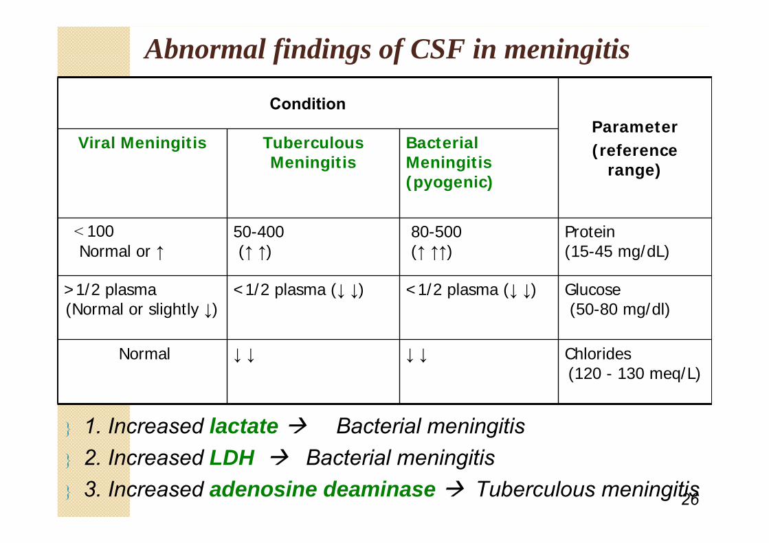
Abnormal findings of CSF in meningitis
ConditionParameter(reference
range)
Viral Meningitis Tuberculous Meningitis
Bacterial Meningitis(pyogenic)
˂ 100 Normal or ↑
50-400 (↑ ↑)
80-500 (↑ ↑↑)
Protein (15-45 mg/dL)
>1/2 plasma(Normal or slightly ↓)
<1/2 plasma (↓ ↓) <1/2 plasma (↓ ↓) Glucose (50-80 mg/dl)
Normal ↓ ↓ ↓ ↓ Chlorides(120 - 130 meq/L)
1. Increased lactate Bacterial meningitis 2. Increased LDH Bacterial meningitis 3. Increased adenosine deaminase Tuberculous meningitis
27


29

30
31

32
33


![Laboratory Diagnosis of Bacterial Meningitis - cmr.asm.org · Meningitis occurs in the subarachnoid space (between the arachnoid [including the trabeculae] andthepia mater). The subarachnoid](https://static.fdocuments.us/doc/165x107/5c4a91b493f3c31760718c27/laboratory-diagnosis-of-bacterial-meningitis-cmrasmorg-meningitis-occurs.jpg)