
J. (1992), 66, Ltd., Press Ltd., and studies 'V'-bottomed
6
Br. J. Cancer (1992), 66, 660 665 © Macmillan Press Ltd., Growth, morphology and chemosensitivity studies on postconfluent cells cultured in 'V'-bottomed microtiter plates P.E. Pizaol, D.M. Lyaruu2, G.J. Peters', J. van Ark-Otte', B. Winograd3'5, G. Giacconel & H.M. Pinedo" 4 'Department of Oncology, Free University Hospital, PO Box 7057, 1007 MB Amsterdam; 2Department of Oral Cell Biology, ACTA - Vrije Universiteit, Amsterdam; 'EORTC - New Drug Development Office, Amsterdam; 4The Netherlands Cancer Institute, Amsterdam, Netherlands. Summary This study assessed the growth pattern, cellular organisation and chemosensitivity of established human tumour cell lines growing as postconfluent cultures in 'V'-bottomed, 96-well microtiter plates. Cross- sections of the colon (HT29, SW620, SWI 116), ovarian (A2780) and head and neck (UM-SCC-22B) car- cinoma microcultures allowed in situ evaluation of the cellular organisation in the wells. After 5 days of growth, every cell line had reached confluence, but each of them displayed a specific pattern of cell stacking which ranged from two to ten layers. Postconfluent HT29 cells displayed morphologic features suggestive of some degree of enterocytic differentiation. Growth and cytotoxicity could be studied reliably and reproducibly in this system with the sulforhodamine B protein assay. Against HT29 postconfluent cultures, the EC50's (drug concentrations producing absorbance readings 50% lower than those of non-treated wells) of 5-fluorouracil and of the ether lipid, hexadecylphosphocholine, were 1 mm and 50 gM respectively. The possibility to perform chemosensitivity tests using semiautomated microtiter plate technology supports further evaluation of this system as an alternative antitumour drug testing model. In the past four decades, worldwide efforts to discover and develop new anticancer agents using murine leukaemias as screening systems have produced drugs which are mainly effective against rapidly proliferating tumours. In view of the limited discovery of new anticancer agents for the treatment of the most common human solid malignancies (e.g., lung, colon, mammary carcinomas), current research is directed towards the study of alternative laboratory models. In a critical evaluation of the predictive value of in vitro chemosensitivity assays, Phillips et al. (1990) pointed out that: (a) besides the inherent chemosensitivity of tumour cells, other factors such as drug pharmaco-kinetics/-dynamics and the biology of solid tumours significantly influence the success rate of chemotherapy in vivo; and, (b) the lack of simulation of those conditions in vitro is a major limitation to the improvement of the predictability of this type of tests. Therefore, an ideal in vitro tumour model should mimic the organisational structure and the heterogeneity in biologic characteristics found in human tumours. Several investigators have shown that multicellular spheroids display a degree in complexity which is intermediate between the in vivo neoplas- tic cell deposits and monolayered tumour cell cultures (reviewed by Mueller-Klieser, 1987 and Sutherland, 1988). However, the use of spheroids specifically for new antitumour drug screening and chemosensitivity testing has been carried out only on a limited scale because of technical limiting-factors. Other investigators have used either postconfluent (Kruse & Miedema, 1965; Skehan et al., 1986; Pelletier et al., 1990) or plateau-phase (Twentyman, 1976; Drewinko et al., 1981) tumour cell cultures in order to increase the degree with which in vivo cell-cell interactions, growth physiology and microenvironment conditions are simulated in vitro. It has been suggested that results generated with these systems more closely resemble those obtained with spheroids and with in vivo models than those obtained with monolayer cell cul- tures. The objective of this study was to evaluate the growth pattern and cellular organisation of human tumour cell lines growing as postconfluent cultures (two or more layers of cells) in 'V'-bottomed, 96-well microtiter plates. We also assessed the feasibility of using the sulforhodamine B (SRB) assay to study cytotoxicity in this system. Materials and methods Chemicals and drugs Dulbecco's Modified Eagle's Medium (DMEM) was pur- chased from Flow Laboratories (Irvine, Scotland); foetal calf serum (FCS) was from Gibco (New York, USA); 3-(4,5- dimethylthiazol-2-yl)-2,5-diphenyltetrazolium bromide (MIT), 5-fluorouracil (5FU) and sulforhodamine B were from Sigma Chemical Co. (St Louis, USA); spectrophotometric graded dimethyl sulfoxide (DMSO) was from Baker Chemicals B.V. (Deventer, Holland). HistoresinR was from Reichert/Jung (Salzburg, Austria). Hexadecylphosphocholine (HPC), also known as miltefosine, was a kind gift of Dr P. Hilgard (Asta Pharma, Bielefeld, Germany). All other chemicals were of standard analytical quality commercially available. Cell culture and plating HT29, SW620 and SWl 116 human colon adenocarcinoma cell lines were obtained from the American Type Culture Collection (Rockville, USA). UM-SCC-22B (22B) human head and neck squamous cell carcinoma line was a gift of Dr T. Carey (University of Michigan, USA). The A2780 human ovarian carcinoma cell line was obtained from Dr R.F. Ozols (NCI, Bethesda, USA) and MCF-7 mammary carcinoma cells from Dr K. Cowan (NCI, Bethesda, USA). Detailed description of routine cell culture and plating procedures used during these experiments were reported elsewhere (Keepers et al., 1991). Briefly, mycoplasma- negative cells were maintained without antibiotics in DMEM supplemented with 5% FCS and 1 mM L-glutamine in a 37'C, 5% C02, 95% humidified air incubator. Exponentially growing cells were trypsinised and resuspended in antibiotic- containing medium (50 ,lg gentamicin ml-'); single cell suspensions displaying > 90% viability by trypan blue dye exclusion were subsequently counted and seeded (15,000 cells/50 il/well) in 96-well plates with 'V'-shaped bottoms (Greiner Labortechnik, Solingen, Germany). Twenty-four h after seeding, 100 ,l4 of medium were added to each well. From the second day after plating until the end Correspondence: G. Giaccone. 5Present address: Bristol-Myers Squibb Pharmaceutical Research Ins- titute, Brussels, Belgium. Received 25 October 1991; and in revised form 16 June 1992. %'." Macmillan Press Ltd., 1992 Br. J. Cancer (1992), 66, 660-665
Transcript of J. (1992), 66, Ltd., Press Ltd., and studies 'V'-bottomed

Br. J. Cancer (1992), 66, 660 665 © Macmillan Press Ltd.,
Growth, morphology and chemosensitivity studies on postconfluent cellscultured in 'V'-bottomed microtiter plates
P.E. Pizaol, D.M. Lyaruu2, G.J. Peters', J. van Ark-Otte', B. Winograd3'5, G. Giacconel & H.M.Pinedo" 4
'Department of Oncology, Free University Hospital, PO Box 7057, 1007 MB Amsterdam; 2Department of Oral Cell Biology,ACTA - Vrije Universiteit, Amsterdam; 'EORTC - New Drug Development Office, Amsterdam; 4The Netherlands CancerInstitute, Amsterdam, Netherlands.
Summary This study assessed the growth pattern, cellular organisation and chemosensitivity of establishedhuman tumour cell lines growing as postconfluent cultures in 'V'-bottomed, 96-well microtiter plates. Cross-sections of the colon (HT29, SW620, SWI 116), ovarian (A2780) and head and neck (UM-SCC-22B) car-cinoma microcultures allowed in situ evaluation of the cellular organisation in the wells. After 5 days ofgrowth, every cell line had reached confluence, but each of them displayed a specific pattern of cell stackingwhich ranged from two to ten layers. Postconfluent HT29 cells displayed morphologic features suggestive ofsome degree of enterocytic differentiation. Growth and cytotoxicity could be studied reliably and reproduciblyin this system with the sulforhodamine B protein assay. Against HT29 postconfluent cultures, the EC50's (drugconcentrations producing absorbance readings 50% lower than those of non-treated wells) of 5-fluorouraciland of the ether lipid, hexadecylphosphocholine, were 1 mm and 50 gM respectively. The possibility to performchemosensitivity tests using semiautomated microtiter plate technology supports further evaluation of thissystem as an alternative antitumour drug testing model.
In the past four decades, worldwide efforts to discover anddevelop new anticancer agents using murine leukaemias as
screening systems have produced drugs which are mainlyeffective against rapidly proliferating tumours. In view of thelimited discovery of new anticancer agents for the treatmentof the most common human solid malignancies (e.g., lung,colon, mammary carcinomas), current research is directedtowards the study of alternative laboratory models.
In a critical evaluation of the predictive value of in vitrochemosensitivity assays, Phillips et al. (1990) pointed outthat: (a) besides the inherent chemosensitivity of tumourcells, other factors such as drug pharmaco-kinetics/-dynamicsand the biology of solid tumours significantly influence thesuccess rate of chemotherapy in vivo; and, (b) the lack ofsimulation of those conditions in vitro is a major limitationto the improvement of the predictability of this type of tests.Therefore, an ideal in vitro tumour model should mimic theorganisational structure and the heterogeneity in biologiccharacteristics found in human tumours. Several investigatorshave shown that multicellular spheroids display a degree incomplexity which is intermediate between the in vivo neoplas-tic cell deposits and monolayered tumour cell cultures(reviewed by Mueller-Klieser, 1987 and Sutherland, 1988).However, the use of spheroids specifically for newantitumour drug screening and chemosensitivity testing hasbeen carried out only on a limited scale because of technicallimiting-factors.Other investigators have used either postconfluent (Kruse
& Miedema, 1965; Skehan et al., 1986; Pelletier et al., 1990)or plateau-phase (Twentyman, 1976; Drewinko et al., 1981)tumour cell cultures in order to increase the degree withwhich in vivo cell-cell interactions, growth physiology andmicroenvironment conditions are simulated in vitro. It hasbeen suggested that results generated with these systems moreclosely resemble those obtained with spheroids and with invivo models than those obtained with monolayer cell cul-tures.The objective of this study was to evaluate the growth
pattern and cellular organisation of human tumour cell linesgrowing as postconfluent cultures (two or more layers of
cells) in 'V'-bottomed, 96-well microtiter plates. We alsoassessed the feasibility of using the sulforhodamine B (SRB)assay to study cytotoxicity in this system.
Materials and methods
Chemicals and drugs
Dulbecco's Modified Eagle's Medium (DMEM) was pur-chased from Flow Laboratories (Irvine, Scotland); foetal calfserum (FCS) was from Gibco (New York, USA); 3-(4,5-dimethylthiazol-2-yl)-2,5-diphenyltetrazolium bromide (MIT),5-fluorouracil (5FU) and sulforhodamine B were from SigmaChemical Co. (St Louis, USA); spectrophotometric gradeddimethyl sulfoxide (DMSO) was from Baker Chemicals B.V.(Deventer, Holland). HistoresinR was from Reichert/Jung(Salzburg, Austria). Hexadecylphosphocholine (HPC), alsoknown as miltefosine, was a kind gift of Dr P. Hilgard (AstaPharma, Bielefeld, Germany). All other chemicals were ofstandard analytical quality commercially available.
Cell culture and platingHT29, SW620 and SWl 116 human colon adenocarcinomacell lines were obtained from the American Type CultureCollection (Rockville, USA). UM-SCC-22B (22B) humanhead and neck squamous cell carcinoma line was a gift of DrT. Carey (University of Michigan, USA). The A2780 humanovarian carcinoma cell line was obtained from Dr R.F. Ozols(NCI, Bethesda, USA) and MCF-7 mammary carcinomacells from Dr K. Cowan (NCI, Bethesda, USA).
Detailed description of routine cell culture and platingprocedures used during these experiments were reportedelsewhere (Keepers et al., 1991). Briefly, mycoplasma-negative cells were maintained without antibiotics in DMEMsupplemented with 5% FCS and 1 mM L-glutamine in a37'C, 5% C02, 95% humidified air incubator. Exponentiallygrowing cells were trypsinised and resuspended in antibiotic-containing medium (50 ,lg gentamicin ml-'); single cellsuspensions displaying > 90% viability by trypan blue dyeexclusion were subsequently counted and seeded (15,000cells/50 il/well) in 96-well plates with 'V'-shaped bottoms(Greiner Labortechnik, Solingen, Germany).
Twenty-four h after seeding, 100 ,l4 of medium were addedto each well. From the second day after plating until the end
Correspondence: G. Giaccone.5Present address: Bristol-Myers Squibb Pharmaceutical Research Ins-titute, Brussels, Belgium.Received 25 October 1991; and in revised form 16 June 1992.
%'." Macmillan Press Ltd., 1992Br. J. Cancer (1992), 66, 660-665
POSTCONFLUENT CELL CULTURES 661
of the experiments, culture medium was gently aspirated andreplaced by fresh medium (150 lI/well) once daily.
Histological procedures
On days 5 and 10 after plating, culture medium from platesassigned for cross-sections was aspirated, wells were rinsedonce with PBS and cultures were fixed in situ with 1%phosphate-buffered gluteraldehyde (pH = 7.3) for at least 2 h.Fixed cultures were washed twice with the same buffer,dehydrated in ascending ethanol series and embedded in150 ,sl glycol-methacrylate-containing plastic resin (His-toresinR), using the protocol supplied by the manufacturer.After polymerisation, individual wells (n = 6) were sawedapart from the microtiter plate and re-embedded en bloc in alarger mould with the same resin. Each block was thensagitally cross-sectioned (Jung-K Microtome; Reichter-Jung)in two regions: in the centre of the 'V'-shaped bottom and ina region corresponding approximately to one third of thedistance between the centre and the edge of the well bottom.At least twenty-four, 5 fm thick, serial cross-sections fromeach of these regions were collected on microscope glassslides and stained with dilute toluidine blue or with PAS(periodic acid-Schiff)/hematoxylin. A PAS-positive staining(red colour) indicates the presence of cellular carbohydrates(e.g., mucins) (Cook, 1990). Cross-sectioning experimentswere repeated at least three times.
Chemosensitivity assessment
Chemosensitivity tests were carried out using HT29 cells andtwo drugs: 5FU and the ether lipid, HPC (Hilgard et al.,1988). These two drugs were specifically selected because theyproduced dose-response curves with markedly differentprofiles in a previous series of experiments usingmonolayered cultures. Stock solutions and test concentra-tions were prepared and used essentially as described in thatmanuscript (Keepers et al., 1991). Plates received drug(150 .LI/well) on day 5 following cell seeding. After 24 h ofdrug exposure, wells were rinsed once with fresh medium andthen incubated for another 4 days in the absence of drugsand with daily medium renewal until cytotoxicity wasassessed. The drug-response patterns of 5FU and HPC wereobtained with the MTT and SRB assays and compared withcell numbers from a third set of microplates where mediumwas aspirated, cells from replicate wells (n = 6/drug concent-ration) were trypsinised (15 min/37°C), carefully resuspendedand immediately counted with an automatic cell counter(Sysmex, CC-i 10, Tokyo, Japan).
SRB assay
The SRB assay was performed essentially as described before(Skehan et al., 1990), however, some variations in the basicprotocol were also tested in order to optimise the assay forpostconfluent cultures. Briefly, cells were fixed by adding50 Ill/well of 50% trichloroacetic acid and incubating for 1 or2 h at 4°C. Plates were then rinsed five times with tap waterand air dried. Microcultures were stained for either 0.5, 1, 1.5or 2 h after the addition of 50 j.l of 0.4% SRB. In order toremove unbound dye, a different number of washing stepswith 1% acetic acid was tested. To solubilise bound dye,wells received 150 lAl of 10 mM Tris base and were exposed to15 min of ultrasonic vibration while floating in an ultrasoniccleaner (Branson 5200). Absorbance of each well was read at450 nm (a suboptimal wavelength required to produce absor-bance readings within the linearity range of the assay) usinga microtiter plate reader (Titertek Multiskan MCC/340; FlowLaboratories) interfaced with an Olivetti PC M19 microcom-puter.
In order to evaluate the range of linearity between cellnumbers and absorbance readings, exponentially growingHT29 cells were trypsinised from culture flasks, counted andplated at the highest cell density in 200 fl medium/well.Serial dilutions were prepared by transporting 100 tlI cell
suspension into immediately neighbouring wells containing100 1l medium. Cells were allowed to attach for 4h beforethe SRB assay was performed.
MTT assay
The MTT assay was performed using a modification of theprotocol reported by Alley et al. (1988). After 4 h incubationwith MTT solution, the large numbers of formazan crystalsin postconfluent cultures were solubilised with 150 lI ofDMSO/0.5% FCS plus 15 min of ultrasound vibration ap-plied to water floating plates in an ultrasonic cleaner (Bran-son 5200). The absorbance of each well was measured at540 nm using a microtiter plate reader.
Data management and analysisData from the microplate reader were transferred to floppydisks using Titersoft E.I.A. software (Flow Laboratories).Subsequent data analysis was performed within Symphonysoftware (Lotus, Cambridge, USA). A P value (Student'st-test) of less than 0.05 was considered to be statisticallysignificant.
Results
Microculture histologyPlates observed under the light microscope 4 h after seedingrevealed that cell sedimentation had taken place preferen-tially in the centre of the 'V'-shaped bottom, independentlyof the specific tumour cell type. When cells were seeded inlarger volumes, e.g., 100 LIl medium, they sedimented in thecentre and in the periphery of the well to the same extent(data not shown). A confluent cell monolayer was seen onthe third day after plating, except in the case of 22B andSWI 116 which needed another 24-48 h to become globallyconfluent. In all cell types tested, culture confluence andmultilayering followed a centrifugal pattern, always startinginitially in the centre of the wells.On the 4th day after plating, MCF-7 cells became very
weakly attached to the plastic substratum and were strippedoff the well whenever medium substitution was performed.This could not be avoided by plate centrifugation prior tomedium manipulation. Therefore, MCF-7 was excluded fromfurther studies.The postconfluent status of the microcultures and
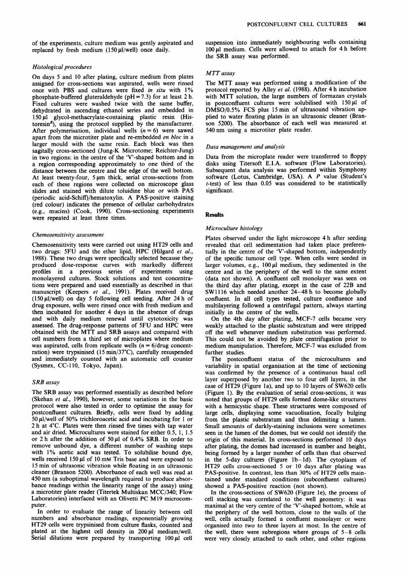
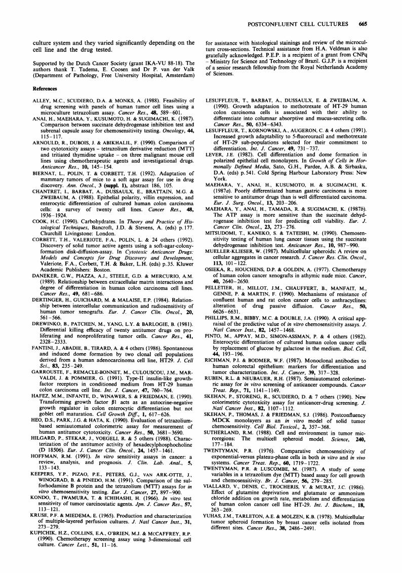
variability in spatial organisation at the time of sectioningwas confirmed by the presence of a continuous basal celllayer superposed by another two to four cell layers, in thecase of HT29 (Figure la), and up to 10 layers of SW620 cells(Figure 1). By the evaluation of serial cross-sections, it wasnoted that groups of HT29 cells formed dome-like structureswith a hemicystic shape. These structures were composed oflarge cells, displaying some vacuolisation, focally bulgingfrom the plastic substratum and thus delimiting a lumen.Small amounts of darkly-staining inclusions were sometimesseen in the lumen of the domes, but we could not identify theorigin of this material. In cross-sections performed 10 daysafter plating, the domes had increased in number and height,being formed by a larger number of cells than that observedin the 5-day cultures (Figure lb-Id). The cytoplasm ofHT29 cells cross-sectioned 5 or 10 days after plating wasPAS-positive. In contrast, less than 30% of HT29 cells main-tained under standard conditions (subconfluent cultures)showed a PAS-positive reaction (not shown).
In the cross-sections of SW620 (Figure le), the process ofcell stacking was correlated to the well geometry: it wasmaximal at the very centre of the 'V'-shaped bottom, while atthe periphery of the well bottom, close to the walls of thewell, cells actually formed a confluent monolayer or wereorganised into two to three layers at most. In the centre ofthe well, there were subregions where groups of 5-8 cellswere very closely attached to each other, and other regions
662 P.E. PIZAO et al.
where the inter-cellular spaces were relatively large. SW620cells grown as sub- or postconfluent cultures were PAS-negative and did not display any morphological features of adifferentiated phenotype. A2780 cells displayed a globallyconfluent monolayer superimposed by compact cell clustersof different sizes, containing up to 15 cells without any signsof differentiation (Figure If). These cell clusters were alsomore frequent in the centre of the wells, while a single layerof cells was observed at the periphery of the plastic subs-tratum. SWI116 and 22B displayed similar microculture or-ganisation 5 days after plating: a confluent monolayer of cellswas the consistent feature; a second layer of cells was seen inless than 50% of the cross-sections and occupied small areasrestricted to the centre of the wells (not shown).
The cross-sections obtained after 10 days of culture did notshow any significant increase in the maximum number of celllayers of any of the tumour lines studied. Nonetheless, onday 10, the process of multiple cell layering was prevalent inlarger areas of the plastic substratum.
Growth and chemosensitivity
According to the protocol described by Skehan et al. (1990),cells were optimally stained if exposed to the SRB solutionfor 30 min after 1 h of TCA fixation. In our experiments,there was no need for a longer incubation with TCA. Never-theless, absorbance readings from 10-day old HT29 post-confluent cultures stained for only 30 min were considerably
a b
c d
e f
Figures la-lf Photomicrographs of in situ cross-sections of postconfluent microcultures prepared from individual 'V'-bottomedwells as described in 'Materials and methods'. a, shows PAS-positive HT29 cells after 5 days in culture. The cells are tightly packedinto a maximum of 3-5 layers. At this time point, dome-like structures of hemicysts could be observed, focally bulging from theplastic substratum and delimiting a lumen sometimes containing darkly-staining inclusions of indeterminate origin. After 10 days inculture, the number and size of these domes increased: b-d, depict serial cross-section taken from a single HT29 dome,demonstrating the 3-dimensional architecture of this structure. b, shows the cross-section at the edge of the dome which appears asa cluster of cells without a lumen; further inwards, c, shows the beginning of the formation of a lumen containing somedarkly-staining inclusions; d, reveals the structure of the same dome at the region where it reaches its maximum height. e, Platescontaining SW620 cells and cross-sectioned at 5 days after seeding showed a maximum of ten layers of cells at the centre of the'V'-shaped wells. Five days after plating, A2780 cells formed confluent monolayers superimposed by clusters containing up to 15cells showing no signs of morphological differentiation f. a-d, PAS/hematoxylin-stained; e and f, toluidine blue-stainedBar= IOism.
POSTCONFLUENT CELL CULTURES 663
lower than those obtained after 1 h of SRB staining,independently of the number of washing steps under acidicconditions (data not shown). No improvement in the assayresults was gained with staining periods longer than 1 h andmore than five washing steps to remove unbound dye (notshown). Therefore, the optimised SRB test used in the addi-tional experiments reported here included 1 h of TCAfixation followed by 1 h of SRB staining and five washingsteps with 1% acetic acid.The necessity for daily medium substitutions to support
the cell growth during 10 days of incubation was initiallyconsidered as a probable source of artifacts (cell loss). How-ever, an absolute need for plate centrifugation immediatelybefore medium aspiration was only seen with A2780 cells.Another problem was the occasional aspiration of large cellclumps that took place whenever the Pasteur pipet wouldaccidentally touch the bottom of the well. Such involuntarycell aspiration could be easily detected by macroscopicexamination of air-dried, SRB-stained wells and marked tobe excluded of the results later.
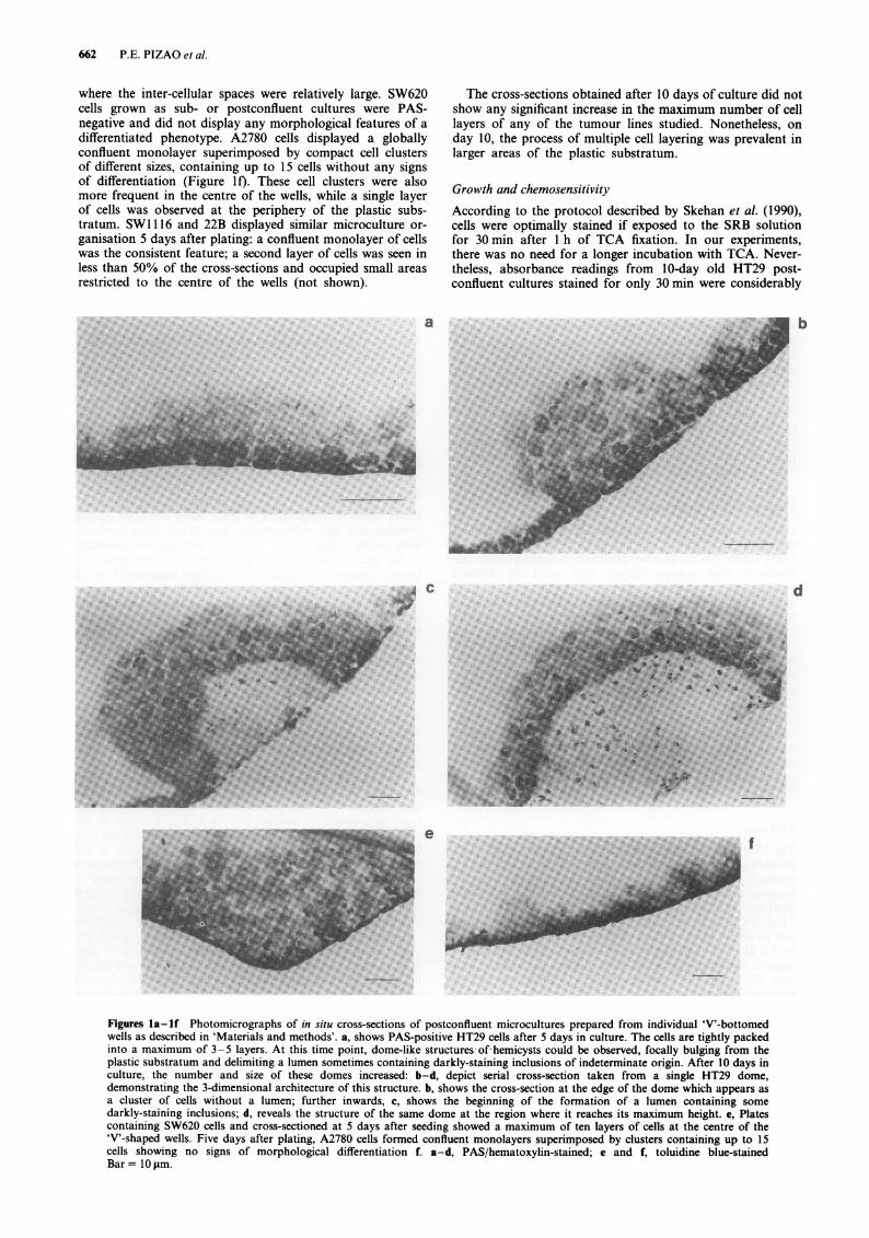
Linearity between increasing HT29 cell numbers and SRBabsorbance readings was observed until cell counts reachedapproximately 106 cells/well (Figure 2). Since hemacytometercountings of 10-day old microcultures of the tumour linestested never exceeded 0.9 x 10⁶ cells/well, this protocol forthe SRB assay was ultimately adopted for use with post-confluent microcultures.
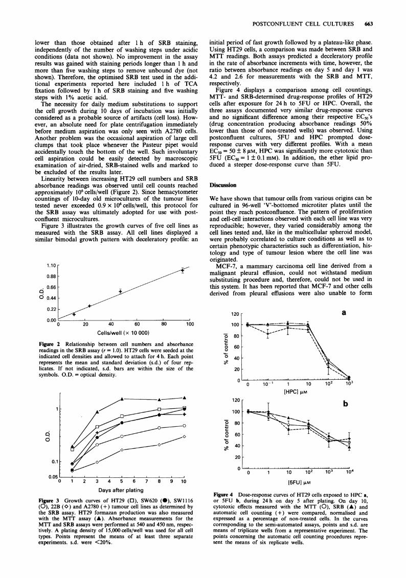
Figure 3 illustrates the growth curves of five cell lines asmeasured with the SRB assay. All cell lines displayed asimilar bimodal growth pattern with deceleratory profile: an
1.10
0.88
nA. v.vv
o 0.44
0.22
0.00 + I
0 20 40 60 80 100
Cells/well (x 10 000)
Figure 2 Relationship between cell numbers and absorbancereadings in the SRB assay (r = 1.0). HT29 cells were seeded at theindicated cell densities and allowed to attach for 4 h. Each pointrepresents the mean and standard deviation (s.d.) of four rep-licates. If not indicated, s.d. bars are within the size of thesymbols. O.D. = optical density.
initial period of fast growth followed by a plateau-like phase.Using HT29 cells, a comparison was made between SRB andMTT readings. Both assays predicted a deceleratory profilein the rate of absorbance increments with time, however, theratio between absorbance readings on day 5 and day 1 was4.2 and 2.6 for measurements with the SRB and MTT,respectively.
Figure 4 displays a comparison among cell countings,MTT- and SRB-determined drug-response profiles of HT29cells after exposure for 24 h to 5FU or HPC. Overall, thethree assays documented very similar drug-response curvesand no significant difference among their respective EC50's(drug concentration producing absorbance readings 50%lower than those of non-treated wells) was observed. Usingpostconfluent cultures, 5FU and HPC prompted dose-response curves with very different profiles. With a meanEC50 = 50 ± 8 gM, HPC was significantly more cytotoxic than5FU (EC50 = 1 ± 0.1 mM). In addition, the ether lipid pro-duced a steeper dose-response curve than 5FU.
Discussion
We have shown that tumour cells from various origins can becultured in 96-well 'V'-bottomed microtiter plates until thepoint they reach postconfluence. The pattern of proliferationand cell-cell interactions observed with each cell line was veryreproducible; however, they varied considerably among thecell lines tested and, like in the multicellular spheroid model,were probably correlated to culture conditions as well as tocertain phenotypic characteristics such as differentiation, his-tology and type of tumour lesion where the cell line wasoriginated.MCF-7, a mammary carcinoma cell line derived from a
malignant pleural effusion, could not withstand mediumsubstituting procedure and, therefore, could not be used inthis system. It has been reported that MCF-7 and other cellsderived from pleural effusions were also unable to form
,o
0C)4-o
[HPCI FML.
T D
.. T.
~~~~~i I I T
-I
0 1 10 102 103 1040 1 2 3 4 5 6 7 8 9 10
Days after plating
Figure 3 Growth curves of HT29 (0), SW620 (0), SWI116(0), 22B (O) and A2780 (+) tumour cell lines as determined bythe SRB assay. HT29 formazan production was also measuredwith the MTT assay (A). Absorbance measurements for theMTT and SRB assays were performed at 540 and 450 nm, respec-tively. A plating density of 15,000 cells/well was used for all celltypes. Points represent the means of at least three separateexperiments. s.d. were <20%.
[5FU] FM
Figure 4 Dose-response curves of HT29 cells exposed to HPC a,
or 5FU b, during 24 h on day 5 after plating. On day 10,cytotoxic effects measured with the MTT (0), SRB (A) andautomatic cell counting (+) were compared, normalised andexpressed as a percentage of non-treated cells. In the curves
corresponding to the semi-automated assays, points and s.d. are
means of triplicate wells from a representative experiment. Thepoints concerning the automatic cell counting procedures repre-
sent the means of six replicate wells.
120
6
0.1
n nr%
100
80
60
40
C4-1
00
0
20
0
U.vU
F
CA.
664 P.E. PIZAO et al.
spheroids (Yuhas et al., 1978). Interestingly, MCF-7 cellsselected with increasing concentrations of doxorubicin anddisplaying the multidrug-resistance phenotype could begrown as postconfluent cultures and did not present anyproblems of detachment from the 'V'-bottomed wells (notshown). As observed with other models of cell aggregates, itis unlikely that postconfluent culture systems can be emp-loyed with an infinite number of tumour cell lines, despite ofthe technical simplicity of the described system.The choice of 'V'-bottomed microtiter plates allowed us to
develop culture conditions favouring confluence and multiplecell-layering in the centre of the well bottom at an earliertime point than it would take place on a flat-bottomed well.Ultimately, the conic geometry of the wells (instead of a flatbottom) also became a technical advantage for it allowedfaster solubilisation of MTT formazan crystals and SRBstain, possibly because 'V'-shaped bottoms submerged in thewater were more efficiently exposed to ultra-sound vibra-tion.The HT29 cell line displays anaplastic features under stan-
dard culture conditions, however, it can differentiate intoenterocyte-like cells when grown as xenografted tumours innude mice (Osieka et al., 1977) and if cultured in glutamine-(Viallard et al., 1986) or glucose-deprived medium in vitro(Chantret et al., 1988; Pinto et al., 1982). The appearance ofdomes has been considered as a functional differentiationprocess in HT29 cell cultures because these structures are aconsequence of transepithelial fluid transport with subsequententrapment of fluids between the cells and the substratum(Fantini et al., 1986; Lever, 1982). More recently, it has beendemonstrated that postconfluent cultures of HT29 are in factheterogeneous and formed by a majority of undifferentiatedcells and a small proportion of columnar absorptive andmucus cells (Lesuffleur et al., 1990). Using monoclonalantibodies to human colorectal epithelium, Richman &Bodmer (1987) have demonstrated that HT29 cells, but notSW620 (cell line derived from a metastatic lymphnodelesion), show reactivity with mucus-related and columnarcell-specific antibodies. Our results showed that HT29 cellsalso formed dome-like structures in 'V'-bottomed wells after5 days in culture. In addition, cross-sections of postconfluentHT29, but not of SW620, stained positively with PAS, ahistological staining procedure used to demonstrate thepresence of cellular carbohydrates, including mucins. Only aminority of HT29 cells (<30%) kept as subconfluent culturesyielded a positive PAS reaction, while SW620 cells remainedPAS-negative if cultured under such conditions. These dataindicated that, under our experimental conditions, post-confluent cultures of HT29 cells displayed some features of adifferentiated phenotype. The growth curves of postconfluentmicrocultures obtained with the SRB assay showed thatHT29 cells were entering a plateau by the time cross-sectionswere made. From day 1 to day 5 after plating, the numbersof HT29 and SW620 cells in 'S -phase, studied by 3H-thymidine incorporation and autoradiography techniques,were reduced by 40%-50% (manuscript submitted for pub-lication). Cells grow exponentially or semi-exponentiallywhen medium conditions are ideal. As cell density increases,medium exhaustion (including glucose) is one of the condi-tions prompting growth deceleration and was possibly cor-related to HT29 differentiation in our system. In addition,complex autocrine and/or paracrine growth factor loops(Garrouste et al., 1991; Hafez et al., 1990), cell/extracellular-matrix interactions (Daneker et al., 1989) and the presence ofspecific intercellular junctional structures (Dertinger et al.,1984) are processes intrinsically correlated to cellular spatialorganisation and can modulate differentiation and othercharacteristics (e.g., growth and chemosensitivity) of post-confluent cultures. Taking into account that subpopulationsof HT29 which are committed to differentiation also showgreater growth adaptability to methotrexate and 5FU(Lesuffleur et al., 1991), our culture system may pose as analternative to study particular biochemical conditions whichmight be involved in the process of anticancer drugmetabolism and resistance.
Several investigators have reported that results obtainedwith the MTT assay are directly proportional to a limitedrange of cell numbers (Twentyman & Luscombe, 1987;Arnould et al., 1990; Heo et al., 1990; Ruben & Neubauer,1987; Keepers et al., 1991). Under the conditions we emp-loyed, the linearity between MTT readings and HT29 cellcounts was lost by the time microcultures reached post-confluence (data not shown). Our results demonstrated thatthe SRB assay can be successfully employed as a simple, fastand reproducible test to assess the chemosensitivity of heavilydense postconfluent microcultures. Dose-response curvesobtained with the SRB assay were almost identical to thoseobtained with cell counts and the MTT assay. If in one handthe SRB values may be directly correlated with microcultureprotein content and number of cells, the same interpretationmay not be attributed here to MTT results. However, it hasbeen documented that the MTT assay allows the assessmentof cell activation even in the absence of cell proliferation(Kondo et al., 1966; Mitsudomi et al., 1990; Maehara et al.,1987a, 1987b; Anai et al., 1987; Kupichik et al., 1990). Thefact that we found no statistically significant differenceamong EC50's determined simultaneously by automatic cellcounting, MTT and SRB tests may be considered as anindication that, under certain circumstances, the tetrazolium-based assay can be used to estimate cytotoxicty measuringcell activation indices instead of cell numbers. It has to beestablished if this will remain true when other drugs, othertreatment protocols and other cell lines are tested.
The study of anticancer drug absorption, distribution,metabolism, excretion and toxicity ultimately requires the useof preclinical in vivo models. Nevertheless, primary screeningof dozens of thousands of candidate compounds usinganimal tumour models is technically and ethicallyinconceivable. Thus the need for adequate in vitro screeningsystems. The advantages and problems of the most com-monly used in vitro chemosensitivity assays have beenrecently reviewed by Hoffman (1991). His study pointed outthat common denominators among the methods whichattempt to simulate or preserve the complex biology andarchitecture of solid tumours (e.g., spheroids and 3-dimensional histocultures) are labour intensiveness and tech-nical difficulties. The two-tumour assay developed by Corbettet al. (1992) has the advantage of allowing the simultaneoustesting of leukaemia- and carcinoma-derived cells in order toidentify potential solid-tumour-specific drugs. However, thismethod is likely to present the same technical limitations ofother types of clonogenic assays. In addition, it has only beenused with a limited number of solid tumour types, namely:colon, pancreatic, and, more recently, mammary carcinomacells (Biernat et al., 1992). Ongoing experiments in ourlaboratories have shown that it is possible to obtain mul-tilayered postconfluent cultures using a large number of otherhuman and murine cell lines of different origins: gliomas,squamous cell carcinoma and prostate, lung, and colontumours, in addition to cells selected for the multidrug resis-tance phenotype. The opportunity to use such a diversity ofestablished cell lines is an advantage in the context of in vitrochemosensitivity testing. The inconvenience of daily mediumsubstitutions required by postconfluent cultures can be tack-led by automation of this procedure. In that case, the systemdescribed in this paper would represent a simpler and fastermethod to obtain cell cultures displaying some degree of3-dimensional cell-cell interactions. It must be established ifthe use of this system will also result in any improvement inthe reliability and predictability of in vitro chemosensitivitytests. The possibility of combining postconfluent cell cultureswith semiautomated microtiter plate technology to assesschemosensitivity supports further evaluation of this system.In an initial study including three cell lines, we have nowcompared the cytotoxic effects of three conventional and fourinvestigational chemotherapeutic agents on sub- and post-confluent cultures (manuscript in preparation). These testshave corroborated the good reproducibility of experimentsusing postconfluent cultures. In addition, marked differencesin chemosensitivity have been noted between the two types of
POSTCONFLUENT CELL CULTURES 665
culture system and they varied significantly depending on thecell line and the drug tested.
Supported by the Dutch Cancer Society (grant IKA-VU 88-18). Theauthors thank T. Tadema, E. Coosen and Dr P. van der Valk(Department of Pathology, Free University Hospital, Amsterdam)
for assistance with histological stainings and review of the microcul-ture cross-sections. Technical assistance from H.A. Veldman is alsogratefully acknowledged. P.E.P. is a recipient of a grant from CNPq- Ministry for Science and Technology of Brazil. G.J.P. is a recipientof a senior research fellowship from the Royal Netherlands Academyof Sciences.
References
ALLEY, M.C., SCUDIERO, D.A. & MONKS, A. (1988). Feasibility ofdrug screening with panels of human tumor cell lines using amicroculture tetrazolium assay. Cancer Res., 48, 589-601.
ANAI, H., MAEHARA, Y., KUSUMOTO, H. & SUGIMACHI, K. (1987).Comparison between succinate dehydrogenase inhibition test andsubrenal capsule assay for chemosensitivity testing. Oncology, 44,115-117.
ARNOULD, R., DUBOIS, J. & ABIKHALIL, F. (1990). Comparison oftwo cytotoxicity assays - tetrazolium derivative reduction (MTT)and tritiated thymidine uptake - on three malignant mouse celllines using chemotherapeutic agents and investigational drugs.Anticancer Res., 10, 145-154.
BIERNAT, L., POLIN, T. & CORBETT, T.H. (1992). Adaptation ofmammary tumors of mice to a soft agar assay for use in drugdiscovery. Ann. Oncol., 3 (suppl. 1), abstract 186, 105.
CHANTRET, I., BARBAT, A., DUSSAULX, E., BRAT`TAIN, M.G. &ZWEIBAUM, A. (1988). Epithelial polarity, villin expression, andenterocytic differentiation of cultured human colon carcinomacells: a survey of twenty cell lines. Cancer Res., 48,1936- 1924.
COOK, H.C. (1990). Carbohydrates. In Theory and Practice of His-tological Techniques, Bancroft, J.D. & Stevens, A. (eds) p. 177.Churchill Livingstone: London.
CORBETT, T.H., VALERIOTE, F.A., POLIN, L. & 24 others (1992).Discovery of solid tumor active agents using a soft-agar-colony-formation disk-diffusion-assay. In Cytotoxic Anticancer Drugs:Models and Concepts for Drug Discovery and Development,Valeriote, F.A., Corbett, T.H. & Baker, L.H. (eds) p.35. KluwerAcademic Publishers: Boston.
DANEKER, G.W., PIAZZA, A.J., STEELE, G.D. & MERCURIO, A.M.(1989). Relationship between extracellular matrix interactions anddegree of differentiation in human colon carcinoma cell lines.Cancer Res., 49, 681-686.
DERTINGER, H., GUICHARD, M. & MALAISE, E.P. (1984). Relation-ship between intercellular communication and radiosensitivity ofhuman tumor xenografts. Eur. J. Cancer Clin. Oncol., 20,561-566.
DREWINKO, B., PATCHEN, M., YANG, L.Y. & BARLOGIE, B. (1981).Differential killing efficacy of twenty antitumor drugs on pro-liferating and nonproliferating tumor cells. Cancer Res., 41,2328-2333.
FANTINI, J., ABADIE, B., TIRARD, A. & 4 others (1986). Spontaneousand induced dome formation by two clonal cell populationsderived from a human adenocarcinoma cell line, HT29. J. CellSci., 83, 235-249.
GARROUSTE, F., REMACLE-BONNET, M., CULOUSCOU, J.M., MAR-VALDI, J. & POMMIER, G. (1991). Type-II insulin-like growth-factor receptors in conditioned medium from HT-29 humancolon carcinoma cell line. Int. J. Cancer, 47, 760-764.
HAFEZ, M.M., INFANTE, D., WINAWER, S. & FRIEDMAN, E. (1990).Transforming growth factor P1 acts as an autocrine-negativegrowth regulator in colon enterocytic differentiation but notgoblet cell maturation. Cell Growth Dif., 1, 617-626.
HEO, D.S., PARK, J.G. & HATA, K. (1990). Evaluation of tetrazolium-based semiautomated colorimetric assay for measurement ofhuman antitumor cytotoxicity. Cancer Res., 50, 3681-3690.
HILGARD, P., STEKAR, J., VOEGELI, R. & 5 others (1988). Charac-terization of the antitumor activity of hexadecylphosphocholine(D 18506). Eur. J. Cancer Clin. Oncol., 24, 1457-1461.
HOFFMAN, R.M. (1991). In vitro sensitivity assays in cancer: areview, analysis, and prognosis. J. Clin. Lab. Anal., 5,133-143.
KEEPERS, Y.P., PIZAO, P.E., PETERS, G.J., VAN ARK-OTTE, J.,WINOGRAD, B. & PINEDO, H.M. (1991). Comparison of the sul-forhodamine B protein and the tetrazolium (MTT) assays for invitro chemosensitivity testing. Eur. J. Cancer, 27, 897-900.
KONDO, T., IWAMURA, T. & ICHIHASHI, H. (1966). In vitro testsensitivity of tumor carcinostatic agents. Jpn. J. Cancer Res., 57,113- 121.
KRUSE, P.F. & MIEDEMA, E. (1965). Production and characterizationof multiple-layered perfusion cultures. J. Natl Cancer Inst., 31,273-279.
KUPICHIK, H.Z., COLLINS, E.A., O'BRIEN, M.J. & MCCAFFREY, R.P.(1990). Chemotherapy screening assay using 3-dimensional cellculture. Cancer L~ett., 51, 11:-16.
LESUFFLEUR, T., BARBAT, A., DUSSAULX, E. & ZWEIBAUM, A.(1990). Growth adaptation to methotrexate of HT-29 humancolon carcinoma cells is associated with their ability todifferentiate into columnar absorptive and mucus-secreting cells.Cancer Res., 50, 6334-6343.
LESUFFLEUR, T., KORNOWSKI, A., AUGERON, C. & 4 others (1991).Increased growth adaptability to 5-fluorouracil and methotrexateof HT-29 sub-populations selected for their commitment todifferentiation. Int. J. Cancer, 49, 731-737.
LEVER, J.E. (1982). Cell differentiation and dome formation inpolarized epithelial cell monolayers. In Growth of Cells in Hor-monally Defined Media, Sato, G.H., Pardee, A.B. & Sirbasku,D.A. (eds) p.541. Cold Spring Harbour Laboratory Press: NewYork.
MAEHARA, Y., ANAI, H., KUSUMOTO, H. & SUGIMACHI, K.(1987a). Poorly differentiated human gastric carcinoma is moresensitive to antitumor drugs than is well differentiated carcinoma.Eur. J. Surg. Oncol., 13, 203-206.
MAEHARA, Y., ANAI, H., TAMADA, R. & SUGIMACHI, K. (1987b).The ATP assay is more sensitive than the succinate dehyd-rogenase inhibition test for predicting cell viability. Eur. J.Cancer Clin. Oncol., 23, 273-276.
MITSUDOMI, T., KANEKO, S. & TATEISHI, M. (1990). Chemosen-sitivity testing of human lung cancer tissues using the succinatedehydrogenase inhibition test. Anticancer Res., 10, 987-990.
MUELLER-KLIESER, W. (1987). Multicellular spheroids: A review oncellular aggregates in cancer research. J. Cancer Res. Clin. Oncol.,113, 101-122.
OSIEKA, R., HOUCHENS, D.P. & GOLDIN, A. (1977). Chemotherapyof human colon cancer xenografts in athymic nude mice. Cancer,40, 2640-2650.
PELLETIER, H., MILLOT, J.M., CHAUFFERT, B., MANFAIT, M.,GENNE, P. & MARTIN, F. (1990). Mechanisms of resistance ofconfluent human and rat colon cancer cells to anthracyclines:alteration of drug passive diffusion. Cancer Res., 50,6626-6631.
PHILLIPS, R.M., BIBBY, M.C. & DOUBLE, J.A. (1990). A critical app-raisal of the predictive value of in vitro chemosensitivity assays. J.Nat! Cancer Inst., 82, 1457-1468.
PINTO, M., APPAY, M.D., SIMON-ASSMAN, P. & 4 others (1982).Enterocytic differentiation of cultured human colon cancer cellsby replacement of glucose by galactose in the medium. Biol. Cell,44, 193-196.
RICHMAN, P.I. & BODMER, W.F. (1987). Monoclonal antibodies tohuman colorectal epithelium: markers for differentiation andtumor characterization. Int. J. Cancer, 39, 317-328.
RUBEN, R.L. & NEUBAUER, R.H. (1987). Semiautomated colorimet-ric assay for in vitro screening of anticancer compounds. CancerTreat. Rep., 71, 1141-1149.
SKEHAN, P., STORENG, R., SCUDIERO, D. & 7 others (1990). Newcolorimetric cytotoxicity assay for anticancer-drug screening. J.Natl Cancer Inst., 82, 1107-1112.
SKEHAN, P., THOMAS, J. & FRIEDMAN, S.J. (1986). PostconfluencyMDCK monolayers as an in vitro model of solid tumorchemosensitivity. Cell Biol. Toxicol., 2, 357-368.
SUTHERLAND, R.M. (1988). Cell and environment in tumor mic-roregions: The multicell spheroid model. Science, 240,177-184.
TWENTYMAN, P.R. (1976). Comparative chemosensitivity ofexponential-versus plateau-phase cells in both in vitro and in vivosystems. Cancer Treat. Rep., 60, 1719-1722.
TWENTYMAN, P.R. & LUSCOMBE, M. (1987). A study of somevariables in a tetrazolium dye (MTT) based assay for cell growthand chemosensitivity. Br. J. Cancer, 56, 279-285.
VIALLARD, V., DENIS, C., TROCHERIS, V. & MURAT, J.C. (1986).Effect of glutamine deprivation and glutamate or ammoniumchloride addition on growth rate, metabolism and differentiationof human colon cancer cell line HT-29. Int. J. Biochem., 18,263-269.
YUHAS, J.M., TARLETON, A.E. & MOLZEN, K.B. (1978). Multicellulartumor spheroid formation by breast cancer cells isolated fromdifferent sites. Cancer Res., 38, 2486-2491.