INTRODUCTION TO INTRODUCTION TO MEDICAL MYCOLOGY C
71
INTRODUCTION TO INTRODUCTION TO MEDICAL MYCOLOGY SOFYAN LUBIS DEPT.OF MICROBIOLOGY UNIV.OF SUMATERA UTARA MEDAN MEDAN 2008
Transcript of INTRODUCTION TO INTRODUCTION TO MEDICAL MYCOLOGY C

INTRODUCTION TOINTRODUCTION TO MEDICAL MYCOLOGYC CO OG
SOFYAN LUBISDEPT.OF MICROBIOLOGY
UNIV.OF SUMATERA UTARAMEDANMEDAN
2008


DefinitionsDefinitions
• Mycologists--scientists who study fungiMycologists scientists who study fungi • Mycology--scientific discipline dealing
with fungiwith fungi • Mycoses--diseases caused in animals by
f ifungi• Mykos = mycete = fungus

I. FUNGI Diverse group of heterotrophs.– Many are ecologically important saprophytes (consume dead and decaying matter)
Oth it– Others are parasites.
Most are multicellular, but yeasts are unicellular.Most are aerobes or facultative anaerobes.Cell walls are made up of chitin (polysaccharide)Cell walls are made up of chitin (polysaccharide).Over 100,000 fungal species identified. Only about 100 are human or animal pathogens.
– Most human fungal infections are nosocomial and/or occur in immunocompromised g pindividuals (opportunistic infections).
Fungal diseases in plants cause over 1 billion dollars/year in losses.

Fungal StructureFungal Structure
• Thallus-”body”• Thallus- body– Molds & fleshy fungi have these structures
• Long filaments of cells (hyphae):• Long filaments of cells (hyphae):» Septate hyphae (cross wall) :most fungi» Aseptate hyphae (coenocytic ) :no cross» Aseptate hyphae (coenocytic ) :no cross
wall, continous mass with many nuclei .

General knowledge of the fungi
• Eukaryotic microorganismsEukaryotic microorganisms• Rigid cell walls: chitin, glucans, mannans
Pl b t l• Plasma membranes: ergosterol• Lysine synthesis by L-α amino adipic acid
(AAA) pathway and other organisms synthesize lysine by diaminopimelic acid (DAP) pathway


General knowledge of the fungi
• Both sexual and asexual spore may beBoth sexual and asexual spore may be produced
• Store their food as glycogen (plant; starch)• Store their food as glycogen (plant; starch)• Fungi are heterotrophic organisms, lack of
hl h ll ( l t t t hi )chlorophyll (plant; autotrophic)

General knowledge of the fungi
• Yeast : unicellular 370CYeast : unicellular, 37 C• Mold : multicellular, hyphae, 250C
Di hi f i (th ll di hi• Dimorphic fungi (thermally dimorphic fungi) : mold phase & yeast phase

YeastsYeasts
Facultative AnaerobesFacultative Anaerobes• Fermentation : ethanol and CO2
• Non-filamentous unicellular fungi– Spherical or oval
• Reproduction:a) by fission ora) by fission, orb). by budding

Yeast ReproductionYeast Reproduction
• FISSIONFISSION• “even” reproduction, nucleus divides
forming two identical cells like bacteriaforming two identical cells, like bacteria• BUDDING• “uneven” reproduction, parent cell’s
nucleus divides and migrates to form a bud and then breaks away

YEASTYEAST
• UnicellularUnicellular• Micr.: Oval to round
(Diameter : 3-15 µm)
• Macr.: Pasty colonies ( bl b t i )(resemble bacteria)

MOULDMOULD
• MulticellularMicr : Hypha(e) (dia: 2 10 µm)Micr.: . Hypha(e) (dia: 2-10 µm)
. Spores / conidia.Macr : Surface texture: Cottony/ powdery/Macr.: .Surface texture: Cottony/ powdery/
wooly/velvety/granular/glabrousPigmentation :obverse & reverse.Pigmentation :obverse & reverse

MOULDMOULD
• Hypha : septate aseptate• Hypha : septate, aseptate
• Mycelium: a. Vegetativeb. Aerial /fertile/
reproductivereproductive

Classification of HyphaeClassification of Hyphae
A. Existence of septaSeptatepNonseptate ( coenocytic )
B. Shape and Morphologyp p gyRacquet SpiralNodular Root-like (rhizoid)( )Pectinate Favic chandelier

Dimorphic FungiDimorphic Fungi
• Capable of growing in mould or Capable of growing in mould or yeast form under different environmental conditions environmental conditions (temperature, CO2, nutrients)
• Thermal dimorphism (a group of • Thermal dimorphism (a group of pathogenic fungi)

Fungal ClassificationFungal Classification
Four groups of true fungiFour groups of true fungi
Z t– Zygomycetes (common bread mold—Rhizopus)
– Basidiomycetes (puffballs & common mushrooms)
– Ascomycetes (Dutch elm disease/rye smut)
– Deuteromycetes (fungi imperfectiDeuteromycetes (fungi imperfecti

Classification ( con’t) :Classification ( con t) :• First three groups :
• Zygomycetes• Zygomycetes• Ascomyetes• Basidiomycetes
is based on their method of sexual d tireproduction
• 4th group, the Deuteromycetes, have NOsexual reproduction

Asexual reproductionAsexual reproduction
• Progeny will be identical to parent:S– Spores
– Conidiospores– Blastospores,p ,– Chlamydospores– Sporangiospores
– Fragmentation of hypahe (Arthrospores)

Examination of asexual sporesExamination of asexual spores
• Fungal spores are different than theFungal spores are different than the endospores of bacteria
• Endospores of bacteria are not for• Endospores of bacteria are not for reproduction, but an environmentally resistant life stageresistant life stage
• Fungal spores are for reproduction, d t i t t i t ldo not ensure resistance to environmental conditions

Asexual SporesAsexual SporesConidiosporesp
Chlamydosporesy p
Sporangiospores
Blastospores
Arthrospore

Asexual spores ( con’t)Asexual spores ( con t)
ConidiosporeConidiospore– Multiple (chains) or single spores formed at
the end of an aerial hyphathe end of an aerial hypha – Not enclosed within a sac
• Aspergillus sppAspergillus spp.• Penicillium spp
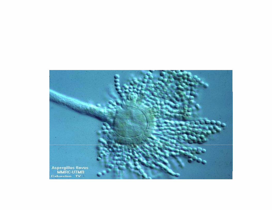
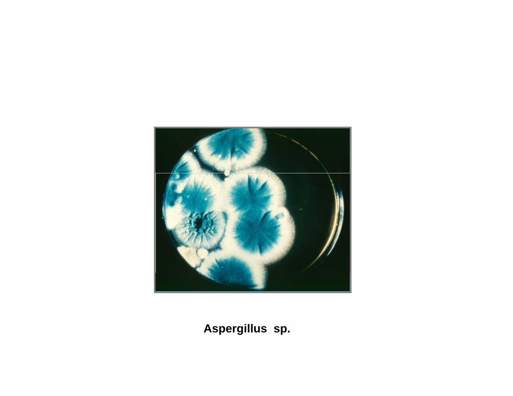
Aspergillus sp.

Columella
Conidiophore
Conidial FungusConidial Fungus
• Reproduces by means of asexual sporesReproduces by means of asexual spores called conidia
• Conidia vary greatly in shape size and• Conidia vary greatly in shape, size and colorM t f th h h ld ld &• Most of the common household molds & mildews are conidial fungi

Asexual spores ( cont)p ( )
• Blastospores– A bud coming off the parent cellA bud coming off the parent cell
• ChlamydosporeChlamydospore– Thick -walled spore– Intercalary lateral terminalIntercalary, lateral , terminal

Chlamydospore of C.albicansy p

ChlamydosporesChlamydospores
• The chlamydospore is a method ofThe chlamydospore is a method of producing a substantial resting spore very quicklyq y
• Nutrient is shunted from adjacent cells into a preferred cell and it swells up, converts p pnutrient materials to oil droplets for efficient storage, then rounds off with a thi k ft h d t ll fthick, often roughened outer wall for protection

Asexual sporesAsexual spores
• SporangiosporesSporangiospores– Hundreds formed within a sac (sporangium)
at the end of an aerial hyphaat the end of an aerial hyphaRhizopus spp.Mucor sppMucor sppAbsidia spp


MYCOSESMYCOSES
• Superficial ( skin, hair, cornea)C t ( D t h t i )• Cutaneous ( Dermatophytosis )
• SubcutaneousSubcutaneous• True systemic (endemic)• Opportunistic

Reproduction of FungiReproduction of Fungi
1. Sexual reproduction --Sexual spores
2. Asexual reproduction--Asexual spores
3 Parasexual reproduction--Genetic 3. Parasexual reproduction Genetic exchange

SEXUAL SporesSEXUAL Spores
1 Zygospore1. Zygospore2. Ascospore3. Basidiospore4 Oospore4. Oospore

Asexual SporesAsexual Spores
1 Arthrospore1. Arthrospore2. Blastospore3 Chl d3. Chlamydospore4. Sporangiosporep g p4. Conidium ( pl conidia ):
macroconidiamacroconidiamicroconidia

Fungi-Taxonomic classificationFungi Taxonomic classification
SEXUAL SPORE CLASSSEXUAL SPORE CLASSZygospore----------ZygomycetesB idi B idi tBasidiospore--------BasidiomycetesAscospore----------AscomycetesNone/Unknown---- Deuteromycetes
(“Fungi Imperfecti”)( Fungi Imperfecti )

Laboratory to diagnosis of fungal infectionLaboratory to diagnosis of fungal infectionLaboratory to diagnosis of fungal infectionLaboratory to diagnosis of fungal infection
• Specimen collection and transportSpecimen collection and transport• Specimen processing• Direct examination• Direct examination• Selection and inoculation of culture media• Identification• Identification
LABORATORY DIAGNOSIS OF MYCOSESMYCOSES
• Direct microscopic examinationDirect microscopic examination
• Culture • Culture
• Serology• Serology

Specimen collection and transportSpecimen collection and transportSpecimen collection and transportSpecimen collection and transport
• must be material from the actual infection sitemust be material from the actual infection site• must be carefully the contamination
b bli h d f h b h f• must be established for the best chance of recovery of causative microorganisms (optimal i )times)

Specimen collection and transport Specimen collection and transport ( con’t)( con’t)
• must be obtained to perform the culture or othermust be obtained to perform the culture or other techniques request (sufficient quantity)
• must be used to ensure optimal recovery of p ymicroorganisms
• obtain cultures before the treatment• the culture container must be properly labeled

Specimen processingSpecimen processing
• specimen should be examined as soon asspecimen should be examined as soon as possible
• direct examination :• direct examination : • KOH mount• Calcofluor white• Calcofluor white• India ink
• culture media• culture media

Selection and inoculation of culture Selection and inoculation of culture mediamedia
• Culture media for recovery of fungi fromCulture media for recovery of fungi from clinical specimens need not be elaborate.
• The recovery rate may be somewhat enhanced• The recovery rate may be somewhat enhanced by using a variety of isolation media, considerations of cost storage incubator spaceconsiderations of cost, storage, incubator space and technologist time.

Initial observations Initial observations in the study of fungus isolatesin the study of fungus isolates
1 Appearance of the growth1.Appearance of the growth2. Rate of growth3 C i i3. Colony pigmentation4. Growth on media containing antifungal
agents5.Dimorphic fungip g

Initial observations Initial observations in the study of fungus isolatesin the study of fungus isolates
1.Appearance of the growth1.Appearance of the growth- surface and reverse surface of colony
were observedwere observed- delicate or hairlike hyphae
2 Rate of growth2. Rate of growth- saprophytes : 3-5 days
di hi f i 10 d- dimorphic fungi : 10 days or more- dermatophytes : 14 days or more

Initial observations Initial observations in the study of fungus isolatesin the study of fungus isolatesin the study of fungus isolatesin the study of fungus isolates
3. Colony pigmentation
4. Growth on media containing antifungal agents
most strains of the dimorphic fungi can grow- most strains of the dimorphic fungi can grow- most strains of the rapidly growing saprobes are
inhibited
5. Dimorphic growth- mold form (the environmental and infective form) ;mold form (the environmental and infective form) ;
ambient or room temperature (22-25 OC)- yeast form (invasive form) ;
near body temperature (30 35 OC)near body temperature (30-35 OC)

Preparation of mounts for studyPreparation of mounts for studyPreparation of mounts for studyPreparation of mounts for study
• The tease mountThe tease mount • Scotch tape preparation
h i lid l h i• The microslide culture technique( slide culture )

Terms useful in Terms useful in the examination of fungithe examination of fungi
• hypha and pseudohyphaehypha and pseudohyphae• mycelium
( i ) h h• septate or aseptate (or coenocytic) hyphae• vegetative mycelium• aerial mycelium• reproductive myceliumreproductive mycelium

Types of asexual sporesTypes of asexual spores

Fungal ReproductionFungal Reproduction
• fungi replicate by mitosis rather than thefungi replicate by mitosis rather than the binary fission employed by bacteria.
• Types of fungal reproduction :• Types of fungal reproduction, : – budding
fi i– fission – hyphae fragmentation – sporulation

Fungal ReproductionFungal Reproduction
• fungi replicate by mitosis rather than thefungi replicate by mitosis rather than the binary fission employed by bacteria.
• Types of fungal reproduction :• Types of fungal reproduction, : – budding
fi i– fission – hyphae fragmentation – sporulation

Sexual & AsexualSexual & AsexualFungal spores come in two varieties: asexual spores and sexual spores – Spores are used extensively to identify fungi.
• Asexual sporesAsexual spores – asexual spores are formed by a single parental fungi and
therefore genetically identical to the parental fungi. Asexual spores come in a variety of types formed by a variety– Asexual spores come in a variety of types formed by a variety of mechanisms including:
• Arthrospores (sliced bread pieces)• Blastospores (buds on a twig)Blastospores (buds on a twig)• Chlamydospores (giant cell with oil)• Conidiospores (fingers)• Sporangiospores (sac)Sporangiospores (sac)


AscomycetesAscomycetes
• Asexual phase-Asexual phase• Conidiospores (Penicillium and Aspergillus)• budding yeastbudding yeast
• Sexual phase (morels lichens )• Sexual phase (morels, lichens )

ZygomycetesZygomycetes
• Asexual phase—Sporangium—bread moldAsexual phase Sporangium bread mold (Rhizopus stolonifer)
• Sexual phase sporgangium shotgun• Sexual phase--- sporgangium ---shotgun fungus (lives on dung) it shoots its sporgangium explosively towards light or flysporgangium explosively towards light or fly pathogen (Entomophthora muscae—--these types of fungi have been used asthese types of fungi have been used as agents for biological control of insects

BasidiomycetesBasidiomycetes
• BasidiosporeBasidiospore• Examples: boletes, puffballs,smuts,
stinkhorns and tooth fungistinkhorns and tooth fungi


Aspergillus spp.

Spora-spora aseksualSpora spora aseksual

SporangiosporeSporangiospore
1.Mucor
2.Rhizopus
3 Absidia3.Absidia
Cunninghamella

1.Mucor
2.Rhizopus
3 Absidia3.Absidia

Cunninghamella


(1).arthrospore (2).chlamydospor(3).phialospore( ) c a ydospo (3) p a ospo e


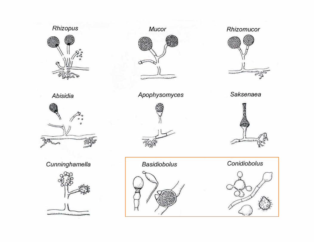
ZygomycosisZygomycosis
• Disease caused by fungi that areDisease caused by fungi that are classifiable as Zygomycetes
Mucormycosis : Order Mucorales– Mucormycosis : Order Mucorales– Entomophthoromycosis : Order
EntomophthoralesEntomophthorales

Zygomycosis (cn’t)Zygomycosis (cn t)
• Presents as a spectrum of diseases, depending on the portal of entry and thedepending on the portal of entry and the predisposing risk factors of the patient:
• Rhinocerebral zygomycosisRhinocerebral zygomycosis• Pulmonary zygomycosis• Gastrointestinal zygomycosisyg y• Cutaneous zygomycosis• Disseminated zygomycosis

Material from the periorbital tissue of a pwoman with poorly controlled diabetes mellitus with facial and periorbital swelling due to zygomycosis (see right picture) is stained with periodic acid Schiff stain (X 560)stained with periodic acid-Schiff stain (X 560). The material demonstrates the classic appearance of irregularly shaped broad hyphae with right-angle branching (arrow).
http://www.emedicine.com/med/topic2438.htm