Mycology – Introduction Student Lab Division of Medical Technology Carol Larson MSEd, MT(ASCP)
35
Mycology – Introduction Student Lab Division of Medical Technology Carol Larson MSEd, MT(ASCP)
-
date post
21-Dec-2015 -
Category
Documents
-
view
216 -
download
0
Transcript of Mycology – Introduction Student Lab Division of Medical Technology Carol Larson MSEd, MT(ASCP)
Mycology –Introduction
Student Lab
Division of Medical Technology
Carol Larson MSEd, MT(ASCP)
Mycoses
• Superficial
• Subcutaneous
• Systemic
• Opportunistic
Characteristics of fungi
• Eukaryotic
• Growth requirements
• Forms– Mold– Yeast
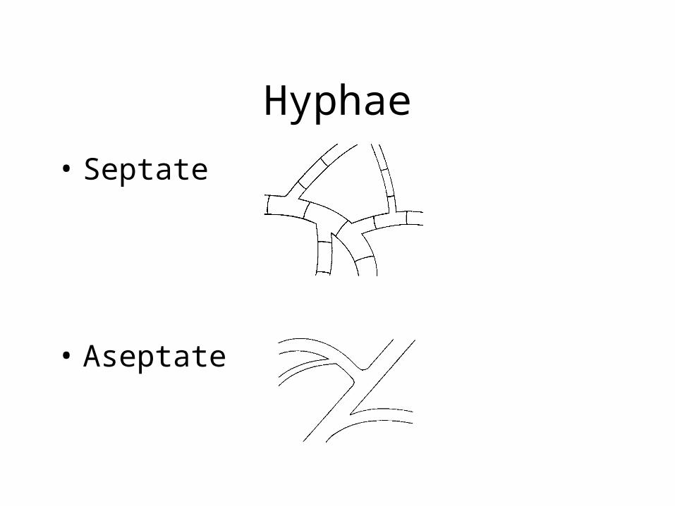
• Aseptate
Hyphae
• Septate
Hyphae
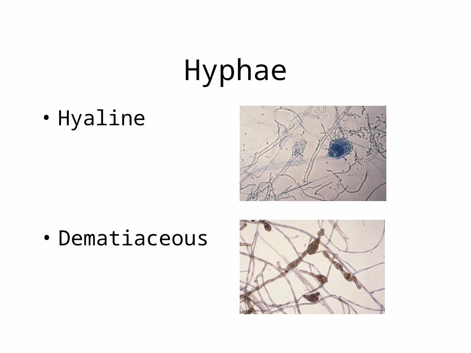
• Hyaline
• Dematiaceous
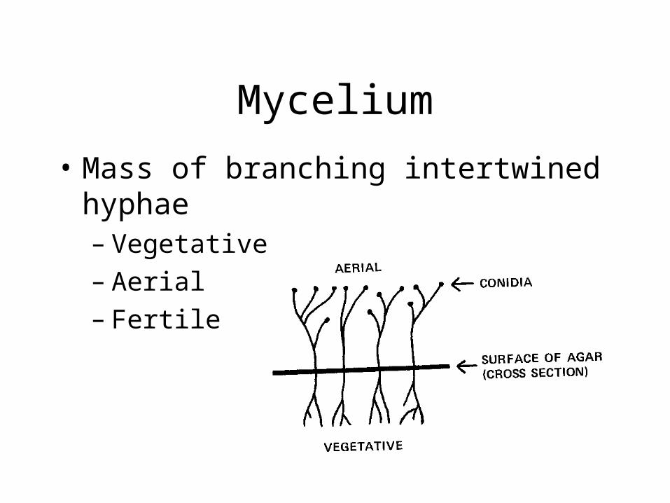
Mycelium
• Mass of branching intertwined hyphae– Vegetative– Aerial– Fertile
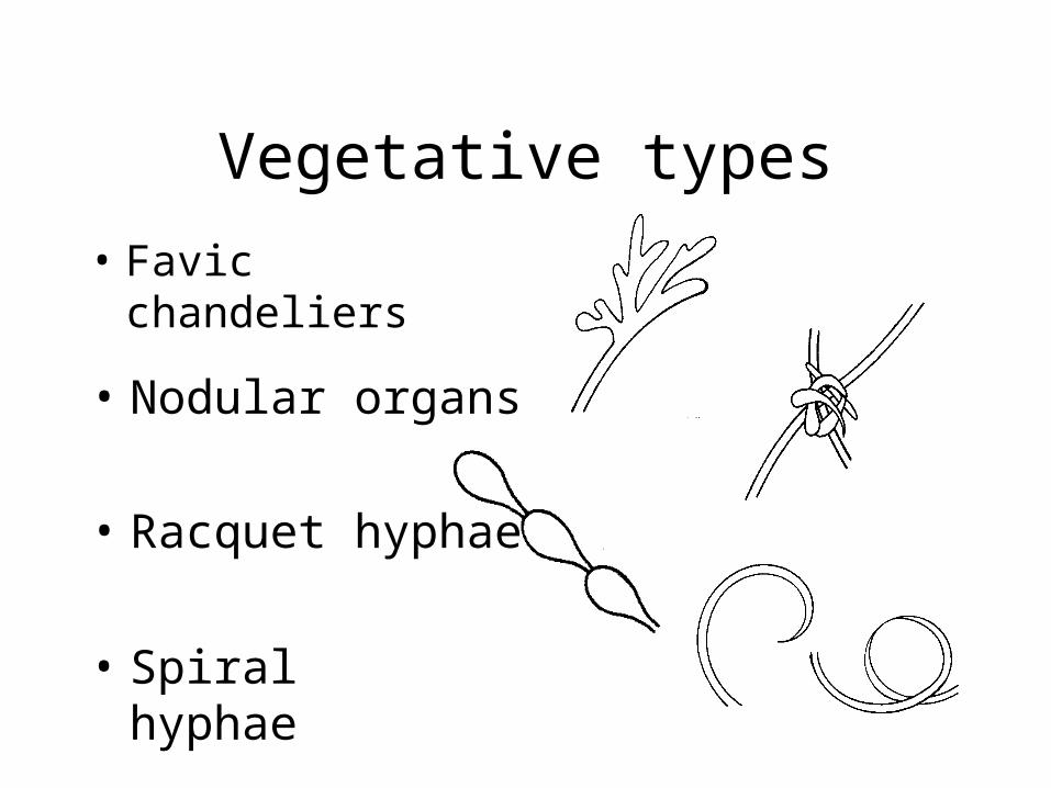
Vegetative types
• Favic chandeliers
• Nodular organs
• Racquet hyphae
• Spiral hyphae
Reproduction
• Identify fungi by:– Morphology of reproductive structures
– Spores from vegetative mycelium or aerial fruiting bodies
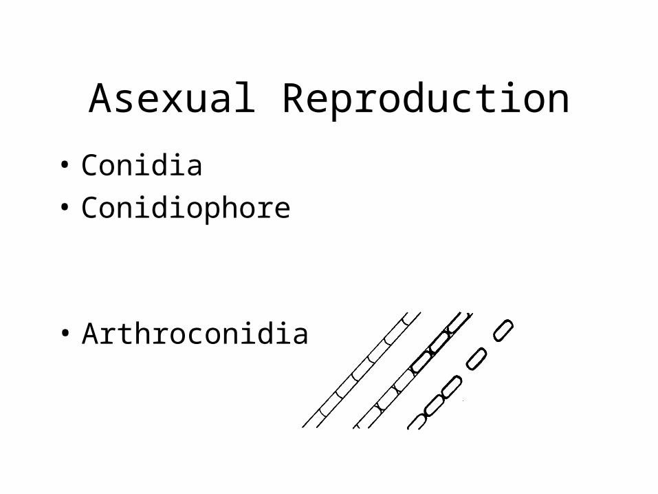
Asexual Reproduction
• Conidia
• Conidiophore
• Arthroconidia
Asexual Reproduction
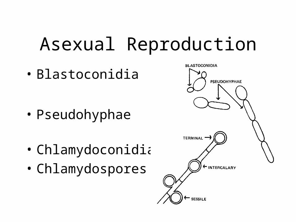
• Blastoconidia
• Pseudohyphae
• Chlamydoconidia
• Chlamydospores
Asexual Reproduction
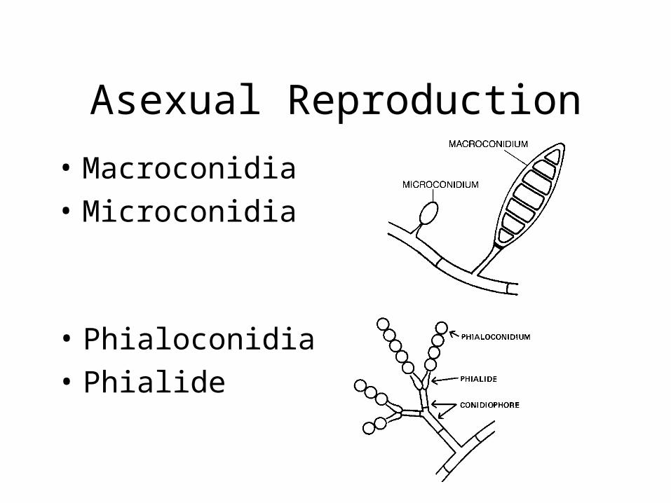
• Macroconidia
• Microconidia
• Phialoconidia
• Phialide
Asexual Reproduction
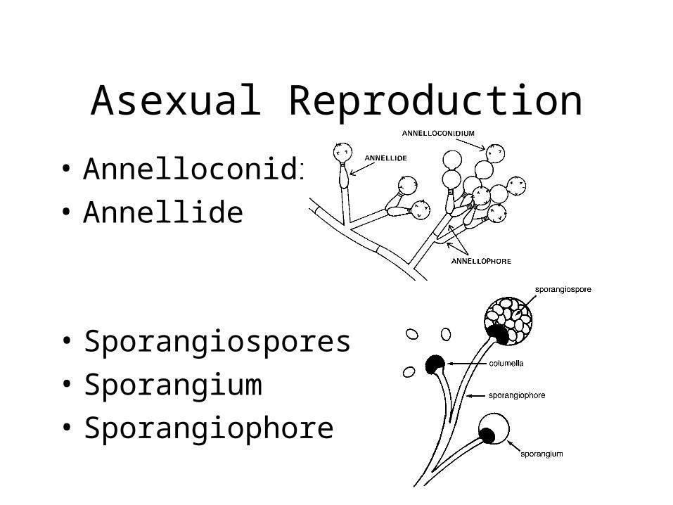
• Annelloconidia
• Annellide
• Sporangiospores
• Sporangium
• Sporangiophore
Sexual Reproduction
• Perfect Fungi – has a sexual stage
• Fungi Imperfecti – no know sexual stage
• Spores
Sexual Reproduction
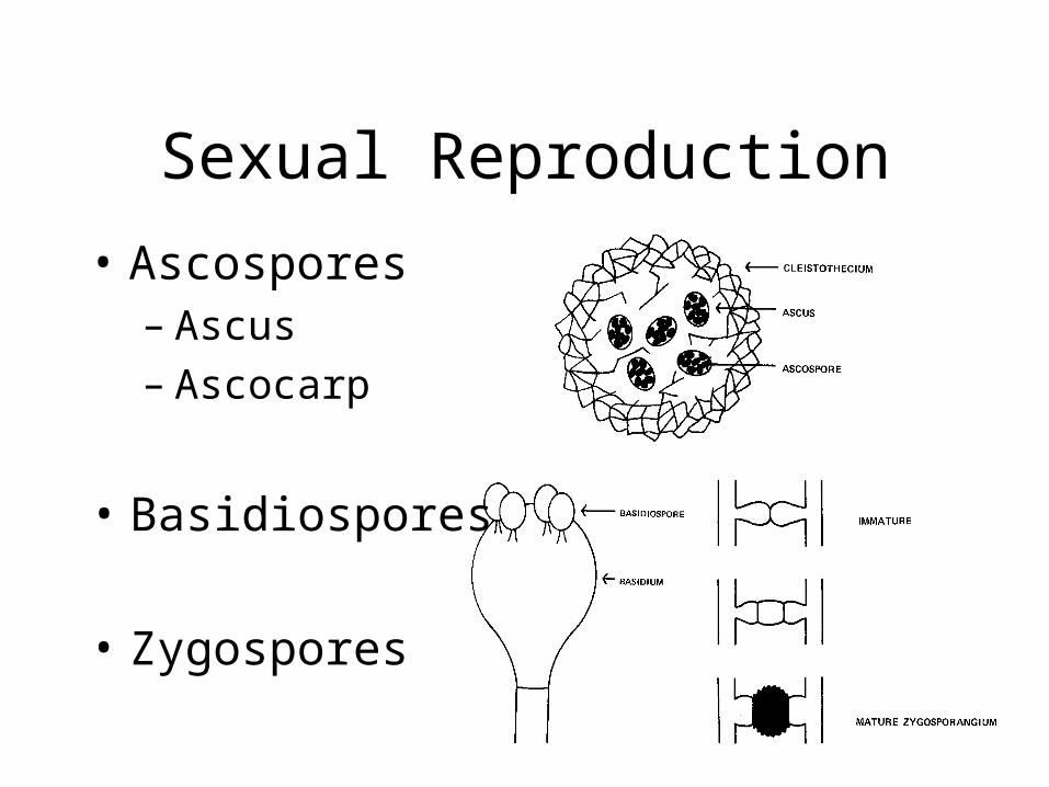
• Ascospores– Ascus– Ascocarp
• Basidiospores
• Zygospores
In review …
• Mycoses – fungal diseases
• Characteristics of fungi– Growth requirements– Forms (mold, yeast)– Structures– Reproduction
• Asexual• Sexual
Fungal Culture Process
• Specimen collection and transportation
• Direct examination of specimen
• Selection and inoculation of media
• Evaluation of fungal growth
• Serological testing
• Antifungal susceptibility testing
Specimen Collection
• Specimen types
• Collect from area most likely infected
• Use sterile technique
• Keep specimen moist
• Label container properly
• Transport right away
• Process right away
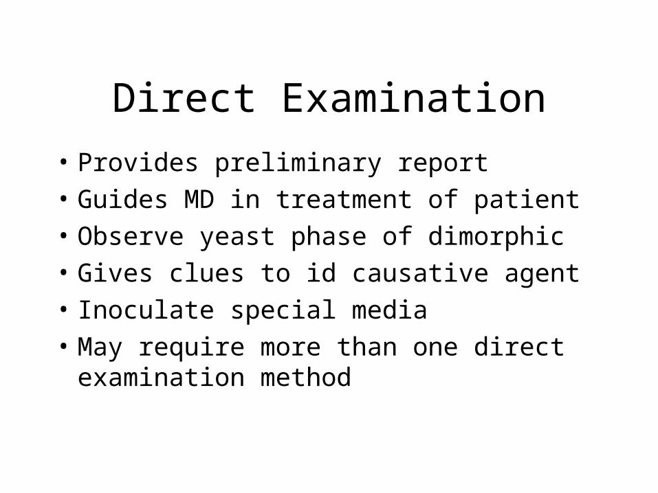
Direct Examination
• Provides preliminary report
• Guides MD in treatment of patient
• Observe yeast phase of dimorphic
• Gives clues to id causative agent
• Inoculate special media
• May require more than one direct examination method
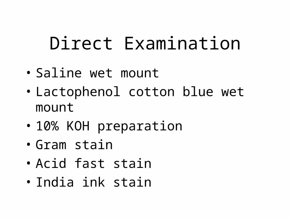
Direct Examination
• Saline wet mount
• Lactophenol cotton blue wet mount
• 10% KOH preparation
• Gram stain
• Acid fast stain
• India ink stain
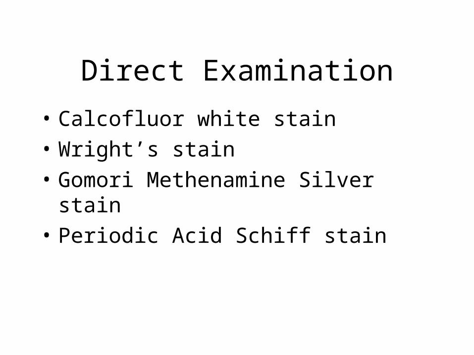
Direct Examination
• Calcofluor white stain
• Wright’s stain
• Gomori Methenamine Silver stain
• Periodic Acid Schiff stain
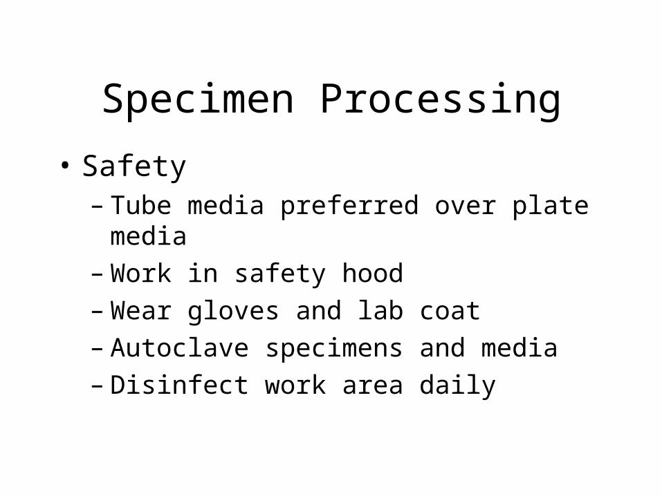
Specimen Processing
• Safety– Tube media preferred over plate media– Work in safety hood– Wear gloves and lab coat– Autoclave specimens and media– Disinfect work area daily
Specimen Processing
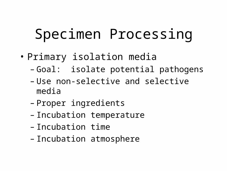
• Primary isolation media– Goal: isolate potential pathogens– Use non-selective and selective media– Proper ingredients– Incubation temperature– Incubation time– Incubation atmosphere
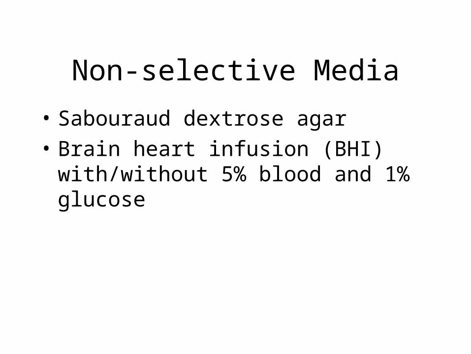
Non-selective Media
• Sabouraud dextrose agar
• Brain heart infusion (BHI) with/without 5% blood and 1% glucose
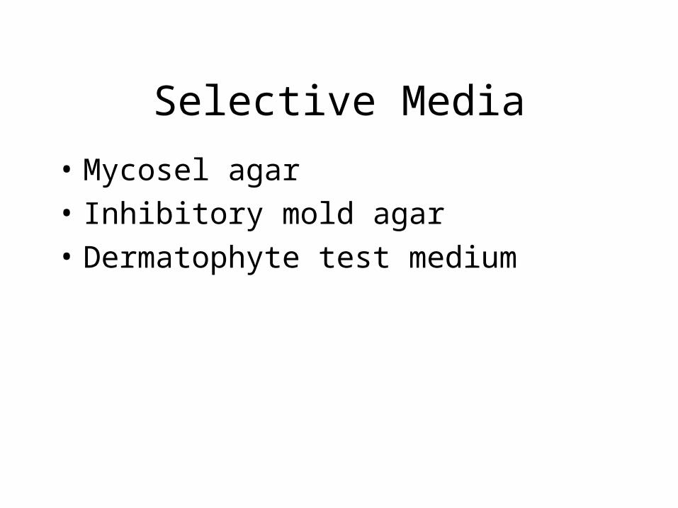
Selective Media
• Mycosel agar
• Inhibitory mold agar
• Dermatophyte test medium
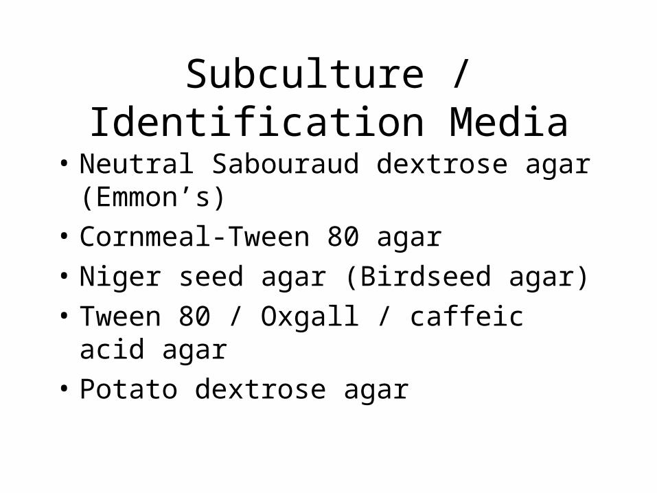
Subculture / Identification Media
• Neutral Sabouraud dextrose agar (Emmon’s)
• Cornmeal-Tween 80 agar
• Niger seed agar (Birdseed agar)
• Tween 80 / Oxgall / caffeic acid agar
• Potato dextrose agar
Examination of Culture Growth
• Potential pathogens– Slow growers– Growth on Mycosel– Color: dull buff, brown, mousy gray– Dimorphic
Examination of Culture Growth
• Growth rate– Rapid growers: 1-5 days– Intermediate growers: 6-10 days– Slow growers: >10 days
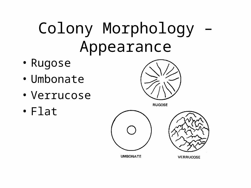
Colony Morphology –Appearance
• Rugose
• Umbonate
• Verrucose
• Flat
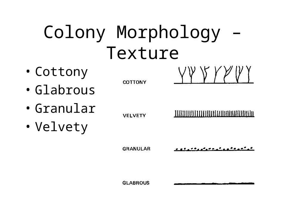
Colony Morphology –Texture
• Cottony
• Glabrous
• Granular
• Velvety
Colony Morphology –Pigmentation
• Surface
• Reverse
Microscopic Morphology
• Definitive means of identification
• Evaluate:– Shape– Method of production– Arrangement of conidia/spores– Size and color of hyphae
Microscopic Techniques
• Tease mount
• Scotch tape preparation
• Slide culture
Serological Diagnosis
• Immunodiffusion
• Complement fixation
• EIA
• Latex agglutination
Antifungal Susceptibility
• Determine appropriateness– Standardization of testing– Methods– Predictability in vivo
• Antifungal agents
In Summary …
• Specimen collection and transport
• Specimen processing and culture
• Direct examination of specimen
• Examination of culture
• Serological testing
• Antifungal susceptibility