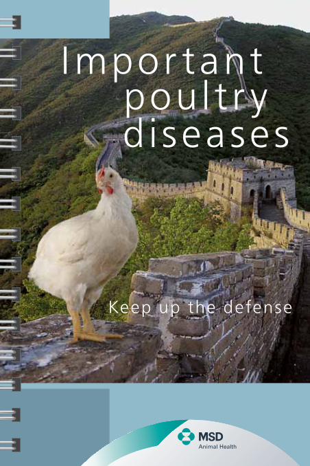
Important poultry diseases
62
Important poultry diseases Keep up the defense
Transcript of Important poultry diseases
Impor tantpoultr ydiseases
Keep up the defense
3 2
IMPORTANT POULTRY DISEASES
5 4
Infectious Respiratory diseasesAspergillosis 10
Avain Influenza 12
Avian Metapneumovirus Rhinotracheitis/TRT 14
Infectious Bronchitis 16
Infectious Coryza 20
Infectious Laryngotracheitis 22
Mycoplasma Gallisepticum “CRD” 27
Mycoplasma Synoviae 30
Newcastle Disease 32
Neoplastic DiseasesLymphoid Leucosis 36
Marek’s disease 38
Avian Adeno virus DiseasesEgg Drop Syndrome ‘76 44
Inclusion Body Hepatitis 48
Miscellaneous Viral DiseasesAvian Encephalomyelitis 54
Chicken Anaemia Virus 58
Fowl Pox 60
Infectious Bursal Disease 62
Malabsorbtion Syndrome/ Runting Stunting Syndrome 66
Reo virus infections 68
Contents Contents
Miscellaneous Bacterial DiseasesColibacilosis 72
Fowl Cholera 76
Infectious Synovitis 78
Necrotic Enteritis 80
Ornithobacterium Rhinotracheale (OR) 84
Pullorum disease/Fowl Typhoid 86
Parasitic DiseasesBlackhead 90
Coccidiosis 92
Red Mite 96
Worms 98
Deficiency DiseasesRiboflavin 102
VitaminD3 103
VitaminE 104
Food Safety in PoultryIntroduction 106
Salmonellosis 106
Campylobacter 111
Diagnostics and Sampling 115

7 6
Foreword Foreword
Foreword
The first edition of Intervet’s “Important Poultry Diseases” was in
1972 and still it is one of our most wanted publications.
An easy to handle and practical booklet for basic understanding of
the most important poultry diseases for people working in poultry
management.
This is the fifth updated version printed in 2013 with new
additional information based on the new developments in Poultry
Diseases and progress of the MSD Animal Health Poultry Research
in developing additional new products.
MSD Animal Health Research is committed to co-operate with the
poultry industry worldwide to develop and support solutions to
control poultry diseases. MSD Animal Health is more than vaccines
alone.
For detailed information of any of our products please contact the
local MSD Animal Health representative or Intervet International
BV– part of MSD Animal Health.
Intervet International BV
Boxmeer- Holland, P.O. Box 31
5830 AA Boxmeer, The Netherlands
Phone +31 485587600 - Fax +31 485577333
E-mail [email protected] - www.merck.com
9 8
Infectious Respiratory diseases Infectious Respiratory diseases
Infectious Respiratory diseases
11 10
Infectious Respiratory diseases Aspergillosis Infectious Respiratory diseases Aspergillosis
Treatment and controlThere no specific treatment for infected birds. The best is to remove and destroy affected birds.Strict hygiene in breeder (hatching eggs) and hatchery management is necessary. Choice and quality of litter is also important to prevent that spore bearing wood shavings or straw are used.Hatchery control with anti-fungal disinfectant may be critical to cleaning and disinfection procedures to control fungus infection.
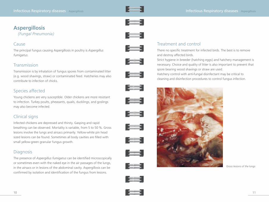
Gross lesions of the lungs
Aspergillosis (Fungal Pneumonia)
CauseThe principal fungus causing Aspergillosis in poultry is Aspergillus fumigatus.
TransmissionTransmission is by inhalation of fungus spores from contaminated litter (e.g. wood shavings, straw) or contaminated feed. Hatcheries may also contribute to infection of chicks.
Species affectedYoung chickens are very susceptible. Older chickens are more resistant to infection. Turkey poults, pheasants, quails, ducklings, and goslings may also become infected.
Clinical signsInfected chickens are depressed and thirsty. Gasping and rapid breathing can be observed. Mortality is variable, from 5 to 50 %. Gross lesions involve the lungs and airsacs primarily. Yellow-white pin head sized lesions can be found. Sometimes all body cavities are filled with small yellow-green granular fungus growth.
DiagnosisThe presence of Aspergillus fumigatus can be identified microscopically or sometimes even with the naked eye in the air passages of the lungs, in the airsacs or in lesions of the abdominal cavity. Aspergillosis can be confirmed by isolation and identification of the fungus from lesions.
13 12
Infectious Respiratory diseases Avian Influenza Infectious Respiratory diseases Avian Influenza
Avian Influenza (AI)
CauseAvian Influenza is caused by an Orthomyxovirus; there are several serotypes.Currently we know there are 16 H- types and 9 N-types and they can show up in all kinds of combinations. For poultry the most important ones are H5, H7 and H9. Pathogenicity varies with the strains HPAI and LPAI (high or low pathogenic AI).
TransmissionAI virus is excreted from nares, mouth, conjunctiva and cloaca. Airborne virus particles from the respiratory tract, droppings, and people carrying virus on their clothing and equipment are the main routes of transmission. Migratory water fowl and other wild birds infected with AI virus may be a source of infection.
Species affectedAvian Influenza viruses have been shown to naturally infect a wide variety of wild and domestic birds. In poultry production main problems are in chickens, turkeys and ducks.
Clinical signsClinical signs will vary, depending on the pathogenicity (HPAI and LPAI) of AI virus involved and other factors as host species, sex, concurrent infections, acquired immunity and environmental factors.LPAI shows generally mild symptoms: respiratory coughing sneezing, wet eyes, nasal discharge depression, lethargy limited reduction of feed intake and limited drop in egg production; low mortality rate.HPAI shows fast onset with increased mortality even before clinical signs are seen, depression, drop in feed and water intake, severe drop in egg production and mortality can vary between 50-90%.
DiagnosisClinical signs are indicative for AI; final confirmation by laboratory testing:- Direct detection of AI proteins or Nucleic acids(RNA) using PCR.- Virus isolation from infected organs, tracheal or cloacal swabs.- Serology from blood samples after infection and for routine
monitoring showing specific AI antibodies.
TreatmentThere is no treatment for Avian Influenza. Antibiotics will help to control secondary bacterial infections.
Prevention and controlIn many countries AI is a notifiable disease with specific local regulations on its control.In AI free areas the disease(LPAI and HPAI) is controlled by monitoring and stamping out.In case of LPAI infected areas countries can decide to allow vaccination only for LPAI.In case of endemic HPAI and/or LPAI vaccination might be allowed.
Vaccination is generally done with inactivated AI vaccines based on the strain H-type causing the outbreaks.
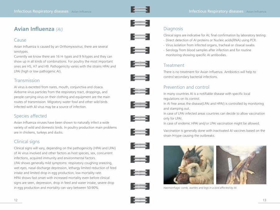
Haemorrhagic comb, wattles and legs in a bird a!ected by AI.
15 14
Infectious Respiratory diseases Avian Metapneumovirus Infectious Respiratory diseases Avian Metapneumovirus
Avian Metapneumovirus (aMPV)
“Rhinotracheitis” (RT) Turkey Rhinotracheitis (TRT)“Swollen Head Syndrome (SHS)“
CauseAvian Metapneumovirus, belongs to the subfamily Pneumovirus from the family Paramyxoviridae. Within the aMPV 4 subtypes (A-B-C-D) are identified.
TransmissionThe virus spreads horizontally via direct contact from bird to bird and via contaminated personnel, water and equipment.
Species affectedTurkeys and chickens.
Clinical signs and lesionsIn both turkeys and chickens.In young birds the respiratory clinical signs; snicking, rales, sneezing, nasal discharge , foamy conjunctivitis, swollen infraorbital and periorbital sinus “swollen head” are most prominent; when birds become older head shaking and coughing can be added. In adult laying hens there may a drop in egg production up to 70% with an increased incidence of poor shell quality.Morbidity is general high (up to 100%); mortality varies from 0.5% up to 50% depending on secondary infections (such as IBV, E.coli) and farm climate conditions. At post mortem the lesions vary. In an uncomplicated infection: rhinitis, tracheitis and sinusitis will dominate , in case of Swollen Head Syndrome, oedema at the head often in combination with purulent or caseous subcutaneous exudate. In complicated cases with E.coli polyserositis including fibrinous exudate in abdominal cavity can be found.
DiagnosisClinical signs can give a good indication for aMPV but need to be confirmed by laboratory testing. Identification of virus by isolation or PCR from nasal secretions and/or tracheal or sinus scraping. After infection several serological methods (VN, Elisa, IFT) can be used to detect antibodies. Be aware that vaccines also induce antibodies.
TreatmentThere is no treatment for aMPV infections. Treatment with antibiotics can be given to control secondary bacterial infections.
PreventionVaccination is the most effective prevention method. Over the years live and inactivated vaccines have been developed and are very efficacious in controlling infections. Short living birds will only be vaccinated with a live vaccine(s). Long living birds are advised to be primed with live- and boosted with inactivated vaccines. In this way the birds will have good long lasting local and systemic protection.
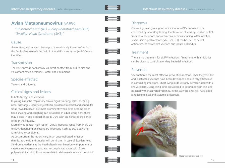
Nasal discharge; wet eye
17 16
Infectious Respiratory diseases Infectious Bronchitis Infectious Respiratory diseases Infectious Bronchitis
Infectious Bronchitis (IB)
CauseInfectious Bronchitis(IB) is present worldwide, it is a highly contagious, acute, and economically important disease. IB is caused by an Avian Coronavirus. In the field, several different IB serotypes have been identified including the classic Massachusetts type and a number of variants such as IB 4/91, QX, Arkansas and Connecticut.
TransmissionThe virus is transmitted rapidly from bird to bird through the airborne route. The virus can also be transmitted via the air between chicken houses and even from farm to farm.The incubation period is only 1-3 days.
Species affectedChickens are the primary poultry species that is susceptible to IB-virus, but quail and pheasants can be affected.Recent discovery of IB virus in other species without clinical signs indicates that other species may act as vectors.
Clinical signsIn young chickens the respiratory form appears with gasping, sneezing, tracheal rales and nasal discharge. Generally chicks are depressed and show reduced feed consumption. Mortality in general is low unless infection gets complicated with secondary bacterial infections (like E.coli).In case of a nephropathogenic type of IB virus generally birds, after initial respiratory signs, are more depressed, show wet droppings resulting in wet litter, increased water intake and increased mortality.
In adult “laying” birds (layers and breeders) after initial respiratory signs the affected flocks show a drop in egg production and a loss of egg quality (shell deformation and internal egg changes) resulting in more second class eggs, affecting the hatchability rate of fertile eggs and day-old chick quality.A specific condition, called “false layers” is related to the QX type of IB; usually flocks do not peak in egg production and many birds show a “penguin-like posture” .
Post mortem lesionsIn young chicks a yellow cheesy plug at the tracheal bifurcation is indicative of IB infection. In case of nephropathogenic infections pale and swollen kidneys and distended ureters with urates are found In older birds mucus and redness in the trachea, exudate in the air sacs, and various changes in the oviduct depending on the time and severity of infection. In case of “false layers” permanent lesions in the oviduct make egg production impossible. The oviduct will be blocked and filled with fluid (cysts) or never developed into an active oviduct.
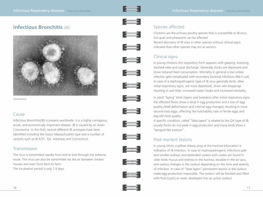
Coronavirus
19 18
Infectious Respiratory diseases Infectious Bronchitis Infectious Respiratory diseases Infectious Bronchitis
DiagnosisClinical signs and post mortem lesions in a flock followed by laboratory confirmation based on virus isolation and identification with PCR.Serology based on paired blood samples using HI, Elisa or VN tests.
TreatmentThere is no treatment for IB.Antibiotics are used to control secondary bacterial infections.
PreventionVaccination with strain specific or cross protective live vaccines, and for layers and breeders the addition of inactivated vaccines at point of lay to induce long lasting systemic immunity.
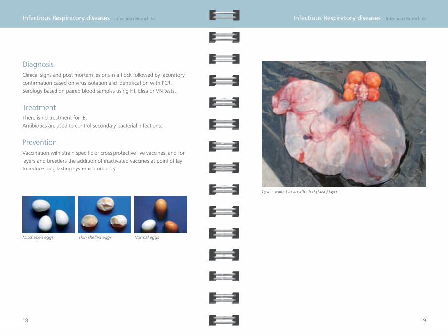
Misshapen eggs Thin shelled eggs Normal eggs
Cystic oviduct in an a!ected (false) layer
21 20
Infectious Respiratory diseases Infectious Coryza Infectious Respiratory diseases Infectious Coryza
Infectious Coryza
CauseThis is a bacterial disease caused by Avibacterium paragallinarum, (in the past known as Haemophilus paragallinarum). There are 3 common serotypes, representing different immunotypes: A, B, C.
TransmissionThe disease spreads from bird to bird and flock to flock by contact and airborne infected dust particles and via the drinking water. Spread by equipment and personnel have also been reported. The incubation period varies from 1 to 3 days.
Species affectedThe chicken is the natural host for Avibacterium paragallinarum. All ages are susceptible, but the disease is usually less severe in juvenile birds.
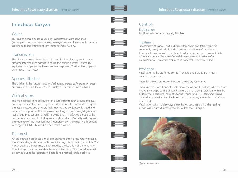
Clinical signsThe main clinical signs are due to an acute inflammation around the eyes and upper respiratory tract. Signs include a serous to mucoid discharge in the nasal passage and sinuses, facial edema and conjunctivitis. Feed and water consumption will be decreased resulting in loss of weight gain and loss of egg production (10-40%) in laying birds. In affected breeders, the hatchability and day-old chick quality might decline. Mortality will vary with the virulence of the infection, but is generally low. Complicating infections with eg IB, ILT, MG, MS and ND can make it worse.
DiagnosisA field infection produces similar symptoms to chronic respiratory disease, therefore a diagnosis based only on clinical signs is difficult to establish. The most certain diagnosis may be obtained by the isolation of the organism from the sinus or airsac exudate from affected birds. This procedure must be carried out in the laboratory. There is no practical serological test.
Control:EradicationEradication is not economically feasible.
TreatmentTreatment with various antibiotics (erythromycin and tetracycline are commonly used) will alleviate the severity and course of the disease. Relapse often occurs after treatment is discontinued and recovered birds will remain carriers. Because of noted drug resistance of Avibacterium paragallinarum, an antimicrobial sensitivity test is recommended.
PreventionVaccination is the preferred control method and is standard in most endemic Coryza areas.
There is no cross protection between the serotypes A, B, C.
There is cross protection within the serotypes A and C, but recent outbreaks due to B serotype strains showed there is partial cross protection within the B- serotype. Therefore, besides vaccines made of A, B, C serotype strains, a broader multivalent vaccine based on serotypes A, B, B-variant and C was developed. Vaccination with multi-serotype inactivated vaccines during the rearing period will reduce clinical signs/control Infectious Coryza.
Typical facial edema
23 22
Infectious Respiratory diseases Infectious Laryngotracheitis Infectious Respiratory diseases Infectious Laryngotracheitis
Infectious Laryngotracheitis (ILT)
CauseILT is caused by a Herpesvirus, only one serotype is known.
TransmissionThe natural entry of ILT is via the upper respiratory tract and ocular route. Field spread occurs via direct contact from bird to bird and/or transmission by contaminated people or equipment (visitors, shoes, clothing, egg boxes, transport crates).Incubation period varies from 4 -12 days.
Species affectedChickens are the primary natural host but other species (pheasants) can also be affected.
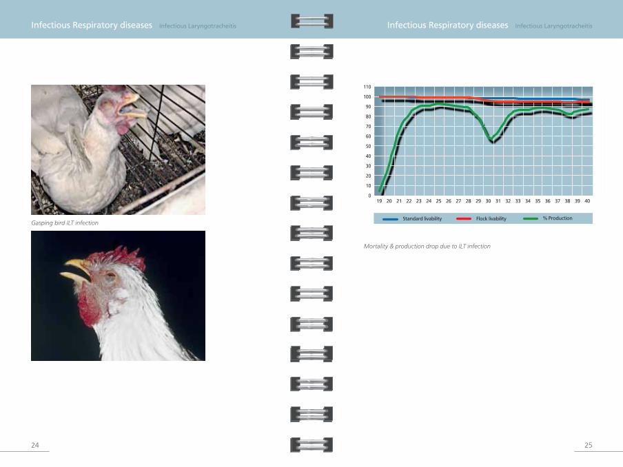
Clinical signsAn acute respiratory disease with nasal discharge and moist rales followed by gasping, marked respiratory distress and expectoration of blood-stained mucus in laying birds. Egg production can drop 10 -50% but will return to normal after 3-4 weeks. Mortality can vary from 5 -70%.Spread through a chicken house is slower compared to IB and ND.
Post mortem lesionsLesion are found throughout the respiratory tract but most pronounced in the larynx and trachea.Depending on the severity of the infection you can find tracheitis with haemorrhagic and/or diphteric changes.
DiagnosisClinical picture with birds showing respiratory distress and expectoration of bloody mucus are indicative for ILT. Laboratory confirmation with: histopathology showing intranuclear inclusion bodies in tracheal epithelial cells, virus isolation from tracheal swabs on embryonated chicken eggs, virus detection with PCR or IFT on tracheal samples.Detecting antibodies from blood samples after infection.
TreatmentThere is no treatment for ILT; emergency vaccination in the early stage of an infected flock may reduce the spread and limit the outbreak.
Prevention and controlIn many countries vaccination is the preferred control method. Though in some countries it is not allowed to vaccinate or only under restriction.
The existing live conventional CEO ILT vaccines are effective in controlling clinical problems but have the risk of spreading and reversion to virulence after multiple passage through chickens. Recent outbreaks show often the relation to the live vaccine strains used in an area. Therefore, the new generation of recombinant vector vaccines are more suitable in the control and prevention of ILT. Recombinant vaccines based on HVT-vector carrying inserts of important immunogenic ILT proteins show good efficacy and do not spread and cannot revert to virulence because there is not a full ILT virus involved.
25 24
Infectious Respiratory diseases Infectious Laryngotracheitis Infectious Respiratory diseases Infectious Laryngotracheitis
Gasping bird ILT infectionStandard livability Flock livability % Production
19 20 21 22 23 24 25 26 27 28 29 30 31 32 33 34 35 36 37 38 39 40
110
100
90
80
70
60
50
40
30
20
10
0
Mortality & production drop due to ILT infection
27 26
Infectious Respiratory diseases
Mycoplasma gallicepticum (CRD)
CauseThe underlying cause of CRD is Mycoplasma gallisepticum (MG). The condition is frequently triggered by respiratory viruses such as ND and IB and subsequently complicated by bacterial invasion. The main agents involved in the infection are Mycoplasma gallisepticum and E. coli. Stress caused by moving the birds, by debeaking, other operations/handlings or other unfavorable conditions e.g. cold or bad ventilation, make the birds more susceptible.
TransmissionThe main problem is that parent birds infected with Mycoplasma gallisepticum can transmit the organism through the egg to their offspring (vertical transmission). In addition, infection can occur by contact or by airborne dust or droplets (horizontal transmission). The incubation period varies from 4 days to 3 weeks.
Species affectedChickens and Turkeys.
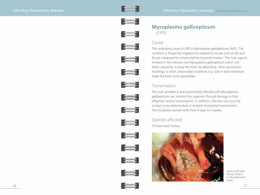
Airsacculitis and foamy content in the abdominal cavity
Infectious Respiratory diseases Mycoplasma gallicepticum
29 28
Infectious Respiratory diseases Mycoplasma gallicepticum Infectious Respiratory diseases Mycoplasma gallicepticum
Clinical signsYoung chickens (broilers or layer pullets) will show respiratory distress. The birds frequently show lack of appetite, decreased weight gain and increased feed conversion ratios.
In adult birds the most common signs are sneezing and general respiratory distress. In laying birds a drop in egg production between 20-30 % can occur. In breeders hatchability can be affected and day-old chick quality produced from hatching eggs coming from infected flocks will be reduced.
CRD does not normally cause an alarming number of deaths. The effect is more of a chronic nature causing reduced weight gain and higher feed conversion ratios in broilers and lower egg production in breeders and layers. In this way the overall economic losses can be very high.
Internal lesionsA reddish inflamed trachea and/or frothy, cheesy exudate in the airsacs, especially in complicated cases (e.g. with secondary E. coli infections) are observed. In mild MG infections the only lesion might be slight mucus in the trachea and a cloudy or light froth in the airsacs.
DiagnosisDiagnosis of MG infection can be made based on clinical signs and post mortem lesions followed by confirmation in the laboratory using blood (serum) samples for serology or organs swabs for identification by PCR or mycoplasma isolation.
Differential diagnosisRespiratory virus infection (Newcastle disease or infectious bronchitis) with secondary infection (E. coli, etc.) can give similar lesions.
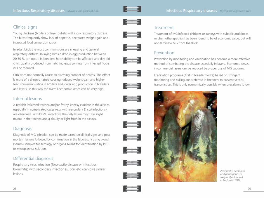
Pericarditis, peritonitis and perihepatitis is frequently observed in birds with CRD
TreatmentTreatment of MG-infected chickens or turkeys with suitable antibiotics or chemotherapeutics has been found to be of economic value, but will not eliminate MG from the flock.
PreventionPrevention by monitoring and vaccination has become a more effective method of combating the disease especially in layers. Economic losses in commercial layers can be reduced by proper use of MG vaccines.
Eradication programs (first in breeder flocks) based on stringent monitoring and culling are preferred in breeders to prevent vertical transmission. This is only economically possible when prevalence is low.
31 30
Infectious Respiratory diseases Mycoplasma Synoviae Infectious Respiratory diseases Mycoplasma Synoviae
Mycoplasma Synoviae
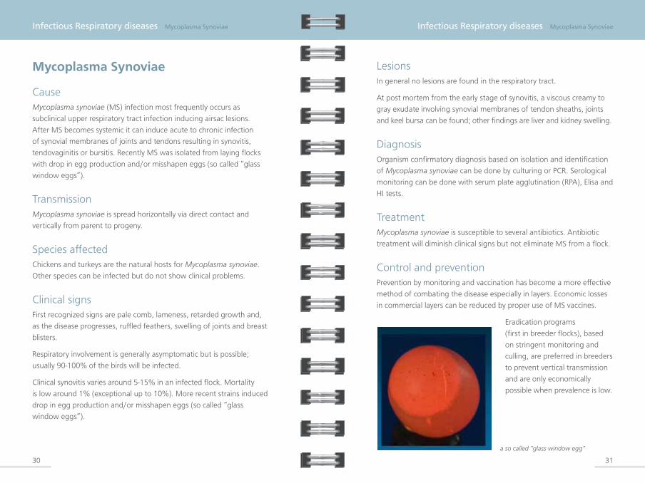
CauseMycoplasma synoviae (MS) infection most frequently occurs as subclinical upper respiratory tract infection inducing airsac lesions. After MS becomes systemic it can induce acute to chronic infection of synovial membranes of joints and tendons resulting in synovitis, tendovaginitis or bursitis. Recently MS was isolated from laying flocks with drop in egg production and/or misshapen eggs (so called “glass window eggs”).
TransmissionMycoplasma synoviae is spread horizontally via direct contact and vertically from parent to progeny.
Species affectedChickens and turkeys are the natural hosts for Mycoplasma synoviae.Other species can be infected but do not show clinical problems.
Clinical signsFirst recognized signs are pale comb, lameness, retarded growth and, as the disease progresses, ruffled feathers, swelling of joints and breast blisters.
Respiratory involvement is generally asymptomatic but is possible; usually 90-100% of the birds will be infected.
Clinical synovitis varies around 5-15% in an infected flock. Mortality is low around 1% (exceptional up to 10%). More recent strains induced drop in egg production and/or misshapen eggs (so called “glass window eggs”).
LesionsIn general no lesions are found in the respiratory tract.
At post mortem from the early stage of synovitis, a viscous creamy to gray exudate involving synovial membranes of tendon sheaths, joints and keel bursa can be found; other findings are liver and kidney swelling.
DiagnosisOrganism confirmatory diagnosis based on isolation and identification of Mycoplasma synoviae can be done by culturing or PCR. Serological monitoring can be done with serum plate agglutination (RPA), Elisa and HI tests.
TreatmentMycoplasma synoviae is susceptible to several antibiotics. Antibiotic treatment will diminish clinical signs but not eliminate MS from a flock.
Control and preventionPrevention by monitoring and vaccination has become a more effective method of combating the disease especially in layers. Economic losses in commercial layers can be reduced by proper use of MS vaccines.
Eradication programs (first in breeder flocks), based on stringent monitoring and culling, are preferred in breeders to prevent vertical transmission and are only economically possible when prevalence is low.
a so called “glass window egg”
33 32
Infectious Respiratory diseases Newcastle Disease Infectious Respiratory diseases Newcastle Disease
Newcastle Disease (ND)
CauseNewcastle disease is caused by a Paramyxovirus (APMV-1). Only one serotype of ND is known. ND virus has mild strains (lentogenic), medium strength strains (mesogenic), and virulent strains (velogenic). The strains used for live vaccines are mainly lentogenic.
TransmissionNewcastle disease virus is highly contagious through infected droppings and respiratory discharge between birds. Spread between farms is by infected equipment, trucks, personnel, wild birds or air. The incubation period is variable but usually about 3 to 6 days.
Species affectedChickens and turkeys.
Clinical signsHighly pathogenic strains (velogenic) of ND cause high mortality with depression and death within 3 to 5 days. Affected chickens do not always exhibit respiratory or nervous signs. Mesogenic strains cause typical signs of respiratory distress.
Labored breathing with wheezing and gurgling, accompanied by nervous signs, such as paralysis or twisted necks (torticollis) are the main signs. Drop in egg production 30 to 50 % or more, returning to normal levels in about 2-3 weeks is observed. Besides also egg shell quality will be affected (thin, loose color). In well-vaccinated chicken flocks clinical signs may be difficult to find.
Intestinal lesionsInflamed tracheas, pneumonia, and/or froth in the airsacs are the main lesions. Haemorrhagic lesions are observed in the proventriculus and the intestines.
DiagnosisClinical signs followed by laboratory confirmation. Be aware that other respiratory infections like IB, ILT and AI can give similar signs. Confirmation can be obtained with virus isolation and identification from tracheal or cloacal swabs, or PCR, Serology testing with HI or Elisa measures the antibody response after infection. Be aware that vaccination also induces antibody response.
TreatmentThere is no specific treatment for ND; antibiotic treatment of secondary bacterial infections (eg E.coli) will reduce the losses.
35 34
Infectious Respiratory diseases Newcastle Disease
Neurotropic form of ND(torticollis)
Prevention and controlVaccination has proven to be a reliable control method. But ND is a notifiable disease, and in many countries the control is based on a combination of obliged vaccination and stamping out in case of outbreaks. A wide range of live and inactivated vaccines are used in vaccination programs to prevent ND. “The new generation” of live recombinant HVT-vector vaccines give the opportunity for early hatchery vaccination and can be used to replace conventional ND vaccination, eliminating vaccination reactions and inducing life long protection.
Haemorrhagic proventriculus
Neoplastic Diseases
Neoplastic Diseases
37 36
Neoplastic Diseases Lymphoid Leucosis Neoplastic Diseases Lymphoid Leucosis
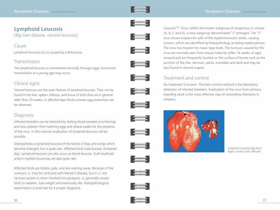
Lymphoid Leucosis (Big liver disease, visceral leucosis)
CauseLymphoid leucosis (LL) is caused by a Retrovirus.
TransmissionThe lymphoid leucosis is transmitted vertically through eggs; horizontal transmission at a young age may occur.
Clinical signsVisceral tumours are the main feature of lymphoid leucosis. They can be found in the liver, spleen, kidneys, and bursa of birds that are in general older than 25 weeks. In affected layer flocks a lower egg production can be observed.
DiagnosisInfected breeders can be detected by testing blood samples (monitoring) and virus isolation from hatching eggs and cloacal swabs for the presence of the virus. In this manner eradication of lymphoid leucosis will be possible.
Osteopetrosis is lymphoid leucosis of the bones of legs and wings which become enlarged, but is quite rare. Affected birds have bowed, thickened legs. Lymphoid leucosis can also occur as blood leucosis. Such erythroid and/or myeloid leucemias are also quite rare.
Affected birds are listless, pale, and are wasting away. Because of the tumours, LL may be confused with Marek’s disease, but in LL the nervous system is never involved (no paralysis). LL generally causes birds to weaken, lose weight and eventually die. Histopathological examination is essential for a proper diagnosis.
Leucosis“J” Virus: within the known subgroup of exogenous LL viruses (A, B, C and D), a new subgroup denominated “J” emerged. The “J” virus shows tropism for cells of the myelomonocytic series, causing tumors, which are identified by histopathology as being myelocytomas. The virus has tropism for meat–type birds. The tumours caused by this virus are normally seen from sexual maturity (after 14 weeks of age) onward and are frequently located on the surface of bones such as the junction of the ribs, sternum, pelvis, mandible and skull and may be also found in visceral organs.
Treatment and controlNo treatment is known. The best control method is the laboratory detection of infected breeders. Eradication of the virus from primary breeding stock is the most effective way of controlling infections in chickens.
Lymphoid Leucosis (big liver) Right: normal Left: a!ected
39 38
Neoplastic Diseases Marek’s disease Neoplastic Diseases Marek’s disease
Marek’s Disease (MD, Neurolymphomatosis)
CauseMarek’s disease is caused by a alphaherpesvirus.
TransmissionThe disease is highly contagious. Main transmission is by infected premises, where day-old chicks will become infected by the oral and respiratory routes. Dander from feather follicles of MD-infected chickens can remain infectious for more than a year. Young chicks are particularly susceptible to horizontal transmission. Susceptibility decreases rapidly after the first few days of age.
Species affectedEspecially chickens, also quail, turkeys and pheasants are susceptible.
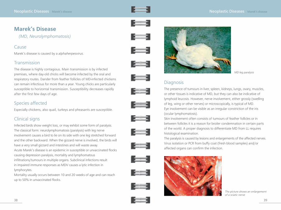
Clinical signsInfected birds show weight loss, or may exhibit some form of paralysis. The classical form: neurolymphomatosis (paralysis) with leg nerve involvement causes a bird to lie on its side with one leg stretched forward and the other backward. When the gizzard nerve is involved, the birds will have a very small gizzard and intestines and will waste away. Acute Marek’s disease is an epidemic in susceptible or unvaccinated flocks causing depression paralysis, mortality and lymphomatous infiltrations/tumours in multiple organs. Subclinical infections result in impaired immune responses as MDV causes a lytic infection in lymphocytes. Mortality usually occurs between 10 and 20 weeks of age and can reach up to 50% in unvaccinated flocks.
DiagnosisThe presence of tumours in liver, spleen, kidneys, lungs, ovary, muscles, or other tissues is indicative of MD, but they can also be indicative of lymphoid leucosis. However, nerve involvement, either grossly (swelling of leg, wing or other nerves) or microscopically, is typical of MD. Eye involvement can be visible as an irregular constriction of the iris (ocular lymphomatosis).Skin involvement often consists of tumours of feather follicles or in between follicles it is a reason for broiler condemnation in certain parts of the world. A proper diagnosis to differentiate MD from LL requires histological examination.The paralysis is caused by lesions and enlargements of the affected nerves. Virus isolation or PCR from buffy coat (fresh blood samples) and/or affected organs can confirm the infection.
MD leg paralysis
The picture shows an enlargement of a sciatic nerve
41 40
Neoplastic Diseases Marek’s disease Neoplastic Diseases Marek’s disease
TreatmentThere is no effective treatment for affected flocks.
ControlVaccination is an effective means of control.It has been demonstrated that MD vaccine only prevents the appearance of Marek’s disease tumours and paralysis. It does not prevent the birds from becoming infected with MD-virus. It is therefore of major importance to maintain high hygienic and sanitary measures by good management to avoid early exposure of young chickens. Multiple age farms are big risk to Marek’s disease.
Vaccination against Marek’s disease is performed in the hatchery; there are two routes of application; In-ovo injection into the18 days embryonated eggs or injection in day-old chickens.For this purpose different MD vaccines exist. Most used are the Rispens (serotype-1), SB1 (serotype-2) and HVT vaccine strains (serotype-3). The choice on the strains for MD vaccination will depend on the virulence of the strains present in the field.
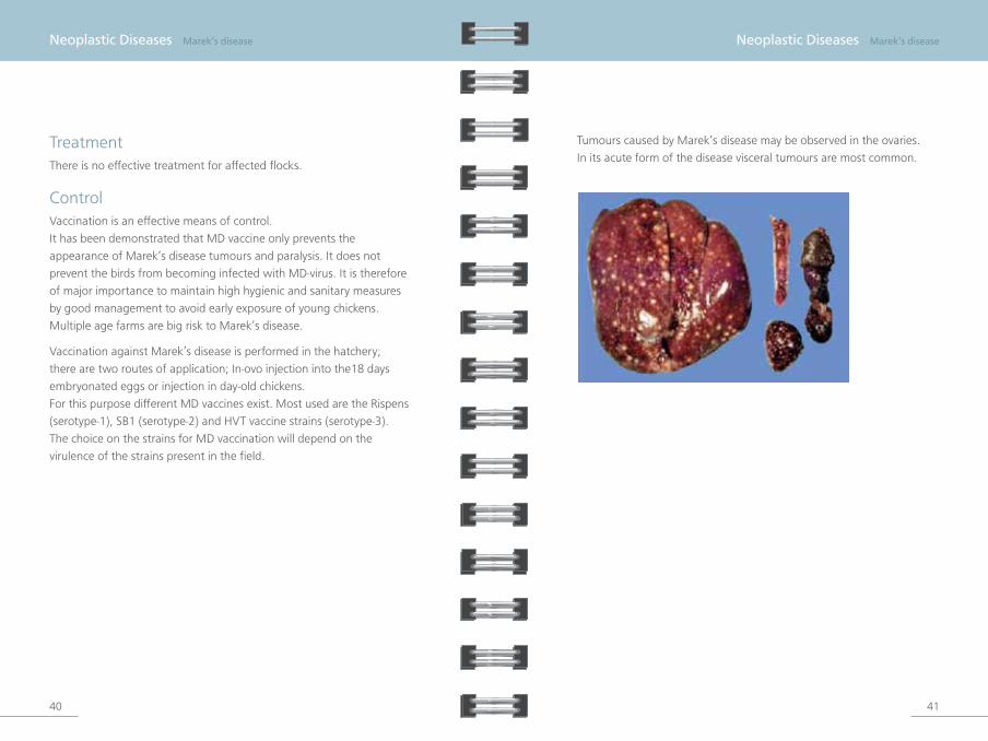
Tumours caused by Marek’s disease may be observed in the ovaries. In its acute form of the disease visceral tumours are most common.
43 42
Avian Adenoviral Diseases Avian Adenoviral Diseases
Avian Adenoviral Diseases
(Hydropericardium-Hepatitis Syndrome)

45 44
Avian Adenoviral Diseases Egg Drop Syndrome Avian Adenoviral Diseases Egg Drop Syndrome
Egg Drop Syndrome (EDS ’76)
CauseThe disease is caused by a virus belonging to the subgroup III of Avian Adenovirus (strain BC14, virus 127), the EDS virus does not belong to any of the 12 Fowl Adenoviruses.
TransmissionThe virus is transmitted through the egg to a few birds in a flock, these birds carry the virus until the flock comes into lay at which time they begin to excrete virus and infect birds kept in the same house. Horizontal spread through infected litter can and does occur once a flock shows the disease but it seems that the virus is not very infectious or the level of virus excretion is low.
Species affectedOnly chickens are susceptible to clinical disease due to EDS virus; however, the virus is widespread in ducks but does not cause any problems.
Clinical signsEDS ’76 affects only layers and breeders at the start of or during their egg production. Affected flocks show a failure to reach peak egg production or a drop in egg production accompanied by an inferior egg shell quality and in the case of brown eggs, a loss of shell color. Affected birds may also appear to be anaemic, may show transient diarrhea and sometimes the food intake may be reduced. No increased mortality or other symptoms are observed.
Internal lesions No specific internal lesions have been observed.
30 35 40
80
40
0Age in weeks
Lay
%
Infection
30 35 40
80
40
0Age in weeks
Lay
% Infection
Non-peaking e!ect of Egg drop by early EDS infection and drop by infection during lay

47 46
Avian Adenoviral Diseases Egg Drop Syndrome Avian Adenoviral Diseases Egg Drop Syndrome
DiagnosisClinical signs maybe indicative for EDS ’76. Virus isolation and antibody tests can confirm this.
Differential diagnosis with Infectious Bronchitis and to a lesser extent Newcastle Disease and Infectious Laryngotracheitis will have to be considered. Proper diagnostic tests to determine the presence of antibodies will confirm infection.
TreatmentThere is no treatment against egg drop syndrome.
ControlVaccination with an inactivated vaccine before point of lay is the only available, effective method for the control of EDS.
Misshapen and soft shelled eggs
49 48
Avian Adenoviral Diseases Inclusion Body Hepatitis Avian Adenoviral Diseases Inclusion Body Hepatitis
Inclusion Body Hepatitis Hydropericardium-Hepatitis Syndrome (HHS)
CauseThe disease is caused by a virus belonging to the group 1 of Avian Adenovirus (for example the Tipton strain) and is usually simultaneously accompanied by other immunosuppressive diseases such as infectious bursal disease or infectious anaemia. There are12 known serotypes of Avian Adenoviruses that may be involved in the development of this disease.
TransmissionEgg transmission is an important factor but also horizontal transmission from bird to bird by contact with droppings can occur. Once the bird becomes immune, the virus can no longer be isolated from the droppings. Progeny of a shedding breeder flock can infect naive progeny of other breeder sources placed in the same house.
Species affectedChickens, turkeys and pheasants and possibly other birds can be affected by Avian Adenoviruses.
Clinical signsChickens with inclusion body hepatitis are affected at usually 5 to 7 weeks of age. The birds are listless, with ruffled feathers. Mortality is usually quite severe, up to 25 % in the first 10 days of the disease. Internal lesions; affected chickens have enlarged and mottled livers, many with pinpoint necrotic and haemorrhagic spots. Pale bone marrow and, in some cases in presence of infectious anemia, gangrenous dermatitis can be seen. Kidneys are pale.
If Gumboro disease (infectious bursal disease) has been present in the birds, even if subclinical, the bursa of Fabricius will be very small (atrophic). Such chickens are immune-suppressed and usually have more severe cases of inclusion body hepatitis and/or infectious anaemia. Mature birds do not have clinical signs of adenovirus infection, but will develop antibodies in their blood.
Hydropericardium-Hepatitis Syndrome (HHS) was reported for the first time in 1987 in Pakistan and was referred to as “Angara disease”. The disease has meanwhile been reported from several other countries, including India and countries in the Middle East and Latin America
Hydropericardium-hepatitis syndrome is caused by a virus belonging to the family of the Fowl Adenoviruses (FAV). Despite of the diversity in the geographical distribution of the disease, in all areas the infection is caused by a virus belonging to FAV serotype 4. There are three features which underline that this condition is a new disease, different from the known IBH. IBH and hydropericardium accompany this syndrome. Once outbreaks of HHS occur, it remains a problem for the poultry industry. And while IBH is shown to be caused by strains belonging to various FAV serotypes, HHS is, contrary to this, caused by FAV serotype 4. The infected flocks show high mortality rates and beside the lesions typical for IBH, a marked hydropericarditis is found in the affected birds.
51 50
Avian Adenoviral Diseases Inclusion Body Hepatitis Avian Adenoviral Diseases Inclusion Body Hepatitis
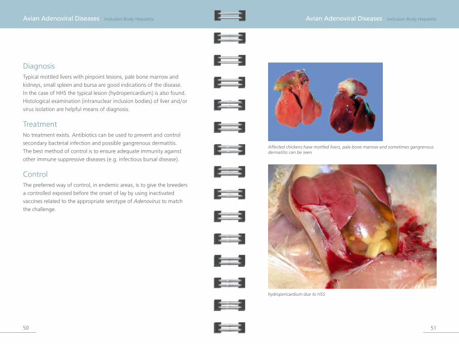
DiagnosisTypical mottled livers with pinpoint lesions, pale bone marrow and kidneys, small spleen and bursa are good indications of the disease. In the case of HHS the typical lesion (hydropericardium) is also found. Histological examination (intranuclear inclusion bodies) of liver and/or virus isolation are helpful means of diagnosis.
TreatmentNo treatment exists. Antibiotics can be used to prevent and control secondary bacterial infection and possible gangrenous dermatitis. The best method of control is to ensure adequate immunity against other immune suppressive diseases (e.g. infectious bursal disease).
ControlThe preferred way of control, in endemic areas, is to give the breeders a controlled exposed before the onset of lay by using inactivated vaccines related to the appropriate serotype of Adenovirus to match the challenge.
A!ected chickens have mottled livers, pale bone marrow and sometimes gangrenous dermatitis can be seen
hydropericardium due to HSS
53 52
Miscellaneous Viral Diseases Miscellaneous Viral Diseases
Miscellaneous Viral Diseases
Stunting Syndrome
55 54
Miscellaneous Viral Diseases Avian Encephalomyelitis Miscellaneous Viral Diseases Avian Encephalomyelitis
Avian Encephalomyelitis AE or Epidemic Tremor
CauseAvian Encephalomyelitis (AE) is caused by an enterovirus belonging to the picornavirus group.
TransmissionEgg transmission is the major route of transmission of AE virus. Infected breeders will show a drop in egg production and in hatchability. Infected chickens that hatch will show clinical signs of the disease and spread the infection in the incubator to other newly hatched susceptible chickens. Young chickens can also be infected on the farm. The incubation period varies from 5 to 14 days depending on the route of infection.
Species affectedPrimarily, chickens are susceptible to AE, but turkeys and pheasants have been reported as natural hosts.
Clinical signsClinical tremors and increased mortality in young chickens, together with a drop in production and hatchability in the breeders or egg production drop in layers are signs related to AE.
The disease is mainly seen in young chickens, between 1 and 3 weeks of age. Affected chickens sit on their hocks, do not move well, and many fall on their sides. A fine, rapid trembling of the head and neck can be seen, but especially felt when affected chickens are held in the hand. In laying and breeding flocks, AE virus infection causes a marked drop in egg production which returns to normal in about 2-3 weeks. Mortality in naturally infected chickens varies and can be as high as 75%.
DiagnosisClinical tremors in young chickens, together with a drop in production and hatchability in the breeders or production drop in layers are indicative for AE infection. Chickens will not have gross lesions, but histological examination of brain, proventriculus and pancreas reveals typical lesions of AE. This will also differentiate the diagnosis of AE from encephalomalacia (Vitamin E deficiency, crazy chicken disease). Laboratory testing of blood serum from breeder flocks for the presence of antibodies, or their hatching eggs, can determine if an infection occurred.
TreatmentThere is no effective treatment for AE.

57 56
Miscellaneous Viral Diseases Avian Encephalomyelitis Miscellaneous Viral Diseases Avian Encephalomyelitis
ControlPreventive vaccination of breeder pullets and layer pullets with live AE vaccine before egg production is the only effective means of AE control. If a breeder flock has not been, or has been inadequately or not vaccinated against AE and an outbreak occurs, it is advisable to stop hatching eggs from the flock for several weeks until the breeders have acquired immunity and no longer transmit AE virus through their eggs.
egg production %
21 22 23 24 25 26 27 28 29 30 31 32 33 34 35
100
80
60
40
20
0
Age weeks
% e
gg p
rodu
ctio
n
AE infected young chickens.
E!ect of AE on egg production
59 58
Miscellaneous Viral Diseases Chicken Anaemia Virus Miscellaneous Viral Diseases Chicken Anaemia Virus
Chicken Anaemia Virus(CAV, Blue wing disease)
CauseChicken Infectious anaemia is caused by a very resistant small virus known as CAV (Chicken Anaemia Virus). CAV is member of the genus Gyrovirus of the Circoviridae.
TransmissionThe major mode of transmission of infectious anemia is vertical transmission from infected breeder hens. Horizontal transmission from bird to bird or by infected equipment, clothing, etc. is also possible.
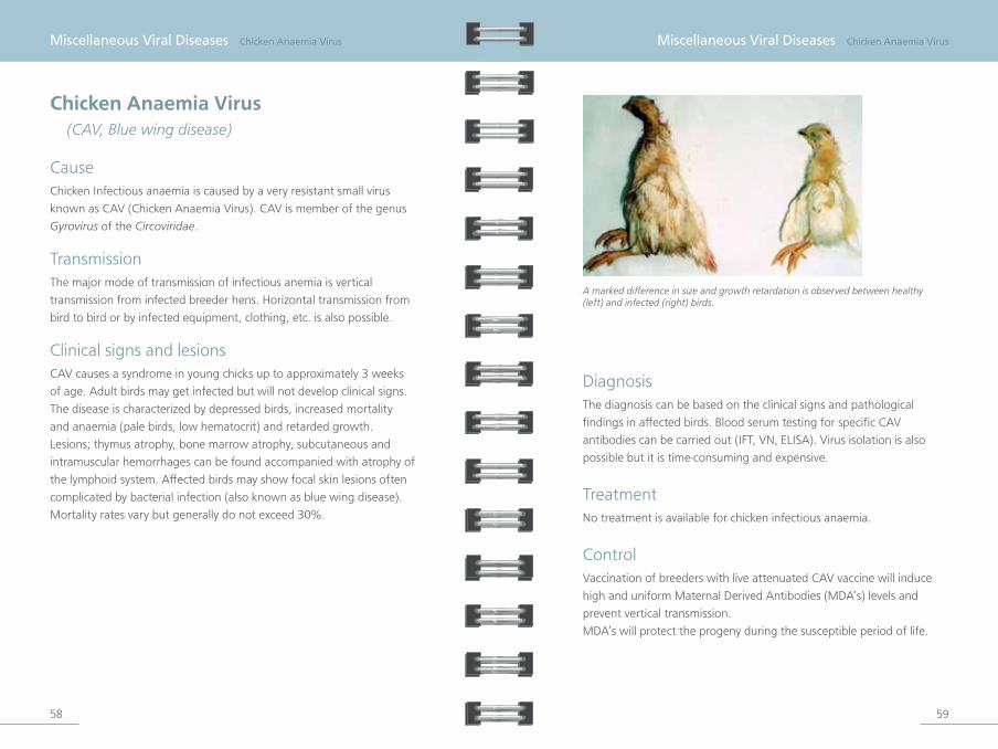
Clinical signs and lesions CAV causes a syndrome in young chicks up to approximately 3 weeks of age. Adult birds may get infected but will not develop clinical signs. The disease is characterized by depressed birds, increased mortality and anaemia (pale birds, low hematocrit) and retarded growth. Lesions; thymus atrophy, bone marrow atrophy, subcutaneous and intramuscular hemorrhages can be found accompanied with atrophy of the lymphoid system. Affected birds may show focal skin lesions often complicated by bacterial infection (also known as blue wing disease). Mortality rates vary but generally do not exceed 30%.
Diagnosis The diagnosis can be based on the clinical signs and pathological findings in affected birds. Blood serum testing for specific CAV antibodies can be carried out (IFT, VN, ELISA). Virus isolation is also possible but it is time-consuming and expensive.
TreatmentNo treatment is available for chicken infectious anaemia.
ControlVaccination of breeders with live attenuated CAV vaccine will induce high and uniform Maternal Derived Antibodies (MDA’s) levels and prevent vertical transmission.MDA’s will protect the progeny during the susceptible period of life.
A marked di!erence in size and growth retardation is observed between healthy (left) and infected (right) birds.
61 60
Miscellaneous Viral Diseases Fowl Pox Miscellaneous Viral Diseases Fowl Pox
Fowl Pox (Avian Pox, Avian Diphtheria)
CauseFowl pox is caused by a Poxvirus.
TransmissionIntroduction of infected or “carrier” birds in a susceptible flock will cause an outbreak by direct contact and water or feed transmission. Mosquitoes and other flying insects can also transmit the virus from bird to bird and also transmit the disease to near-by flocks. The incubation period varies from 4 to 20 days.
Species affectedChickens, turkeys, pheasants and pigeons can be affected by different Fowl Poxvirus strains.
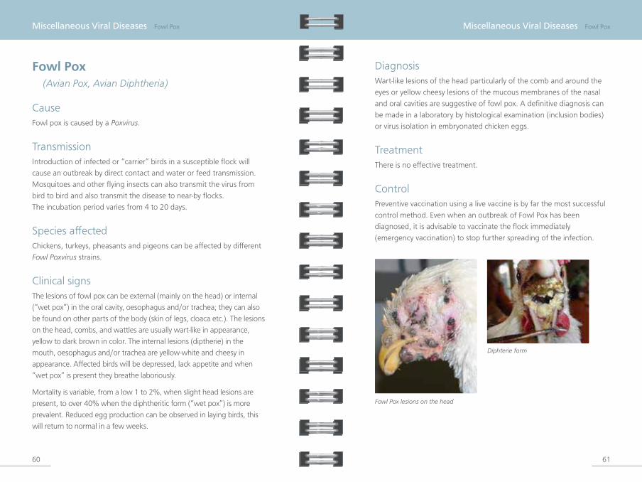
Clinical signsThe lesions of fowl pox can be external (mainly on the head) or internal (“wet pox”) in the oral cavity, oesophagus and/or trachea; they can also be found on other parts of the body (skin of legs, cloaca etc.). The lesions on the head, combs, and wattles are usually wart-like in appearance, yellow to dark brown in color. The internal lesions (diptherie) in the mouth, oesophagus and/or trachea are yellow-white and cheesy in appearance. Affected birds will be depressed, lack appetite and when “wet pox” is present they breathe laboriously.
Mortality is variable, from a low 1 to 2%, when slight head lesions are present, to over 40% when the diphtheritic form (“wet pox”) is more prevalent. Reduced egg production can be observed in laying birds, this will return to normal in a few weeks.
DiagnosisWart-like lesions of the head particularly of the comb and around the eyes or yellow cheesy lesions of the mucous membranes of the nasal and oral cavities are suggestive of fowl pox. A definitive diagnosis can be made in a laboratory by histological examination (inclusion bodies) or virus isolation in embryonated chicken eggs.
TreatmentThere is no effective treatment.
Control Preventive vaccination using a live vaccine is by far the most successful control method. Even when an outbreak of Fowl Pox has been diagnosed, it is advisable to vaccinate the flock immediately (emergency vaccination) to stop further spreading of the infection.
Fowl Pox lesions on the head
Diphterie form
63 62
Miscellaneous Viral Diseases Infectious bursal disease Miscellaneous Viral Diseases Infectious bursal disease
Infectious Bursal Disease(Gumboro disease, IBD)
CauseThe disease is caused by a Birnavirus of serotype 1. Virus strains can be divided in classical and variant strains. The virus is very stable and is difficult to eradicate from an infected farm.
TransmissionIBD virus is very infectious and spreads easily from bird to bird by way of droppings. Infected clothing and equipment are means of transmission between farms.
Species affectedChickens and turkeys appear to be natural hosts.
Clinical signsClinical IBD occurs usually between 3 and 8 weeks of age depending on maternal antibody levels. Affected birds are listless and depressed, pale, huddling producing watery white diarrhea. Mortality varies. Usually new cases of IBD have a mortality rate of about 5 to10% but can be as high as 60% depending on the pathogenicity of the strain involved. Highly pathogenic strains are called “very virulent” IBD (vvIBD) resulting in high mortality.
Subclinical IBD occurs with infections before 3 weeks of age. Early IBD infection result in permanent immunosuppression without mortality. Immunosuppression is economically important due to increased susceptibility to secondary infections especially in the respiratory tract. Gumboro disease related diseases such as inclusion body hepatitis are also more frequent in these birds. In broilers this form of the disease results in bad performance with lower weight gains and higher feed conversion ratios.
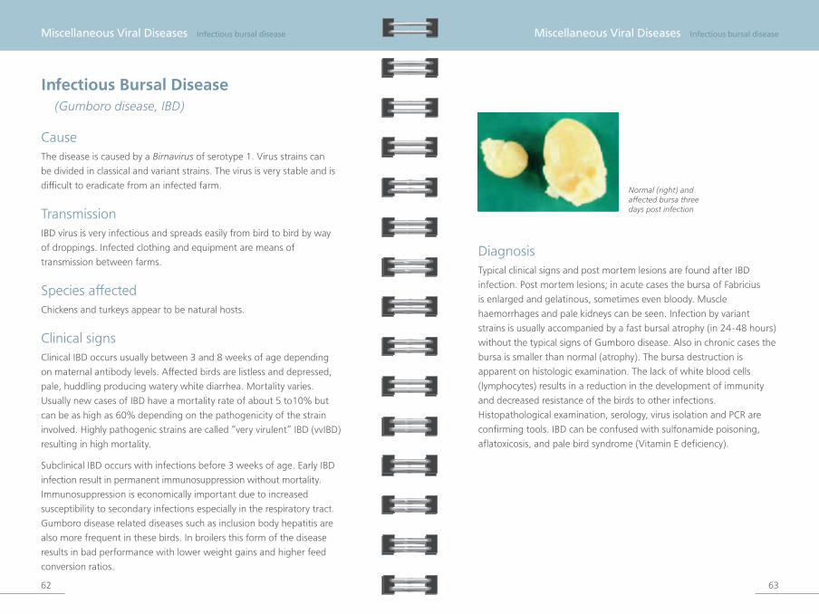
DiagnosisTypical clinical signs and post mortem lesions are found after IBD infection. Post mortem lesions; in acute cases the bursa of Fabricius is enlarged and gelatinous, sometimes even bloody. Muscle haemorrhages and pale kidneys can be seen. Infection by variant strains is usually accompanied by a fast bursal atrophy (in 24 - 48 hours) without the typical signs of Gumboro disease. Also in chronic cases the bursa is smaller than normal (atrophy). The bursa destruction is apparent on histologic examination. The lack of white blood cells (lymphocytes) results in a reduction in the development of immunity and decreased resistance of the birds to other infections. Histopathological examination, serology, virus isolation and PCR are confirming tools. IBD can be confused with sulfonamide poisoning, aflatoxicosis, and pale bird syndrome (Vitamin E deficiency).
Normal (right) and a!ected bursa three days post infection
65 64
Miscellaneous Viral Diseases Infectious bursal disease Miscellaneous Viral Diseases Infectious bursal disease
TreatmentNo treatment is available for IBD.
Control Vaccination of breeders and young chicks is the best means of control. The induction of a high maternal immunity in the progeny of vaccinated breeders, together with the vaccination of the offspring is the most effective approach to successful IBD control. A variety of live and inactivated vaccines have been developed to enhance the control of classical, variant and vvIBD challenges.
Recently a new generation of recombinant vector vaccines based on HVT-vector carrying an insert of the VP2 part of the IBD-virus entered the market for the control of IBD.
Normal chicken(left) and gumboro chicken (right)
67 66
Miscellaneous Viral Diseases Malabsorption Syndrome Miscellaneous Viral Diseases Malabsorption Syndrome
Malabsorption Syndrome(MAS, Runting & Stunting Syndrome)
This complex disease has been reported under various names such as helicopter disease, femoral head necrosis, brittle bone disease, infectious proventriculitis, pale bird syndrome, runting disease and stunting disease.
CauseThe malabsorption syndrome appears to be a disease complex involving Avian Enteric reoviruses and other viral and bacterial agents which may affect the digestive system resulting in nutritional and deficiency signs and lesions.
TransmissionOnly circumstantial evidence is present at the moment to indicate that the causal organism(s) may be vertically transmitted. Horizontal transmission also seems to play a role on infected sites.
Species affectedChickens and possibly turkeys.
Clinical signsThe disease is mainly observed in broiler flocks. Poor performance; increased FCR, retarded growth and uneven flocks. Many affected broiler flocks have a history of diarrhea, beginning as early as a few days of age and lasting until 10-14 days of age. Light or dark brown, foamy droppings can be found with undigested food particles. Several affected broilers in a flock may exhibit malpositioned feathers, especially on the wings. Early rickets with extreme paleness of legs and heads can be observed. Encephalomalacia is also regularly found. Later an important effect is the delayed growth of the affected birds. Mortality is variable and in general as low as 4%
Diagnosis The clinical disease is characterized by one or more of the following lesions: enteritis with watery brown and foaming contents and the presence of undigested food in the intestine. Mucosal and submucosal proventricular lesions. Pancreatic inflammatory infiltration with degenerative changes have been found. Osteoporosis and osteomyelitis, femoral head necrosis whereby the bone of the epiphysis of the femur is unusually soft. Since the causal agent may differ it is difficult to base a diagnose on virus isolation or serology.
TreatmentNo treatment available.
ControlVaccination against reovirus in the breeders helps to reduce problems in the progeny. Strict hygienic and sanitary measures will reduce the incidence of the disease.
69 68
Miscellaneous Viral Diseases Reovirus Infections Miscellaneous Viral Diseases Reovirus Infections
Reovirus Infections(viral arthritis, tenosynovitis)
CauseReovirus infections. Avian reoviruses are members of the Orthoreovirus genus. There are many different reoviruses from pathogenic to apathogenic. In the pathogenic group we also find antigenically different strains, like S1133, 1733, 2408, ERS, which are considered to be most important in meat-type chickens.
TransmissionThe virus may be transmitted by droppings from bird to bird. Egg transmission is also a factor when breeder flocks become infected during egg production.
Species affectedChickens, turkeys and possibly pheasants are natural hosts.
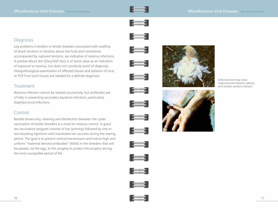
Clinical signsIn young meat–type chickens: increased mortality, arthritis/ tenosynovitis, disuniform flocks resulting in loss of performance and economic losses.The first signs of reovirus infection can be observed in broiler type chickens between 2 and 10 weeks of age. The birds are reluctant to walk and when forced up have a painful, trembling gait. A distinct swelling of the tendons of the shanks and also above the hock joint can be observed. Affected birds have malpositioned feathers, especially on the wings and are called “helicopter chickens”. Internal lesions: The hock joint may be somewhat swollen, but usually not as severely as with Mycoplasma synoviae (MS) or Staphylococcus infections. The tendons usually appear discolored, brown or blood-tinged, with straw colored fluid between them. Ruptured tendons may occur and, in older broiler breeders (29-30 weeks old), one may feel a hard knot in the tendon above the hock joint. When the infection is complicated by Mycoplasma synoviae or Staphylococcus the fluid may appear yellow and creamy. In layer type breeders the disease is not as common as in broiler breeders and broilers.
71 70
Miscellaneous Viral Diseases Reovirus Infections Miscellaneous Viral Diseases Reovirus Infections
DiagnosisLeg problems in broilers or broiler breeders associated with swelling of shank tendons or tendons above the hock joint sometimes accompanied by ruptured tendons, are indicative of reovirus infections. A positive blood test (Elisa/AGP test) is of some value as an indication of exposure to reovirus, but does not constitute proof of diagnosis. Histopathological examination of affected tissues and isolation of virus or PCR from such tissues are needed for a definite diagnosis.
Treatment Reovirus infection cannot be treated successfully, but antibodies are of help in preventing secondary bacterial infections, particularly Staphylococcal infections.
ControlBesides biosecurity, cleaning and disinfection between the cycles vaccination of broiler breaders is a must for reovirus control. A good reo vaccination program consists of live (priming) followed by one or two boosting injections with inactivated reo vaccines during the rearing period. The goal is to prevent vertical transmission and induce high and uniform “maternal derived antibodies” (MDA) in the breeders that will be passed, via the egg, to the progeny to protect the progeny during the most susceptible period of life.
Iinfected birds may show malpositioned feathers (above) and swollen tendons (below)
73 72
Miscellaneous Bacterial Diseases Miscellaneous Bacterial Diseases
Miscellaneous Bacterial Diseases
Fowl Typhoid
75 74
Miscellaneous Bacterial Diseases Colibacillosis Miscellaneous Bacterial Diseases Colibacillosis
Colibacillosis
CauseBacterial infection with avian pathogenic Escherichia coli (APEC).
E.coli bacteria are ubiquitous and various serotypes of E.coli are normal intestinal inhabitants. It is estimated that 10 -15% of the intestinal coliforms are potentially pathogenic. Infection can be primary or secondary after primary host defense damage (eg respiratory viral infections, Mycoplasma infections, parasites, wounds, nutrition).
TransmissionE.coli are normal intestinal inhabitants and are shed via direct or indirect contact and feces. Egg transmission (via egg surface contamination) is also common and can lead to high chick mortality, environmental presence and vectors like house flies and darkling beetles can also spread E.coli.
Species affected Chickens and turkeys, most avian species are susceptible.
Clinical signs and lesions Clinical signs vary depending on specific disease induced by E.coli. From very mild cellulitis to severe disease; lameness, retarded growth, increased mortality, reduced feed and water intake.
Lesions: colisepticaemia in combinations with polyserositis, salpingitis, osteomyelitis/synovitis, enteritis, meningitis and young birds with omphalitis.Both morbidity and mortality are very variable.
Diagnosis Clinical signs in combination with isolation and identification.
TreatmentAntibiotics based on antibiotic sensitivity test.
ControlHygiene management, hatching egg management and E.coli vaccination and vaccination against primary causes like respiratory viruses, coccidiosis, IBD, CAV.
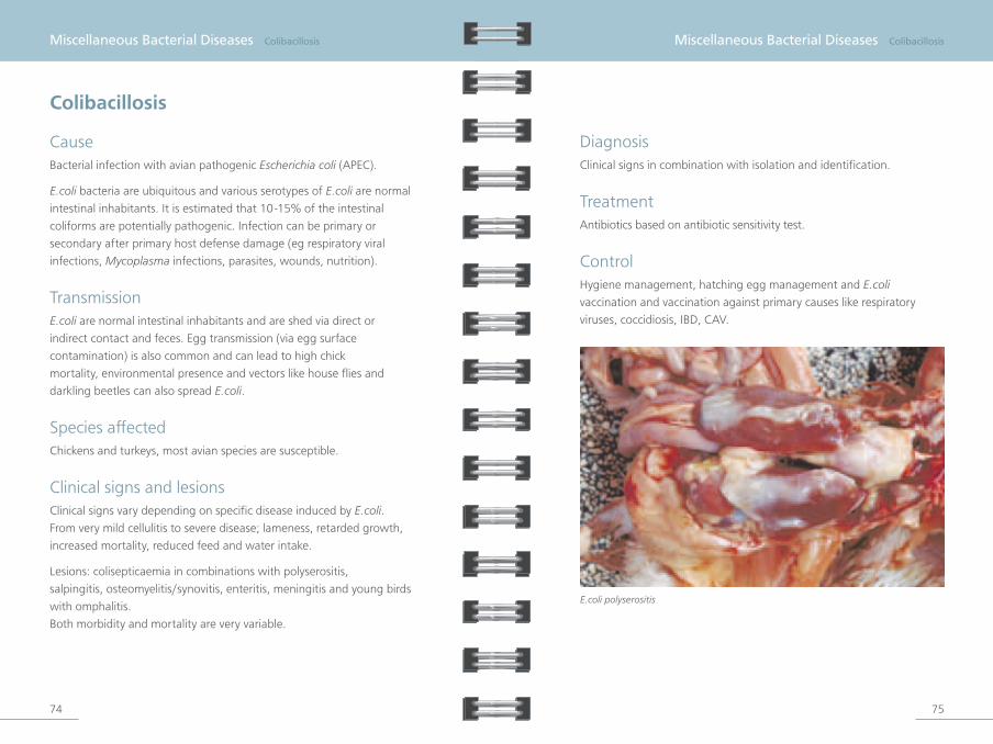
E.coli polyserositis
77 76
Miscellaneous Bacterial Diseases Fowl Cholera Miscellaneous Bacterial Diseases Fowl Cholera
Fowl Cholera(Avian Cholera, Pasteurellosis, Avain hemorrhagic septicaemia)
CauseFowl cholera is caused by a bacterium: Pasteurella multocida (several serotypes).
TransmissionTransmission of fowl cholera is mainly from bird to bird by water or feed contamination. There is no evidence for egg transmission. Vectors like flies and red mite can be carriers and can add to the spread. Rodents (rats and mice) also appear to play a role in contamination of water and feed with Pasteurella multocida.
Species affected Turkeys, chickens, ducks and geese, game birds and other bird species are susceptible.
Clinical signs and lesions Affected birds are depressed and have decreased appetite. Egg production will drop 5-15 % and mortality will be high in acute fowl cholera. Birds that die from acute fowl cholera frequently have bluish combs and wattles. Chronic fowl cholera will not cause high mortality, although there will be an increase in deaths. Swollen wattles is a feature of chronic fowl cholera.
Lesions; acute phase septicaemia, vascular changes in abdominal viscera, hemorrhages, liver swelling with focal necrosis, ovaries appear flaccid and hemorrhagic and show ruptured yolks,Chronic phase; localized infections in conjunctiva, fecial edema, middle ear infection resulting in torticollis, meningeal infection.
Diagnosis Clinical signs in combination with isolation and identification from samples from birds that died of acute Fowl cholera. (Fresh death birds)
TreatmentAntibiotics based on antibiotic sensitivity test, the earlier the diagnosis the better change of a positive effect of an antibiotic treatment.
ControlHygiene management and rodent control to eliminate possible sources of Pasteurella multocida.Vaccination can be considered in areas where Pasteurella multocida is prevalent. Both live and inactivated vaccines are available.
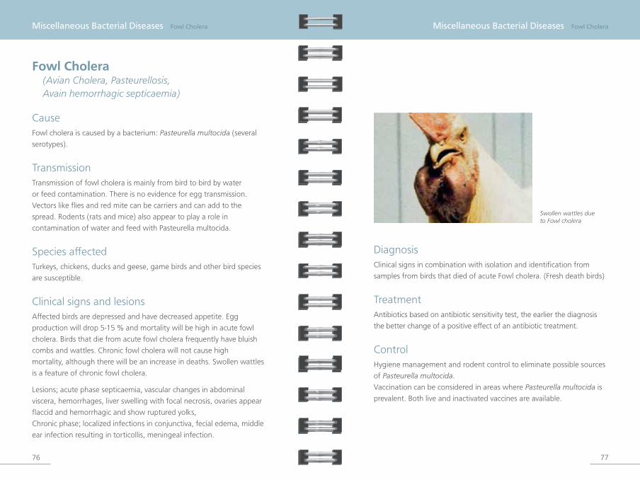
Swollen wattles due to Fowl cholera
79 78
Miscellaneous Bacterial Diseases Infectious Synovitis Miscellaneous Bacterial Diseases Infectious Synovitis
Infectious Synovitis
CauseIs a clinical condition caused by different possible agents: Mycoplasma synoviae, Staphylococcus aureus, E.coli and certain reoviruses. (MS, Reo virus and E.coli information can be found in other chapters of this booklet).
Staphylococcus aureusStaphylococcus infections are common in poultry and mainly caused by Staphylococcus aureus.
Transmission Infections generally occur after the breakdown of natural defense mechanisms, due to mechanical damage (eg wounds) or infectious breakdown of the immune system (eg CAV, IBD, Marek’s Disease).
Species affected Chickens and turkeys
Clinical signs and lesions Staphylococcus aureus is ubiquitous and infections can become manifest after breakdown of the natural defense mechanisms such as wounds, inflammation, hematogenous dissemination.
Signs vary with the site of entry, most frequent affected tissues; bone, tendon sheaths and joints.
Clinical picture includes lameness in one or both legs, ruffled feathers, swollen joints, fever and increased mortality. Lesions; swollen joints, arthritis, peri-arthritis and synovitis, osteomyelitis, septicaemia-swollen liver-spleen.
Diagnosis Clinical signs and gross lesions in combination with isolation from affected tissues will confirm Staphylococcus infection.
TreatmentAntibiotic treatment based on sensitivity test. Success is not guaranteed.
ControlPrevent damage to natural host defense mechanisms. Good litter quality, prevent wounds, hatchery management and sanitation. So far vaccination has not been successful.
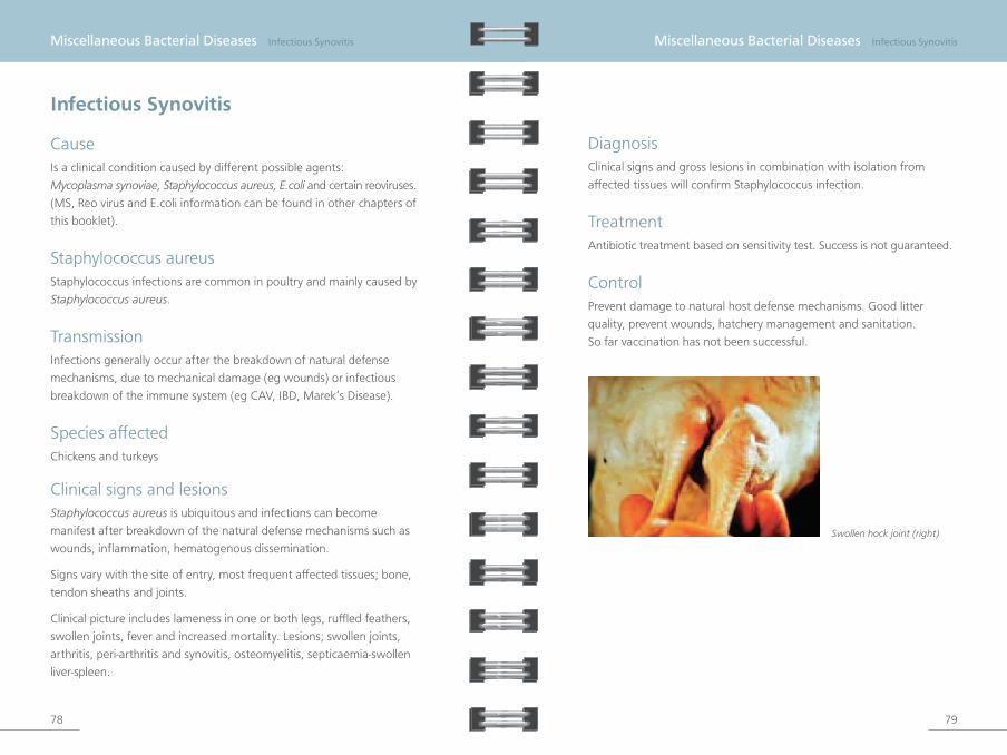
Swollen hock joint (right)
81 80
Miscellaneous Bacterial Diseases Necrotic Enteritis Miscellaneous Bacterial Diseases Necrotic Enteritis
Necrotic Enteritis(NE, Clostridial enteritis, enterotoxemia)
CauseBacterial infection with Clostridium perfringens (Gram+, toxin forming, spore forming, anaerobe).Clostridia including Clostridium perfringens are normal inhabitants of the gut. When the microflora balance in the gut is disturbed, potentially pathogenic clostridia begin to produce toxins and proteolytic enzymes. The toxins will induce cell damage known as Necrotic Enteritis (NE). Factors involved in microflora disturbance include: intestinal infections (eg coccidiosis), nutritional factors (protein source, grain source, diet changes), management: type of litter, timing of feed changes, antibiotic treatments.
TransmissionClostridium perfringens is an ubiquitous organism that can be found in faeces, soil, dust contaminated feed and litter.
Species affected Chickens, turkeys and quail.Natural outbreaks have reported in chickens from 2 weeks to 6 months of age. Majority of NE problems are identified in broilers raised on litter (2 -5 weeks of age).
Clinical signs and lesions There are 2 forms of Necrotic Enteritis (NE), acute clinical form and the subclinical form.Although it can be seen at any age, the acute clinical form is primarily a disease in young chickens, showing severe depression, reluctance to move, diarrhea, ruffled feathers and sudden death and increased mortality. The subclinical form produces not outward signs but has big impact on performance (weight loss, reduced weight gain and impaired FCR).Lesions: necrosis of the mucosa of the small intestine. In the clinical form the necrosis might progress into a fibrinonectoric enteritis forming a diphtheritic membrane. In the mild form, focal areas of intestinal mucosal necrosis without further clinical signs can be found.
DiagnosisClinical signs in combination with typical gross and microscopic lesions and isolation of the causative agent will confirm the clostridial infection.
Treatmentantibiotic
83 82
Miscellaneous Bacterial Diseases Necrotic Enteritis Miscellaneous Bacterial Diseases Necrotic Enteritis
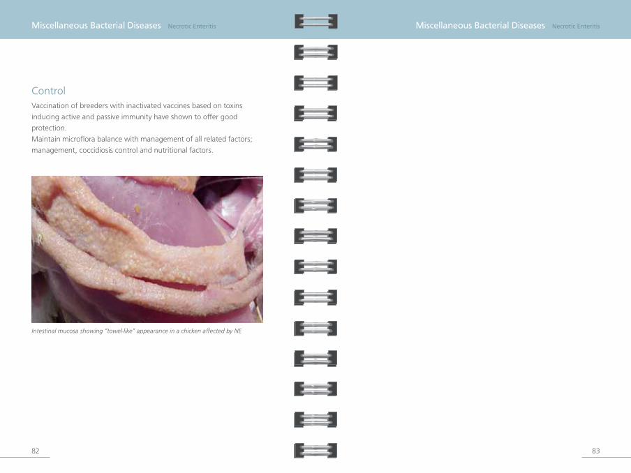
ControlVaccination of breeders with inactivated vaccines based on toxins inducing active and passive immunity have shown to offer good protection.Maintain microflora balance with management of all related factors; management, coccidiosis control and nutritional factors.
Intestinal mucosa showing “towel-like” appearance in a chicken a!ected by NE

85 84
Miscellaneous Bacterial Diseases Ornithobacterium Rhinotracheale Miscellaneous Bacterial Diseases Ornithobacterium Rhinotracheale
Ornithobacterium Rhinotracheale(OR, ORT)
CauseBacterial infection; Ornithobacterium rhinotracheale is a gram-negative rod responsible for causing infections in chickens and turkeys of all ages.
TransmissionHorizontal transmission from bird to bird and by infected fomites. Infected breeder hens may transmit the disease agent vertically to their offspring.
Species affected Chickens and turkeys of all ages.
Clinical signs and lesions OR may cause a respiratory disease in turkeys, usually between 2-6 and 12-20 weeks of age. The birds may show respiratory disease with watery eyes and swelling of the sinus infraorbitalis. Broilers may also show respiratory signs after infection around 4 weeks of age. A severe purulent pneumonia, accompanied by airsacculitis and pericarditis may be found in broilers as well as in turkeys. Affected birds also show growth retardation. Concomitant viral infections may intensify the severity of the lesions.
Diagnosis Blood serum testing using a commercially available ELISA for the detection of specific antibodies against OR and the finding of specific lesions are indicative of OR infection. Isolation of the organism and its biochemical determination may be attempted but care should be taken that these are carried out using appropriate methods in order to avoid unreliable results. Differential diagnose with bacteria causing similar disease patterns is recommended (E. coli, P. multocida, MG).
TreatmentInfections are normally treated with broad-spectrum antibiotics with variable degrees of success. Resistance building might force to perform an Antibiotic Sensitivity test.
ControlAn inactivated vaccine for broiler breeders is available to prevent the disease in the vaccinated birds and provide maternal antibodies to the offspring of the vaccinated breeders.
Pneumonic lung right caused by OR
87 86
Miscellaneous Bacterial Diseases Pullorum disease and Fowl Typhoid Miscellaneous Bacterial Diseases Pullorum disease and Fowl Typhoid
Pullorum disease and Fowl Typhoid
CausePullorum disease is caused by a bacterium, Salmonella pullorum. Fowl typhoid is caused by Salmonella gallinarum, which is related to, but not identical to S. pullorum. Primarily affects chickens and turkeys.
TransmissionPullorum and typhoid can be transmitted horizontally and vertically by infected (carrier) breeder hens through their eggs. Chickens that hatch from such infected eggs will have typical pullorum disease (white diarrhoea) and high mortality. Infected chickens can also infect other chicks via droppings. Fowl typhoid is more a disease of adult chickens, with high mortality and morbidity. Horizontal transmission is important with fowl typhoid through infected droppings, dead bird carcasses, and infected clothing, shoes, utensils and other fomites.
Species affected Chickens and turkeys. Other birds such as quails, pheasants, ducks, peacocks and guinea fowl are susceptible.
Clinical signs and lesions Birds hatched from S.pullorum or S. gallinarum infected eggs appear moribund or dead in the hatching trays and the young birds will show weakness, depressed appetite, poor growth and increased mortality.In older birds depression. diarrhea, ruffled feathers, pale shrunken combs and drop in egg production and increased mortality will be observed. Morbidity and mortality can be highly variable (mortality can reach 25-60%)Lesions; acute phase septicaemia-enlarged and congested liver, spleen and kidneys, pericarditis.Hemorrhages. Livers may show white foci. In adult birds atrophic ovarian follicles may be found.
TreatmentTreatment with antibiotics of pullorum/fowl typhoid disease will not cure but reduce clinical signs and is undesirable from a standpoint of eradication. It is far more practical to control the disease by elimination of infected carrier breeder hens. Blood testing (monitoring) of breeder chickens by the serum plate or tube agglutination test with suitable S. pullorum antigen will detect infected carrier birds which can then be culled. Such control measures will stop the incidence of egg-transmitted pullorum disease/fowl typhoid. If hatching eggs from tested pullorum-free breeders are kept free from contamination through infected eggs from infected breeders or through contaminated equipment, chickens can remain carrier after treatment.
ControlMonitoring and eradication in breeders is the preferred control method.For layers vaccination against fowl typhoid with a special S. gallinarum (9R strain) has been practiced in several countries.
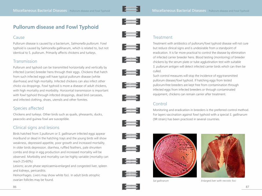
Sal gallinarum Enlarged liver with necrotic foci
89 88
Parasitic Diseases Parasitic Diseases
Parasitic Diseases

91 90
Parasitic Diseases Black head Parasitic Diseases Black head
Black head(Histomonosis, Enterohepatitis)
CauseA protozoan parasite, Histomonas meleagridis.
TransmissionDirect transmission by infected water, feed, or droppings has been proved. Indirect transmission by infected eggs of the caecal worm. Heterakis gallinarum, is also a major factor. Raising turkeys and chickens on wire and indoors decreases the incidence of blackhead.
Species affected Chickens and turkeys and peafowl are natural hosts to blackhead infections.
Clinical signs and lesions Affected birds are depressed, stand or sit with ruffled feathers, and have yellowish diarrhea. Darkening of head parts, especially in turkeys, gave the name to the disease (black head). Gross lesions include circular necrotic areas in livers with a crater-like center and cheesy cores in the caeca. Blackhead can cause high mortality, particularly in young turkey poults, but the disease can also affect older birds. In chickens the mortality from blackhead infection is usually lower, young chickens being the most susceptible.
DiagnosisClinical signs together with post mortem.
TreatmentTreatment with protozoan chemotherapeutics is usually effective. Such drugs can also be given at preventive levels in turkeys starter and grower feed. Growing turkeys on wire and indoors can reduce the incidence of blackhead to a large extent, but even so, strict hygiene and elimination of caecal worms are important control measures.
Necrotic liver infected by Histomonas meleagridis

93 92
Parasitic Diseases Coccidiosis Parasitic Diseases Coccidiosis
Coccidiosis
CauseCoccidiosis is caused by single-celled parasites of the genus Eimeria. In chickens, there are 9 species. The five most economically important species are: E. acervulina, E. maxima, E. tenella, E. necatrix and E. brunetti. Other species can be highly pathogenic (E. mivati) but are less ubiquitous.
TransmissionFeces containing sporulated Eimeria oocysts are the main means of transmission between birds. The incubation period from ingestion to shedding of new oocysts is 4 to 7 days. Clinical signs, such as weight loss, may appear as early as 4-days post-ingestion of infectious oocysts, but bloody droppings from E. tenella infection usually occur at 6 to 7 days post infection.
Clinical signs Clinical signs vary by the species infecting the flock. Some species, such as E. acervulina and E. maxima have a severe negative effect on weight gain and feed conversion, particularly E. maxima; these species do not cause obvious morbidity unless infection is moderately severe. Mortality in field cases of these species is rare, unless complicated by secondary infection with Clostridium perfringens (necrotic enteritis). Other species such as E.tenella, E.necatrix and E.brunetti can induce sick and listless birds in combination with high mortality.
Diagnosis Diagnosis is based on clinical signs and post-mortem examination, including mucosal scrapings of affected birds. E. acervulina is characterized by white spots on the mucosal surface of the duodenum and upper middle intestine. These gross lesions can be scored according to methods described by Johnson and Reid (1970 Exp Parasitol 28:30 – 36).
E. maxima is not easily characterized by gross lesions, although red petechiae on the serosal surface of the middle intestine can sometimes be seen. Proper diagnosis requires microscopic examination of scrapings taken from the mucosa of the middle intestine. Microscopic scoring systems for both E. maxima and E. acervulina have been described by Fitz-Coy in Diseases of Poultry 12th ed. (p. 1070)
E. tenella, E. necatrix and E. brunetti can cause significant morbidity and mortality, characterized by bloody droppings. Hemorrhages of the cecal mucosa, or blood-filled ceca indicate E. tenella; with definitive diagnosis made by mucosal scrapings that may reveal both asexual stages and oocysts. Significant swelling of the middle intestine with both tiny white spots and petechiae on the serosal surface (“salt and pepper”) indicate E. necatrix. E. necatrix is further distinguished by the fact that it tends to affect flocks over 6 weeks of age, although high challenge areas may result in clinical infections before 5 weeks of age. Mucosal scrapings of the middle intestine will reveal only asexual stages (extremely large schizonts), while the oocysts will be found in the ceca.
E. brunetti causes redness and hemorrhage in the colon, neck of the cecum and rectum, but lesions are not diagnostic. Like E. maxima, mucosal scrapings should be viewed microscopically.
95 94
Parasitic Diseases Coccidiosis Parasitic Diseases Coccidiosis
Treatment and preventionAnticoccidial chemicals effectively suppress sensitive Eimeria spp. populations, but complete resistance to these drugs develops very quickly sometimes during a single broiler flock cycle. Therefore, most coccidiosis control programs do not rely on chemical anticoccidials. Chemicals, particularly in liquid preparations, are largely used for treatment of clinical outbreaks.
Most effective coccidiosis prevention and control programs depend upon flock immunity.
One method to develop controlled immunity is via feed medication with ionophore antibiotics. The ionophores reduce coccidiosis multiplication, allowing natural immunity to develop while preventing clinical outbreaks. Over time, the Eimeria field populations become less sensitive to these medications, resulting in subclinical coccidiosis or even clinical outbreaks before complete immunity can develop.
To slow the loss of sensitivity, broiler producers employ shuttle medication programs (a chemical in the starter feed followed by an ionophore in the remaining feeds) and rotational programs (changing the feed medications to slow the loss of sensitivity to a single medication). This strategy was most effective when new medications were periodically introduced. Unfortunately, since the year 2000, no new medications have been introduced to the market and sensitivity to all existing medications has declined.
Another method to develop controlled immunity is coccidiosis vaccination with live attenuated or natural selected strain sporulated oocyst vaccines. Vaccination is already used almost exclusively for coccidiosis control in long-life birds such as breeding and egg-laying populations. Routine vaccination of broilers against coccidiosis is increasing and may eventually replace controlled immunity via feed medication.
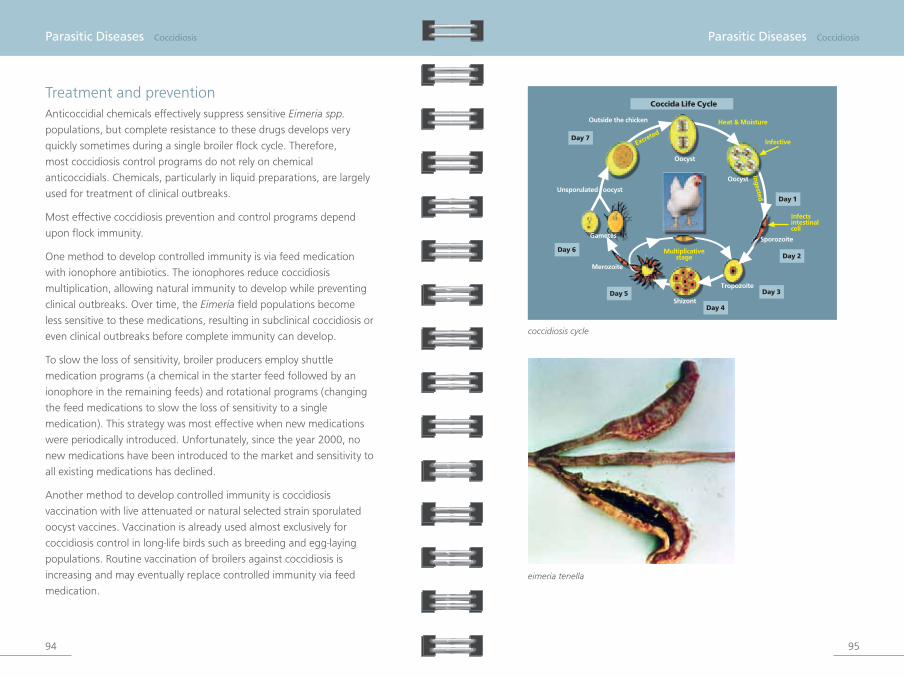
Coccida Life Cycle
Day 1
Day 2
Day 3
Day 4
Day 5
Day 6
Day 7
Outside the chicken
Oocyst
Oocyst
Sporozoite
Tropozoite
Shizont
Merozoite
Gametes
Unsporulated oocyst
Multiplicativestage
Heat & Moisture
Infective
Infectsintestinal cell
Excreted
Ingested
coccidiosis cycle
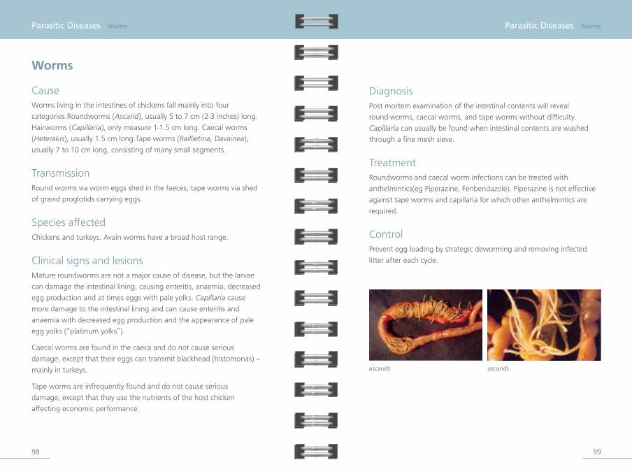
eimeria tenella
97 96
Parasitic Diseases Red Mite Parasitic Diseases Red Mite
Red Mite
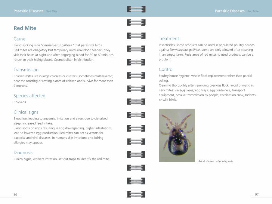
CauseBlood sucking mite “Dermanyssus gallinae” that parasitize birds, Red mites are obligatory but temporary nocturnal blood feeders, they visit their hosts at night and after engorging blood for 30 to 60 minutes return to their hiding places. Cosmopolitan in distribution.
TransmissionChicken mites live in large colonies or clusters (sometimes multi-layered) near the roosting or resting places of chicken and survive for more than 9 months.
Species affected Chickens
Clinical signs Blood loss leading to anaemia, irritation and stress due to disturbed sleep, increased feed intake.Blood spots on eggs resulting in egg downgrading, higher infestations lead to lowered egg production. Red mites can act as vectors for bacterial and viral diseases. In humans skin irritations and itching allergies may appear.
DiagnosisClinical signs, workers irritation, set out traps to identify the red mite.
TreatmentInsecticides, some products can be used in populated poultry houses against Dermanyssus gallinae, some are only allowed after cleaning in an empty farm. Resistance of red mites to used products can be a problem.
ControlPoultry house hygiene, whole flock replacement rather than partial culling.Cleaning thoroughly after removing previous flock, avoid bringing in new mites: via egg cases, egg trays, egg containers, transport equipment, passive transmission by people, vaccination crew, rodents or wild birds.
Adult starved red poultry mite
99 98
Parasitic Diseases Worms Parasitic Diseases Worms
Worms
CauseWorms living in the intestines of chickens fall mainly into four categories.Roundworms (Ascarid), usually 5 to 7 cm (2-3 inches) long.Hairworms (Capillaria), only measure 1-1.5 cm long. Caecal worms (Heterakis), usually 1.5 cm long.Tape worms (Raillietina, Davainea), usually 7 to 10 cm long, consisting of many small segments.
TransmissionRound worms via worm eggs shed in the faeces, tape worms via shed of gravid proglotids carrying eggs.
Species affected Chickens and turkeys. Avain worms have a broad host range.
Clinical signs and lesions Mature roundworms are not a major cause of disease, but the larvae can damage the intestinal lining, causing enteritis, anaemia, decreased egg production and at times eggs with pale yolks. Capillaria cause more damage to the intestinal lining and can cause enteritis and anaemia with decreased egg production and the appearance of pale egg yolks (“platinum yolks”).
Caecal worms are found in the caeca and do not cause serious damage, except that their eggs can transmit blackhead (histomonas) – mainly in turkeys.
Tape worms are infrequently found and do not cause serious damage, except that they use the nutrients of the host chicken affecting economic performance.
DiagnosisPost mortem examination of the intestinal contents will reveal round-worms, caecal worms, and tape worms without difficulty. Capillaria can usually be found when intestinal contents are washed through a fine mesh sieve.
TreatmentRoundworms and caecal worm infections can be treated with anthelmintics(eg Piperazine, Fenbendazole). Piperazine is not effective against tape worms and capillaria for which other anthelmintics are required.
Control Prevent egg loading by strategic deworming and removing infected litter after each cycle.
ascarids ascarids

101 100
Deficiency Diseases Deficiency Diseases
Some important Vitamin Deficiency Diseases
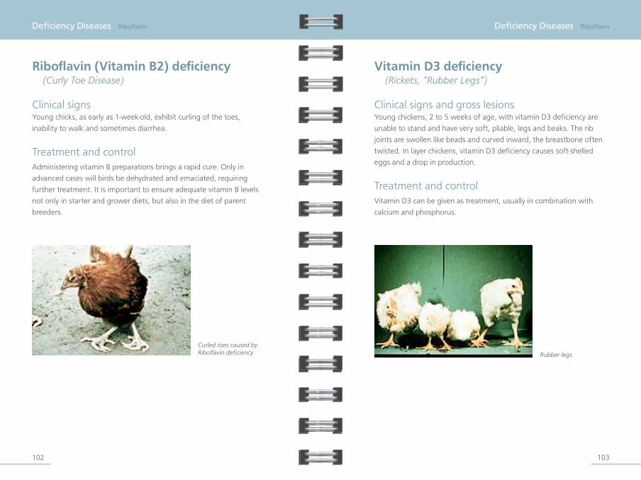
103 102
Deficiency Diseases Riboflavin Deficiency Diseases Riboflavin
Riboflavin (Vitamin B2) deficiency(Curly Toe Disease)
Clinical signsYoung chicks, as early as 1-week-old, exhibit curling of the toes, inability to walk and sometimes diarrhea.
Treatment and controlAdministering vitamin B preparations brings a rapid cure. Only in advanced cases will birds be dehydrated and emaciated, requiring further treatment. It is important to ensure adequate vitamin B levels not only in starter and grower diets, but also in the diet of parent breeders.
Curled toes caused by Riboflavin deficiency
Vitamin D3 deficiency(Rickets, “Rubber Legs”)
Clinical signs and gross lesionsYoung chickens, 2 to 5 weeks of age, with vitamin D3 deficiency are unable to stand and have very soft, pliable, legs and beaks. The rib joints are swollen like beads and curved inward, the breastbone often twisted. In layer chickens, vitamin D3 deficiency causes soft-shelled eggs and a drop in production.
Treatment and controlVitamin D3 can be given as treatment, usually in combination with calcium and phosphorus.
Rubber legs

105 104
Deficiency Diseases Riboflavin
Vitamin E deficiency(Crazy Chick Disease, Encephalomalacia)
Clinical signs and gross lesionsVitamin E deficiency in chickens affects the brain, causing degeneration, edema and hemorrhage, especially in the small brain (cerebellum). Affected young chicks appear unable to walk, they fall on their sides or stand with their heads between their legs. The cerebellum shows gross swelling, with yellow or brown discoloration and pinpoint hemorrhages may be observed. Encephalomalacia can also be found in mature chickens.
Treatment and controlAdequate levels of vitamin E and selenium in the diet of chickens and their parent breeders is of prime importance. Treatment of affected birds with vitamin E preparations (alpha-tocopherol) is effective if the condition is not too advanced.
Brain lesions caused by vitamin deficiency
Food Safety in Poultry
Food Safety in Poultry
107 106
Food Safety in Poultry
IntroductionAccording to the world health organization (WHO), food safety can be defined as a group of actions and policies directed to ensure that the entire food chain, from production to consumption, is as safe as possible. In theory, prevention of foodborne pathogens should be achieved by adequate hygiene and proper handling of food ingredients. However, the presence of human illness associated with the consumption of food contaminated with pathogenic microbes or their metabolic products (toxins) is frequently reported.
Foodborne pathogens, such as Salmonella and Campylobacter species, are commonly present in poultry and poultry products. Several scientifically-based and cost-effective strategies have been implemented by the poultry industry to prevent the transmission of Salmonella and Campylobacter infections to humans.
Public education and proactive interventions by the poultry industry will continue to be in place to reduce pathogen contamination of processed poultry meat and table eggs. Effective management of microbial hazards through Hazard Analysis and Critical Control Point (HACCP) and Microbial Risk Assessment (MRA) systems will continue to be aimed to improving public health.
Salmonellosis (Non S. gallinarum or S. pullorum)Etiologic AgentSalmonellas are ubiquitous, non-spore forming, facultative anaerobic gram negative bacteria. From more than 2500 identified Salmonella serotypes, only a small portion are commonly found in poultry flocks and poultry products. Some Salmonella species present in poultry, such as S. enteritidis, S. typhimurium, S. hadar, S. virchow and S. infantis are epidemiologically important because they cause infections in humans.
Public SignificanceSalmonella infections are usually transmitted to humans by consumption of food contaminated with animal feces. S. enteritidis, in particular, has been considered as the main serotype associated with contamination of table eggs. But also other serotypes S.typhimurium, S.hadar, S.infantis and S. virchow have also been associated with human infections.However, affected persons might require rehydration and antibiotic treatment in cases where systemic infection is present.
Poultry SalmonellosisThe route of contamination is by oral infection followed by colonization of the gastrointestinal tract (involving enterocytes and lymphoid tissue). Soon after colonization, bacteremia is observed, with Salmonella detection in several organs, such as spleen, liver, bone marrow, ovaries and oviduct. Salmonella is excreted in the feces, which constitute an important source of contamination for other birds in a house or other flocks.
In poultry, an age related resistance against Salmonella infections is observed. Factors, such as absence or very naïve intestinal flora and not fully developed immune system favor early age infection. Older birds are less susceptible to infection due to the presence complex and well established flora, which competes for receptors or produce metabolic products (bacteriocins) that inhibit Salmonella growth.
Food Safety in Poultry Food Safety in Poultry

109 108
Control of Poultry Salmonellosis“Keep the Salmonella out of your facilities and increase resistance of the birds.”
General Salmonella control programs involve pre-harvest (on-farm) and post-harvest interventions. The following are some of the pre-harvest interventions common in Salmonella control programs.
disinfection of facilities and equipment. Salmonella status should be monitored after disinfection.
programs (against rodents, flies and beetles).
drinking water.
between 70°C and 90°C.
feed withdrawal programs.
The following, are some post-harvest interventions implemented at the processing plant:
improves the bactericidal efficacy of chemicals.
▸ Use counter current multi-stage scalders with adequate water flow rates.
▸ Addition of chemicals to the scalders (i.e. acetic, lactic and formic/propionic acids).
▸ Maintain adequate water temperature (53°C to 56°C) to avoid Salmonella multiplication.
▸ Prevent feather buildup on equipment.▸ Add chlorine, acetic acid or hydrogen peroxide to the rinse
water.▸ Post-feather removal rinses should be maintained
at recommended temperatures
▸ Establish a regular equipment maintenance and disinfection program.
▸ Use of chlorine (i.e. 20 ppm) for whole carcass rinses.
▸ Immersion Chilling (common in the Unites States)■ If using chlorine, maintain chill water pH between
6.0 and 6.5 at temperatures below 40F with available chlorine content of 1 to 5 ppm.
■ Control of organic material load and use counter-current water flow.
▸ Air Chilling (common in Europe)■ Since chemical interventions are not allowed,
environmental sanitation, cleaning and disinfection of facilities and equipment should be in place.
Food Safety in Poultry Food Safety in Poultry

111 110
Poultry Salmonella Vaccination ProgramsCommercially available vaccines can be classified as live and inactivated (also known as bacterins). Live vaccines (S. enteritidis and S. typhimurium serotypes) serotypes) stimulate humoral and cellular immune responses, reducing colonization of internal organs and gastro intestinal tract and decreasing fecal shedding. Inactivated (bacterins) vaccines stimulate a strong humoral immune response, inducing high levels of circulating antibodies providing protection against internal organ and reproductive tract (ovary and oviduct) infection but also resulting in high levels of maternal derived antibodies (MDA) into the egg providing passive protection to the progenies or table eggs. Modern, commercial inactivated vaccines (Salenvac-line) are more immunogenic and provide excellent protection against field challenge because the vaccine strains S. enteritidis and/or S. typhimurium are grown under iron restriction conditions and forced to express outer membrane Iron Regulating Proteins (IRP’s are associated with iron transport) as an extra set of immunogenic antigens inducing a broader package of antibodies.
Constant Salmonella monitoring Adequate handling of table eggs
CampylobacterEtiologic AgentCampylobacters are ubiquitous. Campylobacters are gram negative and microaerophilic bacteria that belong to the Campylobacterales family, together with Helicobacter pylori (associated with chronic gastritis and gastric ulcers). Thermophilic campylobacters, such as Campylobacter jejuni, C. coli and C. lari play an important role in human intestinal infections.
Public SignificanceCampylobacter species, prevalent in poultry and poultry products, seem to be responsible for the majority of sporadic gastroenteritis in humans. Handling and consumption of undercooked poultry meat is widely considered as a major source of sporadic infection. A low infective dose, as low as 500 colony forming units, is enough to trigger an infection in humans.
Human campylobacteriosis is manifested by an acute watery or bloody diarrhea in association with abdominal pain. The great majority of cases have been associated with C. jejuni, followed by C. coli and C. lari. Most of the infections are self-limiting; however, some cases require medical intervention. Complicated infections, observed at young age or in immunocompromised people, have been associated with post-infection neuropathies such as Guillain-Barre syndrome.
The control of human campylobacteriosis is a constant concern by official health agencies due to the large prevalence of infection and social and economic burdens. Treatment normally involves rehydration, however, additional antimicrobial treatment might be required.
Food Safety in Poultry Food Safety in Poultry

113 112
Campylobacter in PoultryC. jejuni and C. coli are common commensals of the avian gastrointestinal tract. The organisms have been recovered from a wide variety of domestic poultry, including quail, pigeons, turkeys, ducks and chickens, in the absence of clinical disease. C. jejuni and C. coli colonize the mucous layer overlying the intestinal tract, replicating in highest numbers in the ceca. The intestinal tract is the ideal niche for Campylobacter, with the presence of oxygen levels low enough for microaerophilic growth.
Even though vertical transmission has been reported, newly hatched chicks are Campylobacter-negative and remain negative in the presence of maternal antibodies (usually till 2 and 3 weeks of age). At later age, a rapid transmission within a flock has been associated with fecal-oral and water contamination.
Controling Contamination in Poultry ProductsThe main objective of control programs is to reduce the level of poultry contamination, lowering the risk of human illness. When arriving at the processing plant, chickens normally carry a huge concentration of Campylobacter sp. in the gastrointestinal tract (up to 109 CFU per gram of cecal content). This high concentration has been associated with pre-harvest management practices such as feed withdrawal. Furthermore, an increase in Campylobacter populations has been associated with transport and holding before slaughter due to stress associated disturbance of intestinal functions. At the processing plant, Campylobacter contamination can be easily spread from carcass to carcass during defeathering, evisceration and carcass chilling. Currently, there is no treatment to completely remove Campylobacter from poultry carcasses. Control programs, designed to reduce the incidence of Campylobacter contamination, involve pre and post-harvest interventions.
Pre-harvest (on-farm) interventions include:
exclusion (Aviguard) to promote intestinal health.
of poultry drinking water.
Post-harvest interventions include:
(chlorine dioxide, sodium hypochlorite or acidified sodium chlorite), trisodium phosphate.
evisceration machines, to avoid fecal contamination and cross-contamination of chicken carcasses.
Other intervention strategies, such as development of a vaccine, bacteriophage and bacteriocin technologies have been the objective of current research programs. Such interventions have shown promising results and might have commercial applications in the near future.
Proper cleaning, disinfection and calibration of equipment at the processing plant to lower the risk of cross-contamination.
Pre-scalding brush Scalder Picker fingers
Food Safety in Poultry Food Safety in Poultry
115 114
Diagnostics and Sampling Diagnostics and Sampling
Diagnostics and Sampling
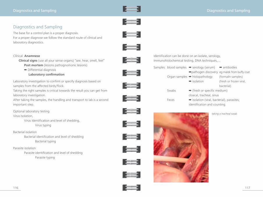
117 116
Diagnostics and Sampling Diagnostics and Sampling
Clinical: AnamneseClinical signs (use all your sense organs) “see, hear, smell, feel”
Post mortem (lesions pathognomonic lesions)°�Differential diagnosis
Laboratory confirmation
Laboratory investigation to confirm or specify diagnosis based on samples from the affected birds/flock.Taking the right samples is critical towards the result you can get from laboratory investigation.After taking the samples, the handling and transport to lab is a second important step.
Optional laboratory testingVirus Isolation,
Virus Identification and level of shedding,Virus typing
Bacterial isolationBacterial identification and level of shedding
Bacterial typing
Parasite isolationParasite identification and level of shedding
Parasite typing
Diagnostics and SamplingThe base for a control plan is a proper diagnosis.For a proper diagnose we follow the standard route of clinical and laboratory diagnostics.
Identification can be done on an isolate, serology, Immunohistochemical testing, DNA techniques,…
Samples: blood samples °�serology (serum) °�antibodies � � °pathogen discovery eg marek from buffy coat Organ samples °�histopathology (formalin samples) °�isolation (fresh or frozen viral, bacterial) Swabs °�(fresh or specific medium) cloacal, tracheal, sinus Feces °�isolation (viral, bacterial), parasites; identification and counting.
taking a tracheal swab

119 118
notes notes

121 120
0600
58 .
01.2
013
. © 2
012
. Int
erve
t Int
erna
tiona
l B.V
. a s
ubsi
diar
y of
Mer
ck &
Co.
, Inc
., W
hite
hous
e St
atio
n, N
J, U
SA. A
ll rig
hts
rese
rved
.
Intervet International bvPO Box 31, 5830 AA Boxmeer The Netherlands Phone: +31 (0)485 587600Fax: +31 (0)485 577333E-mail: [email protected] www.msd-animal-health.com
Impor tantpoultr ydiseases
Impor tantpoultr y
diseases