Important Poultry Diseases · Foreword We first published the “Important Poultry Diseases ” in...
90
Important Poultry Diseases
Transcript of Important Poultry Diseases · Foreword We first published the “Important Poultry Diseases ” in...
Important Poultry Diseases
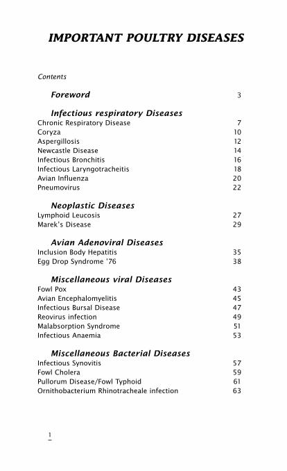
IMPORTANT POULTRY DISEASES
Contents
Foreword 3
InfectiousrespiratoryDiseases Chronic Respiratory Disease 7Coryza 10Aspergillosis 12Newcastle Disease 14Infectious Bronchitis 16Infectious Laryngotracheitis 18Avian Influenza 20Pneumovirus 22
NeoplasticDiseasesLymphoid Leucosis 27Marek’s Disease 29
AvianAdenoviralDiseasesInclusion Body Hepatitis 35Egg Drop Syndrome ’76 38
MiscellaneousviralDiseasesFowl Pox 43Avian Encephalomyelitis 45Infectious Bursal Disease 47Reovirus infection 49Malabsorption Syndrome 51Infectious Anaemia 53
MiscellaneousBacterialDiseasesInfectious Synovitis 57Fowl Cholera 59Pullorum Disease/Fowl Typhoid 61Ornithobacterium Rhinotracheale infection 63
1
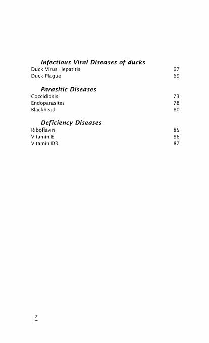
InfectiousViralDiseasesofducksDuck Virus Hepatitis 67Duck Plague 69
ParasiticDiseasesCoccidiosis 73Endoparasites 78Blackhead 80
DeficiencyDiseasesRiboflavin 85Vitamin E 86Vitamin D3 87
2
Foreword
We first published the “Important Poultry Diseases ”in 1972 and, ever since, it has remained the most sought after of all our publications.
However, in the intervening years new causes of disease such as chicken anemia virus and pneumovirus were identified. Therefore, Intervet Research teams developed new vaccines to combat the changed pattern of diseases. Many of these are inactivated vaccines, in either single or combined form, resulting in new vaccination programmes. We have, therefore, produced this fourth edition of the handbook to cover these developments. It is intended as a pocket reference book for people working with poultry. It is not highly academic as it is written from a practical angle to assist with every day problems in the field.
Intervet Research continues to co-operate with the poultry industry, worldwide, to stay ahead of disease problems.
The farmer who uses an Intervet vaccine can do so knowing that it is the best product available.
For detailed information on any of our products contact the local Intervet representative or
Intervet International BVBoxmeer- HollandP.O. Box 315830 AA Boxmeer, The NetherlandsPhone +31 485 58 76 00Fax +31 485 57 73 33Telex 37306 inte nlE-mail [email protected]
3

4
INFEcTIOUS RESPIRATORY
DISEASES
•ChronicRespiratoryDisease(CRD)
•Coryza
•Aspergillosis
•NewcastleDisease(ND)
•InfectiousBronchitis(IB)
•InfectiousLaryngotracheitis(ILT)
•AvianInfluenza
•TurkeyRhinotracheitis/Swollen
HeadSyndrome
5

6
chronic respiratory Disease
(cRD) (Airsacculitis)
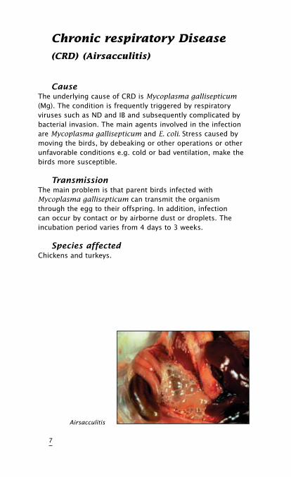
CauseThe underlying cause of CRD is Mycoplasma gallisepticum (Mg). The condition is frequently triggered by respiratory viruses such as ND and IB and subsequently complicated by bacterial invasion. The main agents involved in the infection are Mycoplasma gallisepticum and E. coli. Stress caused by moving the birds, by debeaking or other operations or other unfavorable conditions e.g. cold or bad ventilation, make the birds more susceptible.
TransmissionThe main problem is that parent birds infected with Mycoplasma gallisepticum can transmit the organism through the egg to their offspring. In addition, infection can occur by contact or by airborne dust or droplets. The incubation period varies from 4 days to 3 weeks.
SpeciesaffectedChickens and turkeys.
7
Airsacculitis
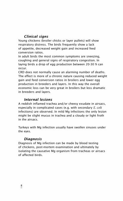
ClinicalsignsYoung chickens (broiler chicks or layer pullets) will show respiratory distress. The birds frequently show a lack of appetite, decreased weight gain and increased feed conversion ratios.In adult birds the most common symptoms are sneezing, coughing and general signs of respiratory congestion. In laying birds a drop of egg production between 20-30 % can occur.CRD does not normally cause an alarming number of deaths. The effect is more of a chronic nature causing reduced weight gain and feed conversion ratios in broilers and lower egg production in breeders and layers. In this way the overall economic loss can be very great in broilers but less dramatic in breeders and layers.
InternallesionsA reddish inflamed trachea and/or cheesy exudate in airsacs, especially in complicated cases (e.g. with secondary E. coli infections) are observed. In mild Mg infections the only lesion might be slight mucus in trachea and a cloudy or light froth in the airsacs.
Turkeys with Mg infection usually have swollen sinuses under the eyes.
DiagnosisDiagnosis of Mg infection can be made by blood testing of chickens, post-mortem examination and ultimately by isolating the causative Mg organism from tracheas or airsacs of affected birds.
8
DifferentialdiagnosisRespiratory virus infection (Newcastle disease or infectious bronchitis) with secondary infection (E. coli, etc.) can give similar lesions.
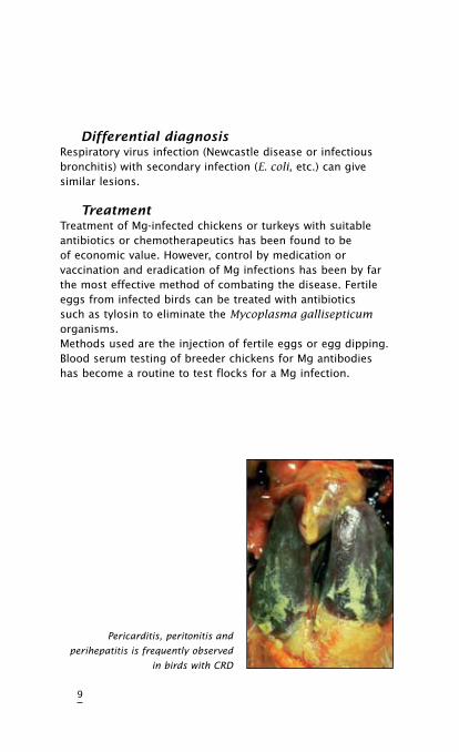
TreatmentTreatment of Mg-infected chickens or turkeys with suitable antibiotics or chemotherapeutics has been found to be of economic value. However, control by medication or vaccination and eradication of Mg infections has been by far the most effective method of combating the disease. Fertile eggs from infected birds can be treated with antibiotics such as tylosin to eliminate the Mycoplasma gallisepticum organisms.Methods used are the injection of fertile eggs or egg dipping. Blood serum testing of breeder chickens for Mg antibodies has become a routine to test flocks for a Mg infection.
9
Pericarditis, peritonitis and
perihepatitis is frequently observed
in birds with CRD
Infectious coryza
CauseThe bacterium causing this disease is Hemophilus paragallinarum.
TransmissionThe disease spreads from bird to bird and flock to flock by contact and airborne infected dust particles and via the drinking water.Spread by equipment and personnel has also been reported. The incubation period varies from 1 to 3 days.
SpeciesaffectedChickens appear to be the only natural hosts of H. paragallinarum.
ClinicalsignsThe main signs of the disease are inflammation of eyes and nose with foul-smelling discharges, conjunctivitis, sneezing and facial swellings. Feed and water intake is reduced, leading to loss of weight. Egg production in laying birds will drop.Mortality will vary with the virulence of the infection but is generally low.
10
11
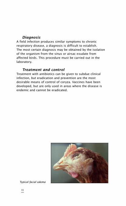
DiagnosisA field infection produces similar symptoms to chronic respiratory disease, a diagnosis is difficult to establish.The most certain diagnosis may be obtained by the isolation of the organism from the sinus or airsac exudate from affected birds. This procedure must be carried out in the laboratory.
TreatmentandcontrolTreatment with antibiotics can be given to subdue clinical infection, but eradication and prevention are the most desirable means of control of coryza. Vaccines have been developed, but are only used in areas where the disease is endemic and cannot be eradicated.
Typical facial edema
Aspergillosis (Fungal Pneumonia)
CauseThe disease is caused by a fungus, Aspergillus fumigatus.
TransmissionTransmission is by inhalation of fungus spores from ontaminated litter (e.g. wood shavings) or contaminated feed. Hatcheries may also contribute to infection of chicks.
SpeciesaffectedYoung chicks are very susceptible, older chickens are more resistant to infection. Turkey poults, pheasant chicks, quail chicks, ducklings, and goslings may also become infected.
ClinicalsignsInfected chicks are depressed and thirsty. Gasping and rapid breathing (“pump handle breathing”) can be observed.Mortality is variable, from 5 to 50 %.Gross lesions involve the lungs and airsacs primarily. Yellow-white pinpoint lesions can be found. Sometimes all body cavities are filled with small yellow-green granular fungus growth.
12
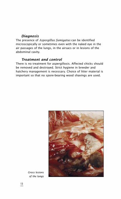
DiagnosisThe presence of Aspergillus fumigatus can be identified microscopically or sometimes even with the naked eye in the air passages of the lungs, in the airsacs or in lesions of the abdominal cavity.
TreatmentandcontrolThere is no treatment for aspergillosis. Affected chicks should be removed and destroyed. Strict hygiene in breeder and hatchery management is necessary. Choice of litter material is important so that no spore-bearing wood shavings are used.
13
Gross lesions
of the lungs
Newcastle Disease (ND)
CauseNewcastle disease is caused by a paramyxovirus. Only one serotype of ND is known. ND virus has mild strains (lentogenic), medium strength strains (mesogenic), and virulent strains (velogenic).The strains used for live vaccines are mainly lentogenic.
TransmissionNewcastle disease virus is highly contagious through infected droppings and respiratory discharge between birds. Spread between farms is by infected equipment, trucks, personnel, wild birds or air. The incubation period is variable but usually about 3 to 6 days.
SpeciesaffectedChickens and turkeys.
ClinicalsignsNewcastle disease causes high mortality with depression and death in 3 to 5 days as major signs. Affected chickens do not always exhibit respiratory or nervous signs.Mesogenic strains cause typical signs of respiratory distress.
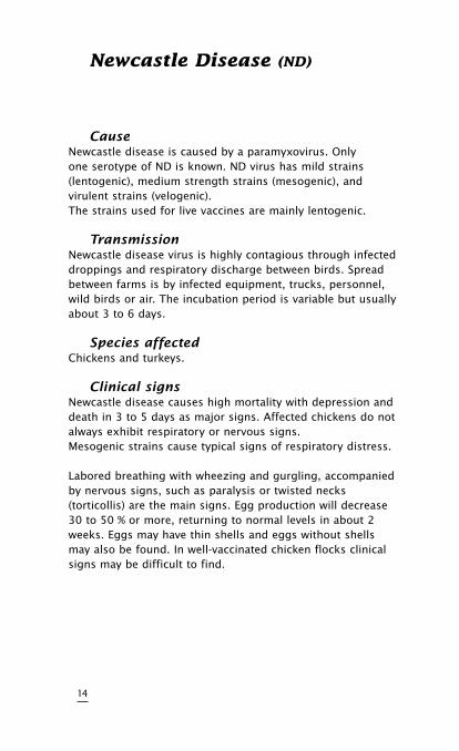
Labored breathing with wheezing and gurgling, accompanied by nervous signs, such as paralysis or twisted necks (torticollis) are the main signs. Egg production will decrease 30 to 50 % or more, returning to normal levels in about 2 weeks. Eggs may have thin shells and eggs without shells may also be found. In well-vaccinated chicken flocks clinical signs may be difficult to find.
14
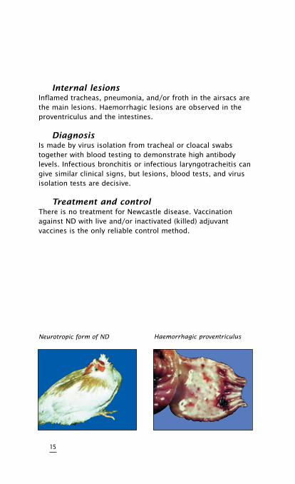
InternallesionsInflamed tracheas, pneumonia, and/or froth in the airsacs are the main lesions. Haemorrhagic lesions are observed in the proventriculus and the intestines.
DiagnosisIs made by virus isolation from tracheal or cloacal swabs together with blood testing to demonstrate high antibody levels. Infectious bronchitis or infectious laryngotracheitis can give similar clinical signs, but lesions, blood tests, and virus isolation tests are decisive.
TreatmentandcontrolThere is no treatment for Newcastle disease. Vaccination against ND with live and/or inactivated (killed) adjuvant vaccines is the only reliable control method.
15
Neurotropic form of ND Haemorrhagic proventriculus
Infectious Bronchitis (IB)
CauseCorona-virus is the causal agent. Several different serotypes of IB virus are known to exist.
TransmissionThe virus is transmitted from bird to bird through the airborne route. The virus can also be transmitted via the air between chicken houses and even from farm to farm.
SpeciesaffectedOnly chickens are susceptible to IB virus.
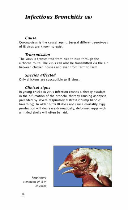
ClinicalsignsIn young chicks IB virus infection causes a cheesy exudate in the bifurcation of the bronchi, thereby causing asphyxia, preceded by severe respiratory distress (“pump handle” breathing). In older birds IB does not cause mortality. Egg production will decrease dramatically, deformed eggs with wrinkled shells will often be laid.
16
Respiratory
symptoms of IB in
chickens
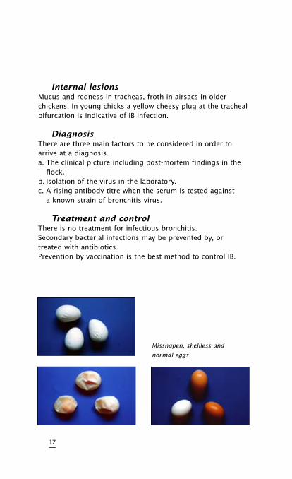
InternallesionsMucus and redness in tracheas, froth in airsacs in older chickens. In young chicks a yellow cheesy plug at the tracheal bifurcation is indicative of IB infection.
DiagnosisThere are three main factors to be considered in order to arrive at a diagnosis.a. The clinical picture including post-mortem findings in the flock.b. Isolation of the virus in the laboratory.c. A rising antibody titre when the serum is tested against a known strain of bronchitis virus.
TreatmentandcontrolThere is no treatment for infectious bronchitis.Secondary bacterial infections may be prevented by, or treated with antibiotics.Prevention by vaccination is the best method to control IB.
17
Misshapen, shellless and
normal eggs
Infectious Laryngotracheitis
(ILT)
CauseILT is caused by a virus belonging to the herpes group. Only one serotype is known.
TransmissionField infection occurs from bird to bird by the respiratory route. Most outbreaks of ILT on farms are traced back to transmission by contaminated people or equipment (visitors, shoes, clothing, egg boxes, used feeders, waterers, cages, crates etc.). The incubation period varies from 4 to 12 days.
SpeciesaffectedChickens and pheasants are natural hosts for ILT.
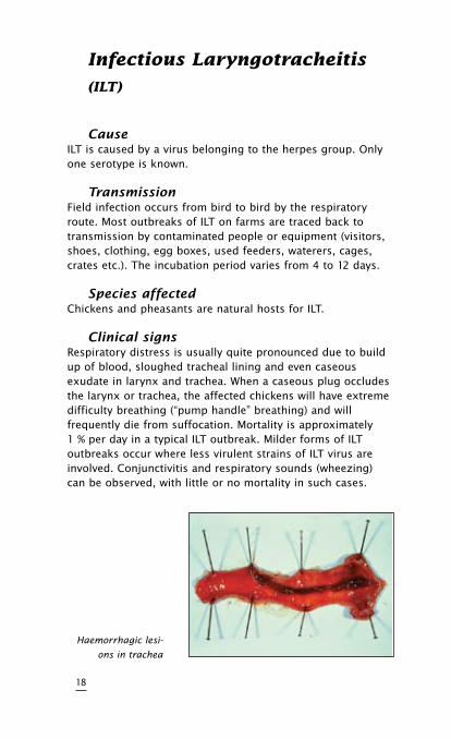
ClinicalsignsRespiratory distress is usually quite pronounced due to build up of blood, sloughed tracheal lining and even caseous exudate in larynx and trachea. When a caseous plug occludes the larynx or trachea, the affected chickens will have extreme difficulty breathing (“pump handle” breathing) and will frequently die from suffocation. Mortality is approximately 1 % per day in a typical ILT outbreak. Milder forms of ILT outbreaks occur where less virulent strains of ILT virus are involved. Conjunctivitis and respiratory sounds (wheezing) can be observed, with little or no mortality in such cases.
18
Haemorrhagic lesi-
ons in trachea
The disease spreads through a chicken house more slowly than either IB or ND. Egg production in laying flocks will usually decrease 10 to 50 %, but will return to normal after 3 to 4 weeks.
DiagnosisIn a chicken flock, spreading of respiratory distress, with possible coughing up of blood and mortality is indicative of ILT.Bloody mucus and cheesy exudate can be found in larynx and trachea. In the laboratory a definite diagnosis can be made by histological examination of tracheal tissues or virus isolation from tracheal mucus in embryonated chicken eggs.
TreatmentandcontrolPrevention of ILT by vaccination with mild eye-drop vaccine is by far the best control method. Sometimes such vaccines are applied by drinking water or spray methods with variable success. Even when an outbreak of ILT has been detected in a chicken flock, immediate vaccination is advisable to stop the spread of infection.
19
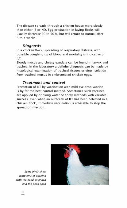
Some birds show
symptoms of gasping
with the head extended
and the beak open
Avian Influenza
CauseAvian influenza is caused by a myxovirus. There are several serotypes.
TransmissionAirborne virus particles from the respiratory tract, droppings, and people-carrying virus on their clothing and equipment are the main routes of transmission.
SpeciesaffectedTurkeys and ducks are mainly affected but chickens, geese, and wild birds can also be infected.
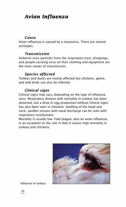
ClinicalsignsClinical signs may vary, depending on the type of influenza virus. Respiratory disease with mortality in turkeys has been observed, but a drop in egg production without clinical signs has also been seen in chickens. Swelling of the head and neck, swollen sinuses with nasal discharge can be seen with respiratory involvement.Mortality is usually low. Fowl plague, also an avian influenza, is an exception to the rule in that it causes high mortality in turkeys and chickens.
20
Influenza in turkeys
DiagnosisA laboratory diagnosis is necessary by serological (agar gel precipitation AGP) or virological methods (virus isolation). Avian influenza can be confused with Newcastle disease, fowl pox, Mycoplasma infection, Staphylococcus, or other respiratory or systemic infections.
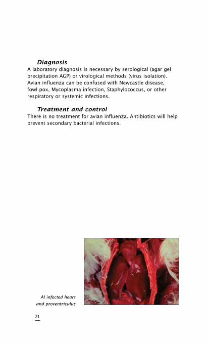
TreatmentandcontrolThere is no treatment for avian influenza. Antibiotics will help prevent secondary bacterial infections.
21
AI infected heart
and proventriculus
Pneumovirus Infections
(Turkey Rhinotracheitis/Swollen Head
Syndrome)
CauseThe disease is caused by a pneumovirus.
TransmissionThe virus may be transmitted horizontally by contaminated water, personnel and equipment as well as from bird to bird.
SpeciesaffectedTurkeys and chickens.
ClinicalsignsandlesionsIn young turkeys sneezing. Rales and nasal discharge, conjunctivitis, swelling of the infraorbital and submandibular sinuses can be seen.In laying flocks a drop in production may occur along with respiratory distress. Morbidity is high whereas mortality may vary being usually higher in young poults. In chickens the pneumovirus may be involved in the so called “swollen head syndrome” (SHS). In such cases affected chickens may show swelling of the periorbital and infraorbital sinuses, torticollis, cerebral disorientation and depression. Marked egg production losses can be associated with SHS.At necropsy the lesions seen may vary due to other microorganisms that may complicate the original picture. In cases of SHS apart from oedema in the head also purulent or caseous subcutaneous exudate can be found. Rhinitis, tracheitis and sinusitis are frequently noted in both chicken and turkeys, hence also the name turkey rhinotracheitis (TRT). Poliserositis affecting the air sacs and pericardium may be due to secondary infections (E. coli). The kidneys may be swollen and congested as well as the lungs which may show a fibrinous exudate in the pleural cavity.
22
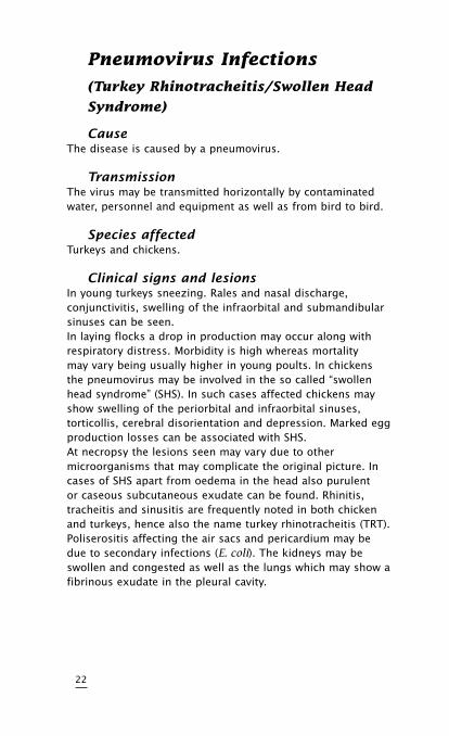
DiagnosisThe diagnosis based only on clinical signs is difficult to establish since other agents may be involved. The most certain diagnosis may be obtained by the isolation of the organism from nasal secretions or tissue scraped from the sinus of affected birds. Antibodies can be detected by several serological methods such as the VN test, IFT and ELISA.
TreatmentandcontrolTreatment with antibiotics can be given to control secondary bacterial infections. The use of vaccines should be the best approach to control the disease.
23
Swollen head
syndrome
Young turkey
with conjunctivitis
and sinusitis

24
NEOPLASTIc DISEASES
•LympoidLeucosis
•Marek’sDisease
25

26
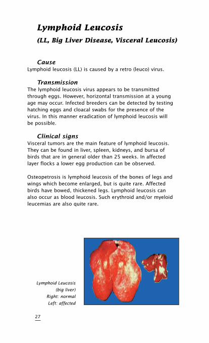
Lymphoid Leucosis
(LL, Big Liver Disease, Visceral Leucosis)
CauseLymphoid leucosis (LL) is caused by a retro (leuco) virus.
TransmissionThe lymphoid leucosis virus appears to be transmitted through eggs. However, horizontal transmission at a young age may occur. Infected breeders can be detected by testing hatching eggs and cloacal swabs for the presence of the virus. In this manner eradication of lymphoid leucosis will be possible.
ClinicalsignsVisceral tumors are the main feature of lymphoid leucosis. They can be found in liver, spleen, kidneys, and bursa of birds that are in general older than 25 weeks. In affected layer flocks a lower egg production can be observed.
Osteopetrosis is lymphoid leucosis of the bones of legs and wings which become enlarged, but is quite rare. Affected birds have bowed, thickened legs. Lymphoid leucosis can also occur as blood leucosis. Such erythroid and/or myeloid leucemias are also quite rare.
27
Lymphoid Leucosis
(big liver)
Right: normal
Left: affected
Affected birds are listless, pale, and are wasting away. Because of the tumours, LL may be confused with Marek’s disease, but in LL the nervous system is never involved (no paralysis). LL generally causes birds to weaken, lose weight and eventually die. Histopathological examination is essential for a proper diagnosis.
“J”VirusRecently, within the known subgroup of exogenous LL viruses (A, B, C and D), a new subgroup denominated “J” has emerged. The new “J” virus shows tropism for cells of the myelomonocytic series, causing tumors, which are identified by histopathology as being myelocytomas. The virus has tropism for meat –type birds. The tumors caused by this virus are normally seen from sexual maturity onward and are frequently located on the surface of bones such as the junction of the ribs, sternum, pelvis, mandible and skull and may be also found in visceral organs.
TreatmentandcontrolNo treatment is known. The best control method is the laboratory detection of infected breeders. Breeding leucosis-free offspring from leucosis-free breeders can eventually lead to eradication of the disease.
28
Marek’s Disease
(MD, Neurolymphomatosis)
CauseMarek’s disease is caused by a herpes virus.
TransmissionMain transmission is by infected premises, where day-old chicks will become infected by the oral and respiratory routes. Dander from feather follicles of MD-infected chickens can remain infectious for more than a year. Young chicks are particularly susceptible to horizontal transmission. Susceptibility decreases rapidly after the first few days of age.
SpeciesaffectedThe domestic fowl.
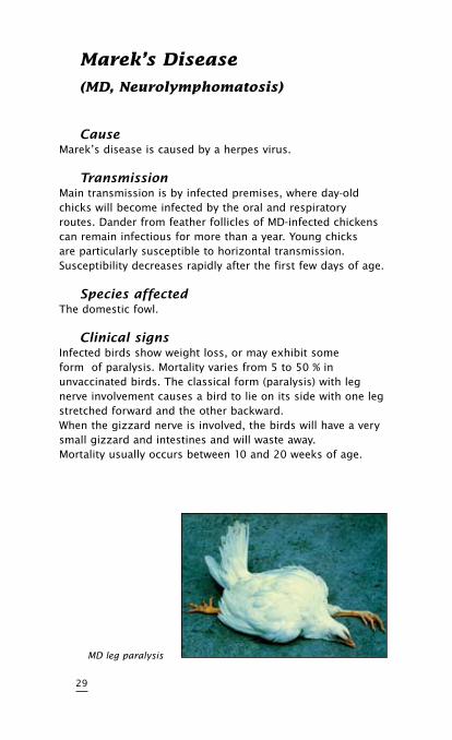
ClinicalsignsInfected birds show weight loss, or may exhibit some form of paralysis. Mortality varies from 5 to 50 % in unvaccinated birds. The classical form (paralysis) with leg nerve involvement causes a bird to lie on its side with one leg stretched forward and the other backward.When the gizzard nerve is involved, the birds will have a very small gizzard and intestines and will waste away.Mortality usually occurs between 10 and 20 weeks of age.
29
MD leg paralysis
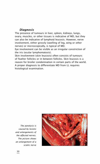
DiagnosisThe presence of tumours in liver, spleen, kidneys, lungs, ovary, muscles, or other tissues is indicative of MD, but they can also be indicative of lymphoid leucosis. However, nerve involvement, either grossly (swelling of leg, wing or other nerves) or microscopically, is typical of MD.Eye involvement can be visible as an irregular constriction of the iris (ocular lymphomatosis).Skin involvement (skin leucosis) often consists of tumours of feather follicles or in between follicles. Skin leucosis is a reason for broiler condemnation in certain parts of the world. A proper diagnosis to differentiate MD from LL requires histological examination.
30
The paralysis is
caused by lesions
and enlargements of
the affected nerves.
The picture shows
an enlargement of a
sciatic nerve
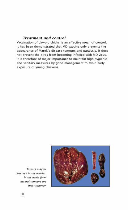
TreatmentandcontrolVaccination of day-old chicks is an effective mean of control. It has been demonstrated that MD vaccine only prevents the appearance of Marek’s disease tumours and paralysis. It does not prevent the birds from becoming infected with MD-virus. It is therefore of major importance to maintain high hygienic and sanitary measures by good management to avoid early exposure of young chickens.
31
Tumors may be
observed in the ovaries.
In the acute form
visceral tumours are
most common

32
AVIAN ADENOVIRAL DISEASES
• InclusionBodyHepatitis
(Hydropericardium-Hepatitis
Syndrome)
• EggDropSyndrome1976
(EDS ’76)
33

34
Inclusion Body Hepatitis
(Hydropericardium-Hepatitis Syndrome)
CauseThe disease is caused by an avian adenovirus (for example the Tipton strain) and is usually simultaneously accompanied by other immunosuppressive diseases such as infectious bursal disease or infectious anaemia. There are 12 known serotypes of avian adenoviruses that may be involved in the development of this disease.
TransmissionEgg transmission is an important factor.Horizontal transmission from bird to bird by contact with droppings. Once the bird becomes immune, the virus can no longer be isolated from the droppings.
SpeciesaffectedChickens, turkeys and pheasants and possibly other birds can be affected by avian adenovirus.
ClinicalsignsChickens with inclusion body hepatitis are affected at usually 5 to 7 weeks of age. The birds are listless, with ruffled feathers. Mortality is usually quite severe, up to 25 % in the first 10 days of the disease.
InternallesionsAffected chickens have mottled livers, many with pinpoint necrotic and haemorrhagic spots. Pale bone marrow and, in some cases in presence of infectious anemia, gangrenous dermatitis can be seen. Kidneys are pale and swollen. The spleen is usually quite small (atrophy).
35
If Gumboro disease (infectious bursal disease) has been present in the birds, even if subclinical, the bursa of Fabricius will be very small (atrophic). Such chickens are immune- suppressed and usually have more severe cases of inclusion body hepatitis and/or infectious anaemia.Mature birds do not have clinical signs of adenovirus infection, they only start showing antibodies in their blood.
Hydropericardium-HepatitisSyndromeHHS was reported for the first time in 1987 in Pakistan and was referred to as “Angara disease”. The disease has meanwhile been reported from several other countries, including India and countries in the Middle East and Latin America. Hydropericardium-hepatitis syndrome is caused by a virus belonging to the family of the fowl adenoviruses (FAV). Despite of the diversity in the geographical distribution of the disease, in all areas the infection is caused by a virus belonging to FAV serotype 4. There are three features which underline that this condition is a new disease, different from the known IBH. IBH and hydropericardium accompany this syndrome. Once outbreaks of HHS occur, it remains a problem for the poultry industry. And while IBH is shown to be caused by strains belonging to various FAV serotypes, HHS is, contrary to this, caused by FAV serotype 4. The infected flocks show high mortality rates and beside the lesions typical for IBH, a marked hydropericarditis is found in the affected birds.
36
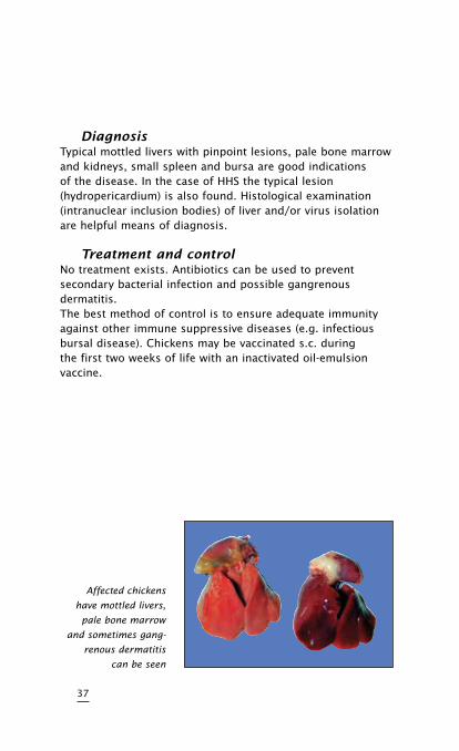
DiagnosisTypical mottled livers with pinpoint lesions, pale bone marrow and kidneys, small spleen and bursa are good indications of the disease. In the case of HHS the typical lesion (hydropericardium) is also found. Histological examination (intranuclear inclusion bodies) of liver and/or virus isolation are helpful means of diagnosis.
TreatmentandcontrolNo treatment exists. Antibiotics can be used to prevent secondary bacterial infection and possible gangrenous dermatitis.The best method of control is to ensure adequate immunity against other immune suppressive diseases (e.g. infectious bursal disease). Chickens may be vaccinated s.c. during the first two weeks of life with an inactivated oil-emulsion vaccine.
37
Affected chickens
have mottled livers,
pale bone marrow
and sometimes gang-
renous dermatitis
can be seen
Egg Drop Syndrome 1976
(EDS ’76)
CauseThe disease is caused by an avian adenovirus (strain BC14, virus 127), the EDS virus does not belong to any of the 12 fowl adenoviruses.
TransmissionThe virus is transmitted through the egg to a few birds in a flock, these birds carry the virus until the flock comes into lay at which time they begin to excrete virus and infect birds kept in the same house.Horizontal spread through infected litter can and does occur once a flock shows the disease but it seems that the virus is not very infectious or the level of virus excretion is low.
SpeciesaffectedOnly chickens are susceptible to clinical disease due to EDS virus; however, the virus is widespread in ducks but does not cause any problems.
38
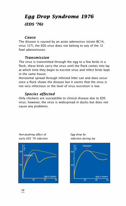
Non-peaking effect of
early EDS ’76 infection
Egg drop by
infection during lay
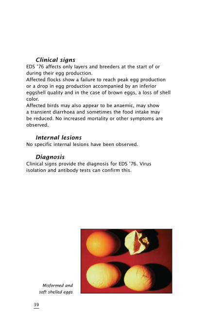
ClinicalsignsEDS ’76 affects only layers and breeders at the start of or during their egg production.Affected flocks show a failure to reach peak egg production or a drop in egg production accompanied by an inferior eggshell quality and in the case of brown eggs, a loss of shell color.Affected birds may also appear to be anaemic, may show a transient diarrhoea and sometimes the food intake may be reduced. No increased mortality or other symptoms are observed.
InternallesionsNo specific internal lesions have been observed.
DiagnosisClinical signs provide the diagnosis for EDS ’76. Virus isolation and antibody tests can confirm this.
39
Misformed and
soft shelled eggs
DifferentialdiagnosisInfectious bronchitis and to a lesser extent Newcastle disease and infectious laryngotracheitis will have to be considered. Proper diagnostic tests such as the antibody tests will eliminate doubt.
TreatmentandcontrolThere is no treatment against egg drop syndrome 1976.Vaccination with an inactivated vaccine before point of lay is the only available, effective method for the control of EDS ’76.
40
MIScELLANEOUS VIRAL
DISEASES
•FowlPox
•AvianEncephalomyelitis
•InfectiousBursalDisease
•ReovirusInfectious
•MalabsorptionSyndrome
•InfectiousAnaemia
41

42
Fowl Pox
(Avian Pox, Avian Diphtheria)
CauseFowl pox is caused by a pox virus.
TransmissionIntroduction of infected or “carrier” birds in a susceptible flock will cause an outbreak by direct contact and water or feed transmission.Mosquitoes and other flying insects can also transmit the virus from bird to bird and also transmit the disease to nearby flocks. The incubation period varies from 4 to 20 days.
Species,affectedChickens, turkeys, pheasants and pigeons can be affected by different fowl pox virus strains.
ClinicalsignsThe lesions of fowl pox can be external (mainly on the head) or internal (“wet pox”) in the mouth, oesophagus and/or trachea, they can also be found on other parts of the body (skin of legs, cloaca etc.). The lesions on the head, combs, and wattles are usually wart-like in appearance, yellow to dark brown in color. The internal lesions in the mouth, oesophagus and/or trachea are yellow-white and cheesy in appearance. Affected birds will be depressed, lack appetite and when “wet pox” is present they breath laboriously.
MortalityMortality is variable, from a low 1 to 2%, when slight head lesions are present, to over 40% when the diphtheritic form (“wet pox”) is more prevalent.Reduced egg production can be observed in laying birds, this will return to normal in a few weeks.
43
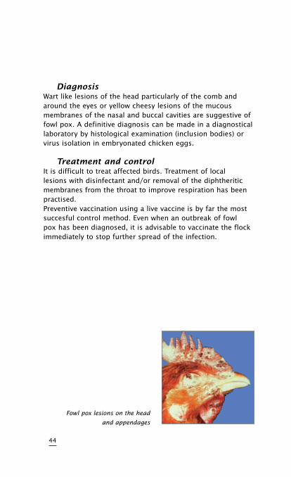
DiagnosisWart like lesions of the head particularly of the comb and around the eyes or yellow cheesy lesions of the mucous membranes of the nasal and buccal cavities are suggestive of fowl pox. A definitive diagnosis can be made in a diagnostical laboratory by histological examination (inclusion bodies) or virus isolation in embryonated chicken eggs.
TreatmentandcontrolIt is difficult to treat affected birds. Treatment of local lesions with disinfectant and/or removal of the diphtheritic membranes from the throat to improve respiration has been practised.Preventive vaccination using a live vaccine is by far the most succesful control method. Even when an outbreak of fowl pox has been diagnosed, it is advisable to vaccinate the flock immediately to stop further spread of the infection.
44
Fowl pox lesions on the head
and appendages
Avian Encephalomyelitis (AE)
or Epedemic Tremor
CauseAvian encephalomyelitis (AE) is caused by an enterovirus belonging to the picornavirus group.
TransmissionEgg transmission is the major route of transmission of AE virus. Infected breeders will transmit the AE virus for several weeks and cause a decrease in egg hatchability. Infected chicks that hatch will show clinical signs of the disease and spread the infection in the incubator to other newly hatched susceptible chicks. Young chicks can also be infected on the farm. The incubation period varies from 5 to 14 days depending on the route of infection.
SpeciesaffectedPrimarily chickens are susceptible to AE, but turkeys and pheasants have been reported as natural hosts.
ClinicalsignsThe disease is mainly seen in young chicks, between 1 and 3 weeks of age. Affected chicks sit on their hocks, do not move well, and many fall on their sides. A fine, rapid trembling of the head and neck can be seen, but especially felt when affected chicks are held in the hand. In laying and breeding flocks, AE virus infection causes a marked drop in egg production which returns to normal in about 2 weeks. Mortality in naturally infected chicks varies and can be as high as 75%.
45
DiagnosisClinical tremors in chicks, together with a drop in production and hatchability in the parent breeders, is indicative of AE infection. Chicks will not have gross lesions, but histological examination of brain, proventriculus and pancreas reveals typical lesions of AE. This will also differentiate the diagnosis of AE from encephalomalacia (Vitamin A deficiency, crazy chick disease). Laboratory testing of blood serum from breeder flocks, or their hatching eggs, can determine if an infection occurred.
TreatmentandcontrolPreventive vaccination of breeder pullets with live AE vaccine before egg production is the only effective means of AE control. If a breeder flock has not been, or has been inadequately, vaccinated against AE and an outbreak occurs, it is advisable to stop hatching eggs from the flock for several weeks until the breeders have acquired immunity and no longer transmit AE virus through their eggs.
46
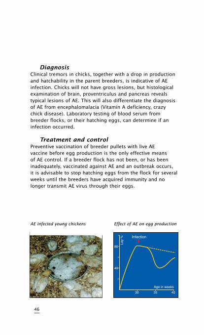
AE infected young chickens Effect of AE on egg production
Infectious Bursal Disease
(IBD, Gumboro Disease)
CauseThe disease is caused by a birna virus of serotype 1. Virus strains can be divided in classical and variant strains. The virus is very stable and is difficult to eradicate from an infected farm.
TransmissionIBD virus is very infectious and spreads easily from bird to bird by way of droppings. Infected clothing and equipment are means of transmission between farms.
SpeciesaffectedChickens and turkeys appear to be natural hosts.
ClinicalsignsClinical IBD occurs usually between 4 and 8 weeks of age. Affected birds are listless and depressed, pale and huddling. Mortality varies. Usually new cases of IBD have a mortality rate of about 5 to10% but can be as high as 60% depending on the pathogenicity of the strain involved. In subsequent infection on the same farm, mortality is lower and eventually, with successive attacks, there is no mortality noted.
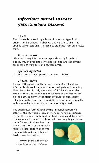
The subclinical form caused by the immunosuppressive effect of the IBD virus is now of more economic importance in that the immune system of the bird is damaged. Gumboro disease related diseases such as inclusion body hepatitis are more frequent in these birds. In broilers this form of the disease results in bad performance with lower weight gains and higherfeed conversion ratios.
47
Normal (right) and affected
bursa three days post infection
DiagnosisIn acute cases the bursa of Fabricius is enlarged and gelatinous, sometimes even bloody. Muscle haemorrhages and pale kidneys can be seen. Infection by variant strains is usually accompanied by a fast bursal atrophy (in 24-48 hours) without the typical signs of Gumboro disease. Also in chronic cases the bursa is smaller than normal (atrophy). The bursa destruction is apparent on histologic examination. The lack of white blood cells (lymphocytes) results in a reduction in the development of immunity and decreased resistance of the birds to other infections. Typical signs and lesions are diagnostic of IBD. Histopathological examination, serology and/or virus isolation are helpful tools. IBD can be confused with sulfonamide poisoning, aflatoxicosis, and pale bird syndrome (Vitamin E deficiency).
TreatmentandcontrolNo treatment is available for IBD. Vaccination of parent breeders and/or young chicks is the best means of control. The induction of a high maternal immunity in the progeny of vaccinated breeders, together with the vaccination of the offspring is the most effective approach to successful IBD control.
48
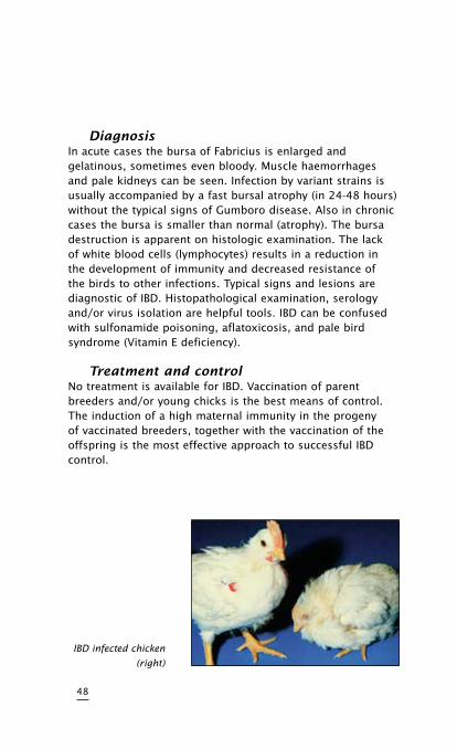
IBD infected chicken
(right)
Reovirus Infections
CauseReovirus infections, also know as viral arthritis/tenosynovitis is caused by an avian reovirus.
TransmissionThe virus may be transmitted by droppings from bird to bird. Egg transmission is also a factor when breeder flocks become infected during egg production.Reovirus is a common inhabitant of the intestines of birds and not all strains are pathogenic.
SpeciesaffectedChickens, turkeys and possibly pheasants are natural hosts.
ClinicalsignsThe first signs of reovirus infection are usually observed in broiler breeder chickens between 6 and 10 weeks of age. The birds are reluctant to walk and when forced up have a painful, trembling gait. A distinct swelling of the tendons of the shanks and also above the hock joint can be observed. Affected birds have malpositioned feathers, especially on the wings.
InternallesionsThe hock joint may be somewhat swollen, but usually not as severely as with Mycoplasma synoviae or Staphylococcus infections. Upon opening the legs the tendons usually appear discolored, brown or blood-tinged, with straw coloured fluid between them.
Ruptured tendons may occur and, in older broiler breeders (29-30 weeks old), one may feel a hard scarry knot in the tendon above the hock joint. When the infection is complicated by Ms or Staphylococcus the fluid may appear yellow and creamy. In commercial egg-laying breeds of chickens, the disease is not as common as in broiler breeders and broilers.
49
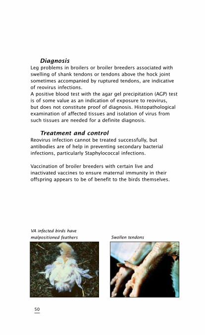
DiagnosisLeg problems in broilers or broiler breeders associated with swelling of shank tendons or tendons above the hock joint sometimes accompanied by ruptured tendons, are indicative of reovirus infections.A positive blood test with the agar gel precipitation (AGP) test is of some value as an indication of exposure to reovirus, but does not constitute proof of diagnosis. Histopathological examination of affected tissues and isolation of virus from such tissues are needed for a definite diagnosis.
TreatmentandcontrolReovirus infection cannot be treated successfully, but antibodies are of help in preventing secondary bacterial infections, particularly Staphylococcal infections.
Vaccination of broiler breeders with certain live and inactivated vaccines to ensure maternal immunity in their offspring appears to be of benefit to the birds themselves.
50
VA infected birds have
malpositioned feathers Swollen tendons
Malabsorption Syndrome
This complex disease has been reported under various names such as helicopter disease, femoral head necrosis, brittle bone disease, infectious proventriculitis, pale bird syndrome, running disease and stunting disease.
CauseThe malabsorption syndrome appears to be a disease complex involving avian Reoviruses and other viral and bacterial agents which may affect the digestive system resulting in nutritional and deficiency signs and lesions.
TransmissionOnly circumstantial evidence is present at the moment to indicate that the causal organism(s) may be vertically transmitted. Horizontal transmission also seems to play a role on infected sites.
SpeciesaffectedChickens and possibly turkeys.
ClinicalsignsThe disease is mainly observed in broiler flocks. Many affected broiler flocks have a history of diarrhoea, beginning as early as a few days of age and lasting until 10-14 days of age. Light or dark brown, foamy droppings can be found with undigested food particles. Several affected broilers in a flock may exhibit malpositioned feathers, especially on the wings. Early rickets with extreme paleness of legs and heads can be observed.
Encephalomalacia is also regularly found. At a later age (5-6 weeks) osteoporosis becomes clinically evident, frequently unilateral causing the birds to limp. Later an important effect is the delayed growth of the affected birds. Mortality is variable and in general as low as 4 %.
51
DiagnosisThe clinical disease is characterized by one or more of the following lesions: enteritis with watery brown and foaming contents and the presence of undigested food in the intestine. Mucosal and submucosal proventricular lesions. Pancreatic inflammatory infiltration with degenerative changes have been found. Osteoporosis and osteomyelitis, femoral head necrosis whereby the bone of the epiphysis of the femur is unusually soft.Since the causal agent may differ it is difficult to base a diagnose on virus isolation or serology.
TreatmentandcontrolTreatment is impossible, vaccination against reovirus in the breeders helps to reduce problems in the progency.Strict hygienic and sanitary measures will reduce the incidence of the disease.
52
Infectious Anaemia
CauseInfectious anaemia is caused by a very resistant small virus known as CAV (Chicken Anaemia Virus).
TransmissionThe major mode of transmission of infectious anemia is vertical transmission from infected breeder hens. Horizontal transmission from bird to bird or by infected equipment, clothing, etc. is also possible.
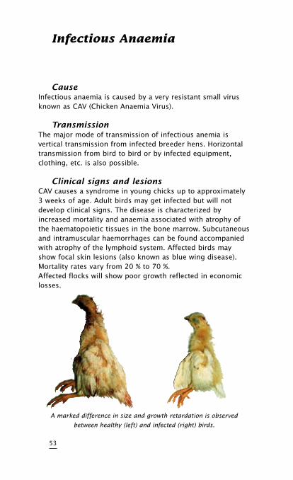
ClinicalsignsandlesionsCAV causes a syndrome in young chicks up to approximately 3 weeks of age. Adult birds may get infected but will not develop clinical signs. The disease is characterized by increased mortality and anaemia associated with atrophy of the haematopoietic tissues in the bone marrow. Subcutaneous and intramuscular haemorrhages can be found accompanied with atrophy of the lymphoid system. Affected birds may show focal skin lesions (also known as blue wing disease). Mortality rates vary from 20 % to 70 %.Affected flocks will show poor growth reflected in economic losses.
53
A marked difference in size and growth retardation is observed
between healthy (left) and infected (right) birds.
DiagnosisThe diagnosis can be based on the clinical signs and pathological findings in affected birds. Blood serum testing for specific CAV antibodies can be carried out (IFT, VN, ELISA). Virus isolation is also possible but it is time-consuming and expensive.
TreatmentandcontrolNo treatment is available for infectious anaemia. Maternally derived antibodies can offer protection. The induction of high maternal immunity in the progency by vaccinating breeders is the best approach to succesful CAV control.
54
MIScELLANEOUS
BAcTERIAL DISEASES
•InfectiousSynovitis
•FowlCholera
•SalmonellaPullorumDisease/
FowlTyphoid
55

56
Infectious Synovitis
CauseThis disease is caused by Mycoplasma synoviae (Ms).
TransmissionThe major mode of transmission of Ms is vertical (egg) transmission from Ms-infected breeder hens. Horizontal transmission from bird to bird and by infected equipment, clothing, shoes, egg boxes and other fomites.
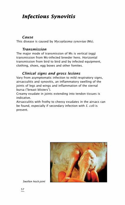
ClinicalsignsandgrosslesionsVary from asymptomatic infection to mild respiratory signs, airsacculitis and synovitis, an inflammatory swelling of the joints of legs and wings and inflammation of the sternal bursa (“breast blisters”).Creamy exudate in joints extending into tendon tissues is indicative.Airsacculitis with frothy to cheesy exudates in the airsacs can be found, especially if secondary infection with E. coli is present.
57
Swollen hock-joint
DiagnosisBlood serum testing for specific Ms antibodies with Ms antigen and the findings of specific lesions are indicative of Ms infections. Isolation of the causative Ms organisms is decisive for diagnosis.
DifferentialdiagnosisStaphylococcus arthritis can also cause swollen joints with a creamy exudate sometimes extending into the tendon sheaths. Reovirus infection can also cause swelling of joints and tendon sheaths, but the exudate is more watery or blood- tinged, unless secondary Staphylococcus infections occur.
TreatmentandcontrolMs infections can be treated with antibiotics with variable degrees or success (tetracycline, erythromycin, tylosin, tiamulin). However, control of Ms has been largely successful by blood testing of breeder chickens and elimination of positive Ms reactors.
58
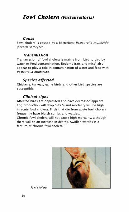
Fowl cholera (Pasteurellosis)
CauseFowl cholera is caused by a bacterium: Pasteurella multocida(several serotypes).
TransmissionTransmission of fowl cholera is mainly from bird to bird by water or feed contamination. Rodents (rats and mice) also appear to play a role in contamination of water and feed with Pasteurella multocida.
SpeciesaffectedChickens, turkeys, game birds and other bird species are susceptible.
ClinicalsignsAffected birds are depressed and have decreased appetite. Egg production will drop 5-15 % and mortality will be high in acute fowl cholera. Birds that die from acute fowl cholera frequently have bluish combs and wattles.Chronic fowl cholera will not cause high mortality, although there will be an increase in deaths. Swollen wattles is a feature of chronic fowl cholera.
59
Fowl cholera
InternallesionsGross lesions in acute cases are mainly internal haemorrhage and congestion of liver, spleen and kidneys. In chronic fowl cholera cheesy exudates can be found between the intestines, and on liver and heart.
TreatmentandcontrolTreatment with appropriate antibiotics or chemotherapeutics can be successful in halting mortality and restoring egg production. However chronic carrier birds have been found in flocks of chickens after treatment. If clinical fowl cholera with mortality reappears in such flocks, one must treat again. Rodent control is also very important to prevent reintroduction of the infection. Vaccines, both inactivated bacterins as well as live vaccines are available.
60
Pullorum Disease and Fowl
Typhoid
CausePullorum disease is caused by a bacterium, Salmonella pullorum. Fowl typhoid is caused by Salmonella gallinarum, which is related to, but not identical to, S. pullorum.
TransmissionPullorum can be transmitted by infected (carrier) breeder hens through their eggs. Chicks that hatch from such infected eggs will have typical pullorum disease (white diarrhoea) and high mortality. Infected chicks can also infect other chicks via droppings.Fowl typhoid is more a disease of adult chickens, with high mortality and morbidity. Horizontal transmission is important with fowl typhoid through infected droppings, dead bird carcasses, and infected clothing, shoes, utensils and other fomites.
SpeciesaffectedChickens, pheasants, ducks, geese and guinea fowl can contract both pullorum and fowl typhoid.
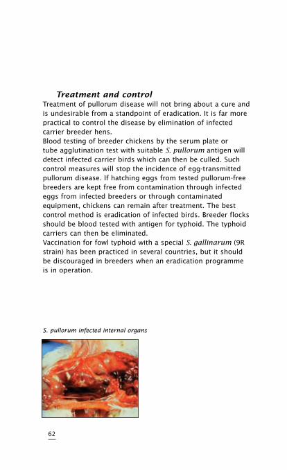
ClinicalsignsPullorum in chicks causes typical white bacillary diarrhoea, with pasted cloacas and high mortality. Infected adult breeders do not have clinical signs of the disease but have internal lesions in the ovary (miss-shaped, dark coloured follicles).Fowl typhoid in adult chickens causes listlessness and sulfurcoloured diarrhoea. The birds have generalized infection with swollen livers, spleens, and kidneys and haemorrhages in such tissues. Mortality is usually high: 25 to 60 %.
61
TreatmentandcontrolTreatment of pullorum disease will not bring about a cure and is undesirable from a standpoint of eradication. It is far more practical to control the disease by elimination of infected carrier breeder hens.Blood testing of breeder chickens by the serum plate or tube agglutination test with suitable S. pullorum antigen will detect infected carrier birds which can then be culled. Such control measures will stop the incidence of egg-transmitted pullorum disease. If hatching eggs from tested pullorum-free breeders are kept free from contamination through infected eggs from infected breeders or through contaminated equipment, chickens can remain after treatment. The best control method is eradication of infected birds. Breeder flocks should be blood tested with antigen for typhoid. The typhoid carriers can then be eliminated.Vaccination for fowl typhoid with a special S. gallinarum (9R strain) has been practiced in several countries, but it should be discouraged in breeders when an eradication programme is in operation.
62
S. pullorum infected internal organs
Ornithobacterium
rhinotracheale (OR) Infection
CauseOrnithobacterium rhinotracheale is a gram-negative rod responsible for causing infections in chickens and turkeys of all ages.
TransmissionHorizontal transmission from bird to bird and by infected fomites. Infected breeder hens may transmit the disease agent vertically to their offspring.
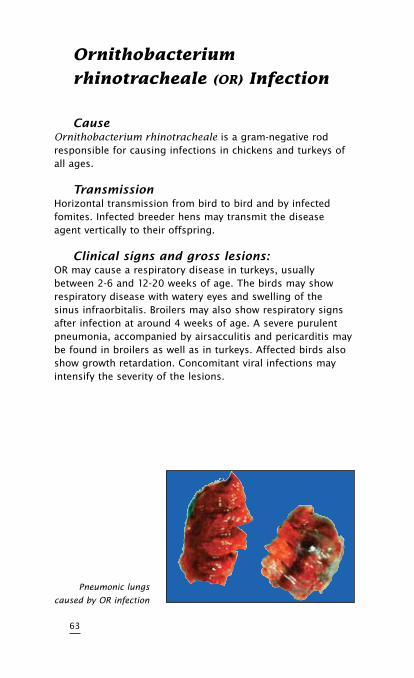
Clinicalsignsandgrosslesions:OR may cause a respiratory disease in turkeys, usually between 2-6 and 12-20 weeks of age. The birds may show respiratory disease with watery eyes and swelling of the sinus infraorbitalis. Broilers may also show respiratory signs after infection at around 4 weeks of age. A severe purulent pneumonia, accompanied by airsacculitis and pericarditis may be found in broilers as well as in turkeys. Affected birds also show growth retardation. Concomitant viral infections may intensify the severity of the lesions.
63
Pneumonic lungs
caused by OR infection
DiagnosisBlood serum testing using a commercially available ELISA for the detection of specific antibodies against OR and the finding of specific lesions are indicative of OR infection. Isolation of the organism and its biochemical determination may be attempted but care should be taken that these are carried out using appropriate methods in order to avoid unreliable results. Differential diagnose with bacteria causing similar disease patterns is recommended (E. coli, P. multocida, MG).
TreatmentandcontrolOR infections may be treated with broad -spectrum antibiotics with variable degrees of success. An inactivated vaccine for broiler breeders and turkey poults is available.
64
INFEcTIOUS VIRAL DISEASES
OF DUckS
•DuckHepatitis
•DuckPlague
65

66
Duck Virus Hepatitis
CauseDuck virus hepatitis is caused by a picornavirus. It is possible that there are more serotypes.
TransmissionThe disease can spread rapidly to all susceptible ducklings in the flock via faeces. The incubation period varies from around 24 hours to a few days.
SpeciesaffectedDucklings under 6 weeks of age are susceptible.
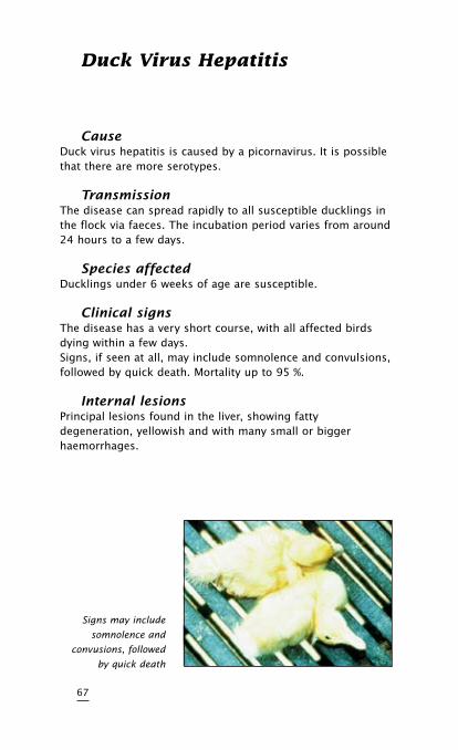
ClinicalsignsThe disease has a very short course, with all affected birds dying within a few days.Signs, if seen at all, may include somnolence and convulsions, followed by quick death. Mortality up to 95 %.
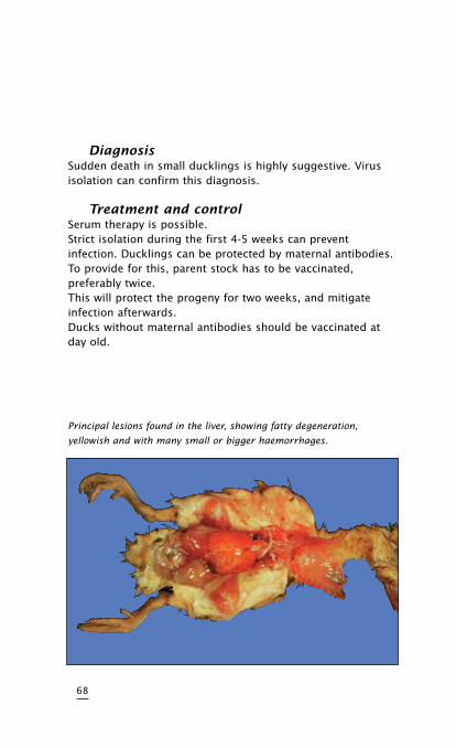
InternallesionsPrincipal lesions found in the liver, showing fatty degeneration, yellowish and with many small or bigger haemorrhages.
67
Signs may include
somnolence and
convusions, followed
by quick death
DiagnosisSudden death in small ducklings is highly suggestive. Virus isolation can confirm this diagnosis.
TreatmentandcontrolSerum therapy is possible.Strict isolation during the first 4-5 weeks can prevent infection. Ducklings can be protected by maternal antibodies. To provide for this, parent stock has to be vaccinated, preferably twice.This will protect the progeny for two weeks, and mitigate infection afterwards.Ducks without maternal antibodies should be vaccinated at day old.
68
Principal lesions found in the liver, showing fatty degeneration,
yellowish and with many small or bigger haemorrhages.
Duck Plague
(Duck Virus Enteritis)
CauseDuck plague is caused by a herpes virus. Only one serotype is known. There is a difference in virulence between strains.
TransmissionDuck plague virus is excreted by affected birds through faeces and other body discharges. Via soiled drinking water, contaminated pound water or open water other birds are infected.
SpeciesaffectedDucks, geese and swans, of all ages.
ClinicalsignsHigh mortality up to 100 %, sometimes sudden death. Droppy appearance, slow movements with hanging wings. Birds show bloody nasal discharges and conjunctivitis, diarrhoea and they may make a hoarse noise. The birds are very thirsty.Birds often die in a rather characteristic position, with the neck twisted downwards, sidewards or backwards.In laying flocks egg production may drop 50 % or more.
69
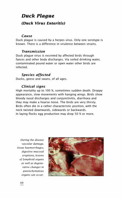
During the disease
vascular damage,
tissue haemorrhages,
digestive mucosal
eruptions, lesions
of lymphoid organs
as well as degene-
rative changes in
parenchymatous
organs can occur.
InternallesionsHaemorrhagic enteritis, haemorrhagic or pseudo-membranic pharingitis, oesophagitis and cloacitis, haemorrhagic ovaritis.
DiagnosisThe gross lesions are rather characteristic for duck plague. Virus isolation and neutralization confirm the diagnosis to distinguish the disease from avian cholera (Pasteurella multocida) or duck hepatitis.
TreatmentandcontrolThere is no treatment known. Prevention should include clean drinking water and keeping wild, free-flying waterfowl away. Vaccination with adapted strains can provide a reliable protection. Even in an infected flock emergency vaccination can limit the damage, due to an interference phenomenon between the vaccine virus and the field virus.
70
PARASITIc DISEASES
•Coccidiosis
•Endoparasites
•Blackhead
71

72
coccidiosis
CauseCoccidiosis is caused by protozoa, unicellular parasites. In chickens there are 9 different species of coccidia of which the main 5 are Eimeria acervulina, Eimeria necatrix, Eimeria tenella, Eimeria maxima and Eimeria brunetti.
TransmissionInfected droppings, containing oocysts of coccidia are the main means of transmission, between birds. The incubation period is 4 to 6 days.
SpeciesaffectedChickens have their own specific coccidiosis types which do not cross-infect other bird species.
73
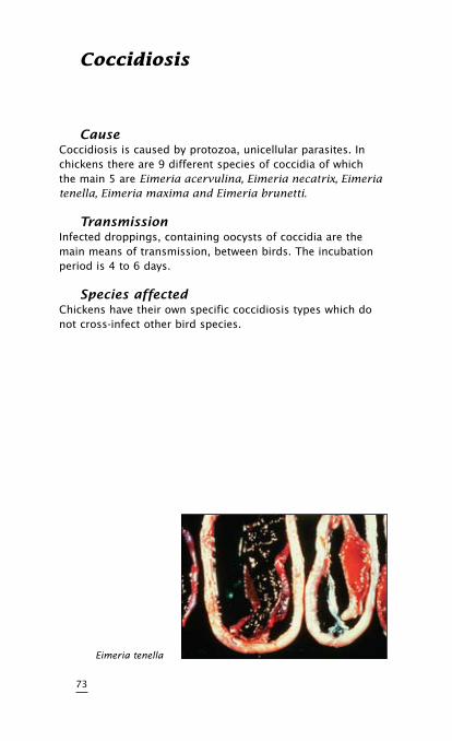
Eimeria tenella
Clinicalsigns/DiagnosisCoccidiosis can be divided into 2 groups:
The caecum is involved (Caecal coccidiosis).Mainly caused by E. tenella in chickens up to 12 weeks. Mortality may run as high as 50 %. Infected birds are listless, have bloody droppings, a pale comb and show a lack of appetite. Laboratory examination will show haemorrhages in the caecal wall. After severe bleeding a core will be formed in the lumen.
The small intestine is involved (small intestinal coccidiosis).Caused by E. acervulina, E. brunetti, E. maxima, E. necatrix.
E. acervulinaMay affect birds of any age.E. acervulina is not normally very pathogenic, but in some cases considerable mortality may be seen.Birds infected show loss of weight, combs may be shriveled and a drop or even cessation of egg production in layers may be seen.At necropsy, haemorrhagic lesions of E. acervulina are seen throughout the upper portion of the affected intestine and also grey or whitish patches may be present.
74
E. brunettiMay affect birds of any age.E. brunetti is definitely pathogenic, in severe infections mortality can be high. Birds infected show emaciation and diarrhoea.At necropsy a white cheese-like material is found in the lumen of the lower intestine and rectum.The caeca and cloaca are inflamed. The gut wall is thickened.
E. maximaMay affect birds of any age.E. maxima is less pathogenic than E. acervulina, necatrix andbrunetti, mortality is generally low.Diarrhoea, loss of weight and a drop in egg production of layers, will be seen; bloody droppings are common.At necropsy the lower portion of the small intestine is dilated and the wall is thickened; the gut is filled with thick mucus, grayish, brownish or pinkish in color.
75
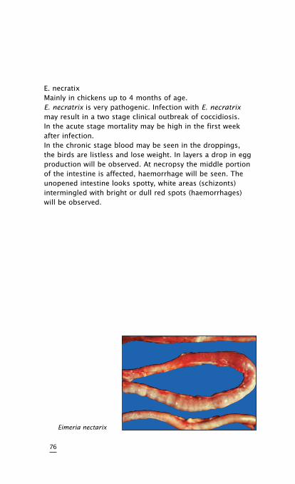
E. necratixMainly in chickens up to 4 months of age.E. necratrix is very pathogenic. Infection with E. necratrix may result in a two stage clinical outbreak of coccidiosis. In the acute stage mortality may be high in the first week after infection.In the chronic stage blood may be seen in the droppings, the birds are listless and lose weight. In layers a drop in egg production will be observed. At necropsy the middle portion of the intestine is affected, haemorrhage will be seen. The unopened intestine looks spotty, white areas (schizonts) intermingled with bright or dull red spots (haemorrhages) will be observed.
76
Eimeria nectarix
TreatmentandcontrolThis heading is most appropriate in the case of coccidiosis as there is no disease group in poultry where both control and treatment are employed more.The well established principles of good management and husbandry are of basic importance.
It is common practice to include low levels of chemotherapeutics in the feed of birds. These chemicals are referred to as coccidiostats and as such keep in check the development of the parasites so that a pathological situation does not develop. It should, however, be taken into account that coccidia can develop a resistance to all chemicals so far used for this purpose and for this reason it is necessary to change from one chemical to another periodically. Treatment of infected flocks may be carried out by the administration of coccidiostats at a higher therapeutic level to the affected birds. There are certain products available which are specifically designed for treatment and which are not satisfactory for prevention. These chemicals are sometimes referred to as coccidiocidal agents.Whenever administering these products, particular attention should be paid to the dosage recommendation of the manufacturer.
77
Endoparasites
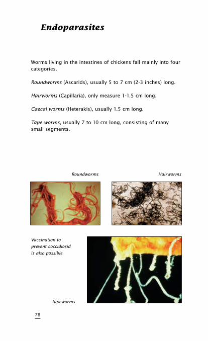
Worms living in the intestines of chickens fall mainly into four categories.
Roundworms (Ascarids), usually 5 to 7 cm (2-3 inches) long.
Hairworms (Capillaria), only measure 1-1.5 cm long.
Caecal worms (Heterakis), usually 1.5 cm long.
Tape worms, usually 7 to 10 cm long, consisting of many small segments.
78
Tapeworms
Vaccination to
prevent coccidiosid
is also possible
HairwormsRoundworms
ClinicalsignsMature roundworms are not a major cause of the disease, but the larvae can damage the intestinal lining, causing enteritis, anaemia, decreased egg production and at times eggs with pale yolks.
Capillaria cause more damage to the intestinal lining and can cause enteritis and anaemia with decreased egg production and the appearance of pale egg yolks (“platinum yolks”).
Caecal worms are found in the caeca and do not cause serious damage, except that their eggs can transmit blackhead – mainly in turkeys (see blackhead, page 80).
Tape worms are infrequently found and do not cause serious damage, except that they use the nutrients of the host chicken.
DiagnosisExamination of the intestinal contents will reveal roundworms, caecal worms, and tape worms without difficulty.Capillaria can usually be found when intestinal contents are washed through a fine mesh sieve.
TreatmentandcontrolRoundworms and caecal worm infections can be treated with piperazine. Piperazine is not effective against tape worms and capillaria for which other anthelmintics are required.
79
Blackhead
(Histomoniasis, Enterohepatitis)
CauseA protozoan parasite, Histomonas meleagridis.
TransmissionDirect transmission by infected water, feed, or droppings has been proved.Indirect transmission by infected eggs of the caecal worm.Heterakis gallinarum, is also a major factor. Raising turkeys and chickens on wire and indoors decreases the incidence of blackhead.
SpeciesaffectedChickens, turkeys, and peafowl are natural hosts to blackhead infection.
ClinicalsignsAffected birds are depressed, stand or sit with ruffled feathers, and have yellowish diarrhoea. Darkening of head parts, especially in turkeys, gave the name to the disease (blackhead). Gross lesions include circular necrotic areas in livers with a crater-like center and cheesy cores in the caeca. Blackhead can cause high mortality, particularly in young turkey poults, but the disease can also affect older birds. In chickens the mortality from blackhead infection is usually lower, young chickens being the most susceptible.
80
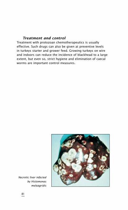
TreatmentandcontrolTreatment with protozoan chemotherapeutics is usually effective. Such drugs can also be given at preventive levels in turkeys starter and grower feed. Growing turkeys on wire and indoors can reduce the incidence of blackhead to a large extent, but even so, strict hygiene and elimination of caecal worms are important control measures.
81
Necrotic liver infected
by Histomonas
meleagridis

82
SOME IMPORTANT
VITAMIN DEFIcIENcY
DISEASES
•Riboflavin
•VitaminE
•VitaminD3
83

84
Riboflavin (Vitamin B2)
Deficiency (curly Toe Disease)
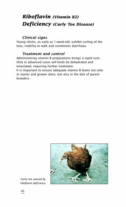
ClinicalsignsYoung chicks, as early as 1-week-old, exhibit curling of the toes, inability to walk and sometimes diarrhoea.
TreatmentandcontrolAdministering vitamin B preparations brings a rapid cure. Only in advanced cases will birds be dehydrated and emaciated, requiring further treatment.It is important to ensure adequate vitamin B levels not only in starter and grower diets, but also in the diet of parent breeders.
85
Curly toe caused by
riboflavin deficiency
Vitamin E Deficiency
(crazy chick Disease, Encephalomalacia)
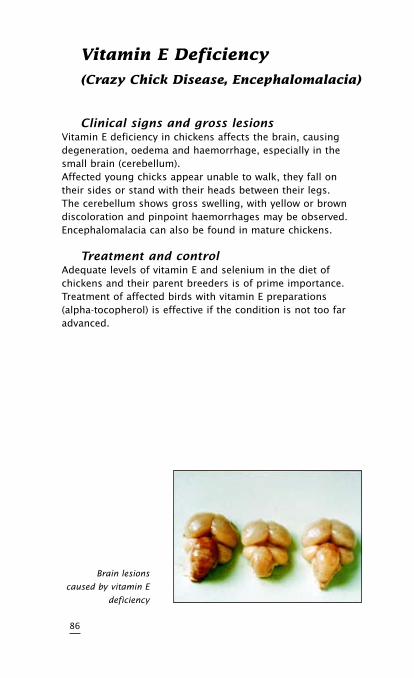
ClinicalsignsandgrosslesionsVitamin E deficiency in chickens affects the brain, causing degeneration, oedema and haemorrhage, especially in the small brain (cerebellum).Affected young chicks appear unable to walk, they fall on their sides or stand with their heads between their legs.The cerebellum shows gross swelling, with yellow or brown discoloration and pinpoint haemorrhages may be observed. Encephalomalacia can also be found in mature chickens.
TreatmentandcontrolAdequate levels of vitamin E and selenium in the diet of chickens and their parent breeders is of prime importance. Treatment of affected birds with vitamin E preparations (alpha-tocopherol) is effective if the condition is not too far advanced.
86
Brain lesions
caused by vitamin E
deficiency
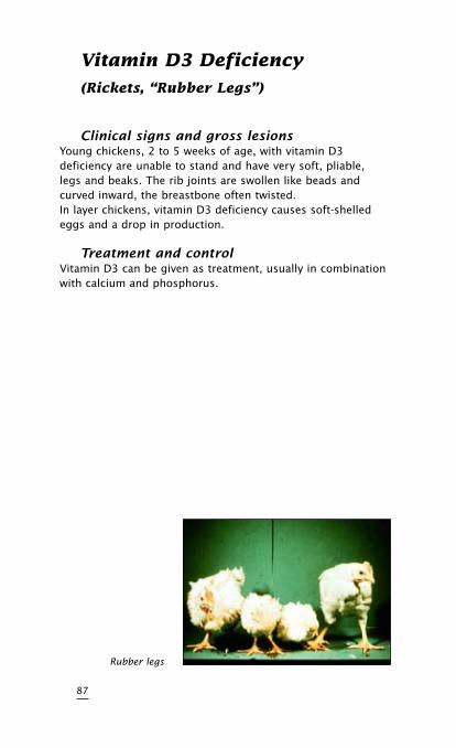
Vitamin D3 Deficiency
(Rickets, “Rubber Legs”)
ClinicalsignsandgrosslesionsYoung chickens, 2 to 5 weeks of age, with vitamin D3 deficiency are unable to stand and have very soft, pliable, legs and beaks. The rib joints are swollen like beads and curved inward, the breastbone often twisted.In layer chickens, vitamin D3 deficiency causes soft-shelled eggs and a drop in production.
TreatmentandcontrolVitamin D3 can be given as treatment, usually in combination with calcium and phosphorus.
87
Rubber legs

No p
art
of
this
bro
chu
re m
ay b
e re
pro
du
ced
in
an
y f
orm
wit
hou
t w
ritt
en p
erm
issi
on
fro
m I
nte
rvet
In
tern
atio
nal
.
Intervet/Schering-Plough Animal Health
91-105 Harpin Street, Bendigo East VIC 3550
Free Call: 1800 033 461 • Free Fax: 1800 817 414
[email protected] • www.intervet.com.au