IHC - BioNordika · to successful • Detection reagents ... SignalStain® Boost IHC Detection...
8
BioNordika (sweden) AB | Norrbackagatan 47 | tel 08 306 010 | www.bionordika.se | [email protected] IHC Volume 1 Issue 2 Immunohistochemistry
-
Upload
phamkhuong -
Category
Documents
-
view
224 -
download
0
Transcript of IHC - BioNordika · to successful • Detection reagents ... SignalStain® Boost IHC Detection...
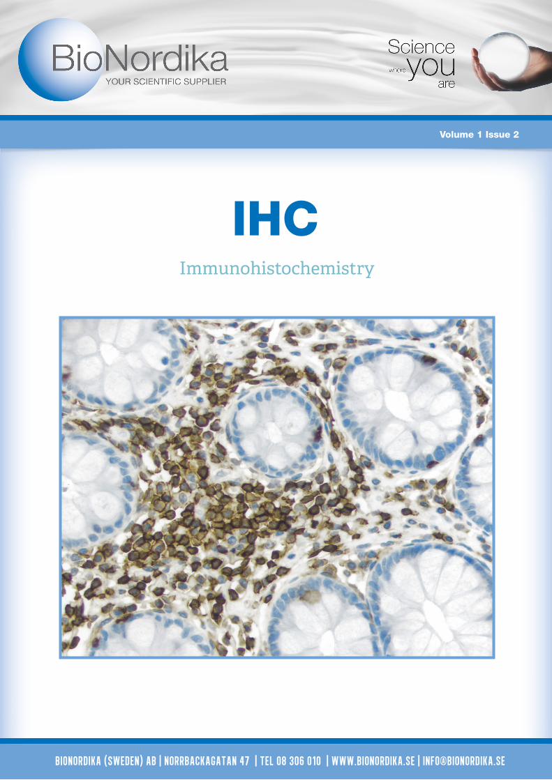
BioNordika (sweden) AB | Norrbackagatan 47 | tel 08 306 010 | www.bionordika.se | [email protected]
IHC
Volume 1 Issue 2
Immunohistochemistry

Complete Staining Kits with Amplified HRP Polymer Technology
The ImmPRESS™ Excel Amplified HRP (peroxi-dase) Polymer Staining Kit is an enzymatic, non-biotin amplification system that produces crisp, highly sensitive, specific staining with low background.
• MP-7601 ImmPRESS™ Excel Amplified HRP Polymer Staining Kit (Anti-Rabbit IgG)
• MP-7602 ImmPRESS™ Excel Amplified HRP Polymer Staining Kit (Anti-Mouse IgG)
The ImmPRESS™ Excel Reagent is conjugated to horseradish peroxidase micropolymers, a novel approach that avoids the use of large dextrans or other macromolecules as a back-bone. The included ImmPACT™ DAB EqV Substrate produces a crisp, dark brown reaction product with the excellent sensi-tivity and low background characteristic of the ImmPRESS™ / ImmPACT™ combination.
The ImmPRESS™ Excel Amplified Staining Kit includes the following reagents:
• BLOXALL™ Endogenous Enzyme Blocking Solution • 2.5% Normal Horse Serum • Amplifier Antibody (goat anti-rabbit or anti-mouse Ig)• ImmPRESS™ Excel Polymer Reagent (horse anti-goat Ig)• ImmPACT™ DAB EqV Reagent 1 & 2
Key advantages of the ImmPRESS™ Polymer System:
• High sensitivity
• Low background
• Non-biotin
• Micropolymer technology
• Enhanced accessibility to nuclear and membrane antigens
• One-step detection
• Ready-to-use in a convenient dropper bottle
• Shorter assay time
• Simplified multiple labeling
Add Primary Antibody 1
Add ImmPRESS™ Excel Polymer Reagent 3 Add Enzyme Substrate 4
Add Amplifier Antibody 2
2 www.bionordika.se

• Advanced avidin/biotin technology The Elite ABC complex is smaller, very uniform, and highly active, allowing more accessibility for binding to a biotinylated target.
• Highest sensitivity, low background The most sensitive avidin/biotin-based peroxidase system. The Elite ABC series is approximately 5 times more sensitive with the same low background.
• Cost effective Higher sensitivity leads to lower cost per slide.
• Available without (Standard kit #PK-6100) or with biotinylated species specific or universal secondary antibodies.
• Available in Ready-To-Use (R.T.U.) formats Prediluted, stabilized working solutions of Elite ABC Kit reagents provide the same high sensitivity and low background as the traditional reagents.
VECTASTAIN Elite ABC System
The Vector® M.O.M.™ Kits are specifically designed to sig-nificantly reduce endogenous mouse Ig staining when using mouse primary antibodies on mouse tissue.
All Vector® M.O.M.™ Kits contain the proprietary M.O.M.™ Mouse Ig Blocking Reagent (#MKB-2213). Vector® M.O.M.™ Kits are available based on either avidin/biotin technology (M.O.M.™ Elite® ABC kit, Fluorescein kit, or Basic kit) or polymer technology (paired with the ready-to-use, non-biotin ImmPRESS™ HRP micro-polymer reagent (#MPX-2402). Excellent staining results for a once difficult application have now become routine with the Vector® M.O.M.™ System.
Left: Without M.O.M.: mouse intestine stained with standard anti-mouse IgG polymer system and Vector DAB. Hematoxylin counter-stain. Note background IgG staining. Right: With M.O.M.: Vector M.O.M. ImmPRESS Kit, Vector DAB and no primary.
Mouse on Mouse (M.O.M) Immunodetection Kits
Add Enzyme Substrate 4
3www.bionordika.se

ALK and ROS1 Antibodies
Cell Signaling Technology has developed two highly sensitive antibodies for detection of ALK and ROS1 full-length proteins and C-terminal fusion proteins.
ALK (D5F3) XP® Rabbit mAb #3633 IHC-P (paraffin) Im-munohistochemical analysis of paraffin-embedded human lung carcinoma using ALK (D5F3) XP® Rab-bit mAb #3633.
ROS1 (D4D6) Rabbit mAb #3287 IHC-P (paraffin) Immu-nohistochemical analysis of paraffin-embedded human lung carcinoma using ROS1 (D4D6) Rabbit mAb #3287.
IHC Companion Products
Features and Benefits
• C-terminal Epitopes – enable detection of full-length protein and C-terminal fusion oncoproteins.
• Highly Sensitive Antibodies – allow detection of endogenous levels of ROS1 or ALK. No Cross-reactivity with Other Family Members – ensures specific detection of ROS1 or ALK protein.
• Rabbit Monoclonal Antibodies – CST’s proprietary XMT® technology allows careful selection of antibodies for relevant applications.
Application Solutions
Get your Guide
to successful
Immunohistochemistry!
• Detection reagents• Substrate• Diluents• Blocking peptides
• Control slides• Isotype controls• Buffers and reagents
Cell Signaling Technology
Highlighted Companion Products
#13747 SignalSlide® PD-L1 IHC Controls
#8112 SignalStain® Antibody Diluent
#8114SignalStain® Boost IHC Detection Reagent (HRP, Rabbit)
#14177 SignalStain® Mounting Medium
#14746 SignalStain® Citrate Unmasking Solution (10X)
#5425 Animal-Free Blocking Solution (5X) #15019
#3900 Rabbit (DA1E) mAb IgG XP® Isotype Control
Application solutions for consistently better immunohisto-chemical analysis. These products are the same reagentsused by CST scientists when validating primary antibodies. CST have the experience to support every step of yourimmunohistochemistry (IHC) experiment.
4 www.bionordika.se

Cancer Immunology Targets
The immune system employs a series of checkpoints to pro-tect normal, healthy tissue from an immune response. These consist of receptors on the surface of activated T cells and their corresponding ligands on the surface of antigen present-ing cells. A key immune checkpoint is triggered when PD-1 (programmed cell death protein 1) engages its ligand PD-L1. As a result of this interaction, T cell activation is attenuated and an active immune response is prevented. This mecha-nism is often co-opted by tumors.
Stimulatory and Inhibitory Receptor-Ligand Complexes
Below is a table of stimulatory and inhibitory receptor-li-gand complexes, which mediate activation or dampening of the T-cell response, respectively. The targets in bold text are available from Cell Signaling Technology.
Explore the Tumor MicroenvironmentRequest the poster now!
Visit cellsignal.com for more information
T-cell Antigen Presenting Cell
Co-s
timul
ator
y
CD28 B7-1 (CD80) or B7-2 (CD86)
CD40L CD40
TLT-2? B7-H3
OX40 (CD134) OX40L
4-1BB (CD137) 4-1BBL
ICOS ICOSL
GITR GITRL
Co-in
hibi
tory
CTLA-4 B7-1 (CD80) or B7-2 (CD86)
PD-1 B7-H1 (PD-L1) or B7-DC (PD-L2)
Unknown B7-H3
Unknown B7-H4
Unknown VISTA
VISTA Unknown
LAG-3 MHC-Class II
TIM-3 Galectin-9
Paraffin-embedded human lung carci-noma using PD-L1 (E1L3N®) XP® Rab-bit mAb #13684.
5www.bionordika.se

UltraMAB®, the Ultra Specific Antibody
Performance and specificity are the pre-requisites for anti-bodies to be used for diagnostic and therapeutic applications. To ensure the superior performance, OriGene validates every UltraMAB® monoclonal antibody according to the scientific findings and the medical records of related diseases.
Some commonly used diagnostic antibodies perform well in applications but cross-react with other unrelated proteins. This cross-reactivity may potentially cause unexpected side effects and generate false diagnostic reports for clinicians.
OriGene is proud to announce that a high density protein microarray chip has been developed for antibody specific-ity testing. Using the chip spotted with 11,088 (10K) unique over-expressed proteins, OriGene has validated the specificity of existing HER2 and ERCC1 diagnostic antibody. This protein microarray technology has also been applied to identify UltraMAB™, the most specific antibodies for cancer biomarkers and other important diagnostic targets.
Over 140,000 human tissue samples have been collected from U.S. medical centers using the industry’s most strict eth-ical requirements and IRB protocols. The diagnosis of every tissue sample, whether cancer, normal or other disease, has been verified by an independent board-certified pathologist. Every tissue block is bar-coded to ensure linkage to tracking
Validated against > 10,000 human antigens
Tissue Blocks, slides, protein lysates, total RNA or genomic DNA
Left: illustrates anti-Her2 staining of breast cancer; Right: shows anti-Ki-67 staining of tonsil tissue. Antibodies were incubated on HIER pretreated paraffin embedded tissue at 1:100 (ALK and Her2) or 1:200 (Ki-67) dilution for 30 minutes at room temperature. Detec-tion was done with RTU polymer detection kit (POLINK-2 Broad HRP) and signal shown DAB chromogen.
OriGene offer 131 UltraMAB validated and tested on the high density protein microarray chip for specific binding.
BCL2 CRABP2 HER2 L1CAM P53 SOX5
CD40 ERCC1 KI67 IRF6 PCAM1 VIMENTIN
IVD Antibodies
SDIX, OriGene’s wholly owned subsidiary, has manu-factured OriGene’s antibodies, including UltraMAB®, in a GMP environment and obtained CE marking for these products. The CE marking provides a level of quality, safety and performance to ensure product reliability and reproducibility. The antibodies are intended for detection of specific protein expression in frozen or formalin fixed human tissues and cells. These antibodies are for in vitro diagnostic (IVD) use*.
* The clinical interpretation of any positive staining or its absence should be complemented by morpho-logical and histological studies with proper controls. Evaluations should be made within the context of the patient’s clinical history and other diagnostic tests by a qualified pathologist.
Complete list www.origene.com/UltraMAB
Human Tissue Source
OriGene
Her2 Ki-67
and matching (RNA, DNA or protein). Donor data is main-tained in a large normalized database. Frozen and FFPE tissue sections can be used for applications such as IHC, ISH, LCM and RNA/DNA/Protein extractions. Each section is freshly cut onto a SuperFrost positively charged glass slide, and offered as a set of 5 slides (each 5 micron thickness).
6 www.bionordika.se

Protect your experiments with Rockland antibodies. Compromise elsewhere. Rockland perform rigorous quality control testing procedures for everything that leaves their facility, whether it is a catalog product or a custom produced solution. They produce highly active antibodies and conjugates for use in immunohisto-
Highlighted Primary antibody tested for IHC (human)
#100-401-403 Angiopoietin 1 Rabbit Polyclonal
#200-301-500 ATM pS1981 Mouse Monoclonal 7C10D8 IgG2a
#600-401-253 Beta Amyloid Rabbit Polyclonal
#200-301-A87 Mesothelin Mouse Monoclonal MB-G10 IgG2a
#100-401-404 Osteopontin Rabbit Polyclonal
#600-401-964 Pdcd4 phospho S457 Antibody
Rabbit Polyclonal
#100-401-E81 Histone H3 Antibody
Rabbit Polyclonal
In addition to primary antibodies, Rockland offer a wide range of secondary antibodies and serum. They produce all of their secondary antibodies in their laboratories near Philadelphia, USA, and have a vast knowledge base for opti-mizing these reagents for Immunohistochemistry. Oftentimes secondary antibodies recognize only one host species of primary antibody or alternatively recognize the entire IgG and any fragment thereof. Choosing the appropriate secondary probe or sufficiently designing the experimental system can overcome this disparity.
Custom IHCRockland offers Custom Immunohistochemistry Studies for clients desiring that service. The most commonly requested IHC study plans are offered by Rockland as service packets for the client’s ease-of-use. Just send them your antibodies and they will return your data! Studies can be customized to in-clude multiple antibodies, to combine tissue lists or to specify the level of interpretation.
Rockland
Common host-specific Secondary Antibodies conjugated with either Alkaline Phophatase (AP) or Horseradish Peroxidase (HRP)
Anti-Rabbit Anti-Goat Anti-MonkeyAnti-Mouse Anti-Donkey Anti-ChickenAnti-Dog Anti-Horse Anti-Guinea PigAnti-Human Anti-Rat Anti-SheepAnti-Swine Anti-Hamster
Human Heart (formalin-fixed, paraffin-embedded) stained with Anti-Beta Amyloid Antibody (#600-401-253) at 5 ug/ml followed by biotinylated goat anti-rabbit IgG secondary antibody, alkaline phosphatase-streptavidin and chromogen.
chemistry experiments, and have a strong portfolio of bioti-nylated or HRP-conjugated primary and secondary antibodies tested for this application. Choose among different formats such as IgG, IgG F(ab´)2, antisera and IgG Fab.
7www.bionordika.se

BioNordika (sweden) AB | Norrbackagatan 47 | tel 08 306 010 | www.bionordika.se | [email protected]


![IHC PPT Ancillary Productsmy1hr-public.s3.amazonaws.com/documents/enroll/IHC PPT Ancillary Products[3].pdfAncillary Products From The IHC Group. The IHC Group Corporate Overview Ø](https://static.fdocuments.us/doc/165x107/5e38c9b5e1bb9a3e4e5b3bd8/ihc-ppt-ancillary-productsmy1hr-publics3-ppt-ancillary-products3pdf-ancillary.jpg)


![Anti-PD-L1 antibody [28-8] · For IHC detection kit, Rabbit specific IHC polymer detection kit HRP/DAB (ab209101) is recommended. Immunohistochemistry (Formalin/PFA-fixed paraffin-embedded](https://static.fdocuments.us/doc/165x107/5f5a978f55ad5f6039367e63/anti-pd-l1-antibody-28-8-for-ihc-detection-kit-rabbit-specific-ihc-polymer-detection.jpg)