FGF1 containing biodegradable device with peripheral nerve grafts ...
12
Restorative Neurology and Neuroscience 30 (2012) 91–102 DOI 10.3233/RNN-2011-0623 IOS Press 91 FGF1 containing biodegradable device with peripheral nerve grafts induces corticospinal tract regeneration and motor evoked potentials after spinal cord resection Jonathan Nordblom a,b , Jonas K.E. Persson a,d , Jonas ˚ Aberg e , Hans Blom f , H˚ akan Engqvist e , Hjalmar Brismar f , Johan Sj ¨ odahl g , Anna Josephson h , Arvid Frostell a , Sebastian Thams a , Lou Brundin a,c , Mikael Svensson a,b,∗ and Per Mattsson a a Department of Clinical Neuroscience, Karolinska Institute, Stockholm, Sweden b Department of Neurosurgery, Karolinska University Hospital, Stockholm, Sweden c Department of Neurology, Karolinska University Hospital, Stockholm, Sweden d Department of Neurophysiology, Karolinska University Hospital, Stockholm, Sweden e Materials in Medicine, Uppsala University, Uppsala, Sweden f Cell Physics, Royal Institute of Technology, Stockholm, Sweden g BioArctic Neuroscience AB, Stockholm, Sweden h Department of Neuroscience, Karolinska Institute, Stockholm, Sweden Abstract. Purpose: Repairing the spinal cord with peripheral nerve grafts (PNG) and adjuvant acidic fibroblast growth factor (FGF1) has previously resulted in partial functional recovery. To aid microsurgical placement of PNGs, a graft holder device was previously developed by our group. In hope for a translational development we now investigate a new biodegradable graft holder device containing PNGs with or without FGF1. Methods: Rats were subjected to a T11 spinal cord resection with subsequent repair using twelve white-to-grey matter oriented PNGs prepositioned in a biodegradable device with or without slow release of FGF1. Animals were evaluated with BBB-score, electrophysiology and immunohistochemistry including anterograde BDA tracing. Results: Motor evoked potentials (MEP) in the lower limb reappeared at 20 weeks after grafting. MEP responses were further improved in the group treated with adjuvant FGF1. Reappearance of MEPs was paralleled by NF-positive fibers and anterogradely traced corticospinal fibers distal to the injury. BBB-scores improved in repaired animals. Conclusions: The results continue to support that the combination of PNGs and FGF1 may be a regeneration strategy to reinnervate the caudal spinal cord. The new device induced robust MEPs augmented by FGF1, and may be considered for translational research. Keywords: Spinal cord injuries, motor evoked potential, rats, peripheral nerves, fibroblast growth factor 1, tracing, neurophysiology, axonal regeneration ∗ Corresponding author: Mikael Svensson, MD, PhD, Depart- ment of Clinical Neuroscience, Karolinska Institute and Department of Neurosurgery, Karolinska University Hospital, SE-171 76 Stock- holm, Sweden. Tel.: +46 8 5177 0000; Fax: +46 8 5177 1778; E-mail: [email protected]. 0922-6028/12/$27.50 © 2012 – IOS Press and the authors. All rights reserved
Transcript of FGF1 containing biodegradable device with peripheral nerve grafts ...

Restorative Neurology and Neuroscience 30 (2012) 91–102DOI 10.3233/RNN-2011-0623IOS Press
91
FGF1 containing biodegradable device withperipheral nerve grafts induces corticospinaltract regeneration and motor evokedpotentials after spinal cord resection
Jonathan Nordbloma,b, Jonas K.E. Perssona,d, Jonas Aberge, Hans Blomf, Hakan Engqviste,Hjalmar Brismarf , Johan Sjodahlg, Anna Josephsonh, Arvid Frostella, Sebastian Thamsa,Lou Brundina,c, Mikael Svenssona,b,∗ and Per Mattssona
aDepartment of Clinical Neuroscience, Karolinska Institute, Stockholm, SwedenbDepartment of Neurosurgery, Karolinska University Hospital, Stockholm, SwedencDepartment of Neurology, Karolinska University Hospital, Stockholm, SwedendDepartment of Neurophysiology, Karolinska University Hospital, Stockholm, SwedeneMaterials in Medicine, Uppsala University, Uppsala, Swedenf Cell Physics, Royal Institute of Technology, Stockholm, SwedengBioArctic Neuroscience AB, Stockholm, SwedenhDepartment of Neuroscience, Karolinska Institute, Stockholm, Sweden
Abstract. Purpose: Repairing the spinal cord with peripheral nerve grafts (PNG) and adjuvant acidic fibroblast growth factor(FGF1) has previously resulted in partial functional recovery. To aid microsurgical placement of PNGs, a graft holder devicewas previously developed by our group. In hope for a translational development we now investigate a new biodegradable graftholder device containing PNGs with or without FGF1.Methods: Rats were subjected to a T11 spinal cord resection with subsequent repair using twelve white-to-grey matter orientedPNGs prepositioned in a biodegradable device with or without slow release of FGF1. Animals were evaluated with BBB-score,electrophysiology and immunohistochemistry including anterograde BDA tracing.Results: Motor evoked potentials (MEP) in the lower limb reappeared at 20 weeks after grafting. MEP responses were furtherimproved in the group treated with adjuvant FGF1. Reappearance of MEPs was paralleled by NF-positive fibers and anterogradelytraced corticospinal fibers distal to the injury. BBB-scores improved in repaired animals.Conclusions: The results continue to support that the combination of PNGs and FGF1 may be a regeneration strategy toreinnervate the caudal spinal cord. The new device induced robust MEPs augmented by FGF1, and may be considered fortranslational research.
Keywords: Spinal cord injuries, motor evoked potential, rats, peripheral nerves, fibroblast growth factor 1, tracing,neurophysiology, axonal regeneration
∗Corresponding author: Mikael Svensson, MD, PhD, Depart-ment of Clinical Neuroscience, Karolinska Institute and Departmentof Neurosurgery, Karolinska University Hospital, SE-171 76 Stock-holm, Sweden. Tel.: +46 8 5177 0000; Fax: +46 8 5177 1778;E-mail: [email protected].
0922-6028/12/$27.50 © 2012 – IOS Press and the authors. All rights reserved

92 J. Nordblom et al. / Spinal cord repair with FGF1 and nerve grafts
1. Introduction
Spinal cord injury (SCI) remains a major challengein neuroscience. A number of promising strategiesto repair CNS injuries have been presented in recentyears and several clinical trials have been or are aboutto be launched in the near future (Fehlings et al.,2011; Kwon et al., 2010; Wu et al., 2011). Manystrategies are focused on optimizing the conditionsafter acute or subacute spinal cord injury. The aimsare mainly to limit secondary reactions, prevention ofdelayed axonal/neuronal damage, regeneration acrossthe injury, improvement of the local milieu etc (Bunge,2008; Cadotte and Fehlings, 2010; Schwab et al.,2006). Recently, new hope was raised for chronicSCI patients when epidural stimulation of the cau-dal spinal cord in combination with locomotor trainingresulted in voluntary movements of the lower extrem-ities (Harkema et al., 2011). The method is thought toutilize previously clinically silent axons crossing thelesion. Potentially, even a complete and chronic SCIwith no spared axons may result in voluntary move-ments of lower extremities if a regeneration strategyis applied in combination with locomotor training andpossibly epidural stimulation.
The use of peripheral nerve grafts (PNG) could bea possible strategy to partly regain functions of theinjured spinal cord (Alilain et al., 2011; Cote et al.,2011). Some experiments use PNGs in combinationwith acidic fibroblast growth factor (FGF1) and prac-tise the strategy of rerouting regenerating fibers in thewhite matter into adjacent grey matter (Cheng et al.,1996; Lee et al., 2008; Lee et al., 2002; Lee et al., 2004;Tsai et al., 2005). The rerouting strategy is intendedto avoid white matter growth inhibitory compoundssuch as NOGO and other myelin associated proteins orrepelling extracellular matrix components (Chen et al.,2000; Fitch and Silver, 2008; Schwab, 2004). Experi-ments presented by Tsai and co-workers favoring thishypothesis showed that longitudinal tracts directed towhite or grey matter on the opposite side of a transec-tion gap in the spinal cord prefer growing into greymatter regardless of guidance (Tsai, et al., 2005).
Meticulous dissection of the PNGs and the adjacentCNS tissue aiming at distinct positioning of PNGsseems to be mandatory in achieving axonal growth.We have in a recent study shown that the precisionof the surgical procedure can be improved and easilyreproduced using a holder for the PNGs (called devicebelow) (Nordblom et al., 2009). Insertion of a spinal
cord device would require a spinal cord resection inthe patient, which theoretically could be performed asa clinical surgical intervention in chronic and completeSCI, provided that no long tract axons are in continuityacross the lesion.
Considering a possible translational development,where a device with PNGs is used in combinationwith FGF1, the patient would likely benefit from abiodegradable device to avoid long-term effects ofimplanted material in the CNS. Delivery of FGF1could be carried out through integration in the device.Therefore, we have developed a device made ofcalcium sulphate, which meets the requirements ofbiodegradability and slow release of FGF1 (Aberg etal., 2010). The biological effect of this new device withPNGs is unknown, with or without FGF1.
The aim of the present study was to investigate ifi) the new device containing autologous PNGs couldresult in a motor cortex derived electrophysiologicalresponse in the hind limb (MEP), ii) if the additionof FGF1 would improve the MEP response, and iii)motor cortex derived hind limb response was paralleledby motor cortex neuron regeneration across the injuryzone.
2. Materials and methods
2.1. The biodegradable graft device
The principles concerning the construction of thegraft device using dental cement was recently doc-umented in detail (Nordblom, et al., 2009). In thepresent study we use the same procedure, howeverchanging the material from dental cement to calciumsulphate, which is biodegradable. The rationale forusing a biodegradable material was based on the ideathat non-dissolving artificial material presented to CNStissue in the long perspective may trigger local immuneresponses with cyst formations and furthermore act asa substrate for potential bacterial infections that mayappear years after surgery.
The �-calcium sulphate hemihydrate powder (BoEhrlander AB, Gothenburg, Sweden) was mixedwith deionized water (liquid/powder ratio 0.35) to ahomogenous paste and injected into a small cisterncontaining plastic wires representing the projections ofthe later inserted peripheral nerve grafts (PNG). Thecistern was vibrated for 90 seconds and left to hardenin room temperature for 24 hours.

J. Nordblom et al. / Spinal cord repair with FGF1 and nerve grafts 93
Fig. 1. a. Schematic view indicating six channels receiving descend-ing motor tracts from white matter (empty circles), projecting themtoward caudal grey matter in the distal spinal cord (grey filled cir-cles represent landing positions in the resection gap 3 mm caudally).1: Dorsal Corticospinal, 2: Lateral Corticospinal, 3: Ventral Corti-cospinal. b: Additional six channels projecting ascending sensorytracts from the caudal white matter (empty circles) to grey mattercranially (grey circles represent landing positions in the resection gap3 mm cranially). 4: Cunate/Gracile, 5: Spinocerebellar, 6: Spinotha-lamic. Bar = 1 mm. c: Hardened calcium sulphate device, cranialview. The 12 visible channels oriented according to Fig. 1a and b,representing entrances of six descending motor tracts and exits of sixascending sensory tracts (see text above). A blue Ethilone® sutureindicates the left dorsal corticospinal tract. For orientation, the devicehad a dorsal indicator (*) that was removed with a small bone rongeurafter placement in the spinal cord gap. Bar = 1 mm. d: photographof implanted device between spinal cord stumps at T11. The dorsalindicator has been removed (*). Bar = 1 mm.
The wires were removed after hardening of the mate-rial, creating channels where peripheral nerve graftscould be inserted. Six channels were positioned rep-resenting descending motor tracts in the white matterprojecting to adjacent grey matter caudally and anothersix channels represented ascending sensory tracts pro-jecting to adjacent grey matter cranially (Fig.1a and b),in accordance with Nordblom and colleagues (Nord-blom, et al., 2009). However in this study we focusedonly on regeneration in the descending corticospinalfibers. The size of the device measured 3.0 mm inheight, 3.0 mm in diameter and contained 12 chan-nels with a diameter of 0.38 mm each (Fig. 1c). Thenanoporous structure of the calcium sulphate permittedloading and a controlled slow release of FGF1 (Aberg,et al., 2010).
2.2. Acidic fibroblast growth factor1 (FGF1)
For growth factor delivery, the device describedabove was incubated to absorb FGF1 in concentra-tions of 0.05 mg/ml or 0.5 mg/ml (Protein Sciences,Meriden, CT, US) during three days at +4◦C in a phos-phate buffered saline solution (PBS, Sigma Aldrich, StLouis, MO, US), with heparin (concentration 1 : 1 w/w,heparin sodium salt from porcine intestinal mucosa,Sigma Aldrich, St Louis, MO, US). Adding heparinto FGF1 has previously been shown to stabilize andenhance the activity of this growth factor (Aberg, et al.,2010). The graft device absorbed approximately 10 �lof FGF1/heparin fluid, corresponding to a total doseof 0.5 �g or 5 �g, respectively. Prepared graft deviceswere stored in –20◦C before surgery.
2.3. Surgery
Adult (270–290 g) female Sprague Dawley rats(Scanbur®, Sollentuna, Sweden) were used in allexperiments. All animals (n = 48) were kept in ven-tilated, humidity- and temperature controlled roomswith 12 hour light per 24-hour cycle and received foodpellets and water ad libitum, according to regulations atKarolinska Institutet. All experiments were approvedby the Stockholm Animal Ethics Committee. Surgerywas carried out as follows:
1) Spinal cord transection and resection (SCI) of3 mm of the cord at T11 followed by position-ing of grafts using the device described abovewithout FGF1 (n = 8), or
2) SCI and grafting using a device with a low doseof absorbed FGF1 (0.5�g, n = 8) or a high doseFGF1 (5 �g, n = 8).
3) A third group was designed to specifically tracecorticospinal axons by injection of BDA in theright motor cortex area. These animals were sub-jected to SCI and grafting as described above (seegroup 2, n = 6), or SCI without grafts or FGF1(n = 6).
Two additional control groups included sham oper-ated animals subjected to laminectomy leaving thespinal cord intact (n = 4) or animals subjected to aSCI as described above, however without grafting andgrowth factors (n = 8).
The animals were anaesthetized with continuousisoflurane inhalation (2.2–2.7%), and kept warm witha thermostatic heating pad. Heart rate and oxygen

94 J. Nordblom et al. / Spinal cord repair with FGF1 and nerve grafts
saturation was measured in the paw, and the rats weregiven extra oxygen together with isoflurane to keepsaturation above 95%.
A Leica operating microscope was used during thewhole procedure. After a dorsal midline skin incision,a laminectomy of the T11 vertebra was performed withhigh-speed diamond drills (1.0 and 0.5 mm in diame-ters, Anspach E-Max 2, Palm Beach Gardens, FL, US).The rats were subjected to a 3 mm resection of thespinal cord at vertebra level T11 (neurological levelT12), performed with micro scissors in high magni-fication. For repair, twelve branches from autologousintercostal nerves were harvested and positioned in thechannels of the graft device. The nerve stumps weretrimmed in high magnification with micro scissors atthe entrances and exits of the channels.
The device was positioned in the spinal cord gap(Fig. 1d) and an indicator in the midline of thedevice provided dorso-ventral and cranio-caudal ori-entation (Fig. 1c). After adequate placement, thedorsal indicator at the back of the device was gen-tly removed with a small bone rongeur (Fig. 1d).Muscle and skin were closed in layers. Antibioticswere administered in the drinking water postop-eratively (Sulfadoxin 1.14 mg/ml and Trimetoprime0.23 mg/ml; BorgalVet®, Intervet International B.V.).Postoperative analgesia was given subcutaneouslyfor three days (Caprofen 5 mg/kg BW × 2 first day,then × 1; Rimadyl®Vet, Pfizer and Buprenorphine0.05 mg/BW × 2; Temgesic®, Schering-Plough). Uri-nary bladders were emptied manually twice daily.
2.4. Functional hind limb tests
Hind limb function was evaluated in all animals bytwo observers once weekly using the Basso, Beattieand Bresnahan score (BBB) (Basso et al., 1995).
2.5. Electrophysiology
Electrophysiological analysis was performed 20weeks post surgery using motor evoked potentials(MEPs). Stimulation thresholds were documented(gradual increase of the current until MEPs were reg-istered), as well as latency time (time from stimulationto response). If no MEPs could be triggered, stimula-tion was interrupted at 30 mA. MEPs from forelimbsas well as hind limbs were registered in all investigatedanimals.
After intraperitoneal ketamine/medetomidineanaesthesia (Ketalar®, 75 mg/kg + Dormitor®,0.5 mg/kg) the rats were placed in a frame with ear pinholders. A midline skin incision was made to exposethe skull bone, which was then drilled to expose theepidural surface of the motor cortex on each side.Medtronic Key Point equipment was used for theelectrophysiology (Software version 5.03, Minneapo-lis, MN, US). The motor cortex was identified usinganatomical landmarks and stimulated via a bipolarstimulator probe (Neurosign® Magstim, UK) placedepidurally on either hemisphere. Recording needleswere inserted bilaterally into hamstring muscles andboth forearm muscles, with the reference needlesinserted into the respective muscle tendon about1-2 cm distal to the active electrodes. The response inall four extremities were simultaneously recorded afterstimulation of the right as well as left motor cortex.Stimulation was given with four pulses, stimulationduration 0.2 ms, interstimulus interval 2 ms, andstimuli were gradually elevated until reproducibleresponses were recorded. Stimulations were stoppedat 30 mA. A subcutaneous needle electrode in the tailroot was used for grounding and a band pass filter setto 10–2000 Hz was used.
Every animal that showed positive MEPs in oneor both hind limbs was re-lesioned with a completeSCI above level T8. New MEP-recordings were thenundertaken to confirm that the previously recordedpotentials had been caused by electrical conductioninitiated and generated above the T8-level, to excludethat the current had traveled outside the spinal cordto the lower limbs. MEP-recordings from the upperextremities were registered to ensure that stimulationwas given correctly also after the re-lesion.
2.6. Anterograde tracing
Seven weeks after surgery animals subjected to trac-ing (group 3, n = 6 + 6) were re-anaesthetized and amidline skin incision was made on the skull. A minorpart of the parietal bone was drilled away to exposethe right motor cortex. An air pressure controlled microglass pipette connected to a stereotactic frame was usedand, in accordance with the previous electrophysio-logical identification of the correct neuron pools bypositive MEPs to the hind limbs, four shallow injec-tions (approximately 1.2 mm deep) of 0.5 �L 10%biotinylated dextran amine (BDA; MW 10000; Molec-ular Probes, Leiden, The Netherlands) were made into
J. Nordblom et al. / Spinal cord repair with FGF1 and nerve grafts 95
the motor cortex, thus labeling motor cortex neuronsprojecting to the hind limbs. Three weeks later (10weeks post grafting) these animals were sacrificed forhistology.
2.7. Tissue processing
Immediately following the electrophysiology, ani-mals were perfused through intracardiac infusion withbody warm isotonic saline (37◦C) and thereafter coldparaformaldehyde (4% w/v). The spinal cords includ-ing the lesion areas were removed by meticulousdissection. In half of the rats a 12 mm segment of thespinal cord, with the injury zone placed in the middle,was cut transversely in 1 mm slices in a tissue matrice(Braintree Scientific, MA, US). Of the transverseslices, every second was put in 1.5% glutaralde-hyde/4% paraformaldehyde for semithin sectioning(0.5–1 �m). Every other transverse slice was postfixedin paraformaldehyde (4% w/v) for two hours, rinsedin phosphate buffer with 17% (w/v) sucrose overnightfor subsequent cryostat sections (14 �m), and mountedon slides. In the other half of the animals the spinalcords were postfixed in paraformaldehyde (4% w/v)for two hours, rinsed in phosphate buffer with 17%(w/v) sucrose overnight and subsequently 60 �m lon-gitudinal free floating cryostat sections were made.
2.8. MRI studies
Five animals were used for magnetic resonanceimaging at one day or 10 weeks postoperatively.After intraperitoneal ketamine/medetomidine anaes-thesia (Ketalar®, 75 mg/kg + Dormitor®, 0.5 mg/kg)the rats were subjected to MRI scannings. MRI record-ings were performed using a Bruker BioSpec Avance47/40 spectrometer with a 4.7 T horizontal magnetwith a 40 cm bore, used with a flat surface coil of20 mm diameter. Images were acquired employing raresequences (Hennig et al., 1986) with a field of view of30 × 30 mm with an effective echo time of 48.62 msand a TR of 2.5 s. A rare-factor of 8 was used with arare maximum of 4. The slice thickness was 1 mm and192 in phase code and 256 in read out. NEX: 4
2.9. Immunohistochemistry
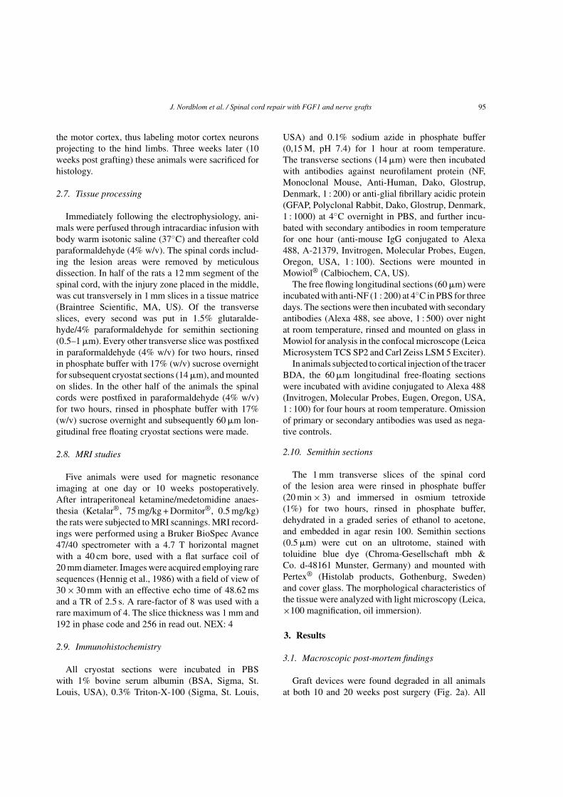
All cryostat sections were incubated in PBSwith 1% bovine serum albumin (BSA, Sigma, St.Louis, USA), 0.3% Triton-X-100 (Sigma, St. Louis,
USA) and 0.1% sodium azide in phosphate buffer(0,15 M, pH 7.4) for 1 hour at room temperature.The transverse sections (14 �m) were then incubatedwith antibodies against neurofilament protein (NF,Monoclonal Mouse, Anti-Human, Dako, Glostrup,Denmark, 1 : 200) or anti-glial fibrillary acidic protein(GFAP, Polyclonal Rabbit, Dako, Glostrup, Denmark,1 : 1000) at 4◦C overnight in PBS, and further incu-bated with secondary antibodies in room temperaturefor one hour (anti-mouse IgG conjugated to Alexa488, A-21379, Invitrogen, Molecular Probes, Eugen,Oregon, USA, 1 : 100). Sections were mounted inMowiol® (Calbiochem, CA, US).
The free flowing longitudinal sections (60 �m) wereincubated with anti-NF (1 : 200) at 4◦C in PBS for threedays. The sections were then incubated with secondaryantibodies (Alexa 488, see above, 1 : 500) over nightat room temperature, rinsed and mounted on glass inMowiol for analysis in the confocal microscope (LeicaMicrosystem TCS SP2 and Carl Zeiss LSM 5 Exciter).
In animals subjected to cortical injection of the tracerBDA, the 60 �m longitudinal free-floating sectionswere incubated with avidine conjugated to Alexa 488(Invitrogen, Molecular Probes, Eugen, Oregon, USA,1 : 100) for four hours at room temperature. Omissionof primary or secondary antibodies was used as nega-tive controls.
2.10. Semithin sections
The 1 mm transverse slices of the spinal cordof the lesion area were rinsed in phosphate buffer(20 min × 3) and immersed in osmium tetroxide(1%) for two hours, rinsed in phosphate buffer,dehydrated in a graded series of ethanol to acetone,and embedded in agar resin 100. Semithin sections(0.5 �m) were cut on an ultrotome, stained withtoluidine blue dye (Chroma-Gesellschaft mbh &Co. d-48161 Munster, Germany) and mounted withPertex® (Histolab products, Gothenburg, Sweden)and cover glass. The morphological characteristics ofthe tissue were analyzed with light microscopy (Leica,×100 magnification, oil immersion).
3. Results
3.1. Macroscopic post-mortem findings
Graft devices were found degraded in all animalsat both 10 and 20 weeks post surgery (Fig. 2a). All

96 J. Nordblom et al. / Spinal cord repair with FGF1 and nerve grafts
Fig. 2. a. Macroscopic post mortem finding of the perfused spinalcord 10 weeks after spinal cord repair with device and peripheralnerve grafts, anterior view, showing well integrated and degradedspinal cord device at level T11. Bar = 3 mm. 2b and c: MRI of thespinal cord repair area in the living animal at one day (b) and 10weeks postoperatively (c). The MRI scans show the location of thedevice in the spinal cord. The implanted (b) and degraded (c) device(low MRI signal) is seen in the middle of the pictures, filling theresection gap. The device/degraded device seemed to be in placedposition at one day as well as 10 weeks postop. Note the longitudinalstructures with similar MRI signal as the spinal cord, most likelyrepresenting nerve grafts within the device (arrows).
individual grafts were fixed to the spinal cord sur-faces on both sides at expected projection spots. Therewere no signs of infection, spine deformities or otherunexpected macroscopic findings (Fig. 2a).
3.2. MRI scanning
One animal was investigated with MRI of the spinalcord at one day postoperatively and four rats wereinvestigated 10 weeks postop. The implanted deviceseemed to be well in place the first post-operative day,projecting an MRI signal describing high water con-tent. At ten weeks postop the device was found tobe completely dissolved at dissection. The MRI at 10weeks postop showed grafts running in the gap betweenin the divided cord. Furthermore, the MRI did notreveal any signs of cyst formations in the cord or otherunexpected pathological findings such as infection orspine deformities (Fig. 2b, c).
3.3. Functional scoring
All animals scored 0 on the BBB scale the firstpostoperative weeks (Fig. 3), except from sham ani-mals. From postoperative week 6–20, the repairedanimals presented with scores between 0 and 4, withsignificantly higher scores in repair groups in com-parison to controls (p = 0.0001, One Way ANOVA,df = 3, Bonferroni’s multiple comparison test). Con-trols without graft and FGF1 scored 0 the first threeweeks and throughout the remaining test period 0-1.Sham operated animals scored 21 points (normal hindlimb function), at all BBB sessions (not shown).
3.4. Electrophysiology
Motor evoked potentials (MEPs) from the forelimbswere found in all animals with a preserved lateraliza-tion, i.e. unilateral motor cortex stimulation resultedin a contralateral response in the leg. MEPs from thehind limbs also presented with a clear lateralization,in some animals only in one leg (unilateral response)and in other animals in both legs (bilateral response).Representative MEP recordings are shown in Fig. 4.
In animals repaired with nerve grafts but withno adjuvant FGF1, five out of eight animals pre-sented MEPs in hamstrings bilaterally (Fig. 5) aftergrafting, i.e. significantly improved in comparisonto control animals subjected to SCI without grafting(p = 0.031, Fisher’s exact test, df = 1). The mean stim-ulatory threshold was 12.2 ± 1.1 mA and mean latencytime was 15.6 ± 2.6 ms. Two animals presented witha unilateral response and one animal failed to triggerpotentials at all in the lower extremities.
All animals subjected to grafting and FGF1 treat-ment presented MEPs in hamstrings bilaterally (Fig. 5).The electrophysiological responses in this group weresignificantly improved compared to controls subjectedto SCI without grafting (p = 0.001, Fisher’s exact test,df = 1), but also better than the group treated withgrafting without FGF1 (p = 0.028, Fisher’s exact test,df = 1). Stimulatory thresholds with low dose adjuvantFGF1 and high dose FGF1 were 14.3 ± 0.75 mA and12.3 ± 0.6 mA respectively and the mean latency time12.3 ± 1.7 ms and 18.5 ± 2.5 ms.
Reproducible MEPs were recorded bilaterally in allfour sham cases (n = 4) with stimulation thresholdsof 5.0 ± 0.2 mA (mean ± SEM) and latency time of11.6 ± 1.1 ms (mean ± SEM) (Fig. 4), which was notsignificantly different from the repair groups.

J. Nordblom et al. / Spinal cord repair with FGF1 and nerve grafts 97
BBB score
0 1 2 3 4 5 6 7 8 9 10 11 12 13 14 15 16 17 18 19
0.0
0.5
1.0
1.5
2.0
2.5
3.0
Repair(n=8)
Repair+FGF10.05 mg/mL(n=7)
Repair+FGF10.5 mg/mL(n=8)
SCI only(n=6)
***
Weeks after SCI and repair
BB
B
Fig. 3. The BBB scale showing functional recovery over the postoperative period (mean ± SEM). The first four to six weeks, no or very littlerecovery was seen. The following weeks a modest, however, significant improvement could be seen in all repair groups, which was not seenin SCI controls (p = 0.0001, One Way ANOVA with Bonferroni’s multiple comparison test). No significant difference was noted among thedifferent repair groups.
MEP response
20 weeks postop after relesion
Fig. 4. Representative oscilloscope curves from animals subjectedto sham operations, three different repair groups after SCI and SCIcontrols. Left column shows clear MEP responses (arrows) at 20weeks postop except for SCI controls where no MEPs were detected.Right column shows absence of MEPs after re-lesion at the levelof T10. Visible stimulation artifacts in some traces are indicated(asterisks).
Bilateral MEP responses could not be induced in anyof the 6 investigated animals in the control group (SCIwithout grafting, Fig. 5). However, 2 of these animals
showed a MEP response in one leg (stimulatory thresh-old 13.0 ± 3.0 mA, latency time 11.1 ± 0.75 ms).
Animals that presented MEPs were re-lesioned threesegments above the repair zone, which in every caseresulted in a complete loss of MEP responses in thehind limbs (Fig. 4), but preserved MEPs in both arms.
3.5. Anterograde tracing
To investigate the possibility of corticospinallyderived regeneration through the spinal cord device,six animals were examined for presence of BDA in thespinal cord three weeks after injection into the motorcortex. Animals without grafting (n = 6) were used ascontrols. In both groups, numerous pyramidal cells pre-sented BDA-IR at the injection site as well as thin,labeled fibers further down in the brain (Fig. 6g). Fur-thermore, we found columns with BDA positive fibersin the spinal cord, contralateral to the injection side,above the lesion area, representing uninjured parts ofthe corticospinal tract (Fig. 6h).
Numerous BDA-containing fibers below the site ofinjury was found in all animals treated with graftingand FGF1 in the lower thoracic cord. These fibers weredetected more than 3 mm caudal to the injury indicat-ing axonal elongation beyond the site of injury in thefollowing one or two cord segments (Fig. 6i). BDAfibers were found in both central and peripheral parts

98 J. Nordblom et al. / Spinal cord repair with FGF1 and nerve grafts
Fig. 5. Bilateral MEP recovery 20 weeks after surgery(mean ± SEM), describing the fraction of animals with bothleft- and right-sided MEP response after contralateral cortexstimulation. All sham-operated animals showed full MEP responsesafter 20 weeks. In the repair groups treated with FGF1 (5 �g or0.5 �g) and nerve grafts, all (100%) animals showed positivebilateral MEPs, which was significantly better (p = 0.028, Fisher’sexact test) than in the repair group not treated with FGF1, wherefive out of eight (62.5%) animals showed positive bilateral MEPs,which was significantly better than the SCI (0%) control group(p = 0.031, Fisher’s exact test).
of the caudal cord, which is line with the findings ofTsai and co-workers, who repaired the spinal cord withobliquely projected nerve graft or spinal cord fusionand found BDA traced fibers in both gray and whitematter. No BDA was detected in the spinal cord belowthe lesion in control animals (Fig. 6j).
3.6. Immunocytochemistry
All nerve grafts within the injury zone containednumerous neurofilament (NF)-positive axons at 20weeks post injury, most likely representing regener-ating axons, either ascending or descending (Fig. 6b).Caudal to the graft area numerous NF labeled axonsappeared in the grey matter (Fig. 6c). These axons hadno clear organization and were projected in all direc-tions in a turning and winding manner, resemblingsprouting axons. We suggest that these fibers repre-sent regenerating axons in line with the tracing resultsabove, even though neurofilament staining cannot sep-arate between sprouting ascending sensory axons fromthe transected caudal surface and descending fibers
entering the caudal spinal cord. Glial fibrillary acidicprotein (GFAP) immunoreactivity (IR) showed scat-tered astrocytes in all groups in the normal spinal cordor more than 1 mm away from any repair area in thevarious treatment groups (Fig. 7). Within the degradedrepair area in treatment groups only few astrocyteswere found. At the cranial and caudal resection bor-ders in spinal cord resected animals, there was anincreased GFAP-IR at study termination regardless ofrepair strategy, with no obvious difference among thegroups.
3.7. Semithin sections
Numerous axons were found in all grafts 20 weekspostoperatively (Fig. 6d–f). The axons were of vary-ing diameter, with different degree of myelination andwere to a large extent found to be arranged in fascicles(Fig 6d–f).
4. Discussion
Although several promising regeneration strategieshave been presented in recent years (Bunge, 2008;Cadotte and Fehlings, 2010; Cote, et al., 2011; Kwon,et al., 2010; Schwab, et al., 2006) spinal cord injury(SCI) remains a major challenge. One is to establishregeneration across a spinal cord gap by letting the longtracts in the spinal cord climb in peripheral nerve grafts(PNG) and enter permissive gray substance on the otherside of the lesion (Cheng, et al., 1996; Cheng et al.,2004; Houle et al., 2006; Lee, et al., 2002; Lee, et al.,2004; Tsai, et al., 2005). To facilitate the microsurgi-cal positioning of the PNGs, we previously developeda graft holder to facilitate reproducible placement ofPNGs (Nordblom, et al., 2009). If there is a poten-tial clinical application using PNGs, literature suggeststhat fibroblast growth factor 1 (FGF1) is needed forfunctional recovery (Cheng, et al., 1996; Lee, et al.,2002; Lee, et al., 2004; Lee et al., 2010; Tsai, et al.,2005). Therefore, the current investigation evaluatedspinal cord repair with PNGs in a newly developedgraft holder made of biodegradable calcium sulphatewith or without slow release of FGF1.
A complete injury of the spinal cord was made byresection of a thoracic segment (T11) in adult ratsfollowed by the implantation of a device carrying12 autologous PNGs with or without FGF1. Post-mortem findings demonstrated that the device was well

J. Nordblom et al. / Spinal cord repair with FGF1 and nerve grafts 99
Fig. 6. Schematic illustration indicating the region (a, d, g, h, i) for micrographs showing neurofilament (NF, a–c) immunoreactivity (IR),semithin sections (d–f) and BDA (g–j) at 20 weeks after spinal cord repair with high dose FGF1. a: Low magnification showing the cranial cord,repair area (b) and the caudal (c) spinal cord. b: High magnification of a peripheral nerve graft in the degraded device, containing numerousNF-positive axons (arrows). Further down, numerous winding and turning NF-positive fibers were found 3 mm into the caudal spinal cord (c),most likely representing sprouting axons of unknown origin. d: Transverse section of the right side of the degraded device showing severaltransplanted nerve grafts at the outer border of the device area (arrows). e: High magnification of one nerve graft (black left corner box in d)revealed numerous well myelinated axons (arrows), the square is found enlarged in f (arrows). The presence of BDA at various locations (seeschematic illustration at top left corner) following injection in the right motor cortex showed clearly labeled pyramidal cells (g) and labeledaxons in the dorsal corticospinal tract in the proximal spinal cord (h, arrows) at 10 weeks after surgery. Caudal to the SCI, there were severalaxons positive for BDA, up to 3 mm into the spinal cord (i, arrows) in animals subjected to spinal cord injury and repair, but not after SCI only(f). Bars = 1 mm (a, d), 50�m (b, e, f), 30�m (c), 0.5 mm (g), 0.2 mm (h, i, j).

100 J. Nordblom et al. / Spinal cord repair with FGF1 and nerve grafts
Fig. 7. Glial fibrillary acidic protein (GFAP) immunoreactivity (IR)in the caudal spinal cord at 20 weeks after sham operation (a), repairwith high dose FGF1 (b–c) or repair without FGF1 (d). In shamoperated animals only scattered astrocytes were found in the cord,whereas at the interface between the degraded device and caudalcord, there was an increased GFAP-IR (b), not noticeably differentfrom animals repaired without FGF1 (d). One mm into the caudalcord (c), the GFAP-IR was similar to the cords of the sham group.Bars (a–d) = 50 �m.
degraded and integrated in the spinal cord at 20 weeksafter repair and that no apparent reaction toward thematerial was observed. Further, FGF1 augmented theelectrophysiological response if the cord was recon-nected with PNGs. According to previous literature,the effect of FGF1 may be attributed to neuroprotec-tion, improved regeneration or local modulation of thespinal cord injury milieu (Giacobini et al., 1991; Guestet al., 1997; Kuo et al., 2011; Lee, et al., 2008; Leeet al., 2011; Tsai et al., 2008).
We also report that the electrophysiological connec-tion between the motor cortex and hind limb muscleswas paralleled by traced corticospinal tract (CST)axons into the caudal spinal cord. This demonstratesthe principle that CST axons may enter and exit PNGsin the spinal cord repair in the presence of FGF1,and serves as an additional piece of evidence that themotor cortex is involved in the recovery after spinalcord repair with PNGs. Putative effect of FGF1 purelyon CST regeneration was not assessed in the currentstudy. SCI alone showed no CST axons in the caudalcord. The positive MEPs were generated from the sameplace as the BDA injections were made in the motorcortex. Even though it is likely that the CST fibers con-duct some of the signals that give rise to the positiveMEPs in this study, other reasonable MEP generat-ing pathways are possible. Such pathways could be
polysynaptic circuits through brainstem derived sig-nals or propriospinal axons in the spinal cord traversingthe injury zone in the PNG, as well as other longtract terminations in neuron pools in the caudal spinalcord before connecting to the secondary motor neuronstargeting hind limb muscles. If CST fibers would termi-nate directly onto alpha motor neurons to the hamstringmuscles (in the rat innervated mainly by the L5 seg-ment) regeneration distance would have to be about20 mm from the cranial injury zone into the caudal cord(post mortem examination of distance to L5 segment,data not shown).
The MEP response in the hind limb muscles wererecorded after increasing stimulation until responsewas registered or terminated at 30 mA. All positiveMEPs disappeared after cord re-transection at the T8level. During MEP sessions, several stimulations weremade with the bipolar electrode on the cortex givinga variation of amplitudes, which is dependent on theamount of depolarized neurons in the cortex (bipolarelectrode placement) as well as the muscle cell depo-larization around a specific volume around the muscleneedle, i.e. needle position dependent. Therefore theMEPs could only be reported as cortex-hind limb con-tact or not (one or zero response), which is in linewith literature on MEPs describing large variations ofamplitudes seen in the same subject at different timepoints depending on stimulation and registration coor-dinates (Wasserman, 2008). Thus, it may be misleadingto compare MEP amplitudes in the current experimen-tal setting.
There is much data to confirm that PNGs is a suc-cessful substrate for regeneration stimulation of theperipheral or central nervous system (Alilain, et al.,2011; Cheng, et al., 1996; Cote, et al., 2011; Richard-son et al., 1980). Therefore the current study onlyevaluated spinal cord bridging repair strategies basedon PNGs in a biodegradable graft holder with two dif-ferent concentrations of FGF1 or without. Thus, thebeneficial effect of FGF1 addition to the graft holdercould only be attributed to PNG/FGF1 in combinationand not separate the individual contribution from PNGor FGF1 alone.
All grafted animals presented with BBB scoresbetween 0 and 4 from the 6th postoperative week. Thiswas significantly better than injured animals withoutrepair suggesting that a limited functional regenerationhad occurred. The reported BBB scores in treated ani-mals are somewhat lower than BBB scores reportedby Tsai and coworkers (BBB of 4-5) (Tsai, et al.,

J. Nordblom et al. / Spinal cord repair with FGF1 and nerve grafts 101
2005) or Lee et al. (BBB around 7) (Lee, et al., 2002;Lee, et al., 2004) after spinal cord injury repair withPNGs and FGF1. However, it is difficult to grade repairstrategies comparing treatments groups from separatepapers, due to possible differences in e.g. origin of rats,care or rehabilitation milieu (Garrison et al., 2011; Lee,et al., 2010). Functional recovery after CNS injury isalso reported to differ depending on rat strain. (Reidet al., 2010). The current groups of rats suffered fromsevere contractures and fixed joints, and a possiblyemerging BBB increase could have been disguised.They were not subjected to physiotherapy of the joints.Efficiency grading among the various reports mayinclude several pitfalls and is currently omitted.
The current paper suggests that the addition of FGF1in the biodegradable graft holder improves the electro-physiological response, but is not designed to evaluatethe effectiveness of PNGs, since nerve grafts were usedin all treatment groups. In the future, there will beinterest in separating the impact of the individual com-ponents FGF1 and PNGs in spinal cord repair, whichwas beyond the scope of the present study.
In conclusion, we report a tolerable method to bridgea spinal cord gap with PNGs with high microsurgi-cal precision in a biodegradable device. If FGF1 wasadded in the calcium sulphate, MEP appearance wasmore robust than without FGF1. The method was par-alleled with a functional improvement and evidenceof CST axons traversing the repair area and into thecaudal spinal cord. If the method enters a coursetoward a translational approach, irreversibly and com-pletely devitalized spinal cord tissue would have to beremoved, which demands an unambiguous diagnosisof complete spinal cord injury. (Steeves et al., 2010;Zariffa et al., 2010).
Acknowledgments
We would like to thank Britt Meijer, Hakan Erikssonand Bjorn Hellberg for excellent laboratory assistance,Brita Robertson for good advices on tracing techniquesand Jeremy Young for MRI assistance. This work wassupported by the Karolinska Institute, Swedish Medi-cal Research Council, Torsten and Ragnar Soderberg’sfoundation, Socialstyrelsen, Magnus Bergwall’s foun-dation, The Swedish Association of Persons withNeurological Disabilities, VINNOVA and BioArcticNeuroscience AB.
Author disclosure statement
J. Sjodahl works for BioArctic AB, which has afinancial interest in spinal cord repair. No other authorof this article has any financial conflict of interest inthis publication.
References
Aberg, J., Eriksson, O., Spens, E., Nordblom, J., Mattsson, P., Sjo-dahl, J., Svensson, M. & Engqvist, H. (2010). Calcium sulfatespinal cord scaffold: A study on degradation and fibroblastgrowth factor 1 loading and release. J Biomater Appl.
Alilain, W.J., Horn, K.P., Hu, H., Dick, T.E. & Silver, J. (2011). Func-tional regeneration of respiratory pathways after spinal cordinjury. Nature, 475(7355), 196-200.
Basso, D.M., Beattie, M.S. & Bresnahan, J.C. (1995). A sensitiveand reliable locomotor rating scale for open field testing in rats.J Neurotrauma, 12(1), 1-21.
Bunge, M.B. (2008). Novel combination strategies to repair theinjured mammalian spinal cord. J Spinal Cord Med, 31(3),262-269.
Cadotte, D.W. & Fehlings, M.G. (2011). Spinal Cord Injury: A Sys-tematic Review of Current Treatment Options. Clin OrthopRelat Res, 469(3), 732-741.
Chen, M.S., Huber, A.B., van der Haar, M.E., Frank, M., Schnell, L.,Spillmann, A.A., Christ, F. & Schwab, M.E. (2000). Nogo-A isa myelin-associated neurite outgrowth inhibitor and an antigenfor monoclonal antibody IN-1. Nature, 403(6768), 434-439.
Cheng, H., Cao, Y. & Olson, L. (1996). Spinal cord repair in adultparaplegic rats: Partial restoration of hind limb function. Sci-ence, 273(5274), 510-513.
Cheng, H., Liao, K.K., Liao, S.F., Chuang, T.Y. & Shih, Y.H. (2004).Spinal cord repair with acidic fibroblast growth factor as atreatment for a patient with chronic paraplegia. Spine, 29(14),E284-E288.
Cote, M.P., Amin, A.A., Tom, V.J. & Houle, J.D. (2011). Periph-eral nerve grafts support regeneration after spinal cord injury.Neurotherapeutics, 8(2), 294-303.
Fehlings, M.G., Theodore, N., Harrop, J., Maurais, G., Kuntz, C.,Shaffrey, C.I., Kwon, B.K., Chapman, J., Yee, A., Tighe, A.& McKerracher, L. (2011). A phase I/IIa clinical trial of arecombinant Rho protein antagonist in acute spinal cord injury.J Neurotrauma, 28(5), 787-796.
Fitch, M.T. & Silver, J. (2008). CNS injury, glial scars, and inflamma-tion: Inhibitory extracellular matrices and regeneration failure.Exp Neurol, 209(2), 294-301.
Garrison, M.K., Yates, C.C., Reese, N.B., Skinner, R.D. & Garcia-Rill, E. (2011). Wind-up of stretch reflexes as a measure ofspasticity in chronic spinalized rats: The effects of passiveexercise and modafinil. Exp Neurol, 227(1), 104-109.
Giacobini, M.M., Hoffer, B.J., Zerbe, G. & Olson, L. (1991). Acidicand basic fibroblast growth factors augment growth of fetalbrain tissue grafts. Exp Brain Res, 86(1), 73-81.

102 J. Nordblom et al. / Spinal cord repair with FGF1 and nerve grafts
Guest, J.D., Hesse, D., Schnell, L., Schwab, M.E., Bunge, M.B. &Bunge, R.P. (1997). Influence of IN-1 antibody and acidic FGF-fibrin glue on the response of injured corticospinal tract axonsto human Schwann cell grafts. J Neurosci Res, 50(5), 888-905.
Harkema, S., Gerasimenko, Y., Hodes, J., Burdick, J., Angeli, C.,Chen, Y., Ferreira, C., Willhite, A., Rejc, E., Grossman, R.G.& Edgerton, V.R. (2011). Effect of epidural stimulation of thelumbosacral spinal cord on voluntary movement, standing, andassisted stepping after motor complete paraplegia: A case study.Lancet, 377(9781), 1938-1947.
Hennig, J., Nauerth, A. & Friedburg, H. (1986). RARE imaging: Afast imaging method for clinical MR. Magn Reson Med, 3(6),823-833.
Houle, J.D., Tom, V.J., Mayes, D., Wagoner, G., Phillips, N. & Silver,J. (2006). Combining an autologous peripheral nervous sys-tem “bridge” and matrix modification by chondroitinase allowsrobust, functional regeneration beyond a hemisection lesion ofthe adult rat spinal cord. J Neurosci, 26(28), 7405-7415.
Kuo, H.S., Tsai, M.J., Huang, M.C., Chiu, C.W., Tsai, C.Y.,Lee, M.J., Huang, W.C., Lin, Y.L., Kuo, W.C. & Cheng, H.(2011). Acid fibroblast growth factor and peripheral nervegrafts regulate Th2 cytokine expression, macrophage acti-vation, polyamine synthesis, and neurotrophin expression intransected rat spinal cords. J Neurosci, 31(11), 4137-4147.
Kwon, B.K., Sekhon, L.H. & Fehlings, M.G. (2010). Emergingrepair. regeneration, and translational research advances forspinal cord injury. Spine (Phila Pa 1976), 35(21 Suppl), S263-S270.
Lee, M.J., Chen, C.J., Cheng, C.H., Huang, W.C., Kuo, H.S., Wu,J.C., Tsai, M.J., Huang, M.C., Chang, W.C. & Cheng, H. (2008).Combined treatment using peripheral nerve graft and FGF-1:Changes to the glial environment and differential macrophagereaction in a complete transected spinal cord. Neurosci Lett,433(3), 163-169.
Lee, M.J., Chen, C.J., Huang, W.C., Huang, M.C., Chang, W.C.,Kuo, H.S., Tsai, M.J., Lin, Y.L. & Cheng, H. (2011). Regulationof chondroitin sulphate proteoglycan and reactive gliosis afterspinal cord transection: Effects of peripheral nerve graft andfibroblast growth factor 1. Neuropathol Appl Neurobiol, 37(6),585-599.
Lee, Y.S., Hsiao, I. & Lin, V.W. (2002). Peripheral nerve grafts andaFGF restore partial hindlimb function in adult paraplegic rats.J Neurotrauma, 19(10), 1203-1216.
Lee, Y.S., Lin, C.Y., Robertson, R.T., Hsiao, I. & Lin, V.W. (2004).Motor recovery and anatomical evidence of axonal regrowthin spinal cord-repaired adult rats. J Neuropathol Exp Neurol,63(3), 233-245.
Lee, Y.S., Zdunowski, S., Edgerton, V.R., Roy, R.R., Zhong, H.,Hsiao, I. & Lin, V.W. (2010). Improvement of gait patterns in
step-trained, complete spinal cord-transected rats treated witha peripheral nerve graft and acidic fibroblast growth factor. ExpNeurol, 224(2), 429-437.
Nordblom, J., Persson, J.K., Svensson, M. & Mattsson, P. (2009).Peripheral nerve grafts in a spinal cord prosthesis result inregeneration and motor evoked potentials following spinal cordresection. Restor Neurol Neurosci, 27(4), 285-295.
Reid, W.M., Rolfe, A., Register, D., Levasseur, J.E., Churn, S.B.& Sun, D. (2010). Strain-related differences after experimentaltraumatic brain injury in rats. J Neurotrauma, 27(7), 1243-1253.
Richardson, P.M., McGuinness, U.M. & Aguayo, A.J. (1980).Axons from CNS neurons regenerate into PNS grafts. Nature,284(5753), 264-265.
Schwab, J.M., Brechtel, K., Mueller, C.A., Failli, V., Kaps, H.P.,Tuli, S.K. & Schluesener, H.J. (2006). Experimental strategiesto promote spinal cord regeneration–an integrative perspective.Prog Neurobiol, 78(2), 91-116.
Schwab, M.E. (2004). Nogo and axon regeneration. Curr Opin Neu-robiol, 14(1), 118-124.
Steeves, J.D., Kramer, J.K., Fawcett, J.W., Cragg, J., Lammertse,D.P., Blight, A.R., Marino, R.J., Ditunno, J.F. Jr, Coleman, W.P.,Geisler, F.H., Guest, J., Jones, L., Burns, S., Schubert, M., vanHedel, H.J. & Curt, A. (2011). Extent of spontaneous motorrecovery after traumatic cervical sensorimotor complete spinalcord injury. Spinal Cord, 49(2), 257-265.
Tsai, E.C., Krassioukov, A.V. & Tator, C.H. (2005). Corticospinalregeneration into lumbar grey matter correlates with locomotorrecovery after complete spinal cord transection and repair withperipheral nerve grafts, fibroblast growth factor 1, fibrin glue,and spinal fusion. J Neuropathol Exp Neurol, 64(3), 230-244.
Tsai, M.C., Shen, L.F., Kuo, H.S., Cheng, H. & Chak, K.F. (2008).Involvement of acidic fibroblast growth factor in spinal cordinjury repair processes revealed by a proteomics approach. MolCell Proteomics, 7(9), 1668-1687.
Wassermann, E.M., Epstein, C.M., Ziemann, U., Walsh, V., Paus, T.& Lisanby, S. (2008). The Oxford Handbook of TranscranialStimulation (1 ed.). New York: Oxford University Press.
Wu, J.C., Huang, W.C., Chen, Y.C., Tu, T.H., Tsai, Y.A., Huang,S.F., Huang, H.C. & Cheng, H. (2011). Acidic fibroblast growthfactor for repair of human spinal cord injury: A clinical trial. JNeurosurg Spine, 15(3), 216-227.
Zariffa, J., Kramer, J.L., Fawcett, J.W., Lammertse, D.P., Blight,A.R., Guest, J., Jones, L., Burns, S., Schubert, M., Bolliger,M., Curt, A. & Steeves, J.D. (2011). Characterization of neu-rological recovery following traumatic sensorimotor completethoracic spinal cord injury. Spinal Cord, 49(3), 463-471.