Eval of Liver Mass ACG 2021 PB LS-FINAL
32
3/4/2021 1 Participating in the Webinar All attendees will be muted and will remain in Listen Only Mode. Type your questions here so that the moderator can see them. Not all questions will be answered but we will get to as many as possible. 1 2 American College of Gastroenterology
Transcript of Eval of Liver Mass ACG 2021 PB LS-FINAL
3/4/2021
1
Participating in the Webinar
All attendees will be muted and will remain in Listen Only Mode.
Type your questions here so that the moderator can see them. Not all questions will be answered but we will get to as many as possible.
1
2
American College of Gastroenterology
3/4/2021
2
How to Receive CME and MOC Points
LIVE VIRTUAL GRAND ROUNDS WEBINAR
ACG will send a link to a CME & MOC evaluation to all attendees on the live webinar.
ABIM Board Certified physicians need to complete their MOC activities by December 31, 2021 in order for the MOC points to count toward any MOC requirements that are due by the end of the year. No MOC credit may be awarded after March 1, 2022 for this activity.
MOC QUESTION
If you plan to claim MOC Points for this activity, you will be asked to: Please list specific changes you will make in your
practice as a result of the information you received from this activity.
Include specific strategies or changes that you plan to implement.THESE ANSWERS WILL BE REVIEWED.
3
4
American College of Gastroenterology

3/4/2021
3
ACG Virtual Grand RoundsJoin us for upcoming Virtual Grand Rounds!
Visit gi.org/ACGVGR to Register
Week 11, 2021Managing Complications of GI Endoscopy Shivangi T. Kothari, MD, FACGMarch 18, 2021 at Noon Eastern
Week 10, 2021How to be an Integrative Gastroenterologist Elena A. Ivanina, DOMarch 11, 2021 at Noon Eastern
Disclosures:
Speaker: Catherine T. Frenette, MDDr. Frenette has no conflicts of interest related to this talk.
Moderator: Anjana A. Pillai, MDSpeaker : Simply Speaking Hepatitis (CME); Medical Advisory Board: Genentech, Eisai Inc, Exelixis
5
6
American College of Gastroenterology
3/4/2021
4
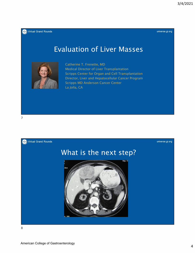
Evaluation of Liver Masses
Catherine T. Frenette, MDMedical Director of Liver TransplantationScripps Center for Organ and Cell TransplantationDirector, Liver and Hepatocellular Cancer ProgramScripps MD Anderson Cancer CenterLa Jolla, CA
What is the next step?
7
8
American College of Gastroenterology
3/4/2021
5
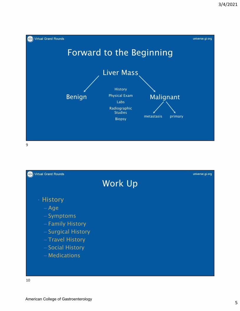
Forward to the Beginning
Liver Mass
Benign MalignantHistory
Physical Exam
Labs
Radiographic Studies
Biopsymetastasis primary
Work Up
• History– Age– Symptoms– Family History– Surgical History– Travel History– Social History– Medications
9
10
American College of Gastroenterology
3/4/2021
6
Work Up
• Physical Exam– Jaundice– Weight loss– Ascites– Abdominal Tenderness
• Labs– Liver panel, CBC– Viral serologies– Tumor Markers (AFP, CA19-9, CEA)
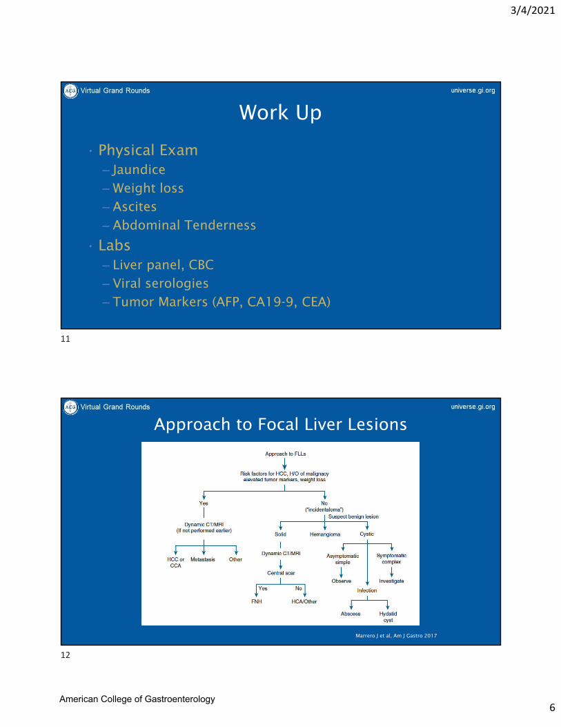
Approach to Focal Liver Lesions
Marrero J et al, Am J Gastro 2017
11
12
American College of Gastroenterology
3/4/2021
7
Benign lesions usually requiring no further intervention
• Cavernous hemangioma
• Focal nodular hyperplasia
• Simple cyst
• Focal fatty change or sparing
Benign lesions requiring further investigation and therapy
• Hepatic adenoma and adenomatosis• Biliary cystadenoma• Hepatic abscess – pyogenic or amebic• Echinococcal cyst• Granulomatous inflammation• Inflammatory pseudotumor
13
14
American College of Gastroenterology
3/4/2021
8
Malignant lesions requiring appropriate management
• Metastasis from other primary sites• Hepatocellular carcinoma• Cholangiocarcinoma• Biliary cystadenocarcinoma• Lymphoma• Hepatic angiosarcoma• Epithelioid hemangioendothelioma
Hemangioma
• The most common benign mesenchymal tumor• Often solitary, may be multiple in up to 40%• Larger than 5 cm are “giant”• Epidemiology from 1-20% depending on the study• 60-80% diagnosed in patients age 30-50• More frequent in woman 3:1
15
16
American College of Gastroenterology
3/4/2021
9
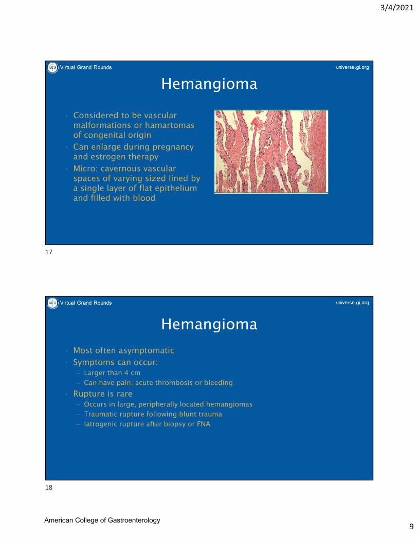
Hemangioma
• Considered to be vascular malformations or hamartomasof congenital origin
• Can enlarge during pregnancy and estrogen therapy
• Micro: cavernous vascular spaces of varying sized lined by a single layer of flat epithelium and filled with blood
Hemangioma
• Most often asymptomatic• Symptoms can occur:
– Larger than 4 cm– Can have pain: acute thrombosis or bleeding
• Rupture is rare– Occurs in large, peripherally located hemangiomas– Traumatic rupture following blunt trauma– Iatrogenic rupture after biopsy or FNA
17
18
American College of Gastroenterology
3/4/2021
10
Hemangioma: Rare things
• Giant: can cause high output cardiac failure– In children this is associated with hypothyroidism
•Presence of high levels of 3 iodothyronin deiodinase activity• Kasabach-Merritt syndrome: in children, a consumptive coagulopathy
can occur• Hepatic Hemangiomatosis: rare in adult patients, associated with
hereditary hemorrhagic telangiectasia and use of metoclopramide (Reglan)
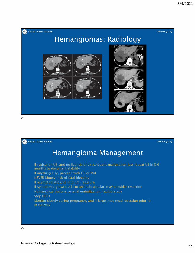
Hemangiomas: Radiology
• US: well demarcated homogenous hypoechoic mass– blood flow seen with doppler in 10-50%
• CT: peripheral nodular enhancement on early phase, followed by centripetal pattern or “filling in” during late phase
• Technetium-99 red blood cell study: initial hypoperfusionduring arterial flow, followed by gradual tracer peaking 30-50 min after injection
19
20
American College of Gastroenterology
3/4/2021
11
Hemangiomas: Radiology
Hemangioma Management
• If typical on US, and no liver dz or extrahepatic malignancy, just repeat US in 3-6 months to document stability
• If anything else, proceed with CT or MRI• NEVER biopsy: risk of fatal bleeding• If asymptomatic and >1.5 cm, reassure• If symptoms, growth, >5 cm and subcapsular: may consider resection• Non-surgical options: arterial embolization, radiotherapy• Stop OCPs• Monitor closely during pregnancy, and if large, may need resection prior to
pregnancy
21
22
American College of Gastroenterology

3/4/2021
12
Adenoma
• Young women, 20-44 years old• Most common in right lobe• 80% solitary• Multiple adenomas
– Think of prolonged OCPs– Glycogen storage disease
• Particularly Type I and III, usually <20 yrs, males, multiple• Can resolve after dietary therapy
– Hepatic adenomatosis
Adenoma
• Strongly associated with use of – OCPs– Anabolic steroids– Pregnancy: increased risk of rupture with 59% maternal and 62% fetal mortality– Diabetes?
• Incidence increasing• Highest risk in women >30 yrs on OCPs for >25 months, particularly
OCPs with high estrogen component• Regression has been seen after d/c of OCPs with recurrence during
readministration or pregnancy
23
24
American College of Gastroenterology

3/4/2021
13
Adenoma: Microscopic
• Large plates of adenoma cells, which are larger than normal hepatocytes and contain glycogen and lipid
• NO normal hepatic architecture• NO septa, portal tracts, bile ductules• Minimal Kupffer cells• Prominent “naked” arteries
Adenoma
• Mostly asymptomatic• Can have abdominal pain in epigastrium/RUQ• Risk of bleeding reported to be as high as 25-41%
– Higher with pain, long OCP use, subcapsular location, larger size
• Risk of malignant transformation: 8-13%, mainly in >5 cm size lesions
25
26
American College of Gastroenterology
3/4/2021
14
Adenomatosis
• More than 10 adenomas• Lack of correlation with steroids/OCPs• Lack of resolution with steroid withdrawal• Lack of association with GSD• Both men/women• Increases in alk phos/GGT• Hemorrhage is common• May need to consider OLT
Adenomas: Diagnosis
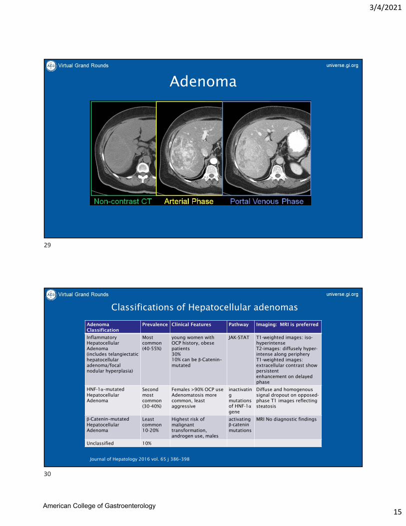
• Biopsy rarely indicated: increased risk of bleeding, difficult to make the diagnosis
• US: well-demarcated and hyperechoic, may be heterogenous• CT: peripheral enhancement during arterial phase with
subsequent centripedal flow during portal venous phase, then iso- or hypo-dense
• MRI: most helpful with sodium gadoxetate (Eovist): has hepatobiliary excretion, which adenomas cannot take up and excrete, so dark spot
27
28
American College of Gastroenterology
3/4/2021
15
Adenoma
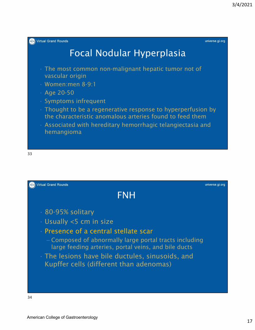
Classifications of Hepatocellular adenomas
Adenoma Classification
Prevalence Clinical Features Pathway Imaging: MRI is preferred
Inflammatory Hepatocellular Adenoma(includes telangiectatic hepatocellular adenoma/focal nodular hyperplasia)
Most common(40-55%)
young women with OCP history, obese patients30% 10% can be β-Catenin–mutated
JAK-STAT T1-weighted images: iso-hyperintenseT2-images: diffusely hyper-intense along periphery T1-weighted images: extracellular contrast show persistentenhancement on delayed phase
HNF-1α–mutated Hepatocellular Adenoma
Second most common(30-40%)
Females >90% OCP useAdenomatosis more common, least aggressive
inactivating mutations of HNF-1αgene
Diffuse and homogenous signal dropout on opposed-phase T1 images reflecting steatosis
β-Catenin–mutated Hepatocellular Adenoma
Least common10-20%
Highest risk of malignant transformation, androgen use, males
activating β-catenin mutations
MRI No diagnostic findings
Unclassified 10%
Journal of Hepatology 2016 vol. 65 j 386–398
29
30
American College of Gastroenterology

3/4/2021
16
Beta-catenin exon 3 mutated adenomas have higher risk of malignancy
Management of Hepatic Adenoma
• Follow up 6 months, q1 yr x5 yrs, then q2 yrs• High risk: resect
– Lesions ≥5 cm (or increasing in size)– Hemorrhage– Male gender (increased malignant risk)– Beta-catenin mutated adenomas– Older females with no history of OCP use
• Low risk: stop OCPs, lose weight– Young females, lesions <5 cm on OCP– Follow with US q6-12 weeks during pregnancy
• Alternatives to resection: RFA, embolization
31
32
American College of Gastroenterology
3/4/2021
17
Focal Nodular Hyperplasia
• The most common non-malignant hepatic tumor not of vascular origin
• Women:men 8-9:1• Age 20-50• Symptoms infrequent• Thought to be a regenerative response to hyperperfusion by
the characteristic anomalous arteries found to feed them• Associated with hereditary hemorrhagic telangiectasia and
hemangioma
FNH
• 80-95% solitary• Usually <5 cm in size• Presence of a central stellate scar
– Composed of abnormally large portal tracts including large feeding arteries, portal veins, and bile ducts
• The lesions have bile ductules, sinusoids, and Kupffer cells (different than adenomas)
33
34
American College of Gastroenterology
3/4/2021
18
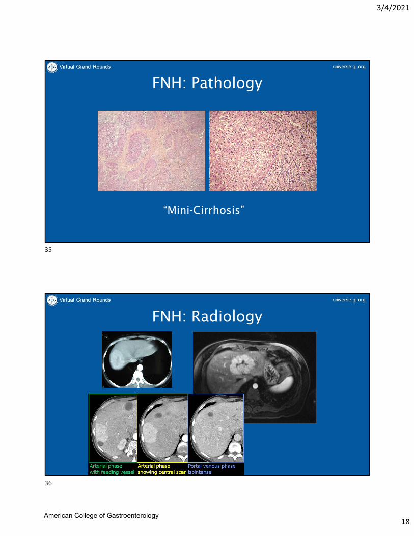
FNH: Pathology
“Mini-Cirrhosis”
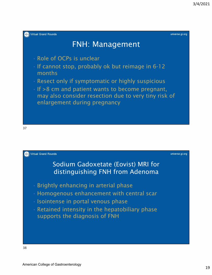
FNH: Radiology
35
36
American College of Gastroenterology
3/4/2021
19
FNH: Management
• Role of OCPs is unclear• If cannot stop, probably ok but reimage in 6-12
months• Resect only if symptomatic or highly suspicious• If >8 cm and patient wants to become pregnant,
may also consider resection due to very tiny risk of enlargement during pregnancy
Sodium Gadoxetate (Eovist) MRI for distinguishing FNH from Adenoma
• Brightly enhancing in arterial phase• Homogenous enhancement with central scar• Isointense in portal venous phase• Retained intensity in the hepatobiliary phase
supports the diagnosis of FNH
37
38
American College of Gastroenterology
3/4/2021
20
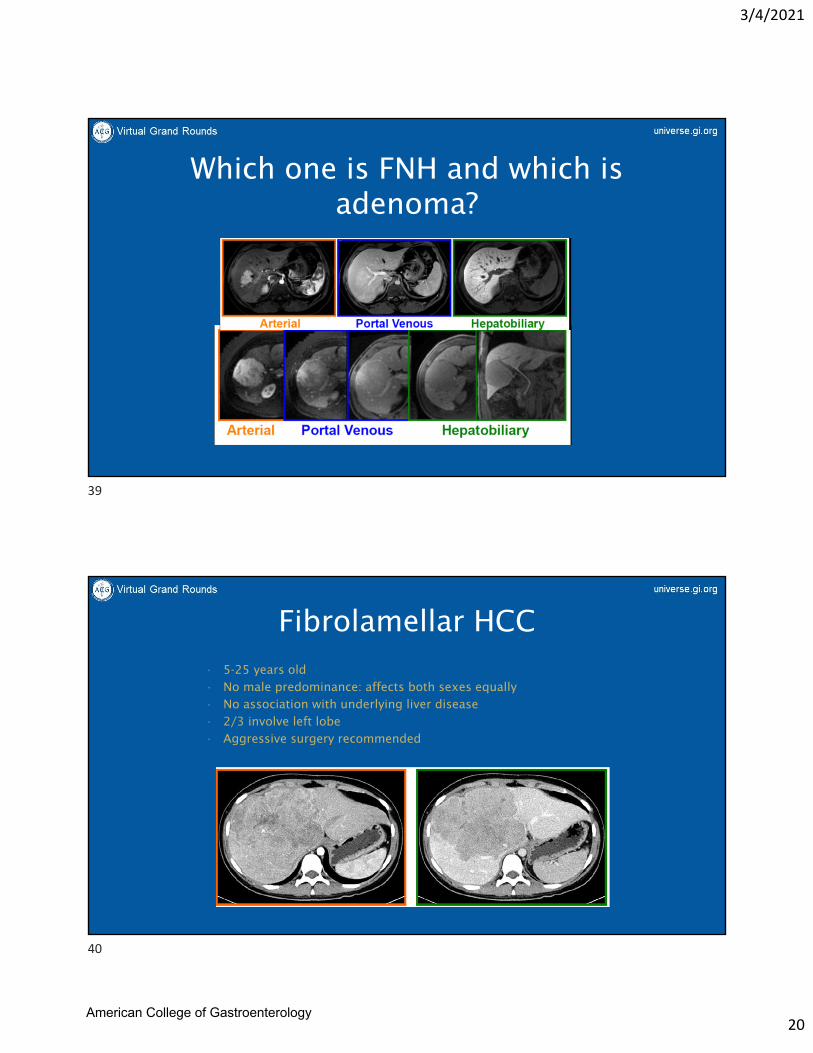
Which one is FNH and which is adenoma?
Fibrolamellar HCC• 5-25 years old• No male predominance: affects both sexes equally• No association with underlying liver disease• 2/3 involve left lobe• Aggressive surgery recommended
39
40
American College of Gastroenterology
3/4/2021
21
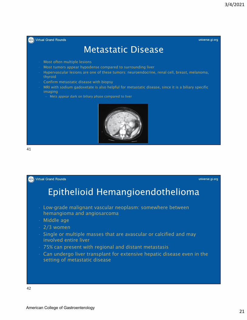
Metastatic Disease• Most often multiple lesions• Most tumors appear hypodense compared to surrounding liver• Hypervascular lesions are one of these tumors: neuroendocrine, renal cell, breast, melanoma,
thyroid• Confirm metastatic disease with biopsy• MRI with sodium gadoxetate is also helpful for metastatic disease, since it is a biliary specific
imaging– Mets appear dark on biliary phase compared to liver
Epithelioid Hemangioendothelioma
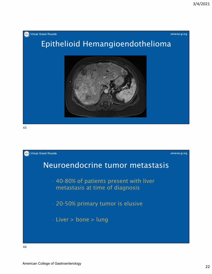
• Low-grade malignant vascular neoplasm: somewhere between hemangioma and angiosarcoma
• Middle age• 2/3 women• Single or multiple masses that are avascular or calcified and may
involved entire liver• 75% can present with regional and distant metastasis• Can undergo liver transplant for extensive hepatic disease even in the
setting of metastatic disease
41
42
American College of Gastroenterology
3/4/2021
22
Epithelioid Hemangioendothelioma
Neuroendocrine tumor metastasis
• 40-80% of patients present with liver metastasis at time of diagnosis
• 20-50% primary tumor is elusive
• Liver > bone > lung
43
44
American College of Gastroenterology
3/4/2021
23
Treatment Options
• Surgery: Resection, liver transplant• Non-surgical liver directed therapies: TACE, Y90,
RFA• Medical therapy:
– Octreotide– Chemo
Liver Transplant Criteria
• Inclusion:– Well- or moderately-differentiated
• Mitotic rate <20 per 10 HPF, <20% ki67 positive markers– Primary tumor drained by the portal system– Resection of primary malignancy and extrahepatic disease
without recurrence >6 months– <50% involvement of the liver– Age <60?
• Exclusion– No previous attempts at major liver resection– No previous upper abdominal exenteration
45
46
American College of Gastroenterology
3/4/2021
24
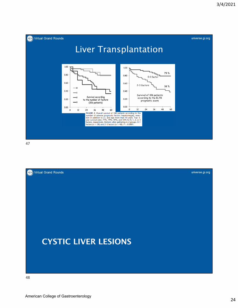
Liver Transplantation
CYSTIC LIVER LESIONS
47
48
American College of Gastroenterology
3/4/2021
25
Classification of Hepatic Cysts• Simple (solitary) cyst• Polycystic liver disease• Parasitic
– Hydatid (echinococcal)• Neoplastic
– Primary• Cystadenoma, cystadenocarcinoma, squamous cell carcinoma
– Secondary• Ovarian, pancreatic, colon, kidney, neuroendocrine
• Duct related– Caroli’s disease– Bile duct duplication
• False cysts– Traumatic intrahepatic hemorrhage– Infarction– Biloma
• Cilated Foregut cyst
More than a simple cyst?Finding Differential diagnosis
Progressive Symptoms Cystadenoma, cystadenocarcinoma, metastasis
Abnormal LFTs Cystadenocarcinoma, metastasis
Rapid growth Cystadenoma, cystadenocarcinoma, metastasis
Calcifications or daughter cysts
Echinococcal cyst
Thick or irregular cyst wall
Cystadenoma, cystadenocarcinoma, metastasis, echinoccocal cyst
Nonhomogeneous cyst content
Cystadenoma, cystadenocarcinoma, metastasis, echinoccocal cyst, bleeding into a simple cyst
Septations or multilocular cyst space
Cystadenoma, cystadenocarcinoma, metastasis, echinoccocal cyst, bleeding into a simple cyst
49
50
American College of Gastroenterology
3/4/2021
26
Simple Cyst
• Cystic formations containing clear fluid• Usually do not communicate with the bile duct• Benign• Rarely large cysts can cause symptoms due to compression
of adjacent structures• Largest cyst ever reported contained 17 liters of fluid!
Simple Cyst
• Tend to occur in right lobe• Female:male 1.5:1 in asymptomatic
– 9:1 in symptomatic
• Huge cysts almost exclusively in women over 50• Huge cysts can cause atrophy of the lobe with
compensatory hypertrophy of the opposite lobe
51
52
American College of Gastroenterology
3/4/2021
27
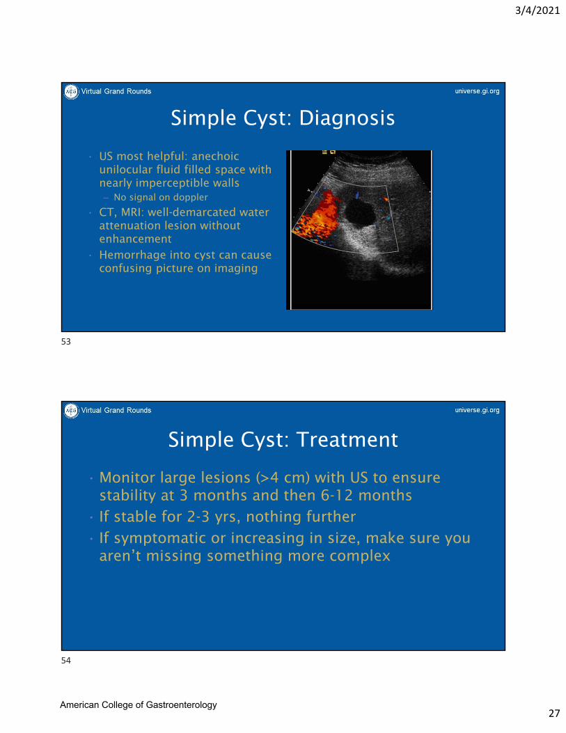
Simple Cyst: Diagnosis
• US most helpful: anechoic unilocular fluid filled space with nearly imperceptible walls– No signal on doppler
• CT, MRI: well-demarcated water attenuation lesion without enhancement
• Hemorrhage into cyst can cause confusing picture on imaging
Simple Cyst: Treatment
• Monitor large lesions (>4 cm) with US to ensure stability at 3 months and then 6-12 months
• If stable for 2-3 yrs, nothing further• If symptomatic or increasing in size, make sure you
aren’t missing something more complex
53
54
American College of Gastroenterology
3/4/2021
28
Simple Cyst: Treatment for Large Cysts
• Aspiration alone not permanent: rapid reaccumulation in near 100%– Can be helpful if trying to decide if symptoms are truly
from the cyst prior to proceeding with surgery
• Aspiration with subsequent injection of sclerosing agent sometimes helps
Simple Cyst: Treatment for Large Cysts
• Surgical resection– Unroofing of cyst– Drainage with cystjejunostomy– Resection of entire cyst
• Surgery associated with recurrence of up to 14% and morbidity up to 15%
• Potential complications– Wound infection– Bile leak– Subphrenic hematoma– Prolonged postoperative drainage
55
56
American College of Gastroenterology
3/4/2021
29
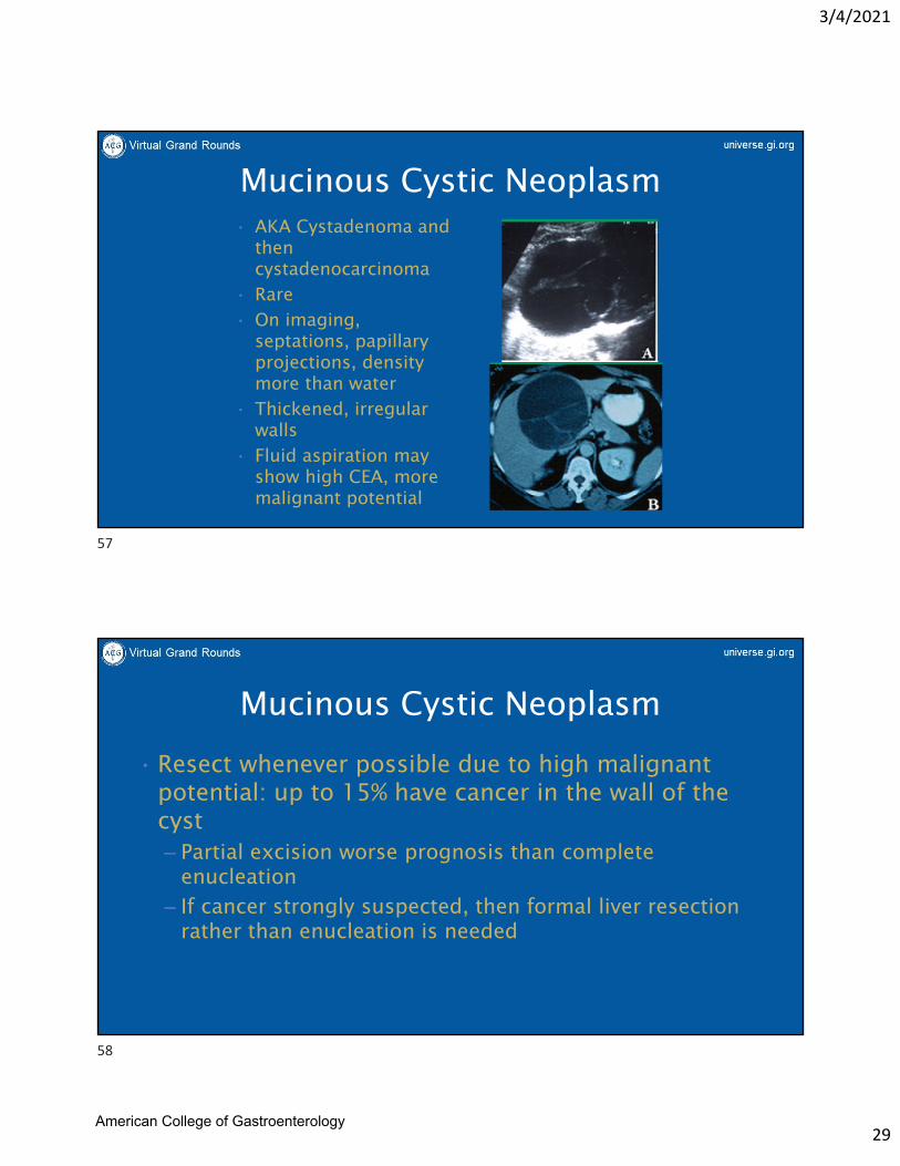
Mucinous Cystic Neoplasm• AKA Cystadenoma and
then cystadenocarcinoma
• Rare• On imaging,
septations, papillary projections, density more than water
• Thickened, irregular walls
• Fluid aspiration may show high CEA, more malignant potential
Mucinous Cystic Neoplasm
• Resect whenever possible due to high malignant potential: up to 15% have cancer in the wall of the cyst– Partial excision worse prognosis than complete
enucleation– If cancer strongly suspected, then formal liver resection
rather than enucleation is needed
57
58
American College of Gastroenterology
3/4/2021
30
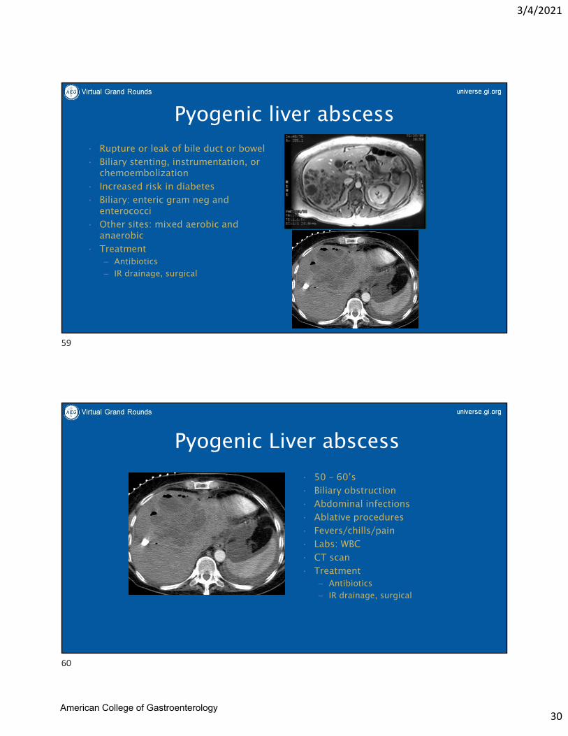
Pyogenic liver abscess
• Rupture or leak of bile duct or bowel• Biliary stenting, instrumentation, or
chemoembolization• Increased risk in diabetes• Biliary: enteric gram neg and
enterococci• Other sites: mixed aerobic and
anaerobic• Treatment
– Antibiotics– IR drainage, surgical
Pyogenic Liver abscess
• 50 – 60’s• Biliary obstruction• Abdominal infections• Ablative procedures• Fevers/chills/pain• Labs: WBC• CT scan• Treatment
– Antibiotics– IR drainage, surgical
59
60
American College of Gastroenterology
3/4/2021
31
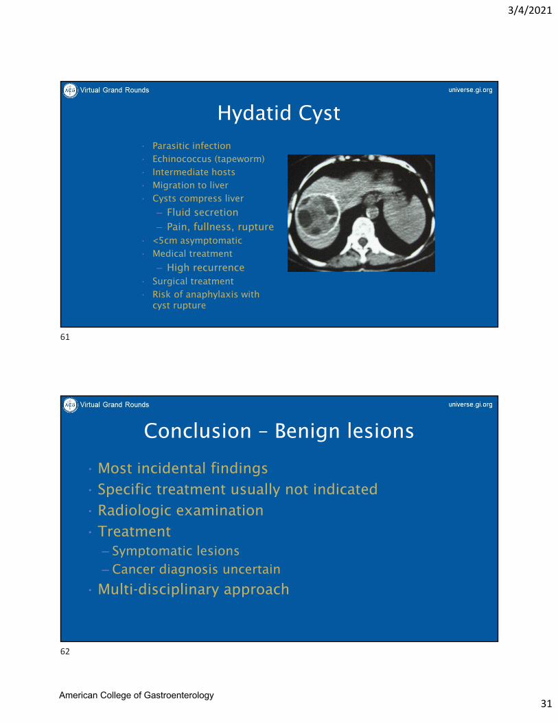
Hydatid Cyst• Parasitic infection• Echinococcus (tapeworm)• Intermediate hosts• Migration to liver• Cysts compress liver
– Fluid secretion– Pain, fullness, rupture
• <5cm asymptomatic• Medical treatment
– High recurrence• Surgical treatment• Risk of anaphylaxis with
cyst rupture
Conclusion – Benign lesions
• Most incidental findings• Specific treatment usually not indicated• Radiologic examination• Treatment
– Symptomatic lesions– Cancer diagnosis uncertain
• Multi-disciplinary approach
61
62
American College of Gastroenterology
3/4/2021
32
Questions?
Speaker: Catherine T. Frenette, MD
Moderator: Anjana A. Pillai, MD
63
64
American College of Gastroenterology