Carol E. Semrad - ACG SmallBowelBleeding2021v5 PB LS
21
5/5/2021 1 Participating in the Webinar All attendees will be muted and will remain in Listen Only Mode. Type your questions here so that the moderator can see them. Not all questions will be answered but we will get to as many as possible. 1 2 American College of Gastroenterology
Transcript of Carol E. Semrad - ACG SmallBowelBleeding2021v5 PB LS

5/5/2021
1
Participating in the Webinar
All attendees will be muted and will remain in Listen Only Mode.
Type your questions here so that the moderator can see them. Not all questions will be answered but we will get to as many as possible.
1
2
American College of Gastroenterology

5/5/2021
2
How to Receive CME and MOC Points
LIVE VIRTUAL GRAND ROUNDS WEBINAR
ACG will send a link to a CME & MOC evaluation to all attendees on the live webinar.
ABIM Board Certified physicians need to complete their MOC activities by December 31, 2021 in order for the MOC points to count toward any MOC requirements that are due by the end of the year. No MOC credit may be awarded after March 1, 2022 for this activity.
MOC QUESTION
If you plan to claim MOC Points for this activity, you will be asked to: Please list specific changes you will make in your
practice as a result of the information you received from this activity.
Include specific strategies or changes that you plan to implement.THESE ANSWERS WILL BE REVIEWED.
3
4
American College of Gastroenterology
5/5/2021
3
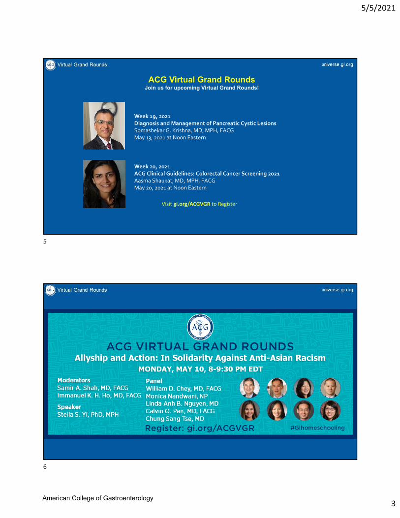
ACG Virtual Grand RoundsJoin us for upcoming Virtual Grand Rounds!
Visit gi.org/ACGVGR to Register
Week 20, 2021ACG Clinical Guidelines: Colorectal Cancer Screening 2021Aasma Shaukat, MD, MPH, FACGMay 20, 2021 at Noon Eastern
Week 19, 2021Diagnosis and Management of Pancreatic Cystic LesionsSomashekar G. Krishna, MD, MPH, FACGMay 13, 2021 at Noon Eastern
OCTOBER 23‐28
5
6
American College of Gastroenterology

5/5/2021
4
Disclosures:
Speaker: Carol E. Semrad, MD, FACGDr. Semrad, faculty for this educational event, has no relevant financial relationship(s) with ineligible companies to disclose.
Moderator: Dejan Micic, MDAdvisory Board: Takeda Pharmaceuticals
*All of the relevant financial relationships listed for these individuals have been mitigated.
Small Bowel Bleeding
Carol E. Semrad, MD, FACG
Professor of Medicine
Director, Small Bowel Disease and Nutrition
7
8
American College of Gastroenterology
5/5/2021
5
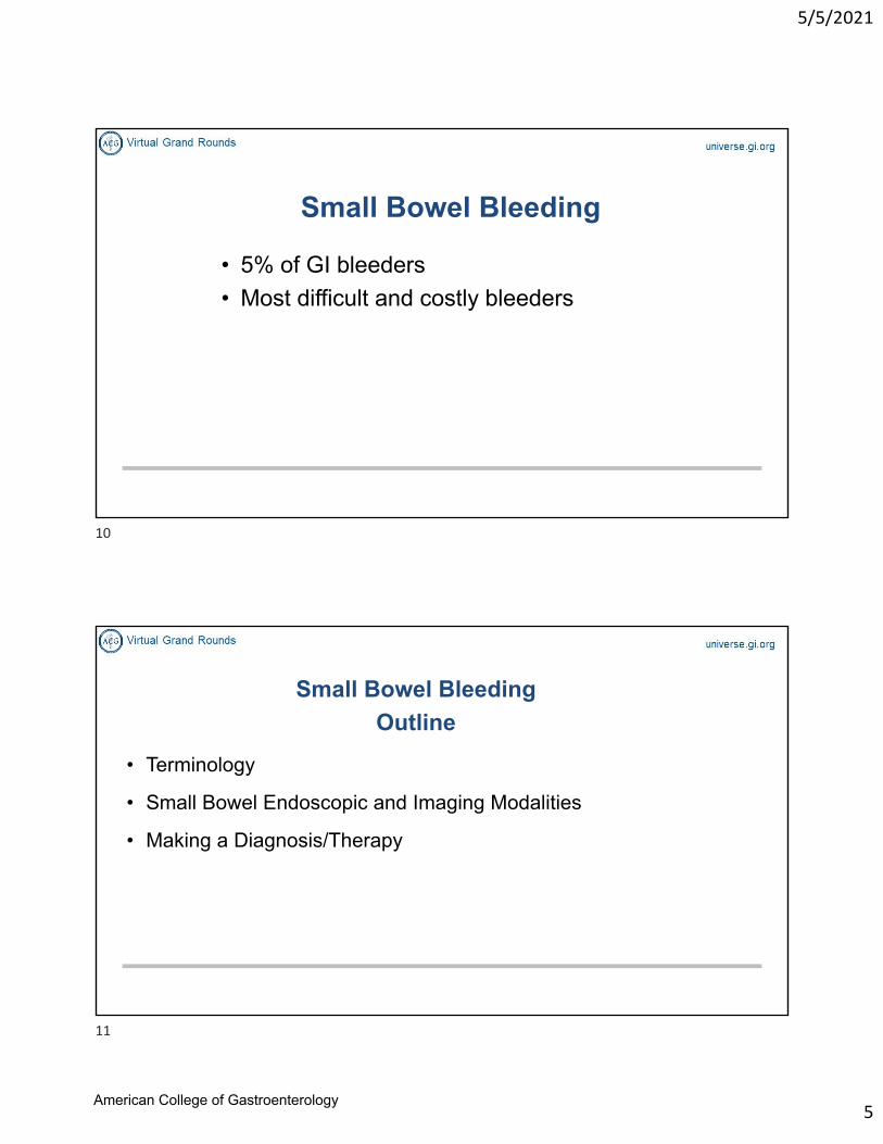
Small Bowel Bleeding
• 5% of GI bleeders
• Most difficult and costly bleeders
Small Bowel Bleeding
Outline
• Terminology
• Small Bowel Endoscopic and Imaging Modalities
• Making a Diagnosis/Therapy
10
11
American College of Gastroenterology

5/5/2021
6
Terminology
Suspected Small Bowel Bleeding
• No source found at upper/lower endoscopy
• Blood in terminal ileum
Obscure GIB
(NEW Definition)
• No source after comprehensive endoscopic and radiologic evaluation of the GI tract
Micic et al. Plos One 2019;20:1‐10
Huprich et al. Radiology 2011;260:744 Huprich et al. AJR 2013;201:65
fat
wall edema
Wireless Device‐Assist Enteroscopy MultiphaseCapsules allows therapy CT Enterography1998 2003 2011
DBE
SBE
Spiral Devices
Radiofrequency
Electric Field Propagation
mass lesion
Arterial phase Dieulafoy
Enteric phase angiectasia
Through Scope Balloon
overtube
12
13
American College of Gastroenterology

5/5/2021
7
Small Bowel Bleeding Imaging Modalities
Test Diagnostic Yield
Small Bowel Barium 5%
Push Enteroscopy 30%
Multi‐Phase CT Enterography 48%
Capsule Endoscopy 38‐83%
Device‐Assist Enteroscopy 51‐80%
Intraoperative Enteroscopy 75‐90%
Triester et al. Am J Gastroenterol 2005;100:2407 Huprich et al. Radiology 2011:260:744Gerson et al. ACG Clinical Guideline, Am J Gastroenterol 2015;110:1265
Small Bowel Capsule Endoscopy What is it good for?
Flat mucosal lesions
Yield highest When performed within first 24-72 hrs in overt bleeding1
Guides therapeutic approach Lesion < 60% SB transit time, upper DAE approach2
Yield of repeat capsule ~ 40% when3
Change from occult to overt bleed
Hemoglobin drop > 4 g/dl
1Rondonotti et al. ESGE guidelines. Endoscopy 2018;50:423.2Li et al. Endoscopy 2009;41:7623Viazis et al. Gastrointest Endosc 2009;69:850
14
15
American College of Gastroenterology

5/5/2021
8
Small Bowel Capsule Endoscopy Limitations
• No sampling, therapy
• Reliability
‐ 30% false positive reads
‐ 20% incomplete studies‐ 18% missed mass lesions‐May miss jejunal/Meckel diverticulum
• Capsule retention in SB
‐ CTE or patency capsule in high risk pt
air bubble‐ polyp
pylorus ‐mass
protrusion ‐mass
lens pressure‐ulcer
Artifacts
Gerson et al. Am J Gastroenterol 2015;110:1265
Push and PullRotational
Single-balloon method Spiral Method
Device-Assist Enteroscopy
16
17
American College of Gastroenterology

5/5/2021
9
Comparison of Enteroscopy Devices Double vs. Single Balloon vs. Spiral
Diagnostic yields similar 50-80%
Summary of small studiesDBE – deepest insertion SBE – easiest set-up Spiral – fastest
Complications similar- Perforation, pancreatitis (0.3%)
All get deeper than push enteroscopy- 80 cm vs 230 cm depth
- 44% vs 62% diagnostic yield
May et al. Am J Gastro 2006;101:2015 May et al. Am J Gastro 2010;105:575 Morgan et al. Gastro Endosc 2010;72:992 Domagk. Endoscopy 2011;43:472 Takano. Gastro Endosc 2011;73:734 Messer. Gastro Endosc 2013;77:241
Device-Assist Enteroscopy
Advantages
• Allows therapy
• Best yield when performed within 24-72 hrs of overt bleed1,2
• Sampling, lesion marking- Minimally invasive surgery
• Best modality for Meckel diverticulum- 40% false negative, adult Meckel scans
Limitations
• Labor intensive
• Steep Learning Curve- 150 cases to achieve total exam3
• Incomplete examinations
1 Aniwan et al. Endosc Int Open 2014;2:E90‐52 Rodrigues et al. Eur J Gastroenterol Hepatol 2018;30:13043 Gross, Stark. Gastrointest Endosc 2008;67:898
18
19
American College of Gastroenterology
5/5/2021
10
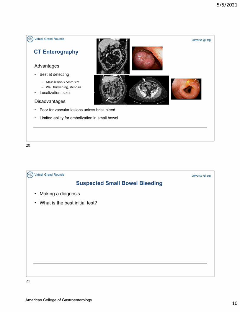
CT Enterography
Advantages
• Best at detecting
– Mass lesion > 5mm size
– Wall thickening, stenosis
• Localization, size
Disadvantages
• Poor for vascular lesions unless brisk bleed
• Limited ability for embolization in small bowel
Suspected Small Bowel Bleeding
• Making a diagnosis
• What is the best initial test?
20
21
American College of Gastroenterology

5/5/2021
11
Causes of Small Bowel Bleeding
Ulcer16%
Tumor 10%
Other14%
AVM60%
N=130
DBE ‐ U.S.A. multi‐center studyMehdizadeh et al. Gastrointest Endosc. 2006;64:740
Other• diverticulum• strictures• varices
Small Bowel Bleeding
Age Guides Best Initial Test for Diagnosis
Young age (< 40 yrs)
• Ulcer (Crohn/NSAID)
• Tumor/polyp
• Meckel diverticulum
• Hereditary vascular lesions
Older age/Co-morbidities
• Vascular lesions
• NSAID injury
22
23
American College of Gastroenterology

5/5/2021
12
Important Physical Findings
• Mucocutaneous telangiectasias- HHT
• Hyperpigmentation lip/skin- Peutz Jeghers Syndrome
• Skin hemangiomas- Blue Rubber Bleb Nevus Syndrome
• SEM of severe aortic stenosis- Anigoectasias, Heyde’s Syndrome
Case
• 26 y.o. woman, 35 wks pregnant
• History of unprovoked GI bleed (melena) 2 yrs ago– EGD, colonoscopy, CE, Meckel scan: All negative
– Told to have CTA if she had recurrent bleeding
• Now with recurrent overt GI bleeding (melena)
• Transfused 8 Units PRBC
• ? Best test for diagnosis
– 40% yield on repeat CE
– CTE/MRE
CE
24
25
American College of Gastroenterology

5/5/2021
13
• Mother given steroid injections to mature fetal lung at 35 wks
• Admitted to hospital for induced delivery
• Standby for emergency C section and tumor resection if GI bleeding with delivery
Lessons
• Beware unexplained overt GI bleeding in the young
• CE misses ~ 20% of small bowel mass lesions
• Retrospective review of her first CE showed debris in proximal SB
• Yield of repeat CE good when recurrent overt bleeding
• Consider CTE/MRE as the first test in young with overt bleeding
26
27
American College of Gastroenterology

5/5/2021
14
What is Safe to Remove in Small Bowel?
Lesion Technique/Outcome
Polyps Hexagonal snareif large, inject epinephrine to shrinkTattoo stalk, clip sitebleeding risk with piecemeal resection
Foreign body Retrieval net for capsulesovertube as shield for sharps
Hemangiomas ? size, depthIf small size, polypectomy, APC, sclerosing agent using EUS
Submucosal mass Perforations reported for polypectomy ofcarcinoid, lipoma
Treatment of Polyps – Hamartomas (PJS)
• Find polyp stalk
• Position polyp
• Use hexagonal snare
• If large polyp– Inject epinephrine 1:100,000, shrink head
– Mark stalk with ink
– Clip stalk after resection
Polyp stalk
28
29
American College of Gastroenterology

5/5/2021
15
Lesion Marking, Laparoscopic Resection
• Indication – Subepithelial mass lesion
– Ulcer/stenosis
• Device‐assist Enteroscopy – Biopsy lesion
– Tattoo at 2 sites
• Surgical resection – Intracorporeal (laparoscopic, internal)
– Extracorporeal (open, mini‐lap, external)
Tapaskar et al Abstract DDW 2018 Yeh et al. Surg Endosc 2009;23:739
Small Bowel Vascular Lesions
• Acquired most common
– Angioectasias
– Dieulafoy lesion
• Hereditary hemorrhagic telangiectasia (HHT)
– Autosomal dominant, 1:5,000 worldwide
– Mutations disrupt TGF‐ pathways in vascular endothelial cells
– Epistaxis most common cause of bleeding/anemia
– GI bleeding in 30%
– AVMs liver, lung, brain
– Juvenile polyposis‐HHT with SMAD4 mutation1
1McDonald et al. Int J Colorectal Dis 2020;35:1963
30
31
American College of Gastroenterology
5/5/2021
16
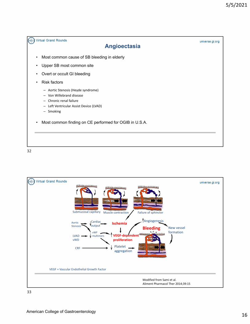
Angioectasia
• Most common cause of SB bleeding in elderly
• Upper SB most common site
• Overt or occult GI bleeding
• Risk factors
– Aortic Stenosis (Heyde syndrome)
– Von Willebrand disease
– Chronic renal failure
– Left Ventricular Assist Device (LVAD)
– Smoking
• Most common finding on CE performed for OGIB in U.S.A.
Submucosal capillary Muscle contraction Failure of sphincter
Aortic Stenosis
LVADvWD
CRF
Cardiac output
vWF multimers
Modified from Sami et al. Aliment Pharmacol Ther 2014;39:15
Ischemia
VEGF‐dependent proliferation
Platelet aggregation
Angiogenesis
Bleeding
VEGF = Vascular Endothelial Growth Factor
New vessel formation
32
33
American College of Gastroenterology
5/5/2021
17
Diagnosis Small Bowel Vascular Lesions
• Capsule Endoscopy
– Best test for flat lesions
– Least invasive, best tolerated
– Guides therapy
• Device-Assisted Enteroscopy
– Invasive
– Allows therapy
• Multiphase CT Enterography
– Uncertain yield for vascular lesions
– Embolization therapy in SB limited due to ischemia risk
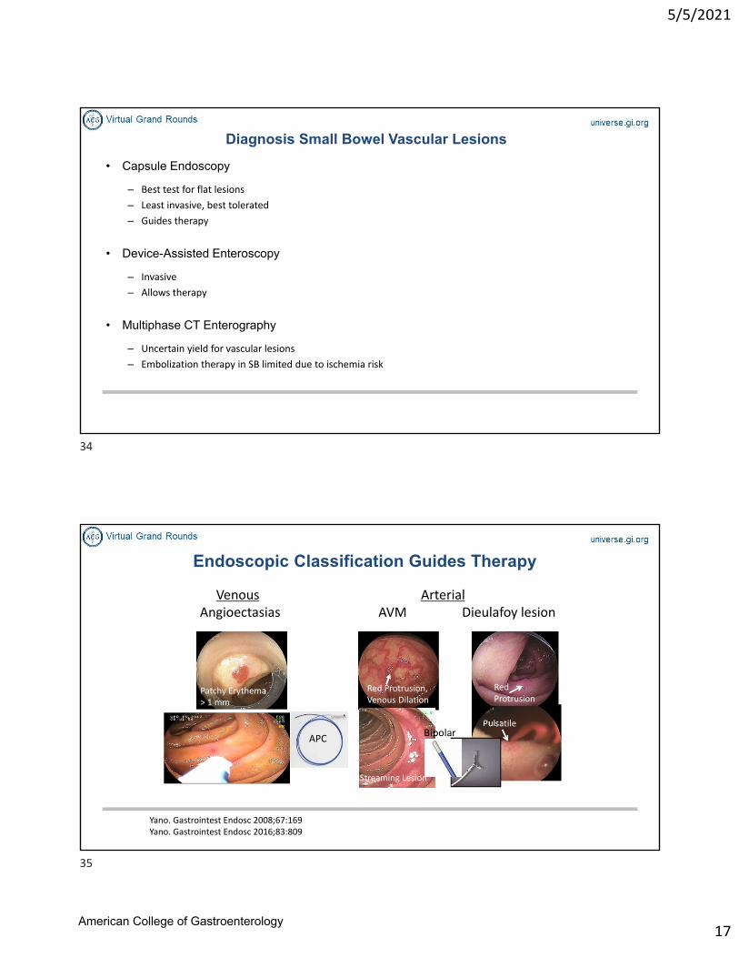
Endoscopic Classification Guides Therapy
VenousAngioectasias
ArterialAVM Dieulafoy lesion
Red Protrusion, Venous Dilation
Patchy Erythema > 1 mm
Red Protrusion
Pulsatile
Yano. Gastrointest Endosc 2008;67:169Yano. Gastrointest Endosc 2016;83:809
APC
Streaming Lesion
34
35
American College of Gastroenterology
5/5/2021
18
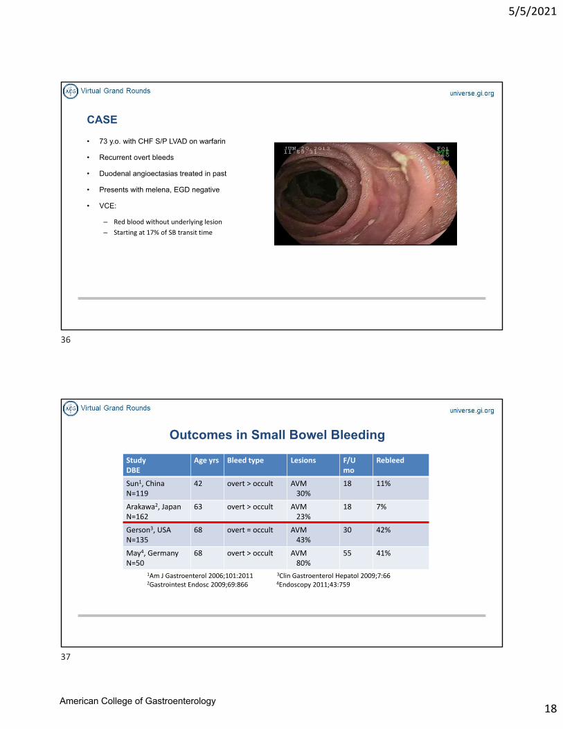
CASE
• 73 y.o. with CHF S/P LVAD on warfarin
• Recurrent overt bleeds
• Duodenal angioectasias treated in past
• Presents with melena, EGD negative
• VCE:
– Red blood without underlying lesion
– Starting at 17% of SB transit time
Outcomes in Small Bowel Bleeding
StudyDBE
Age yrs Bleed type Lesions F/U mo
Rebleed
Sun1, ChinaN=119
42 overt > occult AVM30%
18 11%
Arakawa2, JapanN=162
63 overt > occult AVM 23%
18 7%
Gerson3, USAN=135
68 overt = occult AVM 43%
30 42%
May4, GermanyN=50
68 overt > occult AVM 80%
55 41%
1Am J Gastroenterol 2006;101:2011 3Clin Gastroenterol Hepatol 2009;7:66 2Gastrointest Endosc 2009;69:866 4Endoscopy 2011;43:759
36
37
American College of Gastroenterology
5/5/2021
19
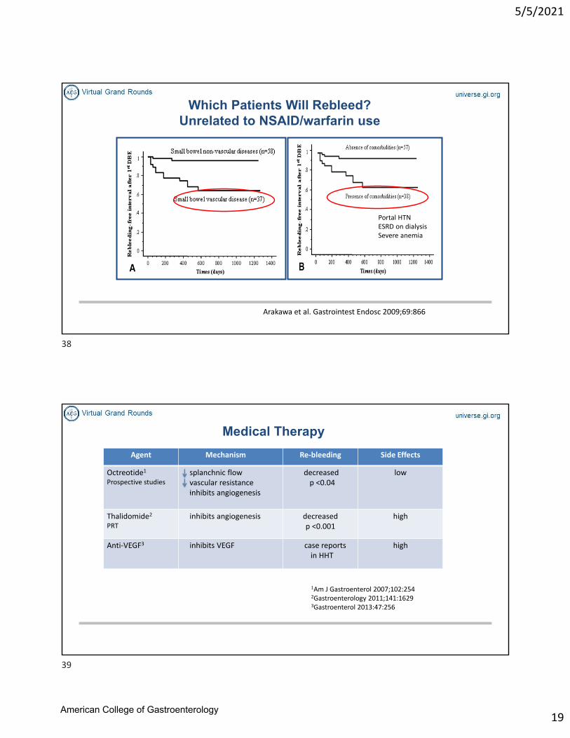
Which Patients Will Rebleed? Unrelated to NSAID/warfarin use
Arakawa et al. Gastrointest Endosc 2009;69:866
Portal HTN ESRD on dialysis Severe anemia
Medical Therapy
Agent Mechanism Re‐bleeding Side Effects
Octreotide1
Prospective studiessplanchnic flowvascular resistanceinhibits angiogenesis
decreased p <0.04
low
Thalidomide2
PRTinhibits angiogenesis decreased
p <0.001high
Anti‐VEGF3 inhibits VEGF case reportsin HHT
high
1Am J Gastroenterol 2007;102:2542Gastroenterology 2011;141:1629 3Gastroenterol 2013:47:256
38
39
American College of Gastroenterology
5/5/2021
20
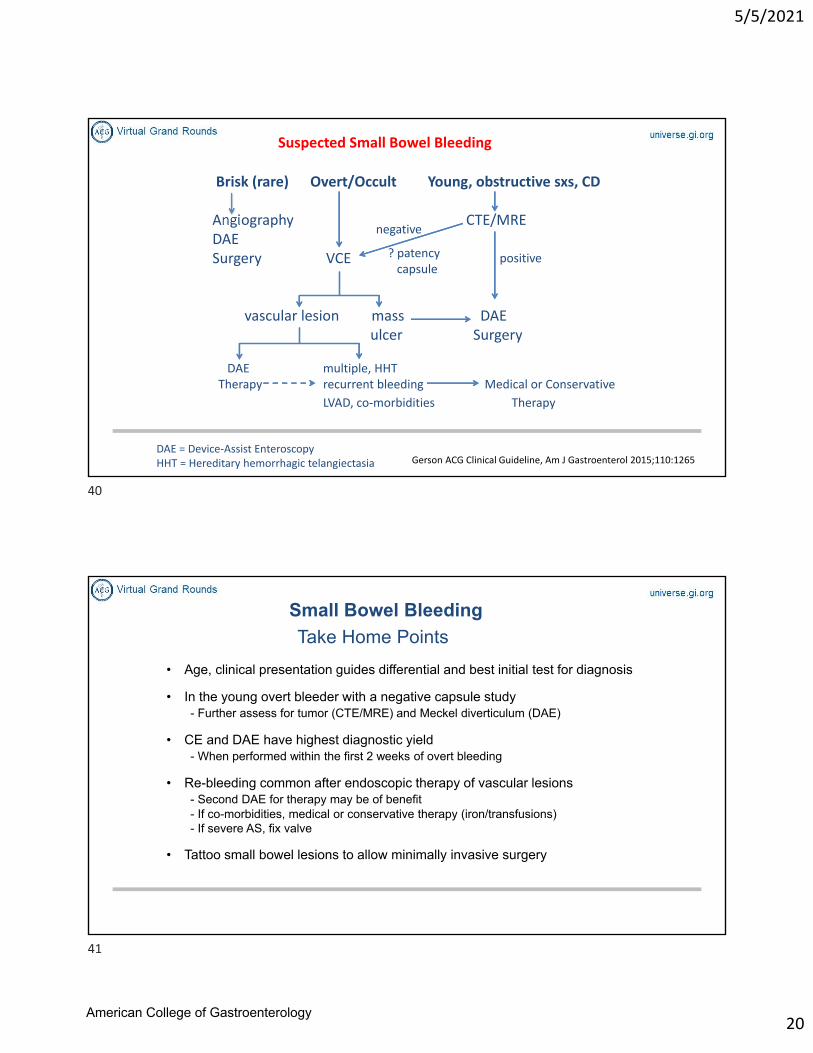
Suspected Small Bowel Bleeding
Brisk (rare) Overt/Occult Young, obstructive sxs, CD
Angiography CTE/MREDAESurgery VCE
vascular lesion mass DAEulcer Surgery
DAE multiple, HHT Therapy recurrent bleeding Medical or Conservative
LVAD, co‐morbidities Therapy
negative
positive? patency capsule
DAE = Device‐Assist Enteroscopy HHT = Hereditary hemorrhagic telangiectasia Gerson ACG Clinical Guideline, Am J Gastroenterol 2015;110:1265
Small Bowel Bleeding
Take Home Points
• Age, clinical presentation guides differential and best initial test for diagnosis
• In the young overt bleeder with a negative capsule study- Further assess for tumor (CTE/MRE) and Meckel diverticulum (DAE)
• CE and DAE have highest diagnostic yield - When performed within the first 2 weeks of overt bleeding
• Re-bleeding common after endoscopic therapy of vascular lesions- Second DAE for therapy may be of benefit- If co-morbidities, medical or conservative therapy (iron/transfusions) - If severe AS, fix valve
• Tattoo small bowel lesions to allow minimally invasive surgery
40
41
American College of Gastroenterology

5/5/2021
21
Questions?
Speaker: Carol E. Semrad, MD, FACG
Moderator: Dejan Micic, MD
42
43
American College of Gastroenterology