Adolescence Traditional views of adolescence Erikson Coleman’s Focal Theory.

CroniconO P E N A C C E S S EC DENTAL SCIENCE
Case Report
Orthopedic Class III Correction in Late Adolescence with Maxillary and Mandible Orthodontic Mini-Implants Combined with Facemask
Therapy: A Novel Approach
Walter A2,4*, Lewington AJ4, Iglesias A1, Bousquet M1, Auladell A1, Wendl B3, Winsauer H4, De la Iglesia F2 Garduño E5
and Puigdollers A6
1Orthodontic Department, Universitat Internacional del Catalunya, Barcelona, Spain2Professor, Orthodontic Department, Universitat Internacional de Catalunya, Barcelona, Spain3Professor, Orthodontic Department, Universität Graz, Austria.4Private Orthodontic Office (Castelldefels, Barcelona, Spain and Bregenz, Austria 5Aligners Institute Invisalign, Ciudad de Mexico6Chairman, Orthodontic Department, Universitat Internacional de Catalunya, Barcelona, Spain
Citation: Andre Walter., et al. “Orthopedic Class III Correction in Late Adolescence with Maxillary and Mandible Orthodontic Mini-Im-plants Combined with Facemask Therapy: A Novel Approach”. EC Dental Science 17.5 (2018): 511-524.
*Corresponding Author: Walter Andre, Professor, Orthodontic and Dento-facial Department, Universitat Internacional de Catalunya, Barcelona, Spain.
Received: March 19, 2018; Published: April 18, 2018
AbstractSkeletal Class III treatment has always been a challenge for orthodontists. Many options of treatment have been described to han-
dle this malocclusion, although the orthopedically treating age of this malocclusion is usually before the growth peak of 8 - 11 years [1]. Nevertheless, skeletal Class III patients in adolescents with malocclusion severity and less craniofacial growth modifications, was considered an impediment for treatment and orthognathic surgery remains the only option. Therefore, it is complicated to identify the best timing and appliance to treat this malocclusion. In these 2 clinical case reports, a novel approach of orthopaedic Class III in late adolescence with the aid of bone anchorage with orthodontic mini-implants (OMIs) is presented.
Keywords: Skeletal Class III; Skeletal Orthopedic Treatment; Late Adolescence; Orthodontic Mini-Implants; Facemask Therapy
The purpose of this study is to measure the skeletal and dento-alveolar changes in two clinical non-growing late adolescent cases treated with an expansion/constriction hybrid expander and facemask therapy (ECHE/FM) combined with indirect mandible bone anchorage (IMBA).
IntroductionIn the past, since 1970 class III malocclusion has been widely described, mostly as a maxillary deficiency rather than a mandible
prognatism. Therefore facemask therapy (FM) remains the main chosen treatment for the correction of skeletal and dental Class III dis-crepancies.
The concomitant use of rapid maxillary expansion with the facemask (RME/FM) treatment has been described to improve the skeletal effects, due to the disruption of the circummaxillary and intermaxillary sutures. This action, could even be increased by the alternation of the expansion and constriction of the sutures to achieve greater sagittal skeletal movements [2].
Much controversy has developed about the orthopedic effects and the appropriate treatment time of the FM. Some authors described the appropriated time in the early mixed dentition, ideally coincidental with the eruption of the upper permanent central incisor [3]. Overall, most of the authors support the statement that the ideal treatment should start no later than 9 years old to produce more skeletal change and less dental movement [4,5].
The great debate about treatment time is based on the desire of achieving greater skeletal changes rather than dental movements, which is the essence of a successful orthopedic treatment. In previous studies there has been a decrease in skeletal effects in those pa-tients who are past the pubertal period with greater dento-alveolar compensations than skeletal movement as result of the treatment [6-8]. One of the explanations for these results is that in older patients the sutures may not be fully disarticulated by a conventional RME [2].

512
Orthopedic Class III Correction in Late Adolescence with Maxillary and Mandible Orthodontic Mini-Implants Combined with Facemask Therapy: A Novel Approach
Citation: Andre Walter., et al. “Orthopedic Class III Correction in Late Adolescence with Maxillary and Mandible Orthodontic Mini-Im-plants Combined with Facemask Therapy: A Novel Approach”. EC Dental Science 17.5 (2018): 511-524.
The alternative RME and constriction (ALT-RAMEC) has been one of the solutions proposed initially by Liou [2] to increase the suture disruption and achieve a greater disarticulation of the maxilla. To reach this disarticulation there is no agreement of the amount of expan-sion necessary, but it seems that with greater expansions, greater stress results in better disarticulation [2]. That is why multiple authors [2,9-12] have developed different protocols with greater expansions and subsequent constriction, both to correct the overexpansion and to produce greater stress to the suture structures.
Skeletal anchorage has broadened the range of movement in many orthodontic treatments, and has also been described [13] to im-prove facemask treatment by the better control of the compensatory side effects. The rational combination of hybrid expanders (HE) with their OMIs located in the anterior palate in combination with FM therapy in children increases skeletal corrections with minor dental side-effects [14]. The so called foot print region in the anterior palate (M4, M5) [15] allows to place several implants in the paramedian region to increase bone anchorage.
However we could not find any investigation that related the effects of skeletal anchorage in late adolescent class III skeletal patients in which the sutures were more ossified and could be re-opened.
The purpose of this study is to measure the skeletal and dento-alveolar changes in two clinical non-growing late adolescent cases treated with an expansion constriction hybrid expander and facemask therapy (ECHE/FM) in combination with indirect mandible bone anchorage (IMBA) with the aid of orthodontic mini-implants and class III elastics force.
Diagnosis and Etiology
The patient is a 13.6 years old female patients with the chief complain of “I have my chin forward”. In the facial analysis, we observed the lower third increased and the chin was deviated 1 mm to the right. The chin musculature was hyperactive when closing lips. The expo-sure of maxillary incisors at rest was insufficient and the smile was non-consonant. From the lateral view, she presented a concave profile with both nasolabial and mentolabial fold opened and a prognatic mandible (Figure 1).
Case Report
Case 1
Figure 1: Pre-treatment facial and intraoral photographs. There is a severe dental-bone discrepancy in the maxilla arch with a complete lack of space for the canine nº 23 eruption.

Citation: Andre Walter., et al. “Orthopedic Class III Correction in Late Adolescence with Maxillary and Mandible Orthodontic Mini-Im-plants Combined with Facemask Therapy: A Novel Approach”. EC Dental Science 17.5 (2018): 511-524.
Orthopedic Class III Correction in Late Adolescence with Maxillary and Mandible Orthodontic Mini-Implants Combined with Facemask Therapy: A Novel Approach
513
The intraoral examination, we observed a molar Class I on the right side and Class III on the left side of 3mm with a non-assess ca-nine relation on both sides. Second upper and lower molars complete erupted and in occlusion. The overjet was negative (-2.5 mm). The dental-bone discrepancy was mild on the lower but severe in the maxilla, with 1.3 in ectopic eruption and 2.3 impacted. The transversal dimension was deficient, with a maxillary compression of 7 mm by Walla [16] (Figure 1).
Concerning the panoramic x-ray, she presented permanent dentition with absence of 1.8, 2.8, 3.8, 4.8 and impaction of 2.3 (Figure 2).
The cephalometric analysis revealed a severe skeletal Class III malocclusion with a ANB -2.7° and Wits -9.3 mm after peak growing and menarche for 2 years ago (Figure 2, Table 1). Lower incisors were normopositioned and normoclined (4.2 mm, 25.3°), the upper incisor procline (4 mm, 28.3°) with a normal mandibular plane of (MP-SN 34.5°).
The objectives for this patient were (1) to improve the Class III profile; (2) correct the anterior crossbite with an anterior displacement of the maxilla bone;(3) create space for the canine eruption; (4) establish a class I molar and canine relationship; achieve an ideal overbite and overjet; and (5) create a stable functional occlusion.
Treatment Objectives
Figure 2: Pre-treatment cephalometric and panoramic radiographs.
Treatment Alternatives
According to the treatment objectives, the conventional proposed treatment plan is extractions of the upper first premolars to allow the eruption of the upper canines and up righting the central incisors in combination with a (1) a Le Fort I osteotomy to move the maxilla bone forward fixated with miniplates in the adult age; or (2) carry out a Le Fort I osteotomy and mobilize the maxilla bone with facemask therapy. Due to the patient young age, the ideal surgical treatment plan was not an option at the time due to the possibility that class III patients can be late growers until young adult age.
Treatment Progress
The treatment plan consisted of a customized hybrid expander with an anchorage support with 2 OMIs (2.5 diameter x 14mm long, Jeil Medical, Seoul, Korea). The attachment of the OMIs with their collar abutments (Tiger Dental, Bregenz, Austria) is carried out with a cemented fixation (Orthodontic reliance, phase II, Itasca, Illinois, USA) [17].

Citation: Andre Walter., et al. “Orthopedic Class III Correction in Late Adolescence with Maxillary and Mandible Orthodontic Mini-Im-plants Combined with Facemask Therapy: A Novel Approach”. EC Dental Science 17.5 (2018): 511-524.
Orthopedic Class III Correction in Late Adolescence with Maxillary and Mandible Orthodontic Mini-Implants Combined with Facemask Therapy: A Novel Approach
514
First, the OMIs were placed half way between bicuspids and midpalatal line [15] and an impression with transference abutment was taken for the customized expander. After bonding of the hybrid expander, instructions for activations were given to the patient follow-ing Alt-Ramec protocol [18]. After a final activation of 8 mm we proceeded with the placement of a lingual arch with soldered hooks designed for class III intermaxillary elastics, also anchored over 2 OMIs (1.6 mm x 12 mm, Jeil Medical, Seoul, Korea) (Figure 3). At this point we started ECHE/FM therapy with extraoral elastics (8oz 8 mm) for 8 - 10 hours of use over-night, followed by a IMBA therapy with intermaxillary class III elastics (4oz 4 mm) placed from the upper molar hooks to the hooks of a lingual arch anchored with 2 OMIs. This intermaxillary class III elastic treatment reinforces the bone anchorage maxillary protraction (BAMP) during the rest of the day (Figure 3).
After one year ECHE/FM therapy, the hybrid expander was removed, leaving a dental class II relation that lead to an upper arch dis-talization with a Top -Jet appliance [19] (Figure 4). The distalizer appliance was activated during 1 year until molar class I was achieved providing space for upper canines. Finally an InvisalingTM treatment was planned for the settling of the final occlusion. Total treatment time was of 38 months.
Figure 3: Class III orthopedic treatment. (a-c) Intermaxillary class III elastics and upper molar hooks for the elastics of the facemask traction, (d) hybrid expander with 2 palatal OMIs and (e) lingual arch with soldered hooks placed in the buc-
cal side for intermaxillary class III elastic placement.
Figure 4: Top-Jet distalizers using the same M4 OMIs position of the hybrid expander. (a) Initial bilateral distalization providing additional space for the canine eruption, and (b)
unilateral distalizer continues to create additional space only in one side.

Citation: Andre Walter., et al. “Orthopedic Class III Correction in Late Adolescence with Maxillary and Mandible Orthodontic Mini-Im-plants Combined with Facemask Therapy: A Novel Approach”. EC Dental Science 17.5 (2018): 511-524.
Orthopedic Class III Correction in Late Adolescence with Maxillary and Mandible Orthodontic Mini-Implants Combined with Facemask Therapy: A Novel Approach
515
In the post-treatment records we observed a favorable aesthetic change both in the frontal and sagittal view of extraoral pictures (Fig-ure 5). The arch smile was corrected, with a curvature following the lower lip at smile with a normal exposure of the upper incisors at rest and at smile. Intraorally, canine and molar class I was achieved with the correction of the negative overjet. Upper arch dimension changed dramatically, solving the compression and the crowding (Figure 5).
Figure 5: Post-treatment facial and intraoral photographs.
In the cephalometric analysis, after 1 year of orthopedic class III treatment (Figure 6a), there was an increase in ANB angle change from -2.7° to 1.0° and the Wits analysis value change from -9.3 mm to -2.5 mm (Table 1). In terms of vertical dimension there was an in-crease on MP-SN of 2°, whereas from the dental point of view the upper and lower incisor showed a slight retrusion without proclination. The general superimposition tracings after 1 year of orthopedic treatment showed a remarkable maxilla protraction with a mandible posterorotation and class III skeletal correction (Figure 6-7, Table 1). The upper lip profile after 1 year of treatment changed dramatically forward in the cephalometric x-ray and in the general superimposition (Figure 6a, and 7).
The final cephalometric x-ray showed minor skeletal relapse with an increase of the upper incisor inclination (Figure 6b, Table 1).
Figure 6: Lateral cephalometric radiographs.(a)After 1 year of orthopedic class III treatment and (b) final cephalometric radiograph.

516
Orthopedic Class III Correction in Late Adolescence with Maxillary and Mandible Orthodontic Mini-Implants Combined with Facemask Therapy: A Novel Approach
Citation: Andre Walter., et al. “Orthopedic Class III Correction in Late Adolescence with Maxillary and Mandible Orthodontic Mini-Im-plants Combined with Facemask Therapy: A Novel Approach”. EC Dental Science 17.5 (2018): 511-524.
Case 1 Norm Pre-treatment Orthop. Treatment (after 1 year) Post-treatmentSNA (º) 82 ± 2 70.9 73.7 74.4SNB (º) 80 ± 2 73.6 72.7 73.4ANB (º) 2 ± 2 -2.7 1 1GoGn- SN (º) 32 ± 4 34.5 36.0 36.5U1-NA (mm) 4.0 ± 1.0 4.0 2.8 5.5L1-NB (mm) 4.0 ± 1.0 4.2 2.9 5.3U1-NA (º) 22 ± 2 28.3 24 26.4L1-NA (º) 25 ± 2 25.3 24 22Wits 0 ± 1 -9.3 -2.5 -2.9
Table 1: Cephalometric analysis of pre-treatment, after 1 year of orthopedic class III treatment and post-treatment.
Figure 7: Superimpositions after 1 year of orthopedic treatment.
Diagnosis and Etiology
Case 2
The second case was a 16.6 years old patient who presented two previous orthodontic treatments (one on going) and was still un-happy with her smile. The patient showed a relapse of the open bite and she wanted to improve the aesthetics of her smile. The frontal view showed proportioned thirds with no exposure of upper incisors at rest and a low nonconsonant smile. In the profile analysis, she presented a convex profile with a normal nasolabial and mentolabial fold (Figure 8).
Figure 8: Pre-treatment facial and intraoral photographs. (The images showed previous bracket placement due to a second diagnostic opinion).

517
Orthopedic Class III Correction in Late Adolescence with Maxillary and Mandible Orthodontic Mini-Implants Combined with Facemask Therapy: A Novel Approach
Citation: Andre Walter., et al. “Orthopedic Class III Correction in Late Adolescence with Maxillary and Mandible Orthodontic Mini-Im-plants Combined with Facemask Therapy: A Novel Approach”. EC Dental Science 17.5 (2018): 511-524.
The intraoral examination showed on the right side a Class I molar and canine and a mild class III malocclusion of 2 mm on the left side. The overjet was edge to edge. Both dental arches were aligned due to previous treatment but we observed dental compensations with upper buccal tipping and lower lingual inclination on posterior segments. Furthermore we found a skeletal compression of 5mm by Walla Ridge [16] (Figure 8).
The cephalometric analysis revealed a skeletal Class III malocclusion (ANB: -0.4 mm, Wits: -3.0 mm), with a slight long face and verti-cal growth tendency. The incisors were protruded and proclined both in the upper at (7.2 mm, 34°) and the lower (6.1 mm, 27.2°). The mandibular plane had an inclination of 33.8° (Figure 9, Table 2).
Figure 9: Pre-treatment cephalometric and panoramic radiographs. (The initial x-rays showed brackets in the images because the above-mentioned reasons).
Treatment Objectives
The objectives for this patient were to improve the cosmetics of her smile, (1) by allowing the upper incisor extrusion and therefore improving the gum display and create an normal overbite, (2) improve skeletally the class III malocclusion and (3) correct the excessive upper incisor proinclination to a normal angle.
According to the treatment objectives, the ideal treatment due to the age of the patient with a skeletal class III malocclusion is a surgi-cal treatment plan consisting in a segmented maxillary advancement with downward movement and 4 premolars extractions to upright the upper incisors to the norm. The patient refused this treatment plan.
First of all, previous orthodontic appliances were removed and we proceeded to the placement of palatal OMIS to assure bone anchor-age for a maxilla orthopedic treatment. The orthopedic treatment consisted of expansion with a hybrid expander anchored on six OMIs [17] placed paramedian to the midpalatal suture (M4, M5 position) due to the bone maturation of the sutures and patients age. To achieve a better disarticulation of the circummaxillary and intermaxillary sutures, expansion/constrictions of the hybrid expander were carried out.
Treatment Alternatives
Treatment Progress
Citation: Andre Walter., et al. “Orthopedic Class III Correction in Late Adolescence with Maxillary and Mandible Orthodontic Mini-Im-plants Combined with Facemask Therapy: A Novel Approach”. EC Dental Science 17.5 (2018): 511-524.
Orthopedic Class III Correction in Late Adolescence with Maxillary and Mandible Orthodontic Mini-Implants Combined with Facemask Therapy: A Novel Approach
518
After a final activation of 6 mm, instructions for ECHE/FM were given as in the previous case following the Alt-Ramec protocol [18] (4 Alt-Ramec cycles in 1 year). Immediately after midpalatal suture opening, we proceeded to the placement of a lingual arch with their buccal hooks for the placement of class III intermaxillary elastics and mandible OMIs (1.6 mm X 12 mm) anchorage to carry out the IMBA therapy (Figure 10).
Figure 10: Orthopedic class III treatment with the combination of the ECHE/FM and the IMBA technique.
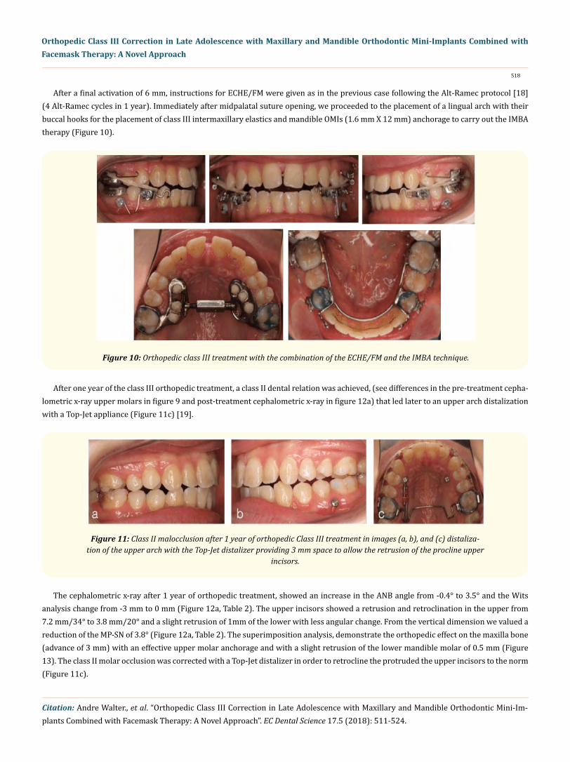
After one year of the class III orthopedic treatment, a class II dental relation was achieved, (see differences in the pre-treatment cepha-lometric x-ray upper molars in figure 9 and post-treatment cephalometric x-ray in figure 12a) that led later to an upper arch distalization with a Top-Jet appliance (Figure 11c) [19].
Figure 11: Class II malocclusion after 1 year of orthopedic Class III treatment in images (a, b), and (c) distaliza-tion of the upper arch with the Top-Jet distalizer providing 3 mm space to allow the retrusion of the procline upper
incisors.
The cephalometric x-ray after 1 year of orthopedic treatment, showed an increase in the ANB angle from -0.4° to 3.5° and the Wits analysis change from -3 mm to 0 mm (Figure 12a, Table 2). The upper incisors showed a retrusion and retroclination in the upper from 7.2 mm/34° to 3.8 mm/20° and a slight retrusion of 1mm of the lower with less angular change. From the vertical dimension we valued a reduction of the MP-SN of 3.8° (Figure 12a, Table 2). The superimposition analysis, demonstrate the orthopedic effect on the maxilla bone (advance of 3 mm) with an effective upper molar anchorage and with a slight retrusion of the lower mandible molar of 0.5 mm (Figure 13). The class II molar occlusion was corrected with a Top-Jet distalizer in order to retrocline the protruded the upper incisors to the norm (Figure 11c).

Citation: Andre Walter., et al. “Orthopedic Class III Correction in Late Adolescence with Maxillary and Mandible Orthodontic Mini-Im-plants Combined with Facemask Therapy: A Novel Approach”. EC Dental Science 17.5 (2018): 511-524.
Figure 12: Cephalometric and panoramic radiographs, (a) showed cephalometric x-ray after 1 year orthopedic class III treatment causing a class II molar relation, (b) cephalometric x-ray
post-treatment and (c) post-treatment panoramic x-ray.
Figure 13: Superimpositions cephalometric tracings of pre-treatment and after 1-year orthopedic class III treatment.
Orthopedic Class III Correction in Late Adolescence with Maxillary and Mandible Orthodontic Mini-Implants Combined with Facemask Therapy: A Novel Approach
519

Citation: Andre Walter., et al. “Orthopedic Class III Correction in Late Adolescence with Maxillary and Mandible Orthodontic Mini-Im-plants Combined with Facemask Therapy: A Novel Approach”. EC Dental Science 17.5 (2018): 511-524.
Orthopedic Class III Correction in Late Adolescence with Maxillary and Mandible Orthodontic Mini-Implants Combined with Facemask Therapy: A Novel Approach
520
For the finishing of the case, multibracket appliances were bonded only to the maxillary teeth and a loop system (0.018 ss. wire) to extrude upper incisors was designed supported by two OMIs (1.6 X 8 mm Jeil Medical, Seoul, Korea) placed between the roots of the teeth nº 1.2 - 1.3 and 2.2 - 2.3. The buccal placement of the upper extrusion loops, from the mechanical point of view facilitates the linguover-sion of the procline incisors (Figure 14). In addition, a loop archwire anchored to the Top-jet distalizer facilitates the closure of the upper distal lateral incisors spaces (Figure 11c). The orthodontic treatment lasted 38 months (Figure 15).
Figure 14: Intraoral images of extrusion and linguoversion of upper incisors with the aid of buccal OMIs (placed between the roots of lateral incisor and canines) and a loop archwire anchored to the Top-jet distalizer to facilitate closing the
spaces of the upper distal incisors.
Post-treatment extraoral pictures showed a noticeable improvement of aesthetics both in the frontal and sagittal view with enhance-ment of malar support and a correct exposure of upper incisors and consonance of the smile. As a result of the maxilla disjunction and protraction, the premolars and molars torque has changed dramatically after the treatment, changing from a negative to a positive torque (see occlusal frontal view of figure 8 and figure 14). In the intraoral view, she finished the treatment with a molar and canine Class I and a correct overjet and overbite (Figure 15).
Figure 15: Post-treatment facial and intraoral photographs.

Citation: Andre Walter., et al. “Orthopedic Class III Correction in Late Adolescence with Maxillary and Mandible Orthodontic Mini-Im-plants Combined with Facemask Therapy: A Novel Approach”. EC Dental Science 17.5 (2018): 511-524.
Orthopedic Class III Correction in Late Adolescence with Maxillary and Mandible Orthodontic Mini-Implants Combined with Facemask Therapy: A Novel Approach
521
Case 2 Norm Pre-treatment Orthop. Treatment (after 1 year) Post-treatmentSNA (º) 82 ± 2 79.7 83.8 83.4SNB (º) 80 ± 2 80.1 79.8 80.2ANB (º) 2 ± 2 -0.4 3.5 3.2GoGn- SN (º) 32 ± 4 33.8 30 33.7U1-NA (mm) 4.0 ± 1.0 7.2 3.8 2.7L1-NB (mm) 4.0 ± 1.0 6.1 5.1 5.0U1-NA (º) 22 ± 2 34 20 19.5L1-NA (º) 25 ± 2 27.2 29.1 27.2Wits 0 ± 1 -3 0 0
Table 2: Cephalometric analysis pre-treatment, after 1 year orthopedic treatment and post-treatment.
Discussion This case-report study shows skeletal and dento-alveolar changes in two non-growing late adolescents that present skeletal class III
malocclusion without the use of surgical treatment.
Several investigators demonstrated the skeletal changes that can be obtained in animals when protraction discontinuous forces with facemask were applied to the maxilla [20-22]. The entire maxilla was displaced anteriorly, suggesting adaptations at the circummaxillary sutures. Many clinicians advocate the use of RME to disarticulate the sutures and facilitate maxillary protraction with a facemask therapy (RME/FM), nevertheless other investigators [23] reported opening of the circummaxillary sutures using the bone anchorage maxillary protraction (BAMP) protocol with a continuous force to the miniplates without RME. These observations suggest that perhaps the direct and continuous force application to the maxillary or zygomatic bones produces suture distraction.
In our study we propose the novel combination of continuous (intermaxillary elastics with indirect bone anchorage) and discontinu-ous forces (nocturne facemask with indirect bone anchorage) non-surgically and without using miniplates to produce favorable changes in the skeletal relation that notably broadens (13.6 and 16.6 years old patients) the conventional treatment age range.
From the point of view of the treatment timing, the RME/FM protocol demonstrates the best outcomes in terms of maxillary protrac-tion in the deciduous or early mixed dentition [24]. Thus, it is recommended that FM therapy should be started before the age of 8 years when possible [7].
Using human autopsy material, Melsen [25] have shown that suture morphology of the palate maxillary region becomes progressively interdigitated with increasing age. Therefore, it is reported that in the late juvenile and adolescent periods, it can be more difficult to disarticulate the palatal bone from the pterygoid process for maxillary protraction. These findings are confirmed in the CBCT study by Garib and coworkers, where it has been seen in children aged from 11 - 14 years, that the effective skeletal midpalatal suture opening in tooth-borne expanders is only 30%, being 70% dento-alveolar compensation [26]. Therefore, a disjunction with bone anchoring using the MARPE technique can be more effective from the skeletal point of view [17,27].
In the two presented clinical cases, the ECHE/FM due to the effective OMIs bone anchorage allows greater forces transmitted to the maxilla bone, thus producing more effective midpalatal suture opening and therefore circummaxillary suture response with a greater suture disarticulation beyond the conventional treatment age. Therefore, the maxilla bone forward movement in both presented cases was possible.
By contrast, BAMP with intermaxillary elastic treatment, is applied more successfully during the late mixed dentition or early perma-nent dentition in the infrazygomatic arch because of the lack of bone quality for OMIs stability in younger patients [28]. However, this

Citation: Andre Walter., et al. “Orthopedic Class III Correction in Late Adolescence with Maxillary and Mandible Orthodontic Mini-Im-plants Combined with Facemask Therapy: A Novel Approach”. EC Dental Science 17.5 (2018): 511-524.
Orthopedic Class III Correction in Late Adolescence with Maxillary and Mandible Orthodontic Mini-Implants Combined with Facemask Therapy: A Novel Approach
522
The use of heavy forces in the maxilla protraction (500 to 1500g) is to stimulate growth and suture remodeling by separating the sutures, especially in the pterygoid process more than would occur otherwise [14,30]. De Clerk., et al. [28] proposed a more favourable maxillary response with a moderate continuous force traction during the day, rather than a heavy interrupted force at night. In our cases, both continuous (250 gr/elastics) and discontinuous (500 gr/FM) forces were applied to take advantage the two techniques.
Toyama Hino., et al. reported that BAMP treatment showed significantly greater orthopedic changes compared with RME/FM [31]. In addition, a third of the RME/FM patients had a predominantly vertical maxillary displacement, in the BAMP sample, only a sixth of the patients had a predominantly vertical response. In the reported cases we can see that in case 1 there is an increase in the MP-SN angle of 2° that could be explained by patients younger age that still had craniofacial residual growth, meanwhile in the second case we observed a reduction of this angle in 3.8°. This suggests that in the second case with pre-treatment slight openbite, the absence of lower brackets placement in the treatment, indirect molar anchorage in both dental arches avoiding molar eruptions side effects, could be a benefit if the anterior maxilla displacement provides greater space for the tongue position and therefore reduces the mandible hinge angle mechani-cally. Nevertheless, in the first case this is not exclusively related mechanically because of the natural growth of the patient and a slight lower molar distalization of 1mm.
Criticisms of FM therapy with dental anchorage claim that this is a camouflage rather than a correction of the skeletal discrepancy, due to teeth movement, instead of achieving true skeletal modification [30]. Skeletal anchorage could be used to avoid the dental side effects by using hybrid expanders [32]. Therefore, maxillary protraction in combination with fixed expansion appliances provide better skeletal results [1].
For the design of the hybrid expander, patient age should be considered to provide enough anchorage to facilitate circummaxillary sutures disarticulation and remodeling, especially in late adolescence. It is known, that high stresses generated in various craniofacial sutures after maxillary protraction with expansion, are responsible for the circummaxillary suture system response, facilitating the or-thopedic effect [33]. To this consideration, for the second case we customized 6 OMIs HE that provided greater stability to withstand opening/constriction forces during the one-year orthopedic treatment period. The superimposition tracings on both cases demonstrated the effectiveness of the OMIs stability to provide the upper molars proper indirect anchorage avoiding molar mesial migration even in late adolescents, being a very useful treatment in skeletal class III patients without surgical procedures with the aid of the mechanical suture response.
With a stable and effective bone anchorage design strategy that allows a complete disarticulation of the maxilla bone and their circum-maxillary sutures, the orthopedic Class III skeletal treatment with maxillary deficiency in late adolescences patients can be an alternative treatment. This opens the range of new treatment possibilities that requires further studies in the future for those patients approaching the late adolescent to adulthood.
protocol requires a surgical intervention for placement and removal of the miniplates that increases morbidity and surgical cost. For this reason, the lower OMIs in the mandible were placed as an indirect anchorage to a lingual arch, with hooks for the class III elastics, on the buccal segment as an alternative of the miniplates. In addition, the OMIs placed in the palate provides enough bone height for their stability and therefore assure indirect anchorage to the upper molars demonstrating in several studies the successful orthopedic maxilla traction [14,29].
Conclusion
Bibliography
1. Ngan P., et al. “Treatment response to maxillary expansion and protraction”. European Journal of Orthodontics 18.2 (1996): 151-168.
2. Liou EJ. “Effective maxillary orthopedic protraction for growing Class III patients : a clinical application simulates distraction osteo-genesis”. Progress in Orthodontics 6.2 (2005): 36-53.
3. Jr MJ. “An orthopedic approach to the treatment of Class III malocclusion in young patients”. Journal of Clinical Orthodontics 21.9 (1987): 598-608.
Citation: Andre Walter., et al. “Orthopedic Class III Correction in Late Adolescence with Maxillary and Mandible Orthodontic Mini-Im-plants Combined with Facemask Therapy: A Novel Approach”. EC Dental Science 17.5 (2018): 511-524.
Orthopedic Class III Correction in Late Adolescence with Maxillary and Mandible Orthodontic Mini-Implants Combined with Facemask Therapy: A Novel Approach
523
4. Proffit W. “Contemporary Orthodontics”. St Louis (1992).
5. Hickham JH. “Maxillary protraction therapy: diagnosis and treatment”. Journal of Clinical Orthodontics 25 (1991): 102-113.
6. Yavuz I., et al. “Face mask therapy effects in two skeletal maturation groups of female subjects with skeletal class iii malocclusions”. Angle Orthodontist 79.5 (2009): 842-848.
7. Cha K-S. “Skeletal Changes of Maxillary Protraction in Patients Exhibiting Skeletal Class III Malocclusion: A Comparison of Three Skeletal Maturation Groups”. Angle Orthodontist 73.1 (2003): 26-35.
8. Baccetti T., et al. “Skeletal effects of early treatment of Class III malocclusion with maxillary expansion and face-mask therapy”. Ameri-can Journal of Orthodontics and Dentofacial Orthopedics 113 (1998): 333-343.
9. Franchi L. “Early Alt-RAMEC and Facial Mask Protocol in Class III Malocclusion”. Journal of Clinical Orthodontics 45.11 (2011): 46-48.
10. Masucci C., et al. “Short-term effects of a modified Alt-RAMEC protocol for early treatment of Class III malocclusion: A controlled study”. Orthodontics and Craniofacial Research 17.4 (2014): 259-269.
11. Wilmes B., et al. “Early class III facemask treatment with the hybrid hyrax and Alt-RAMEC protocol”. Journal of Clinical Orthodontics 48.2 (2014): 84-93.
12. Isci D., et al. “Activation-deactivation rapid palatal expansion and reverse headgear in Class III cases”. European Journal of Orthodon-tics 32.6 (2010): 706-715.
13. Cevidanes L., et al. “Comparison of two protocols for maxillary protraction: bone an- chors versus face mask with rapid maxillary expansion”. Angle Orthodontist 80.5 (2010): 799-806.
14. Nienkemper M., et al. “Maxillary protraction using a hybrid hyrax-facemask combination”. Progress in Orthodontics 14.1 (2013): 1-8.
15. Winsauer H., et al. “Paramedian vertical palatal bone height for mini-implant insertion: a systematic review”. European Journal of Orthodontics 36.5 (2014): 541-549.
16. Roney V., et al. “Mandibular arch form: The relationship between dental and basal anatomy”. American Journal of Orthodontics and Dentofacial Orthopedics 134.3 (2008): 430-438.
17. Winsauer H., et al. “A bone-borne appliance for rapid maxillary expansion”. Journal of Clinical Orthodontics 47.6 (2013): 375-381.
18. Liou EJ-W. “Effective maxillary orthopedic protraction for growing Class III patients: a clinical application simulates distraction os-teogenesis”. Progress in Orthodontics 6.2 (2005): 154-171.
19. Winsauer H., et al. “The TopJet for Routine Bodily Molar Distalization”. Journal of Clinical Orthodontics (2013): 96-107.
20. Kambara T. “Dentofacial changes produced by extraoral forward force in the Macada irus”. American Journal of Orthodontics 71.3 (1977): 249-277.
21. Nanda R. “Protraction of maxilla in rhesus monkeys by controlled extraoral forces”. American Journal of Orthodontics 74.2 (1978): 121-141.
22. Jackson GW and Kokich VG SP. “Experimental and postexperimental response to anteriorly directed extraoral force in young Macaca nemestrina”. American Journal of Orthodontics 75.3 (1979): 318-333.
23. Nguyen T., et al. “Three-dimensional assessment of maxillary changes associated with bone anchored maxillary protraction”. Ameri-can Journal of Orthodontics and Dentofacial Orthopedics 140.6 (2011): 790-798.

Citation: Andre Walter., et al. “Orthopedic Class III Correction in Late Adolescence with Maxillary and Mandible Orthodontic Mini-Im-plants Combined with Facemask Therapy: A Novel Approach”. EC Dental Science 17.5 (2018): 511-524.
Orthopedic Class III Correction in Late Adolescence with Maxillary and Mandible Orthodontic Mini-Implants Combined with Facemask Therapy: A Novel Approach
524
24. Vaughn GA., et al. “The effects of maxillary protraction therapy with or without rapid palatal expansion: a prospective, randomized clinical trial”. American Journal of Orthodontics 128.3 (2005): 299-309.
25. Melsen BMF. “The postnatal development of the palatomaxillary region studied on human autopsy material”. American Journal of Orthodontics 82.4 (1982): 329-342.
26. Garib DG., et al. “Rapid maxillary expansion - Tooth tissue-borne versus tooth-borne expanders: A computed tomography evaluation of dentoskeletal effects”. Angle Orthodontist 75.4 (2005): 548-557.
27. Carlson C., et al. “Microimplant-assisted rapid palatal expansion appliance to orthopedically correct transverse maxillary deficiency in an adult”. American Journal of Orthodontics and Dentofacial Orthopedics 149.5 (2016): 716-728.
28. De Clerck H., et al. “Dentofacial effects of bone-anchored maxillary protraction: A controlled study of consecutively treated Class III patients”. American Journal of Orthodontics and Dentofacial Orthopedics 138.5 (2010): 577-581.
29. Walter A., et al. “Design characteristics, primary stability and risk of fracture of orthodontic mini-implants: Pilot scan electron micro-scope and mechanical studies”. Medicina Oral Patologia Oral y Cirugia Bucal 18.5 (2013): e804-e810.
30. Ngan PW., et al. “Treatment response and long-term dentofacial adaptations to maxillary expansion and protraction”. Seminars in Orthodontics 3.4 (1997): 255-264.
31. Hino CT., et al. “Three-dimensional analysis of maxillary changes associated with facemask and rapid maxillary expansion compared with bone anchored maxillary protraction”. American Journal of Orthodontics and Dentofacial Orthopedics 144.5 (2013): 705-714.
32. Wilmes B., et al. “Application and effectiveness of a mini-implant- and tooth-borne rapid palatal expansion device: the hybrid hyrax”. World Journal of Orthodontics 11.4 (2010): 323-330.
33. Gautam P., et al. “Maxillary protraction with and without maxillary expansion: A finite element analysis of sutural stresses”. American Journal of Orthodontics and Dentofacial Orthopedics 136.3 (2009): 361-366.
Volume 17 Issue 5 May 2018©All rights reserved by Andre Walter., et al.