Correction of congenital ventricular septal defect in a patient with previous surgically produced...
6
Correction of Congenital Ventricular Septal Defect in a Patient with Previous Surgically Produced Pulmonarv Arterv Stenosis* HAROLD MILLS, M.D., F.A.c.c., JEROMEHAROLD KAY, M.D., OSCAR MAGIDSON, M.D., F.A.c.c., ROBERT M. ANDERSON, M.D., and ALICE SCHIFF, M.D. L,os Angeles, California I N 1952, Muller and Dammann’ reported the treatment of large ventricular septal defects in children by the creation of pulmonary artery stenosis. This procedure decreased the ex- cessive pulmonary blood flow which was due to the large left-to-right shunt and thereby in- creased the left ventricular output by diverting more blood systemically instead of through the lesser circulation. The following is a report of an eight and a half year old girl with a large ventricular septal defect who, at the age of twenty months, was in severe heart failure which was alleviated by the creation of pulmonary artery stenosis. Recently, when her cardiac status again deteriorated, she was operated upon and cured by definitive open heart surgery. One other such case has been reported by Sirak, Hosier and Clatworthy.2 CASE REPORT M. F. is an eight and a half year old white girl. At birth a systolic murmur was heard at the lower sternal border, but there was no evidence of heart failure. Although mental development was normal, her growth and physical development were retarded, so that at the age of twelve months she weighed only 17i/2 pounds and her height was 293/s inches. At fifteen months of age, cardiac failure was present as manifested by dyspnea, intolerance to exercise, slight cyanosis and a distended liver without ankle edema. The patient was unable to stand because of dyspnea. The electrocardiogram revealed biventric- ular hypertrophy, predominantly right. Roentgeno- gram of the chest showed cardiomegaly with a large pulmonary artery segment and fluoroscopy demon- strated engorged, pulsating pulmonary arteries. Angiocardiography demonstrated a large ventricu- lar septal defect. Because of the child’s critical condi- tion, Dr. J. Frank Dammann recommended creation of a pulmonary artery stenosis. On October 13,1952, Dr. William H. Muller, Jr. created a pulmonarysteno- sis by removing a small wedge of pulmonary artery and suturing this area together to narrow the main pulmonary artery. In addition, a cellophane band was sutured around the stenosis. Pressures recorded prior to the creation of the pulmonary artery stenosis were 63:/35 mm. Hg in the main pulmonary artery and 87/55 mm. Hg in the aorta. At the completion of this procedure the main pulmonary artery pressure was 102/78 mm. Hg; the distal pulmonary pressure, 49/27 mm. Hg; and the right ventrical pressure, 102/70 mm. Hg. The patient tolerated surgery well and began to improve immediately thereafter. During the subsequent five years she was able to tolerate moderate activity and attended school where she achieved better than average grades. Her growth and physical development, while subnormal, were markedly accelerated as compared to the pre- operative period. During her eighth year she had increasing fatigue, cyanosis upon moderate activity, loss of appetite and marked intolerance to exercise. There was no edema, cough or syncope. Because of this course, re-evaluation of her status was recom- mended, and she was admitted to St. Vincent’s Hospital on February 24, 1959. Physical Examination: The patient was a thin, somewhat pale, alert and intelligent girl weighing 47i/2 pounds and measuring 50 inches in height. The blood pressure in both arms was 90/65 mm. Hg. There was no clubbing and no cyanosis at rest, but following exercise dyspnea and slight cyanosis developed. The venous pressure was not elevated, and neither hepatomegaly nor edema was present. The lungs were normal and all pulses were present and * From the Departments of Medicine, Surgery and Pediatrics, University of Southern California School of Medicine, and the Department of Medicine, University of California School of Medicine, Los Angeles, California. This work was aided in part by a grant from the U. S. Public Health Service to the Department of Surgery, University of Southern California School of Medicine. 976 ‘rHE AMERICAN JOURNAL OF CARDIOI.OGY
-
Upload
harold-mills -
Category
Documents
-
view
212 -
download
0
Transcript of Correction of congenital ventricular septal defect in a patient with previous surgically produced...
Correction of Congenital Ventricular Septal
Defect in a Patient with Previous Surgically
Produced Pulmonarv Arterv Stenosis*
HAROLD MILLS, M.D., F.A.c.c., JEROME HAROLD KAY, M.D., OSCAR MAGIDSON, M.D., F.A.c.c.,
ROBERT M. ANDERSON, M.D., and ALICE SCHIFF, M.D.
L,os Angeles, California
I N 1952, Muller and Dammann’ reported the treatment of large ventricular septal defects
in children by the creation of pulmonary artery stenosis. This procedure decreased the ex- cessive pulmonary blood flow which was due to the large left-to-right shunt and thereby in- creased the left ventricular output by diverting more blood systemically instead of through the lesser circulation.
The following is a report of an eight and a half year old girl with a large ventricular septal defect who, at the age of twenty months, was in severe heart failure which was alleviated by the creation of pulmonary artery stenosis. Recently, when her cardiac status again deteriorated, she was operated upon and cured by definitive open heart surgery. One other such case has been reported by Sirak, Hosier and Clatworthy.2
CASE REPORT
M. F. is an eight and a half year old white girl. At birth a systolic murmur was heard at the lower sternal border, but there was no evidence of heart failure. Although mental development was normal, her growth and physical development were retarded, so that at the age of twelve months she weighed only 17i/2 pounds and her height was 293/s inches. At fifteen months of age, cardiac failure was present as manifested by dyspnea, intolerance to exercise, slight cyanosis and a distended liver without ankle edema. The patient was unable to stand because of dyspnea. The electrocardiogram revealed biventric- ular hypertrophy, predominantly right. Roentgeno- gram of the chest showed cardiomegaly with a large pulmonary artery segment and fluoroscopy demon- strated engorged, pulsating pulmonary arteries.
Angiocardiography demonstrated a large ventricu- lar septal defect. Because of the child’s critical condi- tion, Dr. J. Frank Dammann recommended creation of a pulmonary artery stenosis. On October 13,1952, Dr. William H. Muller, Jr. created a pulmonarysteno- sis by removing a small wedge of pulmonary artery and suturing this area together to narrow the main pulmonary artery. In addition, a cellophane band was sutured around the stenosis. Pressures recorded prior to the creation of the pulmonary artery stenosis were 63:/35 mm. Hg in the main pulmonary artery and 87/55 mm. Hg in the aorta. At the completion of this procedure the main pulmonary artery pressure was 102/78 mm. Hg; the distal pulmonary pressure, 49/27 mm. Hg; and the right ventrical pressure, 102/70 mm. Hg. The patient tolerated surgery well and began to improve immediately thereafter.
During the subsequent five years she was able to tolerate moderate activity and attended school where she achieved better than average grades. Her growth and physical development, while subnormal, were markedly accelerated as compared to the pre- operative period. During her eighth year she had increasing fatigue, cyanosis upon moderate activity, loss of appetite and marked intolerance to exercise. There was no edema, cough or syncope. Because of this course, re-evaluation of her status was recom- mended, and she was admitted to St. Vincent’s Hospital on February 24, 1959.
Physical Examination: The patient was a thin, somewhat pale, alert and intelligent girl weighing 47i/2 pounds and measuring 50 inches in height. The blood pressure in both arms was 90/65 mm. Hg. There was no clubbing and no cyanosis at rest, but following exercise dyspnea and slight cyanosis developed. The venous pressure was not elevated, and neither hepatomegaly nor edema was present. The lungs were normal and all pulses were present and
* From the Departments of Medicine, Surgery and Pediatrics, University of Southern California School of Medicine, and the Department of Medicine, University of California School of Medicine, Los Angeles, California. This work was aided in part by a grant from the U. S. Public Health Service to the Department of Surgery, University of Southern California School of Medicine.
976 ‘rHE AMERICAN JOURNAL OF CARDIOI.OGY

Congenital Ventricular Septal Defect 977
Fro. 1. The phonocardiogram demonstrates the murmurs at the mitral area (MA) and pulmonary area (PA) in relation to the carotid pulse. Note the ejec- tion click (X) and the widely split pulmonary second sound (A and P). The time interval between vertical markings is 0.1 second.
equal. Examination of the heart revealed the apex beat just beyond the mid-clavicular line. There was a moderate right ventricular lift at the left sternal border. A systolic thrill was noted parasternally at the third and fourth intercostal interspaces; a second, more discrete, thrill was present over the pulmonary area. Auscultation showed both heart sounds to be audible in all areas, and the heart rhythm was regular. At the pulmonary area there was a wide splitting of the second sound with a delayed pulmonary element of normal intensity. Along the left sternal border a pulmonary ejection click could be heard. A grade 4 pansystolic murmur was heard along the parasternal border transmitted to the apex, and an ejection type murmur of slightly less intensity was at the pulmonary area. A phono- cardiogram is shown in Figure 1.
Chest X-rq and Electrocardiogram: Cardiac x-ray films showed a transverse diameter of 10.8 cm. and a transthoracic diameter of 20 cm. (Fig. 2). The apex of the heart was elevated with an over-all configuration of right-sided enlargement. There was slight prominence of the main pulmonary artery with normal appearing hilar vessels and slightly diminished vascularity in the peripheral lung fields. The aorta appeared slightly smaller than normal. The electro- cardiogram showed marked right ventricular hyper- trophy (Fig. 3A).
Cardiac Catheterization and Angiocardiography: On February 29, 1959, right heart catheterization was performed. ‘I’hc data obtained at catheterization are shown in Table I. At the same time, selective angiocardiography was performed with the catheter tip in the subvalvular region of the right ventricle
NOVEMBER 1960
(Fig. 4). The data obtained from these studies led to the conclusion that this patient had a large ventricular septal defect with slight bidirectional shunting and a small effective right-to-left shunt. The artificially created pulmonary artery stenosis produced a significant gradient from the right ventricle to the pulmonary artery. ‘I‘he angio- cardiogram accurately shows the stenotic area,
FIG. 2. Posteroanterior roentgenogram of the chest reveals cardiomegaly, predominantly right-sided.

978 Mills et al.
+_C
$P :i 1
--
‘. I ‘3 ”
M ;j,’ ,’ ; -;
AVR AVL
V5
B FIG. 3. A, the preoperative electrocardiogram (June 10, 1959) shows marked hypertrophy of the right ventricle. B, in the postoperative tracing (October 10, 1959) this has diminished substantially.
about 1.5 cm. in length, and reveals slight dilatation of the first inch of pulmonary artery beyond the constriction.
Surgical Treatment: On June 12, 1959, open heart
TABLE I
CARDIAC CATHETERIZATION FINDINGS
Site of Catheterization
Superior vena cava Left superior vena cava Right atrium Right ventricle Outflow tract of right ventricle Right pulmonary artery Arch of aorta Left ventricle
-
. _
-
Pressure (mm. Hg)
7.5/2
$2 100/9
28;6 100/62
100/9
Systemic blood flow = 3.06 L./min. Pulmonary blood flow = 2.83 L./min. Effective shunt = 0.23 L./min. right to left.
-
5
-
Oxygen jaturation
(‘70)
60 59 61 61 66 69 92 93
surgery was performed. Through a median sternot- omy incision, the pericardial sac was opened and examination revealed a constriction of the pulmonary artery approximately l*/l cm. long. This was 5 mm. distal to the pulmonary valve and the lumen of the pulmonary artery at this area was narrowed to 6 mm. in diameter. The diameter of the pulmonary artery was 16 mm. both proximal and distal to the constriction. The patient was placed on cardio- pulmonary by-pass, the right ventricle was incised and the area of constriction of the pulmonary artery was opened in a longitudinal direction along the former suture line. The piece of cellophane used to band the pulmonary artery (Fig. 4B) was completely removed, and the incision in the pulmonary artery was then sewed in a transverse direction, thereby correcting the pulmonary stenosis. A defect, 12 mm. in diameter, was present in the membranous portion of the ventricular septum. This defect was located immediately beneath the aorta behind the tricuspid valve. A compressed Ivalon@ sponge was sewed into the area of defect using interrupted and continuous 4-O sutures. The incision in the right ventricle was closed and the patient was taken off cardio-
THE AMERICAN JOURNAL OF CARDIOLOGY

Congenital Ventricular Septal I)ef~ct
A R
Flo. 4. A, preoperative angiocardiogram demonstrates the pulmonary arter! stenosis. R. sup<.r- impos& cellophane band removed at surg~y.
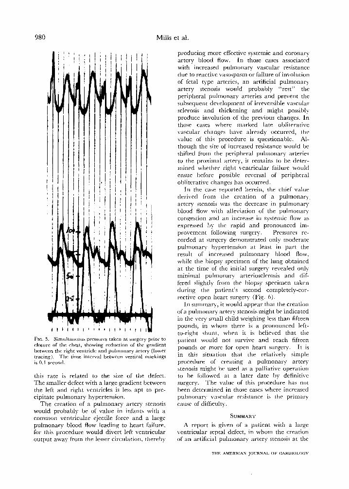
pulmonary by-pass. Simultaneous pressures werr taken in the right ventricle, pulmonary artery and femoral artery just prior to closure of the chest. Although the tracings were not completely satis- factory due to technical difficulties, they show a significant reduction in the gradient between th<* right ventricle and the pulmonary artery (Fig. 5).
Pmtoperatiw Results: The immediate postoperative course was essentially uneventful. Tracheobronchial suctioning was necessary during the first three days. but the patient gradually regained her strength and walked on the fifth postoperative day. There was no evidence of cardiac failure. At present, five months postoperatively, she is completely asympto- matic and tolerates the usual activities of a child her age without difficulty. The patient states that she can never remember feeling so well and being able to do so much. She is now 53 inches tall and weighs 54’,:, pounds. The low systolic murmur and thrill have disappeared and the murmur and thrill over the pulmonary area are much less intense. 4 recent electrocardiogram (Fig. 3B) reveals diminution of the pattern of right ventricular hypertrophy.
COMMENTS
Ventricular septal defects range from the tiny, dynamically insignificant lesions to the very large ventricular septal defects which, in effect, create a single ventricle. The latter defects allow direct transmission of left ventric-
NOVEMRER 1960
ular pressure to the pulmonary vaxulature.
It is this condition, namel)- the larqc, high
pressure ventricular septal defect which pro-
duces disability at birth or shortly tIlerrafter,
that applies to the subject of this report.
In general there are two ma$or sources of circulatory failure in persons with ventricular septal defects: the extremely high pulmonar) blood flow and increased cardiac work and the increased pulmonary resistance leadinq to progressi1.e right \,entricular enlargement and failure. These two factors are often conll)ined. There arc at least two possible mechanisms to account for the high pulmonary resistance so often associated with large defects. First is a persistence of the fetal state of the pullnonar) arteries, imply+ failure of the thick. niuscular fetal vessels to involute to the thin arteries of the normal state.“.? Second is the more frequent situation in which normal involution from the fetal state occurs I)ut is followed 1)~ increased pulmonary resistance as a reaction to the high pressure flow.4,5,6 It is probable that at first the pulmonary arteries react with local con- striction eventually followed l)y thickenin,q and narrowing of the lumen. In this circmnstance the high resistance state will develop at a rate which \.aries from case to case, Ijut in general
Mills et al.
I I I I I I I ’ 1 I 1 I I 1 I ’ I I
FIG. 5. Simultaneous pressures taken at surgery prior to closure of the chest, showing reduction of the gradient between the right ventricle and pulmonary artery (lower tracing). The time interval between vertical markings is 0.1 second.
this rate is related to the size of the defect. The smaller defect with a large gradient between the left and right ventricles is less apt to pre- cipitate pulmonary hypertension.
The creation of a pulmonary artery stenosis would probably be of value in infants with a common ventricular ejectile force and a large pulmonary blood flow leading to heart failure, for this procedure would divert left ventricular output away from the lesser circulation, thereby
producing more effective systemic and coronary artery blood flow. In those cases associated with increased pulmonary vascular resistance due to reactive vasospasm or failure of involution of fetal type arteries, an artificial pulmonary- artery stenosis would probably “rest” the peripheral pulmonary arteries and prevent the subsequent development of irreversible vascular sclerosis and thickening and might possibly produce involution of the previous changes. In those cases where marked late obliterative vascular changes have already occurred, the value of this procedure is questionable. Al- though the site of increased resistance would be shifted from the peripheral pulmonary arteries to the proximal artery, it remains to be deter- mined whether right ventricular failure would ensue before possible reversal of peripheral obliterative changes has occurred.
In the case reported herein, the chief value derived from the creation of a pulmonary artery stenosis was the decrease in pulmonary blood flow with alleviation of the pulmonary congestion and an increase in systemic flow as expressed by the rapid and pronounced im- provement following surgery. Pressures re- corded at surgery demonstrated only moderate pulmonary hypertension at least in part the result of increased pulmonary blood flow, while the biopsy specimen of the lung obtained at the time of the initial surgery revealed only minimal pulmonary arteriosclerosis and dif- fered slightly- from the biopsy specimen taken during the patient’s second completely-cor- rective open heart surgery (Fig. 6).
In summary, it would appear that the creation of a pulmonary artery stenosis might be indicated in the very small child weighing less than fifteen pounds, in whom there is a pronounced left- to-right shunt, when it is believed that the patient would not survive and reach fifteen pounds or more for open heart surgery. It is in this situation that the relatively simple procedure of creating a pulmonary artery stenosis might be used as a palliative operation to be followed at a later date by definitive surgery. The value of this procedure has not been determined in those cases where increased pulmonary vascular resistance is the primary cause of difficulty.
SUMMARY
A report is given of a patient with a large ventricular septal defect, in whom the creation of an artificial pulmonary artery stenosis at the
THE AMERICAN JOURNAL OF CARDIOLOGY

Congenital Ventricular Septal I >efect
I 1
.A B
FIG. 6. A, the biopsy specimen of the lung taken at the time the pulmonary artcry stenosis was created shows minimal pulmonary artcriolar sclerosis. B, the biopsy specimen takrn sewn yrars latrr is cssrntially normal. x 1.50.
age of twent> months reversed severe heart failure and allowed her to live a reasonabl) normal lift for the subsequent five years. At the age of eight )-ears, cardiac failure once again presented a threat and the patient was completely cured I)y definitive open heart surgery.
The creation of a pulmonary artery stenosis may be considered in tiny infants with this condition who arc under fifteen pounds and who arc in circulator)- failure because of increased pulmonary blood flow associated with dimin- ishrd systemic blood flow.
kXiNOWLEDGMENT
‘The authors gratefully acknowledge the cooperation of Dr. J. Frank Dammann who supplied much of the data concerning thr carlier phase of the patient’s illness.
REFERENCES
1. MUL.LER, W. H., JR. and DAMMANN, J. F. The treat- mcmt of crrtain congenital malformations of the
heart by the creation of pulmonic strnosis to rrducc pulmonary hypertension and cxccssivc pulmonary blood flow: a preliminary report. .%I:. f$w. G Obst., 95 : 213, 1952.
2. SIKAK, H. D.. Hosre~, D. Rf. and ~:LA'I\WRTH\.,
H. W., Jr. Defects of the intcrvcntricular srptum in infancy: a two-stagr approach to thcit surgical corrrction. AZzc E&znd J. .\fpd., 260: 147, 1959.
3. I-rxv.snus, J. E. Functional pathology of the pul- monary vascular tree in corqcnital cardiac tlisraw. Circulntion, 15: 164, 1957.
4. FRIEDBERG, c. K. Pulmonary hypcrtrnsion with special refcrencr to its occurrrncc~ in congenital heart discasc. Prq. Cardmmsr. Ih., 1 : 356, 1959.
5. HE~TII, D. and hWARDS, .I. E. The pathology of hyprrtrnsive pulmonary vascular diwasr. :I dc- scription of six grades of structural rhanqcs in the pulmonary artcrirs with sprcial rcfurncc to con- gc=nital cardiac srptal defects. Glru//2/lrv?. 18 : 533, 1958.
6. HEATH, D., HELMEIOIZ, H. F., .rR., BC~KCIWLI.. H. B.,
DUSHANE, W. and EDWARDS, .I. 1:. Graded pul- monary vascular changes and hemodynamic find- ings in cases of atria1 and ventricular wptal defect and patent ductus artcriosus. Cimdotion, 18 : 1155, 1958.
NOVEMBER 1960