Complications ami
30
Surgical treatment of complication of acute MI Dr .HIRALAL PAWAR
-
Upload
hiralal-pawar -
Category
Health & Medicine
-
view
15 -
download
1
Transcript of Complications ami
Surgical treatment of complication of acute MI
Dr .HIRALAL PAWAR
2
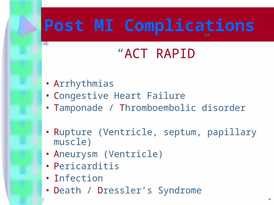
Post MI Complications
“ACT RAPID”
• Arrhythmias• Congestive Heart Failure• Tamponade / Thromboembolic disorder
• Rupture (Ventricle, septum, papillary muscle)• Aneurysm (Ventricle)• Pericarditis• Infection• Death / Dressler’s Syndrome
3
Rupture of the Interventricular Septum
• In 1847 Lathem first described a post MI VSD at autopsy
• In 1923 Brann made diagnosis clinically
• In 1957 Cooly first reported the surgical repair of VSD
4
Natural history
• Early death occurs frequently in patient with post infarct VSD:
Only about 75% survived the first 24 hour
50% the first week Less than 30% in 2 weekOnly 10-20% more than 4 week
5
• ruptured interventricular septum is characterized by the appearance of
a new harsh, loud holosystolic murmur that is heard best at the lower left sternal border and that is usually accompanied by a thrill
Biventricular failure generally ensues within hours to days.
6
• Clinical features associated with an increased risk of rupture of the interventricular septum include:
lack of development of a collateral network,
advanced agehypertension, anterior location of infarction, and possibly fibrinolysis.
7
• Rupture of the septum with an anterior infarction tends to be apical in location, whereas inferior infarctions are associated with perforation of the basal septum and have a worse prognosis than those in an anterior location
8
• In contrast with rupture of the free wall, rupture of the ventricular septum is
more often associated with complete heart block,
right bundle branch block, or atrial fibrillation.
9
Almost all patients have multivessel coronary artery disease, with most exhibiting lesions in all the major vessels.
The likelihood of survival depends on the degree of impairment of ventricular function and the size of the defect

10
The defect can also be recognized by echocardiography with color flow Doppler imaging or
insertion of a pulmonary artery balloon catheter to document the left-to-right shunt.
Catheter placement of an umbrella-shaped device within the ruptured septum may stabilize the condition of critically ill patients with acute septal rupture after STEMI.
MANAGMENT
In such patients, the circulation should at first be supported by intra-aortic balloon pulsation and a positive inotropic agent such as dopamine or dobutamine in combination with a vasodilator, unless the patient is hypotensive
11
12
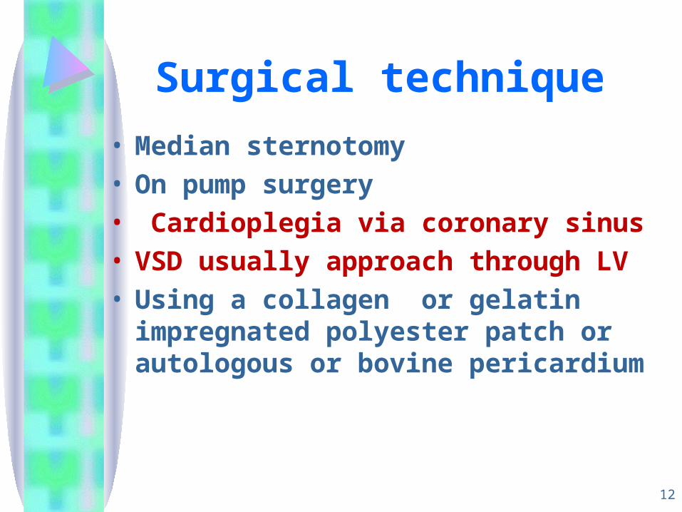
Surgical technique
• Median sternotomy• On pump surgery• Cardioplegia via coronary sinus• VSD usually approach through LV • Using a collagen or gelatin impregnated
polyester patch or autologous or bovine pericardium
13
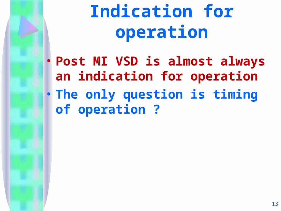
Indication for operation
• Post MI VSD is almost always an indication for operation
• The only question is timing of operation ?
14
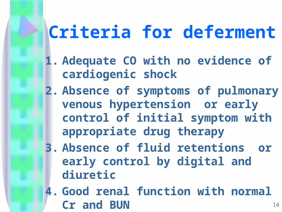
Criteria for deferment
1. Adequate CO with no evidence of cardiogenic shock
2. Absence of symptoms of pulmonary venous hypertension or early control of initial symptom with appropriate drug therapy
3. Absence of fluid retentions or early control by digital and diuretic
4. Good renal function with normal Cr and BUN
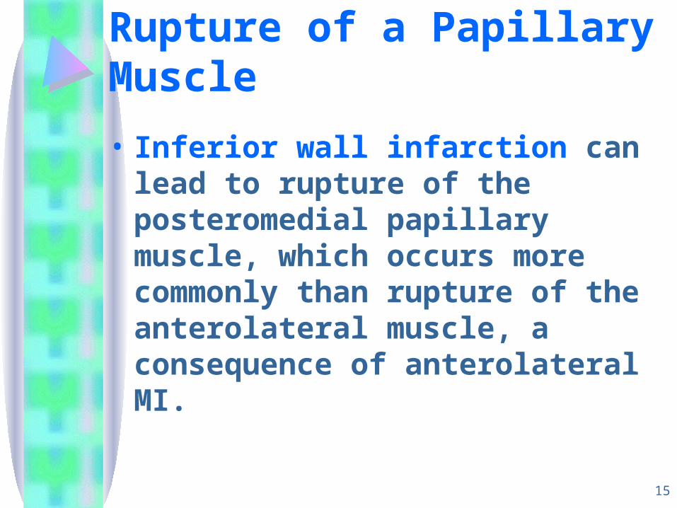
Rupture of a Papillary Muscle
• Inferior wall infarction can lead to rupture of the posteromedial papillary muscle, which occurs more commonly than rupture of the anterolateral muscle, a consequence of anterolateral MI.
15
16
• Complete transection of a left ventricular papillary muscle is incompatible with life because the sudden massive mitral regurgitation that develops cannot be tolerated.
17
• Rupture of a portion of a papillary muscle, usually the tip or head of the muscle, resulting in severe, although not necessarily overwhelming, mitral regurgitation, is much more frequent and is not immediately fatal
18
• Unlike rupture of the ventricular septum, which occurs with large infarcts, papillary muscle rupture occurs with a relatively small infarction in approximately half of the cases seen.
• The extent of coronary artery disease in these patients sometimes is modest as well.
19
• those with papillary muscle rupture manifest a new holosystolic murmur and develop increasingly severe heart failure. In both conditions, the murmur may become softer or disappear as arterial pressure falls
20
Technical operation• The approach is generally through the left
atrium • With acute total papillary muscle rupture
the MV is replaced with a mechanical or bioprosthesis
• When the papillary muscle is not ruptured and the chordal mechnism is intact or only one head is ruptured and there is anular dilation reparative techniques and anuloplasty with or without use of an anoloplasty ring are preferable to valve replacement
21
• When the hemodynamic state is reasonably good one sterategy has been to delay operation 2wk to 2 mo.
FREE RUPTURE
Acute
Pseudoaneurysm
22
23
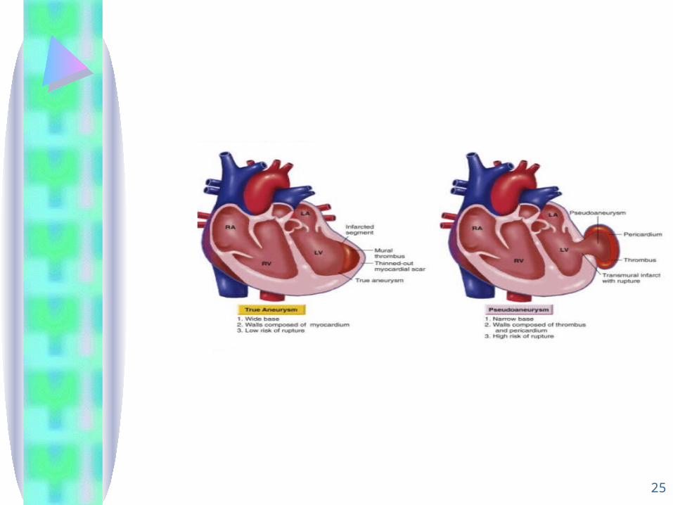
Pseudoaneurysm
• Incomplete rupture of the heart may occur when organizing thrombus and hematoma, together with pericardium, seal a rupture of the left ventricle and thus prevent the development of hemopericardium
24
• Pseudoaneurysms can become large, even equaling the true ventricular cavity in size, and communicate with the left ventricular cavity through a narrow neck.
• Frequently, pseudoaneurysms contain significant quantities of old and recent thrombi, superficial portions of which can cause arterial emboli
25
26
Diagnosis
• The diagnosis of pseudoaneurysm can usually be made by echocardiography and contrast angiography, although at times, differentiation between true aneurysm and pseudoaneurysm can be difficult by any imaging technique
27
• Immediate pericardiocentesis confirms the diagnosis and relieves the pericardial tamponade, at least momentarily.
• If the patient's condition is relatively stable, echocardiography may help in establishing the diagnosis of tamponade.
28
• When rupture is subacute and a pseudoaneurysm is suspected or present, prompt elective surgery is indicated because rupture of the pseudoaneurysm occurs relatively frequently
29
• Surgery should not be delayed in patients with a correctable lesion who agree to an aggressive management strategy and require pharmacologic and/or mechanical (counterpulsation) support. Such patients frequently develop a serious complication (e.g., infection, adult respiratory distress syndrome, extension of the infarct, renal failure) if surgery is delayed.
30
Surgical survival
predicted by early operation short duration of shock mild degrees of right and left
ventricular impairment