REVIEWMACROMOLECULES. The four macromolecules are: carbohydrates proteins lipids nucleic acids.
CHAPTER 5- MACROMOLECULES
5.2 CARBOHYDRATES
= sugars and their polymers
FUNCTIONS:
Energetic fuel source/storage
Structural building blocks
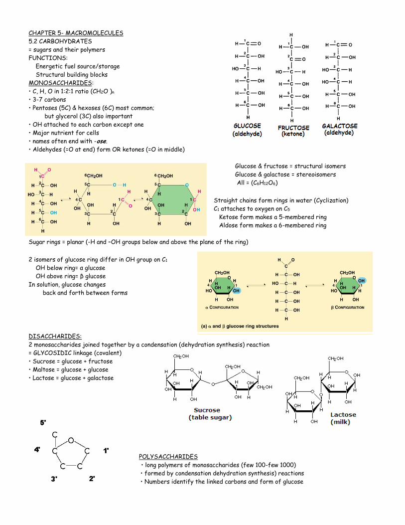
MONOSACCHARIDES:
• C, H, O in 1:2:1 ratio (CH2O )n
• 3-7 carbons
• Pentoses (5C) & hexoses (6C) most common;
but glycerol (3C) also important
• OH attached to each carbon except one
• Major nutrient for cells
• names often end with -ose.
• Aldehydes (=O at end) form OR ketones (=O in middle)
Glucose & fructose = structural isomers
Glucose & galactose = stereoisomers
All = (C6H12O6)
Straight chains form rings in water (Cyclization)
C1 attaches to oxygen on C5
Ketose form makes a 5-membered ring
Aldose form makes a 6-membered ring
Sugar rings = planar (-H and –OH groups below and above the plane of the ring)
2 isomers of glucose ring differ in OH group on C1
OH below ring= α glucose
OH above ring= β glucose
In solution, glucose changes
back and forth between forms
DISACCHARIDES:
2 monosaccharides joined together by a condensation (dehydration synthesis) reaction
= GLYCOSIDIC linkage (covalent)
• Sucrose = glucose + fructose
• Maltose = glucose + glucose
• Lactose = glucose + galactose
POLYSACCHARIDES
• long polymers of monosaccharides (few 100-few 1000)
• formed by condensation dehydration synthesis) reactions
• Numbers identify the linked carbons and form of glucose

OHHO
1-6 linkage
1-4 linkage
STARCH - energy storage in plants (EX: potatoes)
• α-glucose; 1-4 linkage
• forms spirals (helix) stabilized by hydrogen bonds
TWO FORMS OF STARCH
1) AMYLOSE
• straight unbranched glucose chain
• MW= thousands to hundred thousands
2) AMYLOPECTIN
• many linked short amylose chains
• 1-4 links with 1-6 linked branch points
• similar to glycogen but less branched
• MOST STARCH = 10-30% amylose/70-90% amylopectin
• stored as granules in plastids (chloroplasts and amyloplasts)
GLYCOGEN – Energy storage in ANIMALS
• α-glucose; 1-4 linkage
• helix stabilized by hydrogen bonds
• structure similar to amylopectin
but more branched
• MW = hundreds of thousands
• Stored as cytoplasmic granules in liver and muscle
Liver controls blood sugar level
low blood sugar between meals (glycogen→ glucose) (GLUCAGON)
high blood sugar after eating (glucose →glycogen) (INSULIN)
Muscle tissue- source of ATP for muscle contraction
CELLULOSE
• major component in plant cell walls (EX: wood, cotton)
• β-glucose; 1-4 linkages
• straight unbranched chains
• every other glucose upside down
• arrangement allows HYDROGEN BONDING between chains
• Form MICROFIBRILS that give cellulose its structural rigidity
• Dietary fiber in human diet
• Can’t be digested by animals without the help of symbiotic microorganisms
• Don’t have enzymes to break β linkages
CHITIN:
• β 1,4 linkages
• N-acetyl glucosamine (NAG) subunits (sugar + nitrogen group)
• straight unbranched chains
• every other glucose upside down
• Support and protection
EX: Cell walls in fungi; exoskeletons in arthropods
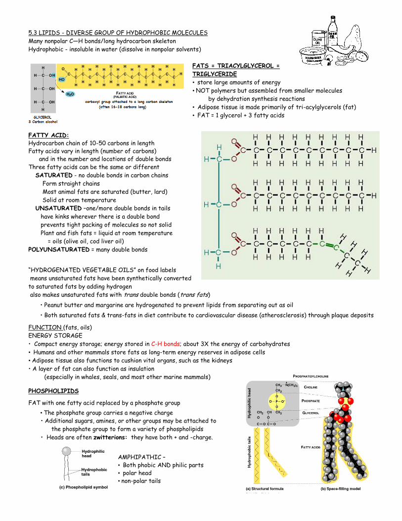
5.3 LIPIDS - DIVERSE GROUP OF HYDROPHOBIC MOLECULES
Many nonpolar C—H bonds/long hydrocarbon skeleton
Hydrophobic - insoluble in water (dissolve in nonpolar solvents)
FATS = TRIACYLGLYCEROL =
TRIGLYCERIDE
• store large amounts of energy
• NOT polymers but assembled from smaller molecules
by dehydration synthesis reactions
• Adipose tissue is made primarily of tri-acylglycerols (fat)
• FAT = 1 glycerol + 3 fatty acids
FATTY ACID: Hydrocarbon chain of 10-50 carbons in length Fatty acids vary in length (number of carbons) and in the number and locations of double bonds Three fatty acids can be the same or different
SATURATED - no double bonds in carbon chains
Form straight chains
Most animal fats are saturated (butter, lard)
Solid at room temperature
UNSATURATED –one/more double bonds in tails
have kinks wherever there is a double bond
prevents tight packing of molecules so not solid
Plant and fish fats = liquid at room temperature
= oils (olive oil, cod liver oil)
POLYUNSATURATED = many double bonds
“HYDROGENATED VEGETABLE OILS” on food labels
means unsaturated fats have been synthetically converted
to saturated fats by adding hydrogen
also makes unsaturated fats with trans double bonds (trans fats)
• Peanut butter and margarine are hydrogenated to prevent lipids from separating out as oil
• Both saturated fats & trans-fats in diet contribute to cardiovascular disease (atherosclerosis) through plaque deposits
FUNCTION (fats, oils)
ENERGY STORAGE
• Compact energy storage; energy stored in C-H bonds; about 3X the energy of carbohydrates
• Humans and other mammals store fats as long-term energy reserves in adipose cells
• Adipose tissue also functions to cushion vital organs, such as the kidneys
• A layer of fat can also function as insulation
(especially in whales, seals, and most other marine mammals)
PHOSPHOLIPIDS
FAT with one fatty acid replaced by a phosphate group
• The phosphate group carries a negative charge • Additional sugars, amines, or other groups may be attached to
the phosphate group to form a variety of phospholipids
• Heads are often zwitterions: they have both + and -charge.
AMPHIPATHIC – • Both phobic AND philic parts • polar head
• non-polar tails
CH3
OH
CH3
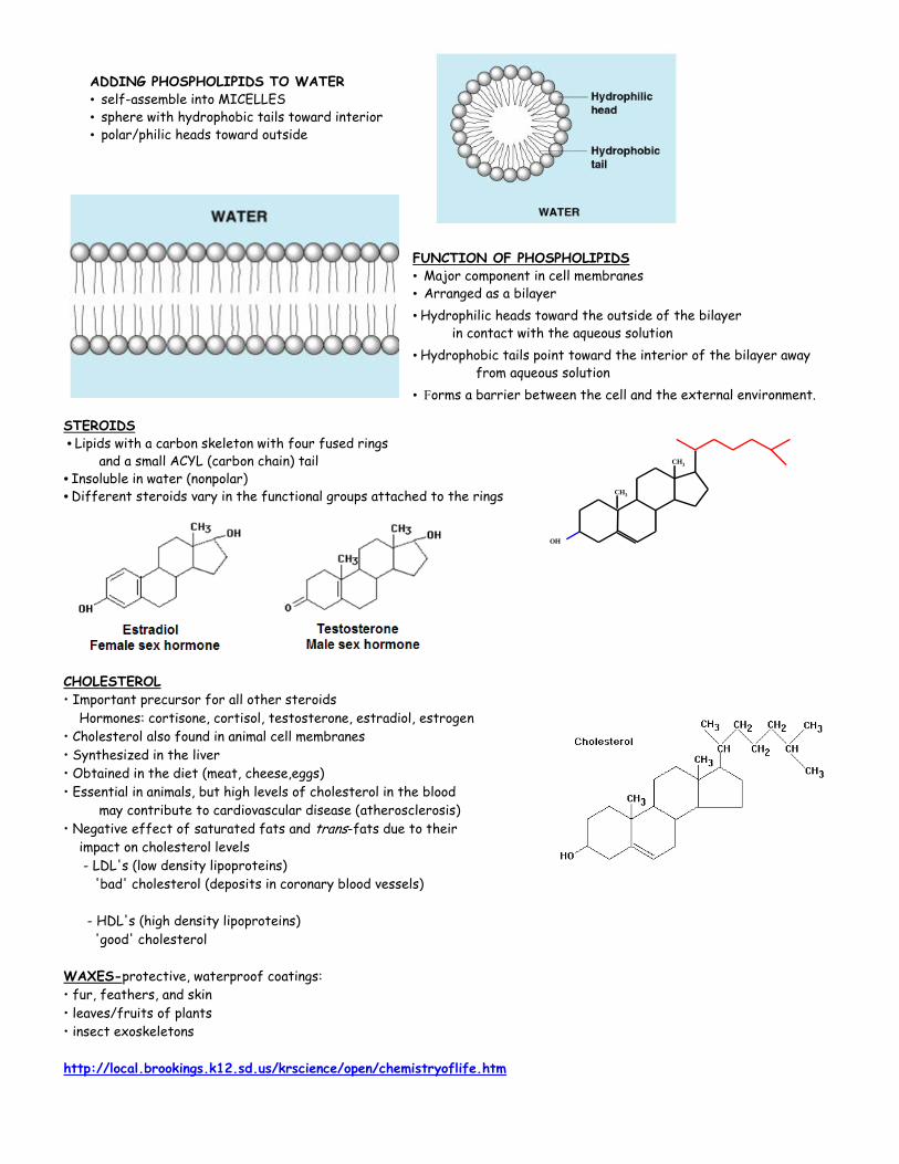
ADDING PHOSPHOLIPIDS TO WATER
• self-assemble into MICELLES
• sphere with hydrophobic tails toward interior
• polar/philic heads toward outside
FUNCTION OF PHOSPHOLIPIDS • Major component in cell membranes • Arranged as a bilayer
• Hydrophilic heads toward the outside of the bilayer in contact with the aqueous solution
• Hydrophobic tails point toward the interior of the bilayer away from aqueous solution
• Forms a barrier between the cell and the external environment.
STEROIDS
• Lipids with a carbon skeleton with four fused rings and a small ACYL (carbon chain) tail • Insoluble in water (nonpolar)
• Different steroids vary in the functional groups attached to the rings
CHOLESTEROL
• Important precursor for all other steroids
Hormones: cortisone, cortisol, testosterone, estradiol, estrogen
• Cholesterol also found in animal cell membranes
• Synthesized in the liver
• Obtained in the diet (meat, cheese,eggs)
• Essential in animals, but high levels of cholesterol in the blood
may contribute to cardiovascular disease (atherosclerosis)
• Negative effect of saturated fats and trans-fats due to their
impact on cholesterol levels
- LDL's (low density lipoproteins)
'bad' cholesterol (deposits in coronary blood vessels)
- HDL's (high density lipoproteins)
'good' cholesterol
WAXES-protective, waterproof coatings:
• fur, feathers, and skin
• leaves/fruits of plants
• insect exoskeletons
http://local.brookings.k12.sd.us/krscience/open/chemistryoflife.htm
Chapter 5.4 PROTEINS- “Cellular toolbox”
• Proteios = Greek “first place” • Make up 50% or more of dray mass of most cells
• Humans have tens of thousands of different proteins
• Typical protein = 200-300 amino acids; biggest known = 34,000
• Know the amino acid sequences of > 875,000 proteins/3D shapes of about 7,000
• Scientists use X-ray cystallography to determine protein conformation
• A protein’s function = EMERGENT PROPERTY determined by its conformation
EXAMPLES OF VARIETY OF PROTEINS/FUNCTIONS:
• Structural: hair, fingernails, bird feathers (keratin); spider silk;
cellular cytoskeleton (tubulin & actin); connective tissue (collagen)
• Storage: egg white (ovalbumin); milk protein (Casein); plant seeds
• Transport: Transport iron in blood (hemoglobin);
• Hormonal: Regulate blood sugar (insulin)
• Membrane proteins (receptors, membrane transport, antigens)
• Movement: Muscle contraction (actin and myosin); Flagella (tubulin & dynein); Motor proteins move vesicles/chromosomes
• Defense: Antibodies fight germs
• Metabolism: Enzymes act as catalysts in chemical reactions
• Toxins (botulism, diphtheria)
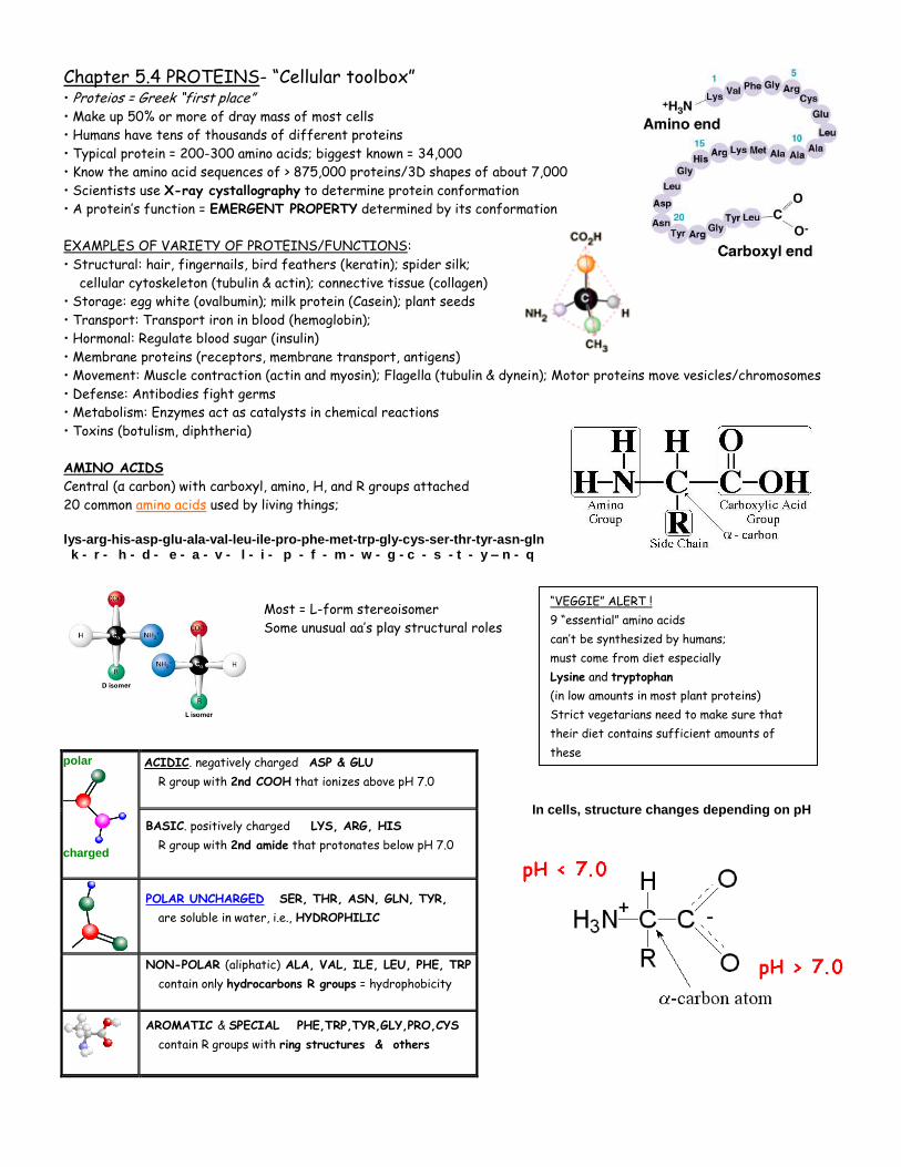
AMINO ACIDS
Central (α carbon) with carboxyl, amino, H, and R groups attached
20 common amino acids used by living things;
lys-arg-his-asp-glu-ala-val-leu-ile-pro-phe-met-trp-gly-cys-ser-thr-tyr-asn-gln k - r - h - d - e - a - v - l - i - p - f - m - w - g - c - s - t - y – n - q
Most = L-form stereoisomer
Some unusual aa’s play structural roles
In cells, structure changes depending on pH
polar
charged
ACIDIC. negatively charged ASP & GLU
R group with 2nd COOH that ionizes above pH 7.0
BASIC. positively charged LYS, ARG, HIS
R group with 2nd amide that protonates below pH 7.0
POLAR UNCHARGED SER, THR, ASN, GLN, TYR,
are soluble in water, i.e., HYDROPHILIC
NON-POLAR (aliphatic) ALA, VAL, ILE, LEU, PHE, TRP
contain only hydrocarbons R groups = hydrophobicity
AROMATIC & SPECIAL PHE,TRP,TYR,GLY,PRO,CYS
contain R groups with ring structures & others
“VEGGIE” ALERT !
9 “essential” amino acids
can’t be synthesized by humans;
must come from diet especially
Lysine and tryptophan
(in low amounts in most plant proteins)
Strict vegetarians need to make sure that
their diet contains sufficient amounts of
these
S
+-
io nic
disu lfid e
hy drog en
hy drop hob ic
hy drop hy llic
S
S
POLYPEPTIDE = polymer of amino acid subunits connected in a specific sequence An enzyme joins the carboxyl of one amino acid and the amino group of another via dehydration synthesis/condensation reaction to form a PEPTIDE BOND
Peptide bonds are rigid, planar structures The -NH bond and the -C=O bond, point away from each other so these groups can hydrogen bond to other parts of chain
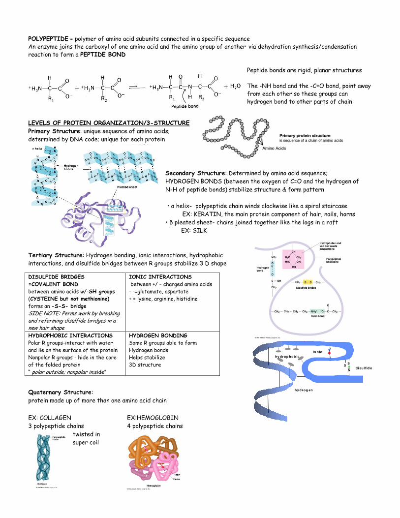
LEVELS OF PROTEIN ORGANIZATION/3-STRUCTURE
Primary Structure: unique sequence of amino acids;
determined by DNA code; unique for each protein
Secondary Structure: Determined by amino acid sequence;
HYDROGEN BONDS (between the oxygen of C=O and the hydrogen of
N-H of peptide bonds) stabilize structure & form pattern
• α helix- polypeptide chain winds clockwise like a spiral staircase
EX: KERATIN, the main protein component of hair, nails, horns
• β pleated sheet- chains joined together like the logs in a raft
EX: SILK
Tertiary Structure: Hydrogen bonding, ionic interactions, hydrophobic
interactions, and disulfide bridges between R groups stabilize 3 D shape
Quaternary Structure:
protein made up of more than one amino acid chain
EX: COLLAGEN EX:HEMOGLOBIN
3 polypeptide chains 4 polypeptide chains
twisted in
super coil
DISULFIDE BRIDGES
=COVALENT BOND
between amino acids w/-SH groups
(CYSTEINE but not methionine)
forms an -S-S- bridge
SIDE NOTE: Perms work by breaking and reforming disulfide bridges in a new hair shape
IONIC INTERACTIONS
between +/ – charged amino acids
- -=glutamate, aspartate
+ = lysine, arginine, histidine
HYDROPHOBIC INTERACTIONS
Polar R groups-interact with water
and lie on the surface of the protein
Nonpolar R groups - hide in the core
of the folded protein
“ polar outside; nonpolar in.side”
HYDROGEN BONDING
Some R groups able to form
Hydrogen bonds
Helps stabilize
3D structure
WHAT DO YOU CALL IT?
• two or more amino acids bonded together = PEPTIDE
• chain of many amino acids = POLYPEPTIDE
• complete folded 3D structure = PROTEIN
Final overall protein shapes
- FIBROUS. - long fiber shape EX: actin or collagen
- GLOBULAR - overall spherical structure EX: hemoglobin,
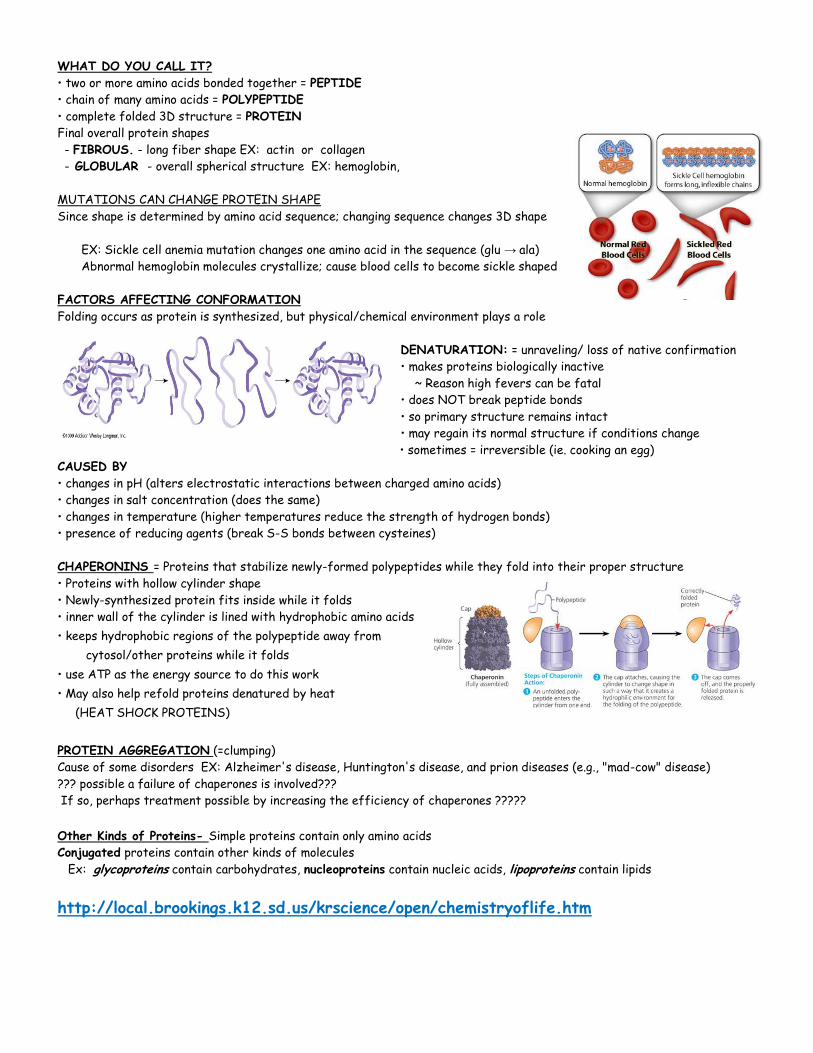
MUTATIONS CAN CHANGE PROTEIN SHAPE
Since shape is determined by amino acid sequence; changing sequence changes 3D shape
EX: Sickle cell anemia mutation changes one amino acid in the sequence (glu → ala)
Abnormal hemoglobin molecules crystallize; cause blood cells to become sickle shaped
FACTORS AFFECTING CONFORMATION
Folding occurs as protein is synthesized, but physical/chemical environment plays a role
DENATURATION: = unraveling/ loss of native confirmation
• makes proteins biologically inactive
~ Reason high fevers can be fatal
• does NOT break peptide bonds
• so primary structure remains intact
• may regain its normal structure if conditions change
• sometimes = irreversible (ie. cooking an egg)
CAUSED BY
• changes in pH (alters electrostatic interactions between charged amino acids)
• changes in salt concentration (does the same)
• changes in temperature (higher temperatures reduce the strength of hydrogen bonds)
• presence of reducing agents (break S-S bonds between cysteines)
CHAPERONINS = Proteins that stabilize newly-formed polypeptides while they fold into their proper structure
• Proteins with hollow cylinder shape
• Newly-synthesized protein fits inside while it folds
• inner wall of the cylinder is lined with hydrophobic amino acids
• keeps hydrophobic regions of the polypeptide away from
cytosol/other proteins while it folds
• use ATP as the energy source to do this work
• May also help refold proteins denatured by heat
(HEAT SHOCK PROTEINS)
PROTEIN AGGREGATION (=clumping)
Cause of some disorders EX: Alzheimer's disease, Huntington's disease, and prion diseases (e.g., "mad-cow" disease)
??? possible a failure of chaperones is involved???
If so, perhaps treatment possible by increasing the efficiency of chaperones ?????
Other Kinds of Proteins- Simple proteins contain only amino acids
Conjugated proteins contain other kinds of molecules
Ex: glycoproteins contain carbohydrates, nucleoproteins contain nucleic acids, lipoproteins contain lipids
http://local.brookings.k12.sd.us/krscience/open/chemistryoflife.htm
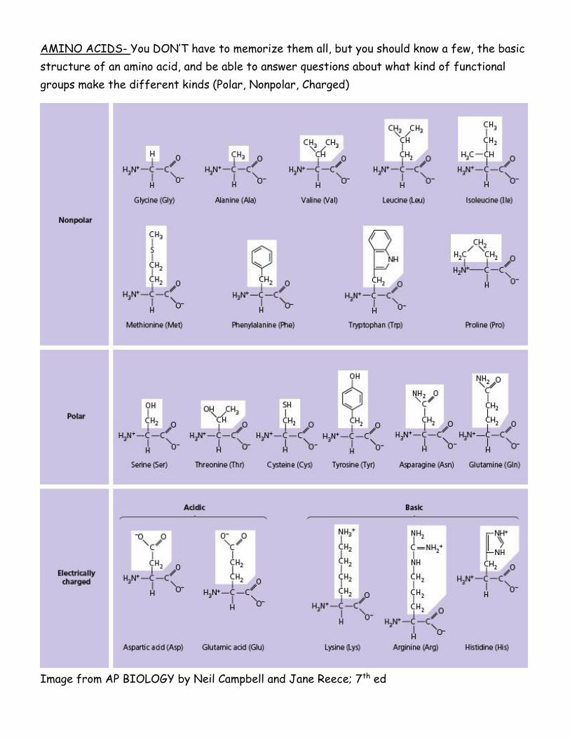
AMINO ACIDS- You DON’T have to memorize them all, but you should know a few, the basic
structure of an amino acid, and be able to answer questions about what kind of functional
groups make the different kinds (Polar, Nonpolar, Charged)
Image from AP BIOLOGY by Neil Campbell and Jane Reece; 7th ed

O O O
-O - P ~ O - P ~ O - P - O -
O- O-O-
Chapter 5.5 NUCLEOTIDES AND NUCLEIC ACIDS
INFORMATION FLOW IN CELLS = “Central Dogma of Molecular Biology”
DNA→ RNA→ PROTEINS
FUNCTION
DNA- genetic code contains info that programs cell activities
RNA-carries message from DNA to cell; protein synthesis
BASIC STRUCTURE
NUCLEOSIDE = nitrogenous base + sugar
NUCLEOTIDE = nitrogenous base + sugar + phosphate group
PURINES = 2 rings; Adenine (A), Guanine (G)
PYRIMIDINES = 1 ring; Cytosine (C), Thymine (T), Uracil (U)
Nitrogen bases
A purine always
bonds with
a pyrimidine.
NUCLEOTIDES
Can have one, two, or three phosphate groups
(mono, di, tri-phosphates)
High energy bond between phosphate groups
is important energy transport
Named for nitrogen base and number of phosphate groups
EX: adenosine triphosphate (ATP)
cytosine diphosphate (CDP)
guanosine monophosphate (GMP)
IMPORTANT NUCLEOTIDES
ADENOSINE TRIPHOSPHATE (ATP) = main energy currency in ALL living things
(GTP & UTP also used)
CYCLIC AMP (cAMP)- “Second messenger”
Important in cell signaling and response to hormones
COENZYMES- Many coenzymes are nucleotides or their derivatives (vitamins)
EX: Flavin adenine dinucleotide (FAD) & nicotinamide adenine dinucleotide (NAD) used in cellular respiration
nicotinamide adenine dinucleotide phosphate (NADP) used in photosynthesis
3’
5’
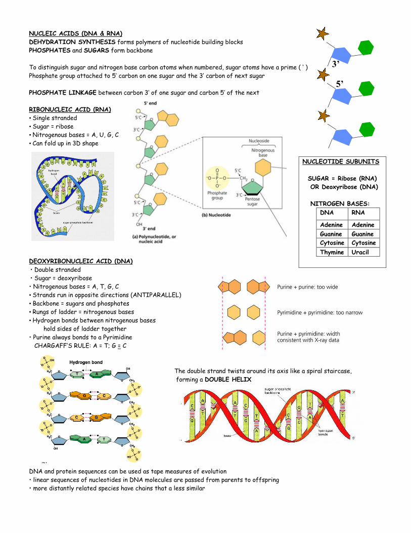
NUCLEIC ACIDS (DNA & RNA)
DEHYDRATION SYNTHESIS forms polymers of nucleotide building blocks
PHOSPHATES and SUGARS form backbone
To distinguish sugar and nitrogen base carbon atoms when numbered, sugar atoms have a prime ( ‘ )
Phosphate group attached to 5’ carbon on one sugar and the 3’ carbon of next sugar
PHOSPHATE LINKAGE between carbon 3’ of one sugar and carbon 5’ of the next
RIBONUCLEIC ACID (RNA)
• Single stranded
• Sugar = ribose
• Nitrogenous bases = A, U, G, C
• Can fold up in 3D shape
DEOXYRIBONUCLEIC ACID (DNA)
• Double stranded
• Sugar = deoxyribose
• Nitrogenous bases = A, T, G, C
• Strands run in opposite directions (ANTIPARALLEL)
• Backbone = sugars and phosphates
• Rungs of ladder = nitrogenous bases
• Hydrogen bonds between nitrogenous bases
hold sides of ladder together
• Purine always bonds to a Pyrimidine
CHARGAFF’S RULE: A = T; G = C
The double strand twists around its axis like a spiral staircase,
forming a DOUBLE HELIX
DNA and protein sequences can be used as tape measures of evolution
• linear sequences of nucleotides in DNA molecules are passed from parents to offspring
• more distantly related species have chains that a less similar
NUCLEOTIDE SUBUNITS
SUGAR = Ribose (RNA)
OR Deoxyribose (DNA)
NITROGEN BASES:
DNA RNA
Adenine Adenine
Guanine Guanine
Cytosine Cytosine
Thymine Uracil