
Brain Imaging in the Courtroom - National Association of ... Imaging in... · Brain Imaging in the...
53
Brain Imaging in the Courtroom: Admissibility and Future Directions Teneille Brown, J.D. NAWJ Conference October 9, 2015
Transcript of Brain Imaging in the Courtroom - National Association of ... Imaging in... · Brain Imaging in the...

Brain Imaging in the Courtroom:Admissibility and Future Directions
Teneille Brown, J.D.
NAWJ Conference
October 9, 2015

Outline
1. How Neuroimaging is Being Used in
Court
2. Introduction to three types: EEG;
QEEG; fMRI
3. 702 and 403 Analysis of fMRI
4. Possibly (?): Future Directions

Outline
1. How Neuroimaging is Being Used in
Court
2. Introduction to three types: EEG;
QEEG; fMRI
3. Admissibility of QEEG and fMRI
4. Possibly (?): Future Directions
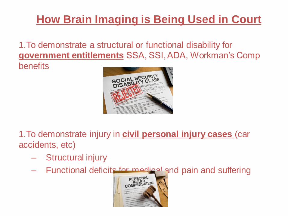
How Brain Imaging is Being Used in Court
1.To demonstrate a structural or functional disability for government entitlements SSA, SSI, ADA, Workman’s Comp
benefits
1.To demonstrate injury in civil personal injury cases (car
accidents, etc)
– Structural injury
– Functional deficits for medical and pain and suffering

How Brain Imaging is Being Used in Court
3. Criminal Trials
– Deciding whether defendants are competent
– Deciding whether to punish (the guilt phase)
– Deciding how much to punish (the sentencing phase)
• Brian Dugan case (serial killer in IL)
• To show developmental population differences
(adolescents are less capable of impulse control,
should not be executed or subjected to life without
parole) i.e., Roper, Jackson, Graham
– After punishment, ineffective assistance of counsel
claims
4. Lie Detection in civil, criminal and probate trials


Outline
1. How Neuroimaging is Being Used in
Court
2. Introduction to three types: eeg;
QEEG; fMRI
3. 702 and 403 Analysis of fMRI
4. Possibly (?): Future Directions

EEG

EEG
• Discovery in 1800s that brain produces electricity
• Cortical activity can be measured on the surface of the scalp with standard sensors, some subcortical
• EEG has remained a crucial clinical and research tool
• Does not require exposure to external radiation (fMRI) or radioactive tracers (like PET/SPECT)

EEG
• Requires subject compliance
• Susceptible to countermeasures
• Less expensive than fMRI
• More mobile
• Great temporal specificity
• Poor spatial and depth resolution

EEG – Clinical Use
• Diagnose epilepsy, coma, brain death
• With digital EEG, all signals are typically
digitized and stored in a particular reference
base
• The EEG can be viewed by the technician
in any display format that is desired
(comparing one recording channel with
average of those around it; comparing
individual to reference class of “normals”)

QEEG (Quantitative EEG)
• A computational analysis of EEG output,
using around 20 channels of EEG
recordings;
• Compare activity to database of
“neurotypicals”
• Show relative differences in
wave/patterns/strength
– For example, some research shows increased
beta waves in left tempo-parietal and right
occipital lobes in psychopaths (small studies
and can’t diagnose just one person, but can
describe differences in group average)

QEEG: generally NOT accepted for
these purposes
• To demonstrate TBI for personal injury claims and insurance coverage– Greene v. State Farm; 2008 WL 6667445
– Smith v. Ryan; 2012 WL 6019055
• For criminal mitigation at sentencing– Mendoza v. State, 87 So. 3d 644 (Fla. 2011)
• For post-conviction habeas and Atkins hearings– United States v. Williams, CR 06-00079 DAE-KSC,
2009 WL 424583 (D. Haw. Feb. 20, 2009)
– Smith v. Ryan, CV-87-234-TUC-CKJ, 2012 WL 6019055 (D. Ariz. Dec. 3, 2012)
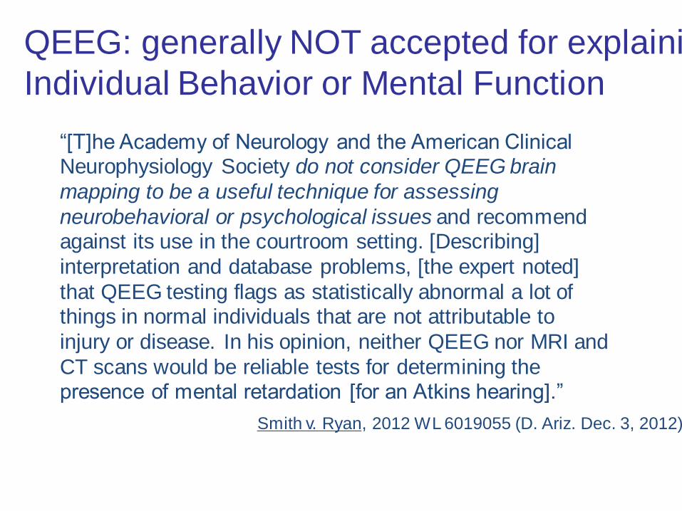
“[T]he Academy of Neurology and the American Clinical Neurophysiology Society do not consider QEEG brain
mapping to be a useful technique for assessing
neurobehavioral or psychological issues and recommend against its use in the courtroom setting. [Describing]
interpretation and database problems, [the expert noted]
that QEEG testing flags as statistically abnormal a lot of things in normal individuals that are not attributable to
injury or disease. In his opinion, neither QEEG nor MRI and
CT scans would be reliable tests for determining the presence of mental retardation [for an Atkins hearing].”
Smith v. Ryan, 2012 WL 6019055 (D. Ariz. Dec. 3, 2012)
QEEG: generally NOT accepted for explaining
Individual Behavior or Mental Function

QEEG: Admitted!
• Grady Nelson, stabbed his wife 61 times, raped her and her daughter
• Judge agreed to allow QEEG results at his sentencing hearing. The jurors were split 6-6 in sentencing; resulting in automatic life sentence
• Some jurors said QEEG did influence their move from the death penalty to a life sentence
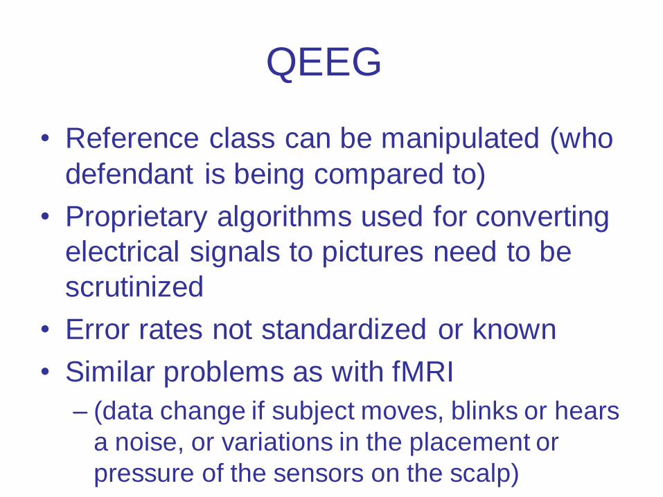
QEEG
• Reference class can be manipulated (who
defendant is being compared to)
• Proprietary algorithms used for converting
electrical signals to pictures need to be
scrutinized
• Error rates not standardized or known
• Similar problems as with fMRI
– (data change if subject moves, blinks or hears
a noise, or variations in the placement or
pressure of the sensors on the scalp)

fMRI: the “f” stands for function

fMRI
• Requires voluntary compliance of subject
• Contraindicated: metal plates / powerful
magnets
• Scanning time is expensive (~$450/hour?)
• Post-scanning analysis is expensive
(~$250/study)
• Great spatial resolution, not great temporal
resolution
Outline
1. How Neuroimaging is Being Used in
Court
2. Introduction to three types: eeg;
QEEG; fMRI
3. 702 and 403 Analysis of fMRI
4. Possibly (?): Future Directions

Federal Admissibility Standards
• FRE 403 – probative and not overly
prejudicial (or a waste of time,
misleading, or cumulative)
• FRE 702 – Daubert
– Valid and reliable data?
• Peer-reviewed
• Falsifiable
• Error rate
• Fit for this purpose (Joiner)
Utah Admissibility Standards
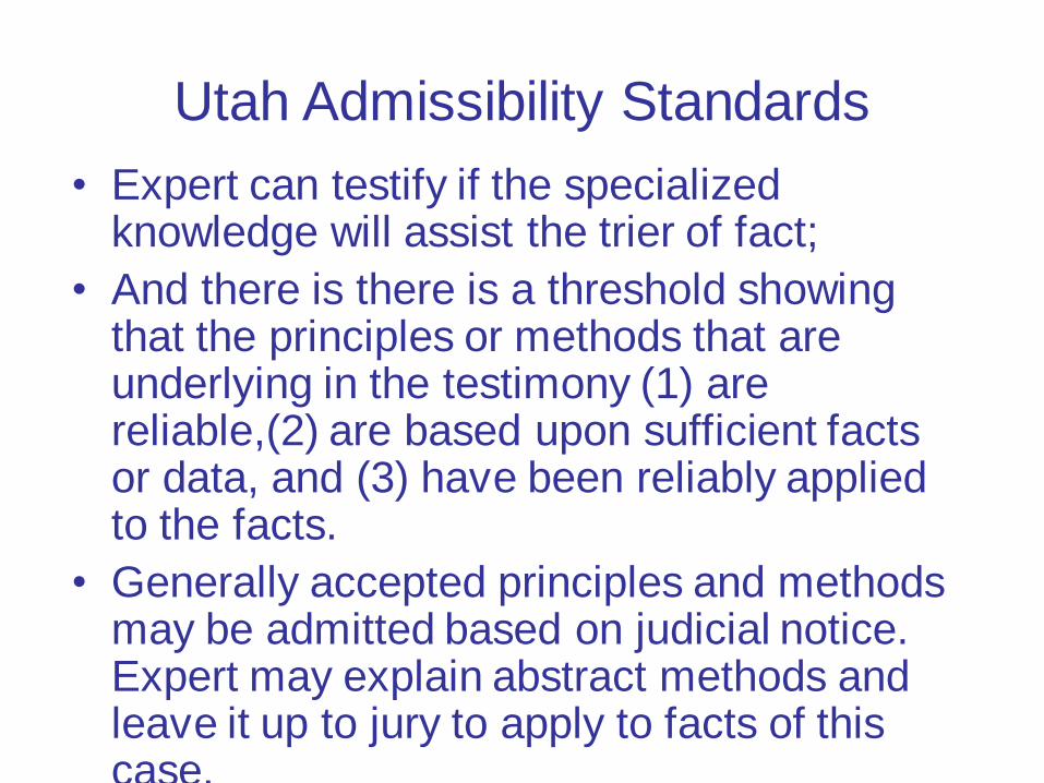
• Expert can testify if the specialized knowledge will assist the trier of fact;
• And there is there is a threshold showing that the principles or methods that are underlying in the testimony (1) are reliable,(2) are based upon sufficient facts or data, and (3) have been reliably applied to the facts.
• Generally accepted principles and methods may be admitted based on judicial notice. Expert may explain abstract methods and leave it up to jury to apply to facts of this case.

Utah Admissibility Standards
“[J]udge must take care to direct her
skepticism to the particular proposition that
the expert testimony is offered to support.
The Daubert court characterized this task
as focusing on the “work at hand”…The
foundation of reliability presented for it
[should] reflect that consideration.”•Mangrum And Benson On Utah Evidence; 1 UTPRAC
RULE 702

Utah Admissibility Standards
The greatest hurdle for most brain imaging
evidence in being admitted is this problem of “fit”;
or Joiner standard in federal courts
Perfectly valid research studies using fMRI, EEG,
or QEEG for tracking stroke victims, seizure
patients, or localizing speech centers before brain
surgeries are being used in inappropriate contexts
to say something about the criminal defendant’s
criminal mental state (such as competence to stand
trial or mens rea)

Because you cannot evaluate
admissibility without knowing
what the evidence is being
introduced to prove, I will focus
my remaining remarks on using
fMRI to prove a criminal
defendant’s past mental state
(mens rea or provocation)

Legal Claims Made About fMRI
• “It’s magical!”
• “Computerized and therefore objective”
• “Capable of reading our innermost
thoughts”
• “Impossible to game”
• “Provides a colorful video in real-time of
someone’s thoughts”

Legal Claims Made About fMRI
• “Computerized and therefore objective”
• “Capable of reading our thoughts”
• “Impossible to Game”
• “Provides a colorful video in real-time of
someone’s thoughts”

ActivationMap
IncreasedNeuronalActivity
IncreasedOxygenatedBlood Flow
HbO2 Hbarterial venous
Performa Task
Cognition/
Behavior
FunctionalBrain Anatomy
What do we know?Functional scans: fMRI
Images courtesy of Gary Glover, Stanford
fMRI measures oxygenated blood flow, not neuronal
activity directly.
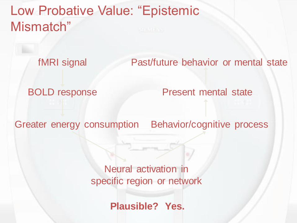
fMRI signal
BOLD response
Greater energy consumption
Neural activation in
specific region or network
Behavior/cognitive process
Present mental state
Past/future behavior or mental state
Plausible? Yes.
Low Probative Value: “Epistemic
Mismatch”

Potential for Prejudice
• Encourage an overly emotional
response?
• Confuse the jury?
• Waste the court’s time/resources
• Cumulative of other evidence

Cumulative of Behavioral Evidence?
Stimulus
Response
A A A AB B B
- =
"A" state images Activation map”B" state images
Images courtesy of Gary Glover, Stanford

Make a database of “normal brains”
?
Comparing an individual subject to the “norm”
“Normal” is a statistical creation
What is normal? Base rates and reference
classes

Individual Differences
• People have different memories and reference
points
• People perform tasks in the scanner at different
speeds and with different skills
• People have different brain architecture
• People have different molecular signaling
pathways (based on genes and environment)
• People use what they have in different ways
Annu Rev Neurosci. 2009 ; 32: 225–247

Individual Differences
Miller et al., J of Cognitive Neuroscience
2002

P < 0.001P < 0.01P < 0.05
“Dialing a defect”
(Images courtesy of Scott Grafton)
Lies, damn lies, and statistical thresholds
Which brain has processing deficits?
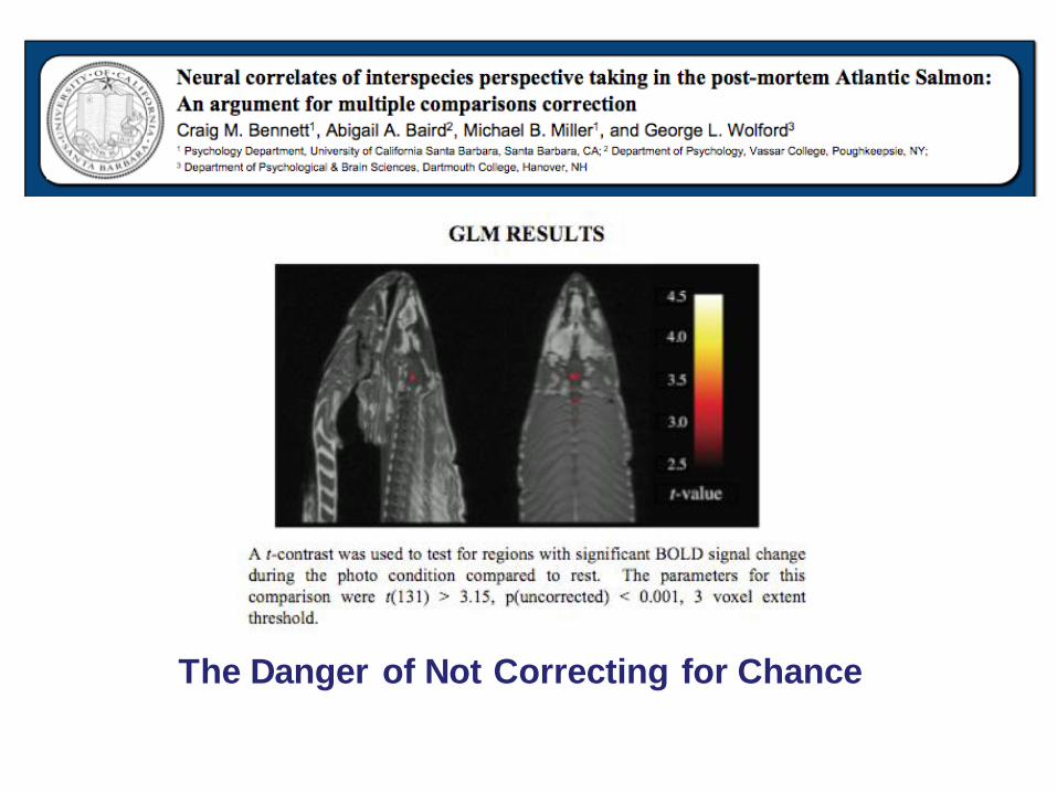
The Danger of Not Correcting for Chance

The Danger of Not Correcting for Chance
How could a dead fish exhibit brain
function?

Evoked Activity
Intrinsic Activity
Other
The brain’s energy budget
The BOLD Response
fMRI relies on a very noisy and subtle signal, and is best
used as a research tool

• The images are only as good as the precision
and relevance of the task the subject is asked to
perform
• If the subject performs a task that is not related to
the relevant legal behavior (such as the specific
intent to kill, deception, feel pain) then the image
will have little probative value and will not be
reliably applied to the facts
• Impossible to replicate criminal behavior in a
scanner
Ecological Validity

• We first need to know something about the
baserate of the phenomenon we are testing
• If the baserate is low, the positive predictive
value (PPV) will also be low (because of false
positives)
• PPV is critical when you REALLY want to know if
someone possesses a trait, like legally relevant
mental state or mental abnormality
Why “Accuracy” in a
Vacuum is Useless

Try this:
Should a fMRI-based “test”
with 90% specificity and
sensitivity pass 702?

Security screening example:
1000 travelers going through SLC airport security
10 of them are lying about carrying explosives (1%)
Brain scan with 90% specificity, 90% sensitivity
Reality Allow Detain Total
Truth 891 99 990
Lying 1 9 10
892 108
Scan result
Positive predictive value: At 90% specificity and 90% accuracy, but a 1% prevalence rate, the scan incorrectly says to detain 99/108
people: wrong 91.7% of the time!
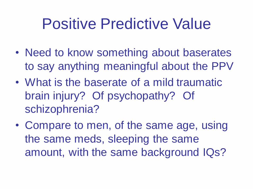
Positive Predictive Value
• Need to know something about baserates
to say anything meaningful about the PPV
• What is the baserate of a mild traumatic
brain injury? Of psychopathy? Of
schizophrenia?
• Compare to men, of the same age, using
the same meds, sleeping the same
amount, with the same background IQs?
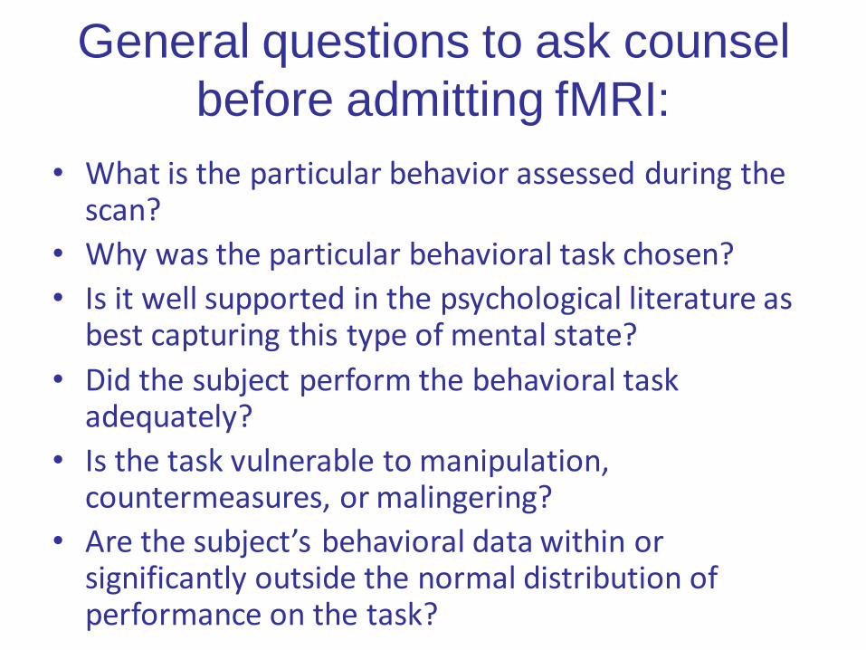
General questions to ask counsel
before admitting fMRI:
• What is the particular behavior assessed during the scan?
• Why was the particular behavioral task chosen?
• Is it well supported in the psychological literature as best capturing this type of mental state?
• Did the subject perform the behavioral task adequately?
• Is the task vulnerable to manipulation, countermeasures, or malingering?
• Are the subject’s behavioral data within or significantly outside the normal distribution of performance on the task?
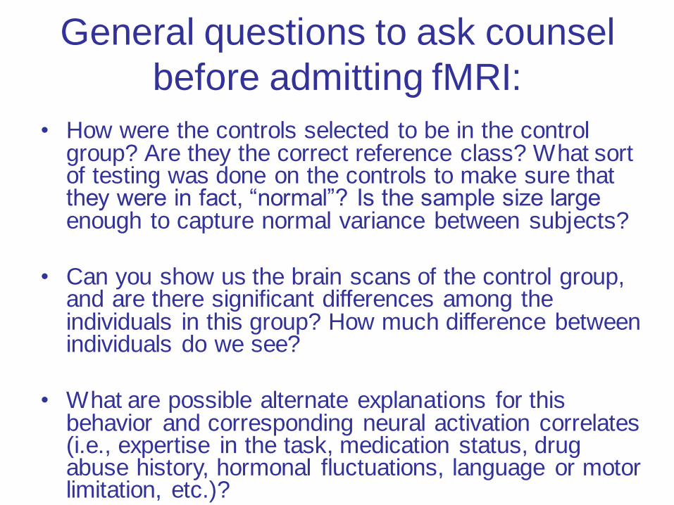
General questions to ask counsel
before admitting fMRI:
• How were the controls selected to be in the control group? Are they the correct reference class? What sort of testing was done on the controls to make sure that they were in fact, “normal”? Is the sample size large enough to capture normal variance between subjects?
• Can you show us the brain scans of the control group, and are there significant differences among the individuals in this group? How much difference between individuals do we see?
• What are possible alternate explanations for this behavior and corresponding neural activation correlates (i.e., expertise in the task, medication status, drug abuse history, hormonal fluctuations, language or motor limitation, etc.)?

For List of Questions to Ask Before Admitting fMRI Evidence
• See the Appendix in
Teneille Brown and Emily Murphy, Through a Scanner Darkly: Functional Neuroimaging as Evidence of a Criminal Defendant’s Past Mental States, 62 Stanford Law Review 1119 (2010)
Outline
1. How Neuroimaging is Being Used in
Court
2. Introduction to three types: EEG;
QEEG; fMRI
3. 702 and 403 Analysis of fMRI
4. Possibly (?): Future Directions

fMRI for Sentencing & Risk Prediction
• Using fMRI to infer mental states requires drawing a
conclusion about an individual:
– Did defendant intend to defraud? Is plaintiff in
pain? Is defendant telling the truth?
– What is the cost of getting this wrong? Jail time?
Not receiving damages?
• Risk prediction, on the other hand, deals with
probabilities for populations
– Not about retribution, but deterrence
– How do we predict which classes of people are
most likely to reoffend?

Neuroprediction of Future Rearrest
• The odds that an offender with relatively low ACC
activity would be rearrested were approximately
double that of an offender with high activity in this
region, holding constant other observed risk factors.
• These results suggest a potential neurocognitive
biomarker for persistent antisocial behavior.
Aharoni et al, 110 PNAS 6223 (2013)

Neuroprediction of Future Rearrest
• Brain activity elicited during performance of an inhibitory task (Go/No-Go) prospectively predicted subsequent rearrest among adult offenders within 4 yr of release (N = 96).
• Inmates were given a simple test asking them to press a button when the letter “X” appeared on a computer screen but to refrain from pressing the button when a “K” appeared.
• Inmates who made mistakes and exhibited low brain activity in the ACC afterward had a harder time controlling their impulses and were more prone to apathetic or aggressive behavior
Aharoni et al, 110 PNAS 6223 (2013)
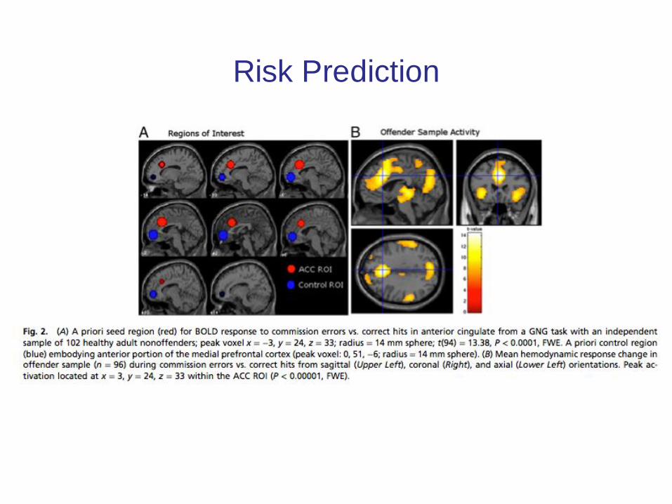
Risk Prediction

Neuroprediction of Future Rearrest
• What are the benefits of this approach?
• What are the concerns?
Aharoni et al, 110 PNAS 6223 (2013)
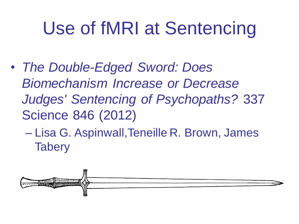
Use of fMRI at Sentencing
• The Double-Edged Sword: Does
Biomechanism Increase or Decrease
Judges' Sentencing of Psychopaths? 337
Science 846 (2012)
– Lisa G. Aspinwall,Teneille R. Brown, James
Tabery