![Muscular Dystrophy Evaluation of Mamsagni Rasayana [MGR] in Evaluation of Mamsagni Rasayana [MGR] in 55 Patients of DMD, BMD & LGMD Mukesh Jain, AMDS.](https://static.fdocuments.us/doc/165x107/56649d1b5503460f949f07b8/muscular-dystrophy-evaluation-of-mamsagni-rasayana-mgr-in-evaluation-of-mamsagni.jpg)
Ayurvedic Amalaki Rasayana promotes improved stress ...Rasayana (AR) is a prominent herbal...
30
1 Ayurvedic Amalaki Rasayana promotes improved stress tolerance and thus has anti-aging effects in Drosophila melanogaster Vibha Dwivedi 1 and Subhash C. Lakhotia * Cytogenetics Laboratory, Department of Zoology, Banaras Hindu University, Varanasi 221005, India 1 Present address: School of Biosciences, University of Birmingham, Birmingham, UK (email: [email protected]) * Corresponding author Email: [email protected] Tele: +91-542-2368145 ORCID Id: 0000-0003-1842-8411 Running Title: Amalaki Rasayana betters stress tolerance in Drosophila Keywords: Cell Stress Response, Hsp83, Hsp70, Hsp27, Life span, Aging, Ayurveda . CC-BY-NC-ND 4.0 International license certified by peer review) is the author/funder. It is made available under a The copyright holder for this preprint (which was not this version posted April 26, 2016. . https://doi.org/10.1101/050476 doi: bioRxiv preprint
Transcript of Ayurvedic Amalaki Rasayana promotes improved stress ...Rasayana (AR) is a prominent herbal...

1
Ayurvedic Amalaki Rasayana promotes improved stress tolerance and thus has
anti-aging effects in Drosophila melanogaster
Vibha Dwivedi1 and Subhash C. Lakhotia*
Cytogenetics Laboratory, Department of Zoology, Banaras Hindu University,
Varanasi 221005, India
1 Present address: School of Biosciences, University of Birmingham, Birmingham,
UK (email: [email protected])
* Corresponding author
Email: [email protected]
Tele: +91-542-2368145
ORCID Id: 0000-0003-1842-8411
Running Title: Amalaki Rasayana betters stress tolerance in Drosophila
Keywords: Cell Stress Response, Hsp83, Hsp70, Hsp27, Life span, Aging, Ayurveda
.CC-BY-NC-ND 4.0 International licensecertified by peer review) is the author/funder. It is made available under aThe copyright holder for this preprint (which was notthis version posted April 26, 2016. . https://doi.org/10.1101/050476doi: bioRxiv preprint

2
Abstract
Ethnopharmacological relevance
Amalaki Rasayana (AR), a common Ayurvedic herbal formulation of Phyllanthus
emblica fruits and other ingredients and used for general good health and healthy
aging, was reported earlier to improve life history traits and suppress
neurodegeneration and induced apoptosis in Drosophila.
Aim of the study
To examine effects of dietary AR supplement on cell stress responses in Drosophila
melanogaster.
Materials and methods
Healthy larvae/flies, reared on normal food or on that supplemented with 0.5% (w/v)
AR, were exposed to crowding, thermal or oxidative stress and examined for survival,
stress tolerance and levels of lipid peroxides, SOD and HSPs.
Results
Wild type larvae/flies reared on AR supplemented food survived the various cell
stresses much better than those reared on normal food. AR-fed mutant park13 or DJ-
1βDelta93 larvae, however, showed only partial or no protection, respectively, against
paraquat-induced oxidative stress. AR feeding reduced the accumulation of reactive
oxygen species (ROS) and lipid peroxidation in 35 day old wild type flies while
enhancing superoxide dismutase (SOD) activity. Interestingly, while Hsp70 or Hsp83
expression under normal or stress conditions was not differentially affected by AR
feeding, levels of Hsp27 were elevated in AR fed wild type control as well as heat
shocked larvae. Therefore, besides the known anti-oxidant activity of Phyllanthus
emblica fruits, dietary AR also enhanced cellular levels of Hsp27.
Conclusion
AR feeding provides better cell stress tolerance through multiple pathways. Taken in
conjunction with our earlier findings, AR promotes "healthy aging" and enhanced life
span through better homeostasis and stress tolerance.
.CC-BY-NC-ND 4.0 International licensecertified by peer review) is the author/funder. It is made available under aThe copyright holder for this preprint (which was notthis version posted April 26, 2016. . https://doi.org/10.1101/050476doi: bioRxiv preprint

3
Graphic Abstract
.CC-BY-NC-ND 4.0 International licensecertified by peer review) is the author/funder. It is made available under aThe copyright holder for this preprint (which was notthis version posted April 26, 2016. . https://doi.org/10.1101/050476doi: bioRxiv preprint

4
1. Introduction
The ancient Ayurvedic literature as available today does not elaborate bases and
mechanisms of actions of Ayurveda's varied therapeutic preparations and procedures
in terms of our contemporary understanding of biology and other sciences. Therefore,
a new class of studies was initiated to rigorously apply the methods of modern
science to understand its major concepts, procedures and mechanistic aspects
(Valiathan, 2006). With a view to get insight into the cell biological and biochemical
bases of actions of one of the classical Rasayana formulation viz. Amalaki Rasayana,
we have used the fruit fly, Drosophila melanogaster, as a model organism. Amalaki
Rasayana (AR) is a prominent herbal formulation in Ayurvedic classics like Charak
Samhita (Sharma, 1994) and Ashtang Hridaya (Murthy, 2000). This Rasayana
formulation is prepared from fruits of Amla or Indian gooseberry (Phyllanthus
emblica, synonym Emblica officinalis) with some other ingredients following a
specific and elaborate process (see Dwivedi et al. 2012). This formulation continues
to be widely used in view of the claim that it enhances life expectancy, body strength,
intellect, fertility and reduces age-related debilities (Puri, 2003).
We reported earlier (Dwivedi et al. 2012) that rearing of wild type Drosophila
melanogaster from the beginning of 1st instar larval stage on food supplemented with
0.5% AR improved various biological parameters like life-span, fecundity, stress-
tolerance etc., which generally agreed with AR's suggested therapeutic applications in
Ayurveda. An interesting finding was that AR significantly increased levels of several
heterogeneous nuclear ribonucleoproteins (hnRNPs) and P300 (CBP), a histone-
acetyl-transferase, in different cell types in the fly model. We also reported that
dietary supplement of AR suppressed neurodegeneration in fly models of polyQ-
disorders or Alzheimer’s disease (AD) without any side-effects (Dwivedi et al. 2013).
Dietary supplement of AR not only reduced the polyQ or the Aβ inclusion bodies, it
also improved the ubiquitin proteasomal system for better protein clearance in
affected cells. Additionally, we also found that AR supplement substantially inhibited
induced but not developmental apoptosis (Dwivedi et al. 2015).
Biological systems are fine-tuned to the specific environmental conditions in which
they live. However, the dynamicity of external as well as internal environmental
conditions continuously challenges the homeostasis of cells and the organism. In
.CC-BY-NC-ND 4.0 International licensecertified by peer review) is the author/funder. It is made available under aThe copyright holder for this preprint (which was notthis version posted April 26, 2016. . https://doi.org/10.1101/050476doi: bioRxiv preprint

5
order to meet these continuing insults, biological systems have evolved a variety of
highly conserved stress responses which attempt to restore homeostasis or induce cell
death to minimize damage to the organism (Arya et al. 2007; Ekengren et al. 2001;
Evgen’ev et al. 2014; Feder and Hoffmann, 1999). It is commonly realized that one of
the factors that brings about aging and age-related debility is the progressively
declining ability to effectively respond to the variety of cell stresses that are
experienced by the organism as part of its normal life (Arya et al. 2007; Haigis and
Yanker, 2015; Phillips et al. 1989; Wang et al. 2013; Yan, 2014; Zhang et al. 2015).
In this study we have examined if rearing of wild type flies on AR supplemented food
affects their tolerance to applied stressors like crowding, severe hyperthermia and
increased reactive oxygen species (ROS) production. Growth under crowded
conditions limits the resources due to decreased availability of food and consequent
increases metabolic waste levels which have detrimental effects on survival of the
organism (Borash and Ho, 2001; Mueller and Barter, 2015). Crowding stress resulting
from rearing of Drosophila larvae at high density causes smaller adult size, reduced
fecundity and life span, besides causing increased larval and pupal mortality (Borash
and Ho, 2001; Chippindale et al. 1993; Mueller and Barter, 2015). Thermal stress
affects many important cellular processes like DNA replication, transcription, post-
transcriptional processing, transport and translation in diverse organisms (Arya et al.
2007; Feder and Hoffmann, 1999; Lindquist, 1986). When wild type Drosophila
melanogaster larvae or flies are exposed under laboratory conditions to temperatures
>370C, they often get immobilized ("knocked down") and show a high incidence of
delayed lethality proportional to the duration and severity of the heat shock, which
increases with age due to reduced efficiency of stress response (Calderwood et al.
2009; Landis et al. 2012; Morrow and Tanguay, 2003). The pro-oxidants, generated
in course of normal life, in the form of reactive oxygen species (ROS) or free radicals
are effectively kept in check by different antioxidant defence systems. However,
exposure to adverse physicochemical, environmental or pathological conditions
disrupts this delicately maintained balance in favour of pro-oxidants resulting in
‘oxidative stresses which can cause extensive cellular damage through DNA damage,
modifications of polypeptides, lipids etc (Adelman et al. 1988; Lushchak, 2014; Sies,
2015; Zhao and Haddad, 2011). Depending upon the extent of inflicted damage,
different pathological conditions, including cancers and autoimmune diseases ensue.
.CC-BY-NC-ND 4.0 International licensecertified by peer review) is the author/funder. It is made available under aThe copyright holder for this preprint (which was notthis version posted April 26, 2016. . https://doi.org/10.1101/050476doi: bioRxiv preprint

6
Pathogenesis of several neurodegenerative diseases, including Parkinson's disease,
Alzheimer's disease, Friedreich's ataxia, amyotrophic lateral sclerosis etc. is also
known to involve generation of ROS and mitochondrial dysfunction (Gandhi and
Abramov, 2012; Greene et al. 2005; Melkani et al. 2013; Shukla et al. 2014).
Oxidative damage is considered to be one of the major factors underlying aging
process (Landis et al. 2012; Schieber and Chandel, 2014; Wang et al. 2013; Yan,
2014; Zhang et al. 2015)
Our results show that dietary AR significantly improves tolerance of wild type flies to
crowding, hyperthermic and paraquat-induced oxidative stress. The AR-fed aged flies
show reduced levels of hydroperoxides, and lipid-peroxidation while levels of SOD
and Hsp27 are elevated. It appears, therefore, that dietary AR improves fly's tolerance
to diverse stresses through multiple paths, the improved stress tolerance in turn
promotes increase in their longevity and fecundity as noted earlier (Dwivedi et al.
2012) and suppresses neurodegeneration in polyQ and Alzheimer's disorders. Since
Drosophila and vertebrates share a large number of genes, possess similar basic
metabolic functions that maintain sugar, lipid and amino acid homeostasis (Baker and
Thummel, 2007; Britton et al. 2002; Engelman, 2006; Leopold and Perrimon, 2007;
Saltiel and Kahn 2001) and have highly conserved PI3K/Akt/mTOR signalling
pathways that play central roles in regulation of oxidative metabolism and aging in
both groups (Wang et al. 2012), the present findings in the fly model provide a
mechanistic basis for the use of AR in traditional Ayurvedic practices for promoting
healthy life and healthy aging.
Materials and Methods
1.1. Drosophila stocks and rearing
The Oregon R+ strain of Drosophila melanogaster was used as wild type. The
following two mutant stocks, w; +/+; park13/Tm6BTb, and w1118; +/+; DJ-1βDelta93
stocks (Bloomington stock no 33601), associated with Parkinson's disease model in
flies, were also used. The park13 is a null allele of parkin in Drosophila (Greene et al.
2005) while DJ-1 βDelta93 is a null allele of DJ-1β (Meulener et al. 2005). A fly stock
(Hsc70Cb-YFP, no. 115570 from DGGR, Kyoto) expressing YFP-tagged Hsc70Cb
was also used.
.CC-BY-NC-ND 4.0 International licensecertified by peer review) is the author/funder. It is made available under aThe copyright holder for this preprint (which was notthis version posted April 26, 2016. . https://doi.org/10.1101/050476doi: bioRxiv preprint

7
All fly stocks were reared on standard agar-cornmeal-sugar-yeast Drosophila food at
240C±10C. Amalaki Rasayana, prepared by Arya Vaidya Sala (Kottakkal, Kerala,
India) was mixed in the standard food (0.5% w/v) for rearing of experimental larvae
and/or flies at 240C±10C as described earlier (Dwivedi et al. 2012), with parallel
controls reared on the regular un-supplemented food. In all experiments, eggs were
collected from fly stocks that had always been reared on the regular food. For each
experiment, the regular (control) and the formulation (AR) supplemented foods were
prepared from the same batch and likewise all larvae/adults for a given experiment
were derived from a common pool of eggs and reared in parallel on the regular or AR
supplemented food.
1.2. Crowding resistance assay
Freshly hatched Oregon R+ larvae were reared in standard food vials, each containing
about 5g of 0.5% AR supplemented or regular (control) food. 25 (normal density), 50
(moderately crowded) or 100 (highly crowded) freshly emerged larvae were
transferred to each food vial and allowed to grow and eclose. The median life span of
flies was calculated (Dwivedi et al. 2012) in each case.
1.3. Thermal Tolerance Assay
Flies of the desired age (3, 15, 30, 45 or 60 day after eclosion), reared at 24±10C
either on regular or on 0.5% AR supplemented food through the larval stages were
lightly etherised and transferred to plastic vials (25 flies per vial). The flies were
allowed to recover from anaesthesia for at least 4 h in food vials before subjecting
them to heat shock (HS) in empty vials in a water bath maintained at 38oC for 60 min.
The number of flies that could not fly or move and fell down in the vial (knocked
down) during the period of HS was monitored every 15 min. After 60 min at 38oC, the
flies were transferred to food vials at 24±10C and the numbers of flies surviving after
24 h were recorded. Eight replicates of 25 flies each were examined for each
experimental condition.
1.4. Paraquat induced oxidative stress
Male flies of the Oregon R+ (wild type), w; +/+; park13/Tm6BTb, or w1118; +/+; DJ-
1βDelta93 stocks of desired age, reared either on formulation supplemented or regular
food since first instar larval stage, were starved in empty vials for 6 hours before
treatment with N, N′-dimethyl -4, 4′-bipyridinium dichloride or paraquat (Sigma-
.CC-BY-NC-ND 4.0 International licensecertified by peer review) is the author/funder. It is made available under aThe copyright holder for this preprint (which was notthis version posted April 26, 2016. . https://doi.org/10.1101/050476doi: bioRxiv preprint

8
Aldrich, India). Flies were transferred to a food-free vial containing a filter paper
soaked in 10mM paraquat in 5% sucrose solution and kept in a moist chamber (Hao et
al. 2007). The flies were monitored for survival at 12 h intervals till all the flies in a
vial died. Parallel control flies were kept in vials with filter paper soaked in 5%
sucrose solution only. Eight replicates of 25 flies each were examined for each
experiment.
1.5. Reactive Oxygen Species (ROS) estimation
To assay cellular ROS levels, the desired tissues of wild type late larvae, 1 or 35 day
old wild type flies fed on regular or AR supplemented food since the first instar stage
were dissected out in 1X PBS and incubated in 1µg/ml 2’,7’-dichlorfluorescein-
diacetate (DCFH-DA) at 240C (DCFD, Sigma-Aldrich, India) for 5 min followed by
washing twice in 1X PBS and immediately viewed in the LSM 510 Meta Zeiss
confocal microscope. Conversion of the non-fluorescent DCFH-DA to highly
fluoresecent compound 2’,7’-dichlorfluorescein (DCF) is directly proportional to
levels of hydroperoxides in cells (Cathcart et al. 1983). Therefore, the total
fluorescent intensity of DCF indicates the level of ROS in the sample.
1.6. Lipid peroxidation measurement
35 day old 10 male and 10 female wild type flies that were fed on formulation
supplemented food or on regular food since the first instar larval stage were
homogenized in 150 µl 1X phosphate buffered saline (PBS, 13mM NaCl,
0.7mMNa2HPO4, 0.3mM NaH2PO4, pH 7.0) at 40C. Lipid peroxidation was measured
following the method of Ohkawa et al. (1979) and levels of lipid peroxides were
expressed in terms of nmoles of malondialdehyde (MDA) formed/hour/mg of protein.
1.7. Superoxide dismutase Assay
35 day old 10 males and 10 wild type females, fed on formulation supplemented or
regular food since first instar stage, were homogenized in 150 µl extraction buffer
(100 mM potassium phosphate and 100 mM EDTA, pH 7.5) at 40C. The Cu–Zn SOD
activity was estimated following Kakkar et al. (1984) and expressed as specific
activity (enzyme units/min/mg protein)
1.8. Immunblotting and immunostaining of tissues
.CC-BY-NC-ND 4.0 International licensecertified by peer review) is the author/funder. It is made available under aThe copyright holder for this preprint (which was notthis version posted April 26, 2016. . https://doi.org/10.1101/050476doi: bioRxiv preprint

9
Western blots of total larval proteins from larvae fed on regular food or AR
supplemented food, and subjected to desired experimental condition, were challenged
with rat anti-Hsp70 (7Fb, 1:1000 dilution, Velazquez and Lindquist, 1984) or rat anti-
Hsc/Hsp70 (7.10.3, 1:500 dilution, Palter et al. 1986) or anti-β-tubulin (E7, 1:200
dilution, Sigma Aldrich, India). The primary antibody binding was detected using
alkaline phosphatase conjugated anti-rat or anti-mouse IgG (Bangalore Genei, India)
secondary antibody, respectively as described earlier (Prasanth et al. 2000). For
immunostaining, the desired tissues were dissected out in Poels’ salt solution (PSS)
(Lakhotia and Tapadia, 1998) and transferred to freshly prepared 3.7%
paraformaldehyde for 20 min at RT and processed further for immunostaining as
described (Prasanth et al. 2000). The different primary antibodies used were: (1) rat anti-
Hsp70 (7Fb, 1:100 dilution), (2) mouse monoclonal anti-Hsp27 (ab49919, 1:100
dilution, Abcam, UK), (3) mouse monoclonal anti-Hsp90 (SPA 830, 1:100 dilution,
Stressgen, USA). Appropriate secondary antibodies conjugated either with Cy3 (1:200,
Sigma-Aldrich) or Alexa Fluor 488 (1:200, Molecular Probes) were used to detect the
given primary antibody. The immunostained tissues were counterstained with DAPI,
mounted in DABCO and examined under LSM510 Meta Zeiss laser scanning confocal
microscope using appropriate laser, dichroic and barrier filters. Quantitative analysis and
colocalization of immuno-fluorescence were carried out using the Histo and Profile tools
in the LSM510 Meta software.
All images were assembled using Adobe Photoshop 7.0.
1.9. RNA isolation and Real Time-PCR
Third instar larvae reared either on regular food or on AR supplemented food were
dissected out after heat shock/recovery in PSS and total RNA was isolated using the TRI
Reagent as per the manufacturer’s (Sigma-Aldrich, India) instructions. RNA pellets were
resuspended in nuclease free water. The cDNA was synthesized and real-time
quantitative PCR (RT-qPCR) was carried out as described previously (Singh and
Lakhotia, 2016), using appropriate primers and SYBR-Green dye on 7500 Real Time
PCR System (Applied Biosystems) with the 7500 software v2.0.4. The primers used
were: (i) G3PDH: Forward: 5'-CCACTGCCGAGGAGGTCAACTA-3', Reverse: 5'-
GCTCAGGGTGATTGCGTATGCA-3', (ii) Hsp70: Forward: 5'-
AGGGTCAGATCCACGACATC-3', Reverse: 5'-CGTCTGGGTTGATGGATAGG-3',
(iii) Hsp27: Forward: 5'-GTCCATGCCCACGATCTGTT-3’, Reverse: 5'-
.CC-BY-NC-ND 4.0 International licensecertified by peer review) is the author/funder. It is made available under aThe copyright holder for this preprint (which was notthis version posted April 26, 2016. . https://doi.org/10.1101/050476doi: bioRxiv preprint

10
CGACACATCCATGCACACCT-3’, (iv) Hsp83 Forward: 5'-
CCTGGACAAGATCCGCTATG-3’, Reverse: 5'-GAAACCCACACCGAACTGAC-3’.
1.10. Statistical Analysis
Sigma Plot 11.0 was used for statistical analyses. All percentage data were subjected
to arcsine square root transformation. One-Way ANOVA was performed for
comparison between the parallel control and formulation fed samples. Data are
expressed as mean ± S.E. of mean (SEM) of several replicates.
2. Results
2.1. AR feeding improved median survival of wild type flies maintained under
crowded conditions
In order to examine effect of crowding stress on median life span, 50 or 100 larvae
and flies were maintained per vial and the proportion of flies that survived on
different day were compared with those maintained under uncrowded condition of 25
of larvae/flies per vial. In agreement with earlier observations (Dwivedi et al. 2012),
under uncrowded condition, AR feeding increased the median life span of flies when
compared with those reared on normal food (Table 1). Crowding stress inflicted by
keeping 50 or 100 larvae/flies per vial reduced the median life span of flies reared on
regular as well as AR supplemented food (Table 1). Interestingly, however, the
median life span of AR fed flies continued to be significantly greater than of those
reared on normal food (Table 1). Further, while maximum crowding (100 flies/food
vial) on normal food reduced the median life span by about 20% of the uncrowded
condition, for the AR-fed flies the reduction was only about 9% of the uncrowded
condition.
Table 1. Flies reared on AR supplemented food show better tolerance to crowding
stress
No. of flies per vial Mean (±S.E.) Median life span of flies (in days) reared on different foods
Control AR supplemented 25 35.4 ± 1.1 41.2 ± 1.5* 50 33.6 ± 1.8 39.8 ± 1.4*
100 28.2 ± 1.3 37.6 ± 1.9* * Values significantly different (P<0.001) from the corresponding control values
N= 8 replicates of 25, 50 or 100 flies as per requirement.
.CC-BY-NC-ND 4.0 International licensecertified by peer review) is the author/funder. It is made available under aThe copyright holder for this preprint (which was notthis version posted April 26, 2016. . https://doi.org/10.1101/050476doi: bioRxiv preprint

11
2.2. AR feeding improved thermo-tolerance in young as well as older flies
It was reported earlier (Dwivedi et al. 2012) that compared to those fed on normal
food, the AR fed third instar larvae and 3 day old flies displayed better tolerance to
heat shock at 380C for 60 minutes. In the present study, we examined if older flies
also show better thermotolerance when reared on AR supplemented food.
Thermotolerance of 3, 15, 30, 45 and 60 day old flies, reared since 1st instar larval
stage on AR supplemented or normal (control) food was assessed by survivorship
after 24 h of the thermal stress and by comparing the proportion of flies knocked
down after 15, 30, 45 or 60 min exposure to 380C. As the data in Table 2 show, the
proportion of flies surviving 24 h after the 60 min exposure to 380C was significantly
higher for the AR-fed flies than for those reared on normal food, although as
expected, older flies showed greater sensitivity to 60 min exposure to 380C. Similarly,
compared to normal food reared flies, the proportion of flies that were knocked down
at different time points of the heat shock was always lower for AR-fed flies, except
for the 100% knock down observed in both sets of 45 and 60 day old flies thermally
stressed for >30 min. These results thus show that even older flies have better thermo-
tolerance when reared on AR supplemented food.
Table 2. Feeding on 0.5% AR supplemented food provides high degree of thermo-
tolerance to the flies of different age groups
Age Duration (min) at 380C
Mean ± S.E. % flies Knocked down
Mean ± S.E. % flies surviving after 24 h
Control AR Control AR 3 Day 15 0 0 64.5 ± 4.2 79.5 ± 2.6*
30 2 ± 1.1 1.5 ± 1.0 45 61 ± 2.6 32 ± 1.8* 60 83 ± 2.5 60 ± 1.4*
15 Day 15 1.5 ± 0.7 1.5 ± 0.5 61.5 ± 3.6 78.5 ± 2.3* 30 3.5 ± 1.4 2 ± 1.1 45 65 ± 2.5 28.5 ± 2.3* 60 84.5 ± 2.6 67 ± 2.1*
30 Day 15 50.5 ± 2.4 29.5 ± 2.9* 41.5 ± 2.3 70.5 ± 3.4* 30 83.5 ± 3.1 55.5 ± 2.4* 45 98.5 ± 1.1 81.5 ± 2.1 * 60 100 100
45 Day 15 93.5 ± 2.0 70.5 ± 2.1* 26 ± 2.1 48.5 ± 2.4* 30 100 91.5 ± 1.8* 45 100 100 60 100 100
60 Day 15 100 92.5 ± 1.5* 17 ± 1.9 36.5 ± 2.8*
.CC-BY-NC-ND 4.0 International licensecertified by peer review) is the author/funder. It is made available under aThe copyright holder for this preprint (which was notthis version posted April 26, 2016. . https://doi.org/10.1101/050476doi: bioRxiv preprint
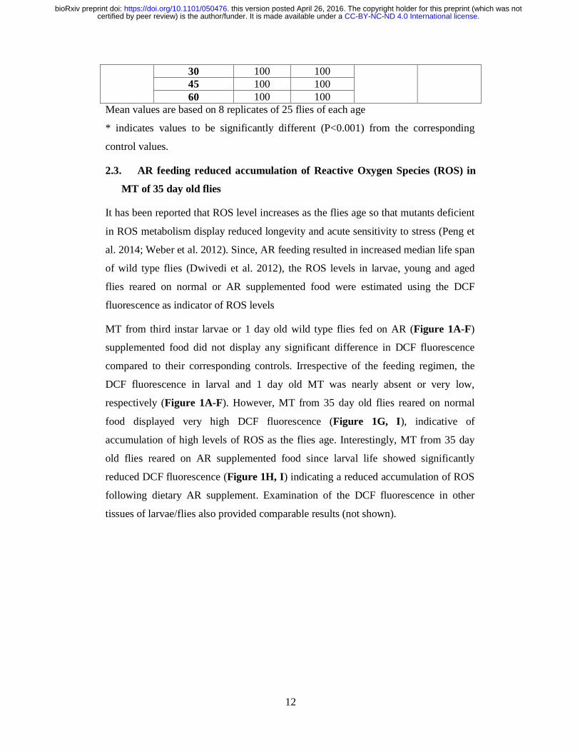
12
30 100 100 45 100 100 60 100 100
Mean values are based on 8 replicates of 25 flies of each age
* indicates values to be significantly different (P<0.001) from the corresponding
control values.
2.3. AR feeding reduced accumulation of Reactive Oxygen Species (ROS) in
MT of 35 day old flies
It has been reported that ROS level increases as the flies age so that mutants deficient
in ROS metabolism display reduced longevity and acute sensitivity to stress (Peng et
al. 2014; Weber et al. 2012). Since, AR feeding resulted in increased median life span
of wild type flies (Dwivedi et al. 2012), the ROS levels in larvae, young and aged
flies reared on normal or AR supplemented food were estimated using the DCF
fluorescence as indicator of ROS levels
MT from third instar larvae or 1 day old wild type flies fed on AR (Figure 1A-F)
supplemented food did not display any significant difference in DCF fluorescence
compared to their corresponding controls. Irrespective of the feeding regimen, the
DCF fluorescence in larval and 1 day old MT was nearly absent or very low,
respectively (Figure 1A-F). However, MT from 35 day old flies reared on normal
food displayed very high DCF fluorescence (Figure 1G, I), indicative of
accumulation of high levels of ROS as the flies age. Interestingly, MT from 35 day
old flies reared on AR supplemented food since larval life showed significantly
reduced DCF fluorescence (Figure 1H, I) indicating a reduced accumulation of ROS
following dietary AR supplement. Examination of the DCF fluorescence in other
tissues of larvae/flies also provided comparable results (not shown).
.CC-BY-NC-ND 4.0 International licensecertified by peer review) is the author/funder. It is made available under aThe copyright holder for this preprint (which was notthis version posted April 26, 2016. . https://doi.org/10.1101/050476doi: bioRxiv preprint

13
Figure 1. AR feeding reduced accumulation of ROS in MT cells in 35 day old flies.
Confocal projections of four medial optical sections from MT of third instar larvae
(A, B), 1 day old flies (D, E) or 35 day old flies (G, H) reared on normal (A, D and
G) or AR (B, E and H) food, show DCF fluorescence following incubation in DCFH-
DA. Scale bar in A represents 20µm and applies to all panels. Histograms in C, F and
I show mean (±S.E.) intensities of DCF fluorescence (N=25 in each case); * in I
indicates P<0.01 when compared with the parallel control reared on regular food.
2.4. AR feeding provided better resistance to reactive oxygen species
generated by paraquat in wild type flies
The N, N′-dimethyl -4, 4′-bipyridinium dichloride or paraquat is commonly used to
generate reactive oxygen species (ROS) in vivo. Three day old wild type male flies,
.CC-BY-NC-ND 4.0 International licensecertified by peer review) is the author/funder. It is made available under aThe copyright holder for this preprint (which was notthis version posted April 26, 2016. . https://doi.org/10.1101/050476doi: bioRxiv preprint

14
fed either on regular or AR supplemented food through their larval stages, were
starved for six hours at 240C and then transferred to vials without food but with a
filter paper soaked in 10mM paraquat in 5% sucrose solution. The ROS accumulation
in the flies following paraquat feeding was examined after 24 h using the DCF
fluorescence (Cathcart et al. 1983) in Malpighian tubule (MT) cells. Compared to the
intensity of DCF fluorescence in Malpighian tubules from paraquat treated flies
reared on normal food, the signal was much less in tissues from similarly treated flies
that were reared on AR supplemented food (Figure 2A-C).
Effect of 10mM paraquat was also examined on survival of flies fed on regular or
formulation supplemented food. In agreement with the reduced levels of ROS
following paraquat exposure, the AR fed flies exhibited a significantly longer median
survival (84.1 h) compared to the 52.4 h median survival of flies grown on regular
food (Figure 2D). As a negative control, flies reared on normal or AR food showed
little death (data not presented) when exposed in parallel to 5% sucrose solution.
2.5. AR feeding partially improved survival of park13 mutant flies but not of
the DJ-1β-null flies exposed to paraquat oxidative stress
Mitochondrial ROS generation is implicated in neurodegenerative diseases like
Parkinson’s disease (PD, Bonifati et al. 2003; Faust et al. 2009; Joselin et al. 2012;
Munoz-Soriano and Paricio, 2011; Narendra et al. 2008, Yang et al. 2005; Yokota et
al. 2003). ROS play a key role in the movement of Parkin into mitochondria in
neurons (Joselin et al. 2012). A dominant null mutant allele of parkin in Drosophila is
park13 (Greene et al. 2005), has been widely used as a fly model of PD. Another PD
associated gene, DJ-1 β, has a role in protection from oxidative stress (Bonifati et al.
2003; Faust et al. 2009; Meulener et al. 2005; Yang et al. 2005; Yokota et al. 2003).
The park13 as well as DJ-1βDelta93 individuals are reported to be highly sensitive to
oxidative stress (Botella et al. 2009; Meulener et al. 2005). Therefore, park13/TM6B
and DJ-1βDelta93 larvae were reared on regular or AR supplemented food and the 3
day old male flies were exposed to 10mM paraquat treatment and monitored every 12
hour to count the number of surviving flies. In agreement with earlier studies (Joselin
et al. 2012), the park13/TM6B flies reared on regular food were very sensitive to
paraquat. AR feeding significantly improved the survival of paraquat treated
park13/TM6B flies although the improved median survival was less than that of the
AR reared wild type flies exposed to paraquat (Figure 2D). Interestingly, however,
.CC-BY-NC-ND 4.0 International licensecertified by peer review) is the author/funder. It is made available under aThe copyright holder for this preprint (which was notthis version posted April 26, 2016. . https://doi.org/10.1101/050476doi: bioRxiv preprint

15
AR feeding did not improve the survival of DJ-1βDelta93 (DJ-1β null) flies following
exposure to paraquat (Figure 2D). Formulation fed or regularly fed flies kept in
parallel on 5% sucrose solution did not show any significant death during the period
of observation (not shown). These results indicate that DJ-1β, is essential for AR
mediated rescue from paraquat-induced oxidative stress.
Figure 2. AR feeding improved the oxidative stress tolerance in wild type flies. A
and B are confocal projections of four medial optical sections of MT showing DCF
fluorescence in 3 day old flies treated for 24 h with 10mM paraquat followed by
incubation in DCFH-DA. Scale bar in A represents 20µm and applies to images in A
and B. Histogram in C shows mean (±S.E., N = 8 for each sample) intensities of DCF
fluorescence in MT principle cells. Histogram in D shows mean (±S.E.) median
survival of WT (Oregon R+), park13 and DJ-1βDelta93 flies (in hours, Y-axis) kept on
10mM paraquat (N for WT Control=14 replicates, for AR=12 replicates and for
park13 and DJ-1βDelta93 Control and AR=8 replicates of 25 male flies each. The bars in
histogram shown in E represent mean (±S.E.) MDA levels (Y-axis) and those in F
represent mean (±S.E.) SOD activity (Y-axis) in 35 day old flies fed either on regular
food (Control) or AR supplemented (AR) food. * in C-F indicate Mean to be
.CC-BY-NC-ND 4.0 International licensecertified by peer review) is the author/funder. It is made available under aThe copyright holder for this preprint (which was notthis version posted April 26, 2016. . https://doi.org/10.1101/050476doi: bioRxiv preprint

16
significantly different (P<0.001) when compared with corresponding Control reared
on regular food.
2.6. AR feeding decreased lipid peroxidation and increased SOD activity in
flies
Aging is associated with an increase in oxidative damage to nucleic acids, sugars,
sterols and lipids (Lushchak, 2014; Peng et al. 2014; Rikans and Hornbrook, 1997;
Sies, 2015; Zhao and Haddad, 2011). Since lipid peroxidation is one of the markers
for age-associated oxidative damage (Kasapoglu and Ozben, 2001, Peng et al. 2014),
we estimated levels of lipid peroxidation in formulation fed and regularly fed 35 day
old flies. Lipid peroxide levels were expressed as nano-moles of malondialdehyde
(MDA) formed per mg of protein following exposure of the fly homogenates to the
assay mixture containing sodium dodecyl sulphate and thiobarbituric acid (Ohkawa et
al. 1979). Compared to the regularly fed flies, AR fed 35 day old flies showed
significantly less concentration of MDA (Figure 2E) reflecting reduced lipid
peroxidation.
An inadequate clearance of ROS like superoxide, hydrogen peroxide and the
hydroxyl radical results in oxidative damage, leading to physiological dysfunction
and many types of age related disorders/diseases (Chaudhari et al. 2007; Park et al.
2005; Tsuzuki et al. 2007; Turner at al. 2009). Glutathione-dependant antioxidants
and enzymes like superoxide dismutase (SOD), catalase (CAT) etc. have an
evolutionarily conserved role in the metabolism and removal of ROS (Muller at al.
2007). We compared the levels of Cu–Zn SOD activity in 35 day old flies reared on
AR supplemented or normal food. Interestingly, AR feeding remarkably enhanced the
Cu–Zn SOD activity (Figure 2F) in aged flies when compared to those reared in
parallel on normal food (Figure 2F).
2.7. AR feeding did not affect the Hsp70/Hsc70 and Hsp83 gene expression
under normal conditions, on heat shock or during recovery
Since different stress-induced heat shock proteins have been implicated in conferring
stress-tolerance and modulating life span (Arya et al. 2007; Feder and Hoffmann,
1999; Lindquist, 1986; Landis et al. 2012; King and MacRae, 2015), we examined
levels of Hsp70, Hsp83 and Hsp27 in unstressed and heat shocked tissues of larvae to
see if their levels are affected by rearing on AR supplemented food.
.CC-BY-NC-ND 4.0 International licensecertified by peer review) is the author/funder. It is made available under aThe copyright holder for this preprint (which was notthis version posted April 26, 2016. . https://doi.org/10.1101/050476doi: bioRxiv preprint

17
Hsp70 is the most strongly induced heat shock protein in Drosophila following cell
stress (Arya et al. 2007; Feder and Hoffmann, 1999; Guertin et al. 2010; Peterson and
Lindquist, 1988), expression pattern and levels of Hsp70 were examined in wild type
third instar larval tissues reared on AR or regular food when maintained at 240C or
subjected to heat shock at 370C for 1h or allowed to recover from the heat shock for 2
h. Levels of hsp70 transcripts in tissues from AR fed larvae, determined by real time
PCR, did not show any significant difference from the corresponding samples of the
regular food reared larvae (Figure 3A). Third instar larval salivary glands (SG) were
immunostained with the stress-induced Hsp70 specific 7Fb antibody (Velazquez and
Lindquist, 1984) following heat shock at 370C or after 2h recovery from heat shock.
SG from AR or regular food fed larvae showed comparable distribution of Hsp70
(Figure 3B, B’, C, C’). Unstressed cells from regular or AR fed larvae did not show
any detectable levels of Hsp70 (not shown). Western blot detection of Hsp70, using
the 7Fb antibody, in whole larval proteins from wild type larvae fed on regular or AR
supplemented food and subjected to heat shock at 370C or allowed to recover at 240C
from the 1 h heat shock confirmed the above immunostaining results that AR feeding
did not alter the levels of induced Hsp70 protein on heat shock or during recovery
(inset in Figure 3B’, C’).
We also examined if the expression of YFP-tagged Hsc70Cb, one of the constitutively
expressed Hsc70 family proteins, is affected by AR feeding. The pattern and intensity
of YFP fluorescence in SG from late Hsc70Cb-YFP larvae fed on regular or AR food
did not differ from each other neither under normal conditions nor after heat shock or
during recovery from heat shock (Figure 3D, D’, E, E’, F, F’). Western blot detection
of all Hsc70/Hsp70, using the 7.10.3 Ab, which detects all constitutive and inducible
members of the Hsp70 family in Drosophila (Palter et al. 1986), in total proteins from
wild type larvae reared either on AR or on regular food confirmed that the levels of
Hsc70/Hsp70 remained similar under all conditions (insets in Figure 3D’, E’, F’).
.CC-BY-NC-ND 4.0 International licensecertified by peer review) is the author/funder. It is made available under aThe copyright holder for this preprint (which was notthis version posted April 26, 2016. . https://doi.org/10.1101/050476doi: bioRxiv preprint

18
Figure 3. Feeding larvae on AR supplemented food did not affect the distribution and
levels of Hsp70/Hsc70 on heat shock and during recovery. A- Histograms show mean
(± S.E.) fold changes of Hsp70 transcripts (Y-axis) in larvae fed on normal (C) or AR
(AR) food at 240C (unstressed) or following heat shock (C/AR-HS) or after 2 h
recovery from heat shock (C/AR-R2); the Hsp70 mRNA levels in unstressed larvae
reared on normal food (C) were taken as 1 for estimating the fold changes in other
samples. B-F’-Confocal projection images showing levels of Hsp70 (B-C’) or
Hsc70Cb-YFP (D-F’) in salivary glands from third instar larvae reared on control
(Con) or AR supplemented (AR) food under different conditions as noted at the
bottom (Hsp70 immunostaining for unstressed condition is not shown). The scale bar
in B (20 μm) applies to B-F’. Insets in B’, C’ are western blots showing levels of
Hsp70 detected by the 7Fb antibody (rows 1) and those in D'-F' show levels of
Hsc70/Hsp70 detected by the 7.10.3 antibody (rows 1) in total proteins from
differently fed (Con and AR) wild type late 3rd instar larvae under unstressed (D') or
after heat shock (B', D') or after 2 h recovery from heat shock (C', F'); β-tubulin
levels detected by the E7 antibody, used as the loading control, are shown in lower
rows (2) in each western blot.
In unstressed cells Hsp83 was abundant in cytoplasm but much less in nuclei of SG
from larvae reared on normal or AR supplemented food (Figure 4A, D). Following
heat shock (Figure 4B, E) as well as during recovery from heat shock (Figure 4C,
F), the Hsp83 was more abundant in SG nuclei than in cytoplasm. However, under all
the three conditions, the distribution pattern and levels of Hsp83 in SG polytene cells
from AR supplemented food reared larvae were not different from those in normal
food reared larvae (Figure 4). This was also confirmed by quantification of Hsp83
fluorescence intensity in different samples (Figure 4G). Similar results were obtained
with MT and eye disc cells (not shown). Levels of hsp83 transcripts in differently fed
.CC-BY-NC-ND 4.0 International licensecertified by peer review) is the author/funder. It is made available under aThe copyright holder for this preprint (which was notthis version posted April 26, 2016. . https://doi.org/10.1101/050476doi: bioRxiv preprint

19
larvae were also examined under unstressed conditions, after heat shock and after 2 h
recovery by real time PCR. It was observed that levels of hsp83 transcripts in larvae
reared on normal food and AR supplemented food were comparable under various
conditions (Figure 4H).
Figure 4. Feeding larvae on AR supplemented food did not affect the distribution and
levels of Hsp83 protein and transcripts. A-F are confocal projections of four medial
optical sections of third instar larval salivary glands immunostained with anti-Hsp83
antibody either at 240C (unstressed, A, D), or after heat shock at 370C for 1 hour (B,
E) or after 2 h recovery from heat shock (C, F). Scale bar in A represents 20µm and
applies to images A-F. Histogram in G shows the mean (± S.E.) fluorescence
intensities of Hsp83 in SG of third instar larvae reared on normal (Control) or AR
supplemented (AR) food and subjected to various conditions. H- Histogram
represents mean (± S.E.) fold change of Hsp83 transcripts in larvae fed on AR or
normal food either kept under normal condition or heat shocked (HS) or recovered for
2 hours (R2).
The Hsp27 protein is normally distributed in nuclei as well as in cytoplasm of wild
type SG from larvae reared on regular food (Figure 5A). Following heat shock,
Hsp27 levels are enhanced in both nucleus as well as cytoplasm (Figure 5B), which
nearly restored to that in unstressed cells after 2 h recovery from heat shock (Figure
5C). Interestingly, SG cells from AR fed larvae showed significantly higher levels of
Hsp27 in nucleus as well as cytoplasm of unstressed cells than in those from regular
food reared larvae (compare Figure 5A and D). Heat shock further elevated levels of
Hsp27 in AR fed larval SG (Figure 5E). Unlike in regular fed larval SG cells, Hsp27
levels in AR larval SG remained higher even after 2 hour recovery from heat shock
.CC-BY-NC-ND 4.0 International licensecertified by peer review) is the author/funder. It is made available under aThe copyright holder for this preprint (which was notthis version posted April 26, 2016. . https://doi.org/10.1101/050476doi: bioRxiv preprint

20
(Figure 5C and F). The above noted differences in levels of Hsp27 between normal
and AR supplemented food confirmed by quantification of the fluorescence intensity
of immunostained cells (Figure 5G). Other tissues like MT, eye and other imaginal
discs also displayed a comparable difference in the levels of Hsp27 in regularly fed or
AR fed larvae (not shown). Examination of levels of hsp27 transcripts by real time
PCR showed that in parallel with the immunostaining patterns, hsp27 transcripts were
significantly more abundant under all conditions (unstressed, heat shock and 2 h
recovery) in AR fed larvae than in those reared on regular food. Heat shock resulted
in greater induction of hsp27 transcripts in AR while after 2 hour recovery, the hsp27
transcripts in AR fed samples still remained higher than that in normal fed recovery
sample (Figure 5H).
Figure 5. Feeding larvae on AR supplemented food elevated the levels of Hsp27
protein and transcripts. A-F are confocal projections of four medial optical sections of
third instar larval salivary glands immunostained with anti-Hsp27 maintained at 240C
(unstressed, A, D), or after heat shock at 370C for 1 h (B, E) or after 2 h recovery
from heat shock (C, F). Scale bar in A represents 20µm and applies to images A-F.
Histogram in G shows the mean (± S.E.) fluorescence intensities of Hsp27 (Y-axis) in
SG of third instar larvae reared on regular (C) or AR supplemented (AR) food
subjected to various conditions (X-axis). H- Histogram represents mean (± S.E.) fold
change in levels of Hsp27 transcripts in larvae fed on regular (C) or AR supplemented
(AR) food (X-axis). In G and H, C and AR indicate unstressed larvae reared on
regular or AR supplemented food, HS indicate heat shocked samples while R2
indicates samples recovered for 2 h from the heat shock. * indicates P<0.001 when
compared with the corresponding control (AR vs C).
.CC-BY-NC-ND 4.0 International licensecertified by peer review) is the author/funder. It is made available under aThe copyright holder for this preprint (which was notthis version posted April 26, 2016. . https://doi.org/10.1101/050476doi: bioRxiv preprint

21
3. Discussion
Ayurvedic system, the oldest among various traditional health care practices in world,
is claimed to provide a holistic health care (Patwardhan et al., 2015; Singh, 2009).
Ayurveda has its own wide range of formulations consisting of herbal, mineral and
other biological materials, which have been compiled as ‘Dravyaguna Sangrah’
(Sharma, 1994; Singh, 2009). However, we know very little about the specific modes
of actions of the various complex Ayurvedic formulations (Valiathan, 2006, Lakhotia,
2013). Natural products present in Ayurvedic formulations have provided interesting
sources of new molecular entities with potential therapeutic applications; this has
stimulated a large number of studies on various herbs to identify the 'active
principles'. However, Ayurvedic herbal and other Rasayanas are complex mixtures of
multiple ingredients which work in a synergistic manner and, therefore, the
reductionist approach to identify the 'active principle' does not always provide
information on the mechanisms through which the classical formulations exert their
effects (Lakhotia, 2013; Patwardhan et al., 2015).
Earlier studies from our laboratory (Dwivedi et al. 2012, 2013, 2015) established
Drosophila as a model organism to understand the mechanisms and modes of actions
of the complex Ayurvedic formulations. As noted earlier, one of the major usages of
AR in humans is to rejuvenate and thus improve longevity and maintain youthfulness.
Earlier studies showed that feeding flies on 0.5% AR supplemented food enhanced
their life span, improved fecundity and enhanced cellular levels of key regulatory
molecules like the hnRNPs, CBP300 (Dwivedi et al. 2012), suppressed
neurodegeneration associated with polyQ disorders and Alzheimer’s Disease
(Dwivedi et al. 2013), and prevented induced apoptosis without affecting
developmental cell death (Dwivedi et al. 2015). A study in mouse model has shown
that the genomic integrity in aging mice provided with AR is significantly better than
in control sibs (Swain et al. 2012). Present studies provide further insights into the
reported beneficial and rejuvenation affects of dietary AR.
Besides the earlier reported improved tolerance of AR-fed flies to starvation and
thermal stress (Dwivedi et al. 2012), present studies clearly show that AR feeding
significantly improves tolerance to crowding and oxidative stresses. A more
significant finding is that AR-feeding makes even older wild type flies more tolerant
to a severe heat shock (380C). Such improved capability to face various external
.CC-BY-NC-ND 4.0 International licensecertified by peer review) is the author/funder. It is made available under aThe copyright holder for this preprint (which was notthis version posted April 26, 2016. . https://doi.org/10.1101/050476doi: bioRxiv preprint

22
environmental stresses as well as internal metabolic oxidative stresses seem to
promote the longer life span displayed by the AR fed flies. The improved starvation-
tolerance (Dwivedi et al. 2012) and resistance to crowding displayed by AR-fed
larvae/flies and their increased longevity agrees with the earlier reported correlation
that better larval physiology promotes better starvation tolerance and longevity of
adults (Chippindale et al. 1996; Mueller and Barter, 2015; Prasad and Joshi, 2003;
Service 1987).
Several studies have reported very strong anti-oxidant activity of extracts of Amla or
Indian Gooseberry fruit, the principal component of AR (Chatterjee et al. 2011;
Govindarajan et al. 2004; Khan, 2009; Krishnaveni and Mirunalini, 2010; Poltanov et
al. 2009; Scartezzini and Speroni, 2000). In agreement, present results show that AR
feeding significantly improved the oxidative stress tolerance of wild type flies and
remarkably reduced the accumulation of ROS with age, reflecting an improved
cellular redox homeostasis. The partially improved survival of park13 flies but no
improvement in survival of paraquat exposed DJ-1β mutant flies shows that the
improved management of ROS by AR feeding requires functional Parkin and more so
the DJ-1β protein. The DJ-1β protein plays a significant role in managing oxidative
stress and the Phosphatidylinositol 3-Kinase/Akt signalling pathway (Meulener et al.
2005; Yang et al. 2005).
Aging is strongly correlated with sensitivity to components that increase the oxidative
stress and thus cause greater damage to macromolecules like DNA and lipids (Haigis
and Yanker, 2015; Holmbeck et al. 2014; Lagouge et al. 2013; Mallikarjun et al.
2014; Weber et al. 2012). AR fed flies had reduced levels of ROS and reduced levels
of lipid peroxidation. Populations of flies with longer life span exhibit higher
frequencies of the high-activity allele of the Cu–Zn SOD which has an important role
in conferring resistance to oxidative stressors through removal of superoxides (Peng
et al. 2014; Tyler et al. 1993). The significantly improved SOD activity in AR fed
older flies contributes to reduced oxidative damage, which in turn would improve
fecundity and life span. The increased resistance to oxidative stress following AR
feeding appears to also underlie the reported reduced DNA damage seen in aged mice
(Swain et al., 2012).
The significantly greater thermotolerance exhibited by older flies that were reared on
AR supplemented food is remarkable. Our results show that unlike the generally
.CC-BY-NC-ND 4.0 International licensecertified by peer review) is the author/funder. It is made available under aThe copyright holder for this preprint (which was notthis version posted April 26, 2016. . https://doi.org/10.1101/050476doi: bioRxiv preprint

23
strong correlation (Krebs and Feder, 1998; Lin et al. 2014; Stetina et al. 2014)
between thermotolerance and expression of heat shock proteins, especially the Hsp70,
AR did not significantly affect expression of Hsp83 and Hsp70 proteins or transcripts.
On the other hand we found that basal as well as heat shock induced levels of Hsp27
were significantly more elevated in AR fed larvae. It is interesting that although
Hsp27 is reported to be unessential for surviving heat shock (Hao et al., 2007), it
plays significant roles in tolerance to other insults like starvation, oxidative stress etc
and also in determining the life span of flies (Hao et al. 2007, Pandey et al. 2015; Shi
et al. 2011; Wang et al. 2004). Thus the improved thermo-tolerance of AR fed larvae
and flies does not appear to be due to their stronger heat shock response. It may be
noted that the heat shock response is a transient process and its long-term activation
may actually be harmful (Lamech and Haybes, 2014). The improved thermo-tolerance
that follows rearing on AR supplemented food may thus be due to better management
of oxidative stress as also to the overall improved physiology and homeostasis
(Dwivedi at al., 2012, 2013, 2015) promoted by AR through enhanced levels of key
regulatory molecules like different hnRNPs and CBP300 (histoneacetyl transferase)
and proteasomal activity that have global roles in maintaining gene expression.
Various environmental insults (external as well as internal) also induce cells to
apoptotic pathways, which is one of the factors underlying aging (Lu et al. 2012;
Pérez-Garijo and Steller, 2015). Our earlier finding that AR feeding suppresses
induced but not the developmental apoptosis also indicates a better cellular tolerance
promoted by AR to environmental insults (Dwivedi et al. 2015).
4. Conclusions
The modern medicine and Ayurveda, although have universal attributes and share the
common objective of well being of mankind, seem to differ in their philosophical and
epistemological foundations, conceptual framework and practical outlook
(Patwardhan et al. 2015). Most contemporary scientific studies to understand specific
Ayurvedic formulations/therapies employ the typical reductionist approach of
molecular biology to identify the so called “active principle”. However, this is not in
consonance with the holistic and combinatorial approach of Ayurveda (Lakhotia,
2013; Patwardhan et al. 2015). Therefore, unbiased and a rational understanding of
Ayurvedic Biology in a holistic manner is expected to provide better quality control
of the various formulations. Our findings in the fly model have clinical implications
.CC-BY-NC-ND 4.0 International licensecertified by peer review) is the author/funder. It is made available under aThe copyright holder for this preprint (which was notthis version posted April 26, 2016. . https://doi.org/10.1101/050476doi: bioRxiv preprint

24
since the more robust physiological state following dietary AR supplement in flies
suggest that AR may prevent several diseases and aging conditions and thus
contribute to "healthy aging” in mankind.
5. Acknowledgements
We thank Arya Vaidya Sala, Kottakal (Kerala, India) for providing the Amalaki
Rasayana formulation and Prof. M. S. Valiathan for initiating the coordinated studies
on Science of Ayurveda. We thank Dr. P. K. Tiwari (Gwalior, India) for providing the
anti-Hsp27, Dr. R. M. Tanguay (Canada) for anti-Hsp90 antibody and Dr. M. B.
Evgen’ev (Russia) for 7Fb and 7.10.3 antibodies. Bloomington stock centre is
acknowledged for providing various stocks used in present study. This work was
supported in part by a grant (no. Prn.SA/ADV/Ayurveda/6/2006) from the Office of
the Principal Scientific Advisor to Govt. of India (New Delhi) and by the Raja
Ramanna Fellowship of the Department of Atomic Energy (Govt. of India, Mumbai)
to SCL. The Confocal Facility, established by the Department of Science &
Technology, Govt. of India (New Delhi), is supported by the Banaras Hindu
University. VD is supported by research fellowship from University Grants
Commission (New Delhi).
.CC-BY-NC-ND 4.0 International licensecertified by peer review) is the author/funder. It is made available under aThe copyright holder for this preprint (which was notthis version posted April 26, 2016. . https://doi.org/10.1101/050476doi: bioRxiv preprint

25
6. References
Adelman, R., Saul, R.L., Ames, B.N. 1988. Genetics oxidative damage to DNA: relation to species metabolic rate and life span. Proc. Nail. Acad. Sci. USA. 85. 2706-2708.
Arya, R., Mallik, M., Lakhotia S.C., 2007. Heat shock genes – integrating cell survival and death. J. Biosci. 32, 494–610.
Baker, K.D., Thummel, C.S., 2007. Diabetic larvae and obese flies-emerging studies of metabolism in Drosophila. Cell Metab. 6, 247-266.
Bonifati, V., Rizzu, P., Squitieri, F., Krieger, E., Vanacore, N, et al, 2003. DJ-1 (PARK7), a novel gene for autosomal recessive, early onset parkinsonism. Neurol. Sci. 24, 149-160.
Borash D.J., Ho G.T., 2001. Patterns of selection: stress resistance and energy storage in density-dependent populations of Drosophila melanogaster. J. Insect Physiol. 47, 1349–1356
Britton, J.S., Lockwood, W.K., Li, L., Cohen, S.M., Edgar, B.A., 2002. Drosophila's insulin/PI3-kinase pathway coordinates cellular metabolism with nutritional conditions. Dev. Cell. 2, 239-249.
Calderwood, S.K., Murshid, A., Prince, T., 2009. The shock of aging: molecular chaperones and the heat shock response in longevity and aging – a mini-review. Gerontology. 44, 440–448.
Cathcart, R., Schwiers, E. Ames, B.N., 1983. Detection of picomole levels of hydroperoxides using a fluorescent dichlorofluorescein assay. Anal. Biochem. 134, 111-116.
Chatterjee, U.R., Bandyopadhyay, S.S., Ghosh, D., Ghosal, P.K., Ray, B., 2011. In vitro anti-oxidant activity, fluorescence quenching study and structural features of carbohydrate polymers from Phyllanthus emblica. Int. J. Biol. Macromol. 49, 637-642.
Chaudhari, A.A., Seol, J.W., Kim, S.J., Lee, Y.J., Kang, H.S., et al., 2007. Reactive oxygen species regulate Bax translocation and mitochondrial transmembrane potential, a possible mechanism for enhanced TRAIL-induced apoptosis by CCCP. Oncol. Rep. 18, 71-76.
Chippindale, A.K., Chu, T.J.F., Rose, M.R., 1996. Complex trade-offs and the evolution of starvation resistance in Drosophila melanogaster. Evolution 40, 743–766.
Dwivedi, V., Anandan, E.M., Mony, R.S., Muraleedharan, T.S., Valiathan, M.S., Mutsuddi, M., Lakhotia, S.C., 2012. In vivo effects of traditional Ayurvedic formulations in Drosophila melanogaster model relate with therapeutic applications. PLoS One. 7, e37113.
Dwivedi, V., Tiwary, S., Lakhotia, S.C., 2014. Suppression of induced but not evelopmental apoptosis by Ayurvedic, Amalaki Rasayana and Rasa-Sindoor in Drosophila melanogaster. J. Biosci. 40, 281-297.
.CC-BY-NC-ND 4.0 International licensecertified by peer review) is the author/funder. It is made available under aThe copyright holder for this preprint (which was notthis version posted April 26, 2016. . https://doi.org/10.1101/050476doi: bioRxiv preprint

26
Dwivedi, V., Tripathi, B.K., Mutsuddi, M., Lakhotia, S.C., 2013. Ayurvedic Amalaki Rasayana and Rasa-Sindoor suppress neurodegeneration in fly models of Huntington’s and Alzheimer’s diseases. Curr. Sci. 104, 1711-1723.
Ekengren, S., Tryselius, Y., Dushay, M.S., Liu, G., Steiner, H., Hultmark, D., 2001. A humoral stress response in Drosophila. Curr. Biol. 11, 1479.
Engelman, J.A., Luo, J., Cantley, L.C., 2006. The evolution of phosphatidylinositol 3-kinases as regulators of growth and metabolism. Nat. Rev. Genet. 7, 606-619.
Evgen’ev, M.B., Garbuz, D.G., Zatsepina, O.G., 2014. Heat shock proteins and whole body adaptation to extreme environments. Springer, Dordrecht Heidelberg New York London.
Faust, K., Gehrke, S., Yang, Y., Yang, L., Beal, M.F., Lu, B., 2009. Neuroprotective effects of compounds with antioxidant and anti-inflammatory properties in a Drosophila model of Parkinson's disease. BMC Neurosci. 10, 109, doi:10.1186/1471-2202-10-109.
Feder, M.E., Hofmann, G.E., 1999. Heat-shock proteins, molecular chaperones, and the stress response, evolutionary and ecological physiology. Annu. Rev. Physiol. 61, 243–282.
Gandhi, S., Abramov, A.Y., 2012. Mechanism of oxidative stress in neurodegeneration. Oxidative Med. Cell. Longevity. Article ID 428010, doi:10.1144/2012/428010.
Govindarajan, R., Vijayakumar, M., Pushpangadan, P., 2004. Antioxidant approach to disease management and the role of 'Rasayana' herbs of Ayurveda. J. Ethnopharmacol. 99, 164-178.
Greene, J.C., Whitworth, A.J., Andrews, L.A., Parker, T.J., Pallanck, L.H., 2005. Genetic and genomic studies of Drosophila parkin mutants implicate oxidative stress and innate immune responses in pathogenesis. Human Mol. Genet. 14, 799-811.
Guertin, M.J., Petesch, S.J., Zobeck, K.L., Min, I.M., Lis, J.T., 2010. Drosophila heat shock system as a general model to investigate transcriptional regulation. Cold Spring Harb. Symp. Quant. Biol. 74, 1-9.
Haigis, M.C., Yankner, B.A., 2015. The aging stress response. Mol. Cell. 40, 333-344.
Hao, X., Zhang, S., Timakov, B., Zhang, P., 2007. The Hsp27 gene is not required for Drosophila development but its activity is associated with starvation resistance. Cell Stress Chaperones. 12, 364-372.
Holmbeck, M.A., Rand, D.M., 2014. Dietary fatty acids and temperature modulate mitochondrial function and longevity in Drosophila. J. Gerontol. doi: 10.1093/gerona/glv044.
Joselin, A.P., Hewitt, S.J., Callaghan, S.M., Kim, R.H., Chung,Y.H., Mak, T.W., et al., 2012. ROS-dependent regulation of Parkin and DJ-1 localization during oxidative stress in neurons. Human Mol. Genet. 21, 4888–4903.
Kakkar, P., Das, B., Vishwanathan, P.N., 1984. A modified spectrophotometric assay of superoxide dismutase. Indian J. Biochem. Biophys. 21, 130–132.
.CC-BY-NC-ND 4.0 International licensecertified by peer review) is the author/funder. It is made available under aThe copyright holder for this preprint (which was notthis version posted April 26, 2016. . https://doi.org/10.1101/050476doi: bioRxiv preprint

27
Kasapoglu, M., Ozben, T., 2001. Alterations of antioxidant enzymes and oxidative stress markers in aging. Exp. Gerontol. 36, 209-220.
Khan, K.H., 2009. Roles of Emblica officinalis in medicine, a review. Botany Res. Internatl. 2, 218-228.
King, A.M., MacRae, T.H., 2015. Insect heat shock proteins during stress and diapause. Annu. Rev. Entomology. 60, 49-74. DOI: 10.1146/annurev-ento-011613-162107.
Krebs, R.A., Feder, M.E., 1998. Hsp70 and larval thermotolerance in Drosophila melanogaster: how much is enough and when is more too much? J. Insect. Physiol. 44, 1091-1101.
Krishnaveni, M., Mirunalini, S., 2010. Therapeutic potential of Phyllanthus emblica (Amla): the Ayurvedic wonder, J .Basic Clin. Physiol. Pharmacol. 21, 93-104.
Lagouge, M., Larsson, N.G., 2013. The role of mitochondrial DNA mutations and free radicals in disease and ageing. J. Intern. Med. 273, 429-443.
Lakhotia, S.C., 2013. In-depth basic science studies essential for revival of Ayurveda. Ann. Ayurvedic Med. 2, 58-60.
Lakhotia, S.C., Tapadia, M., 1998. Genetic mapping of the amide response element(s) of the hsrω locus of Drosophila melanogaster. Chromosoma. 107, 127-135.
Lamech, L.T., Haynes, C.M., 2014. The unpredictability of prolonged activation of stress response pathways. J. Cell Biology. 209, 781–787.
Landis, G., Shen, J., Tower, J., 2012. Gene expression changes in response to aging compared to heat stress, oxidative stress and ionizing radiation in Drosophila melanogaster. Aging. 4, 768-789.
Leopold, P., Perrimon, N., 2007. Drosophila and the genetics of the internal milieu. Nature. 440, 186-188.
Lin, H.J., Li, Z., Dang, X.Q., Su, W.J., Zhou, Z.Y., Wang, L.L., 2014. Short-term increased expression of the heat shock protein 70 family members during heat shock is positively correlated with basal thermotolerance in the midgut of three strains of the silkworm, Bombyx mori. African Entomology. 22, 24-29.
Lindquist, S. 1986. The Heat-Shock Response. Ann. Rev. Biochem. 55, 1151-1191.
Lu, B., Chen, H.D., Lu, H.G., 2012. The relationship between apoptosis and aging. Adv. Bioscience Biotech. 3, 705-711,DOI: 10.4236/abb.2012.326091.
Lushchak, A.I., 2014. Free radicals, reactive oxygen species, oxidative stress and its classification. Chemico-Biological Interactions. 224, 164–174.
Mallikarjun, V., Sriram, A., Scialo, F., Sanz, A., 2014. The interplay between mitochondrial protein and iron homeostasis and its possible role in ageing. Exp. Gerontol. 46, 123-134.
Melkani, G.C., Trujillo, A.S., Ramos, R., Bodmer, R., Bernstein. S.I., et al., 2013. Huntington’s disease induced cardiac amyloidosis is reversed by modulating protein folding and oxidative stress pathways in the Drosophila heart. PLoS Genet. 9,e1004024. doi:10.1371/journal.pgen.1004024
.CC-BY-NC-ND 4.0 International licensecertified by peer review) is the author/funder. It is made available under aThe copyright holder for this preprint (which was notthis version posted April 26, 2016. . https://doi.org/10.1101/050476doi: bioRxiv preprint

28
Meulener, M., Whitworth, A.J., Armstrong-Gold, C.E., Rizzu, P., Heutink, P., et al., 2005. Drosophila DJ-1 mutants are selectively sensitive to environmental toxins associated with Parkinson’s disease. Current Biology. 15, 1572–1577.
Morrow, G., Tanguay, R.M., 2003. Heat shock proteins and aging in Drosophila melanogaster. Seminar. Cell Develop. Biol. 14, 291–299.
Mueller, L.D., Barter, T.T., 2015. A model of the evolution of larval feeding rate in Drosophila driven by conflicting energy demands. Genetica. 143, 93-100.
Muller, F.L., Song, W., Jang, Y.C., Liu, Y., Sabia, M., et al., 2007. Denervation-induced skeletal muscle atrophy is associated with increased mitochondrial ROS production. Am. J. Physiol. Regul. Integr. Comp. Physiol. 293, 1149-1168.
Munoz-Soriano, V., Paricio, N., 2011. Drosophila models of Parkinson's disease: discovering relevant pathways and novel therapeutic strategies, Parkinson’s Disease. 2011, Article ID 420640, DOI: 10.4061/2011/420640.
Murthy, K.R.S., 2000. Ashtanga Hridaya (Sanskrit with English Translation), Krishnadas Academy, Varanasi.
Narendra, D., Tanaka, A., Suen, D.F., Youle, R.J., 2008. Parkin is recruited selectively to impaired mitochondria and promotes their autophagy. J. Cell Biol. 183, 794–803.
Ohkawa, H., Ohishi, N., Yagi, K., 1979. Assay for lipid peroxides in animal tissues by thiobarbituric acid reaction. Anal. Biochem. 94 341– 348.
Palter, K.B., Watanabe, M., Stinson, L., Mahowald, A.P., Craig, E.A., 1986. Expression and localization of Drosophila melanogaster Hsp7O cognate proteins. Mol. Cell Biol. 6,1187–1203.
Pandey, A., Sainy, S., Khatoon, R., Sharma, D., Narayan, G., Kar Chowdhuri, D., 2015. Overexpression of hsp27 rescued neuronal cell death and reduction in life- and health-span in Drosophila melanogaster against prolonged exposure to dichlorvos. Mol. Neurobiol. DOI: 10.1007/s12035-015-9221-3
Park, J., Kim, S.Y., Cha, G.-H., Lee, S.B., Kim, S., Chung, J., 2005. Drosophila DJ-1 mutants show oxidative stress-sensitive locomotive dysfunction. Gene. 361, 133–139.
Patwardhan, B., Mutalik, G., Tillu, G., 2015. Integrative Approaches for Health: Biomedical Research, Ayurveda and Yoga. Elsevier/Academic Press, London.
Peng, C., Wang, X., Chen, J., Jiao, R., Wang, L., et al., 2014. Biology of ageing and role of dietary antioxidants, Biomed. Res. Int. 83, 831841, DOI: 10.1144/2014/831841.
Pérez-Garijo, A., Steller, H. 2015. Spreading the word: non-autonomous effects of apoptosis during development, regeneration and disease. Development. 142, 3253-3262, DOI:10.1242/dev.127878.
Peterson, R., Lindquist, S., 1988. The Drosophila hsp70 message is rapidly degraded at normal temperatures and stabilized by heat shock. Gene. 72, 161-168.
Phillips, J.P., Campbell, S.D., Michaud, D., Charbonneaut, M., Hilliker, A.J., 1989. Null mutation of copper/zinc superoxide dismutase in Drosophila confers hypersensitivity to paraquat and reduced longevity. Proc. Natl. Acad. Sci. USA. 86, 2761-2764.
.CC-BY-NC-ND 4.0 International licensecertified by peer review) is the author/funder. It is made available under aThe copyright holder for this preprint (which was notthis version posted April 26, 2016. . https://doi.org/10.1101/050476doi: bioRxiv preprint

29
Poltanov, E.A., Shikov, A.N.,Dorman, H.J., Pozharitskaya, O.N., Makarov, V.G., et al., Chemical and antioxidant evaluation of Indian gooseberry (Emblica officinalis Gaertn, syn. Phyllanthus emblica L.) supplements, Phytother Res 23, 1309-1314, 2009
Prasad, N.G., Joshi, A., 2003. What have two decades of laboratory life-history evolution studies on Drosophila melanogaster taught us? J. Genet. 82, 44–76.
Prasanth, K.V., Rajendra, T.K., Lal, A.K., Lakhotia, S.C., 2000. Omega speckles - a novel class of nuclear speckles containing hnRNPs associated with noncoding hsr-omega RNA in Drosophila. J. Cell Sci. 113, 3484-3497.
Puri, H. S., 2003. Rasayana: ayurvedic herbs for longevity and rejuvenation. Taylor & Frauds, London, New York.
Rikans, L.E., Hornbrook, K.R., 1997. Lipid peroxidation, antioxidant protection and aging. Biochim. Biophys. Acta. 1362, 116-127.
Saltiel, A.R., Kahn, C.R., 2001. Insulin signalling and the regulation of glucose and lipid metabolism. Nature. 414, 799-806.
Scartezzini, P., Speroni, E., 2000. Review on some plants of Indian traditional medicine with antioxidant activity. J. Ethnopharmaco. 71, 23-43.
Schieber, M., Chandel, N.S., 2014. ROS function in redox signaling and oxidative stress. Current Biology. 24, R443–R462.
Service, P.M., 1987. Physiological mechanisms of increased stress resistance in Drosophila melanogaster selected for postponed senescence. Physiol. Zool. 60, 321–326.
Sharma, P.V., Charaka Samhita, (Sanskrit with English Translation), Varanasi, India: Chaukhambha Orientalia, 1994
Shi, Y., Nishida, K., Giammartino, D.C.D., Manley, J.L., 2011. Heat shock-induced SRSF10 dephosphorylation displays thermotolerance mediated by Hsp27. Mol. Cell Biol. 31, 448-464.
Shukla, A.K., Pragya, P., Chaouhan, H.S., Patel, D.K., Abdin, M.Z., Kar Chowdhuri D., 2014. A mutation in Drosophila methuselah resists paraquat induced Parkinson-like phenotypes. Neurobiol. Aging. 34, 2419.e1e2419.e16.
Sies, H., 2015. Oxidative stress: a concept in redox biology and medicine. Redox Biology. 4, 180–183.
Singh, A.K., Lakhotia, S.C., 2016. Expression of hsrω-RNAi transgene prior to heat shock specifically compromises accumulation of heat shock induced Hsp70 in Drosophila melanogaster. Cell Stress Chaperones. 20, 105-121.
Singh, R.H., 2009. Body, mind, spirit- Integrative medicine in Ayurveda Nature and Yoga. Chaukhamba Sanskrit Pratishthan, Varanasi.
Stetina, T., Kostal, V., Korbelova, J., 2014. The role of inducible Hsp70 and other heat shock proteins, in adaptive complex of cold tolerance of the fruit fly (Drosophila melanogaster). PLoS One. 10, e0128976.
Swain, U., Sindhu, K.K., Boda, U., Pothani, S., Giridharan, N.V., Raghunath, M., Rao, K.S., 2012. Studies on the molecular correlates of genomic stability in rat brain cells following Amalakirasayana therapy. Mech. Ageing Dev. 133, 112-117.
.CC-BY-NC-ND 4.0 International licensecertified by peer review) is the author/funder. It is made available under aThe copyright holder for this preprint (which was notthis version posted April 26, 2016. . https://doi.org/10.1101/050476doi: bioRxiv preprint

30
Tsuzuki, T., Nakatsu, Y., Nakabeppu, Y., 2007. Significance of error-avoiding mechanisms for oxidative DNA damage in carcinogenesis. Cancer Sci. 98, 464-470.
Turner, E., Brewster, J.A., Simpson, N.A., Walker, J.J., Fisher, J., Imidazole-based erythrocyte markers of oxidative stress in preeclampsia-an NMR investigation, Reprod Sci 16, 1040-1041, 2009
Tyler, R.H., Brar, H., Singh, M., Latorre, A., Graves, J.L., et al., 1993. The effect of superoxide dismutase alleles on aging in Drosophila. Genetica. 91, 143-149.
Valiathan, M.S., 2006. Ayurvedic biology: a decadal vision document. Indian Academy of Science, Bangalore.
Velazquez, J.M., Lindquist, S., 1984. hsp70: nuclear concentration during environmental stress and cytoplasmic storage during recovery. Cell. 36, 644-662.
Wang, C., Liu, Z.,Huang, X., 2012. Rab32 is important for autophagy and lipid storage in Drosophila, PLoS One 7, e32086.
Wang, C.H., Wu, S.B., Wu, Y.T., Wei, Y.H., 2013. Oxidative stress response elicited by mitochondrial dysfunction: implication in the pathophysiology of aging. Experimental Biology Medicine. 238: 440–460, DOI: 10.1177/1434370213493069.
Wang, H.D., Kazemi-Esfarjani, P., Benzer, S., 2004. Multiple-stress analysis for isolation of Drosophila longevity genes. Proc. Natl. Acad. Sci. USA. 101, 12610-12614.
Weber, A.L., Khan, G.F., Magwire, M.M., Tabor, C.L., Mackay, T.F., Anholt, R.R., 2012. Genome-wide association analysis of oxidative stress resistance in Drosophila melanogaster. PLoS One. 7, e34744.
Yan, L.J., 2014. Positive oxidative stress in aging and aging-related disease tolerance. Redox Biology. 2, 164–169.
Yang, Y., Gehrke, S., Haque, M.E., Imai, Y., Kosek, J., Yang, L., et al., 2005. Inactivation of Drosophila DJ-1 leads to impairments of oxidative stress response and phosphatidylinositol 3-kinase/akt signaling. Proc. Natl. Acad. Sci. USA. 102, 13670-13674.
Yokota, T., Sugawara, K., Ito, K., Takahashi, R., Ariga, H., Mizusawa. H., 2003. Down regulation of DJ-1 enhances cell death by oxidative stress, ER stress and proteasome inhibition. Biochem. Biophys. Res. Commun. 312, 1342-1348.
Zhang, H., Davies, K.J.A., Forman, H.J., 2015. Oxidative stress response and Nrf2 signaling in aging. Free Radical Biology Medicine. 88, 314–336.
Zhao, H.W., Haddad. G.G., 2011. Review: hypoxic and oxidative stress resistance in Drosophila melanogaster. Placenta. 32, Supplement B, Trophoblast Research, 24, S104-S108.
.CC-BY-NC-ND 4.0 International licensecertified by peer review) is the author/funder. It is made available under aThe copyright holder for this preprint (which was notthis version posted April 26, 2016. . https://doi.org/10.1101/050476doi: bioRxiv preprint