X-ray diffraction OJ1112forweb - Pécsi...
5
2011.11.26. 1 X-ray diffraction József Orbán November 2011. X-ray radiation 1895 discovery of X-ray radiation (1901 Nobel prize) Wavelength of X-ray: 0.01-10 nm (10 -11 -10 -8 m) Energy of radiation: 0.1-100 keV (high) Source: inner electron shells of atoms or Bremsstrahlung High energy high ionization effect Wilhelm Röntgen (1845-1923) High energy , high ionization effect Hand of Röntgen’s wife. Spectrum of X-ray Continuous spectrum (Bremsstrahlung) Line spectrum (at discrete, characteristic ) Tipical X-ray spectrum http://www.youtube.com/watch?v=Bc0eOjWkxpU K K K http://www.amptek.com/xrf.html http://jassenbowman.com/science/atomic-structure/ Origin of characteristic spectrum Interference of (electromagnetic) waves Interference is the superposition of coherent waves, that results in a new wave pattern! CONSTRUCTIVE matching phase DESTRUCTIVE Huygen’s principle Huygens' principle states that every point of the wavefront acts as a point source for a secondary radial wave! Christiaan Huygens (1629-1695) One slit interference.
Transcript of X-ray diffraction OJ1112forweb - Pécsi...

2011.11.26.
1
X-ray diffraction
József Orbán
November 2011.
X-ray radiation1895 discovery of X-ray radiation (1901 Nobel prize)
Wavelength of X-ray: 0.01-10 nm (10-11-10-8 m)
Energy of radiation: 0.1-100 keV (high)
Source: inner electron shells of atoms or Bremsstrahlung
High energy high ionization effectWilhelm Röntgen
(1845-1923)
High energy, high ionization effect
Hand of Röntgen’s wife.
Spectrum of X-rayContinuous spectrum (Bremsstrahlung)
Line spectrum (at discrete, characteristic )
Tipical X-ray spectrum
http://www.youtube.com/watch?v=Bc0eOjWkxpU
K
KK
http://www.amptek.com/xrf.htmlhttp://jassenbowman.com/science/atomic-structure/
Origin of characteristic spectrum
Interference of (electromagnetic) wavesInterference is the superposition of coherent waves, that
results in a new wave pattern!
CONSTRUCTIVEmatching phase
DESTRUCTIVE
Huygen’s principleHuygens' principle states that every point of the wavefront acts
as a point source for a secondary radial wave!
Christiaan Huygens (1629-1695)
One slit interference.
2011.11.26.
2
Diffraction (bending of the light)
Thomas Young(1773-1829)
„double slit experiment”
Wave nature of the light!
How is the diffraction pattern created?
nd sinCondition for constructive interference:
diffracted rays are in the same phase!
If we increase the distance (d) between the slits, then the intensity maxima getcloser to each other!
sin
1~d
Optical grating
3000 slits/mm
0.01-0.1 mm height
0.0003 mm width
3 and 5 slits optical grating.
More slits result in sharper and more intense image!
Diffraction pattern of square shape slits.
Diffraction pattern of cross shape slits.
Diffraction patterns of the optical gratings give information about:
1) distances between the slits (lattice constant, d)
2) the geometry of the slits
First step in X-ray diffraction
Max von Laue(1879-1960)
• in 1912 he recognized, that spacing betweenplanes of atoms in crystal is comparable to thewavelength of X-rays.
Crystal as 3D diffraction (optical) grating!
X-ray tube
1914 Nobel prize in Physics!
X-ray diffraction pattern of Copper-Sulphate crystal.
Arrangement of the first X-ray diffraction experiment.
crystal
photographic film
Why can the crystal be used as a grating?A crystal is built up of a periodic three-dimensional arrangement of
atoms, ions, or groups of atoms.
a,b,c could be equal or not…Vary with the chemical composition.
Unit cell: repeating geometrical unit
Lattice point: atom, ion, molecule
a
bc

2011.11.26.
3
Laue equations
kas na )cos(cos 0
CDABs Incident X-ray
Space betweenatoms
Path difference:
Condition for constructive interference:naAB cos 0cos aCD
Only one dimension!
Diffracted X-ray
For all the 3 dimensions:
All equations have to be satisfied at the same time!
lbs nb )cos(cos 0
mcs nc )cos(cos 0
kas na )cos(cos 0
k=0,1,2,3…..
Bragg’s equation
W. L. Bragg(1890-1971)
William.H Bragg(1862-1942)
1915 Nobel prize in Physics!
nd sin2
1915 Nobel prize in Physics!
Bragg’s equation
The distance between the atomic planes (d)can be calculated from the diffracted angle(θ)of the x-ray beam!
Criteria for constructive interference!
Planes of the crystal behave as a mirror!
Diffraction pattern is the result of elastic scattering
X-ray beams elastically scatter on the electron cloud of theatoms (Thomson scattering).
X-ray scattering is determined by the densityof electrons around the atoms!
The first X-ray spectrometer.
Determination of the structure of NaCl crystal.
Discovery of the ionic bond!
Arrangement of X-ray diffraction experimentParts of the diffractometer
•X-ray sources:
- Vacuum tube or
- Synchrotron
• Goniometer (sample rotation)
Goniometer
• Detectors:
- Photographic film
- CCD (couple-charged device) image sensor
2011.11.26.
4
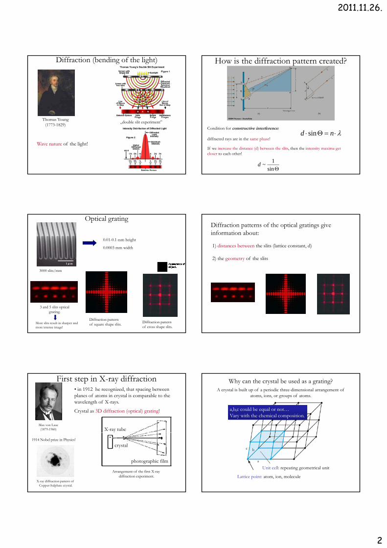
X-ray diffraction methods• Single crystal method (Laue)
- Single crystal (100 μm)- Polychromatic X-ray- Fixed angle* Inorganic solids and macromolecules(pl. proteins, DNA) atomic structure
• Rotating crystal method- Single crystal (100 μm)- Monochromatic X-ray
V i bl l- Variable angle• Powder diffraction
- Polycrystal- Monochromatic X-ray- every possible crystalline orientationis represented in a powdered sample
* Identify unknown crystal samplesInternational Centre for Diffraction Data (ICDD)• Fiber diffraction method
Structure of DNA
Diffractogramm of Zinc-sulphate
2Θ
Back-reflection Laue methodIn the back-reflection method, the film is placed between the x-ray source and the crystal. The beams which are diffracted in a backward direction are recorded. One side of the cone of Laue reflections is defined by the transmitted beam. The film intersects the cone, with the diffraction spots generally lying on an hyperbola.
Transmission Laue methodIn the transmission Laue method, the film is placed behind the crystal to record beams which are transmitted through the crystal. One side of the cone of Laue reflections is defined by the transmitted beam. The film intersects the cone, with the diffraction spots generally lying on an ellipse.
http://www.matter.org.uk/diffraction/x-ray/laue_method.htm
Single crystal
Continous spectrum of x-raysContinous spectrum of x rays
Fixed angle
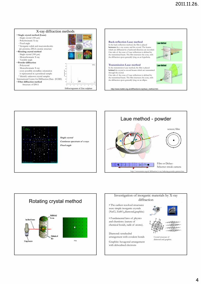
Laue method - powder
screen/film
http://www.matter.org.uk/diffraction/x-ray/indexing_powder_pattern.htm
Film or Debye-Scherrer streak camera
Rotating crystal method
Film
Investigation of inorganic materials by X-ray diffraction
• The earliest resolved structureswere simple inorganic crystals(NaCl, ZnSO4,diamond,graphite)
• Fundamental laws of physicsand chemistry (nature of h i l b d dii f )chemical bonds, radii of atoms).
Crystal structure of diamond and graphite.
Diamond: tetrahedralarrangement with covalent bonds
Graphite: hexagonal arrangementwith delocalised electrons

2011.11.26.
5
Investigation of protein structure by X-raydiffraction.
Study of protein structure by X-ray diffraction
1958 the first atomic structure of a protein
Max Perutz and Sir John Cowdery Kendrew 1962 Nobel Prize in chemistry
Dorothy Crowfoot Hodgkin solvedh X f i li 30
Whale sperm myoglobin
So far ten thousends protein structure weresolved by X-ray diffraction.
The atomic coordinates of the proteins is freely accesible website: www.pdb.org
Designing new drugs.
the X-ray structure of insulin over 30 years!
Insulin
DNA X-ray diffraction pattern
DNA X-ray diffraction pattern
1953
James D. Watson and Francis Crick – DNA model


![EümérnökiMSc Lemorzsolódás v2.pptx [Read-Only])home.mit.bme.hu/~jobbagy/eum/elorehaladas.pdfDebreceni Egyetem 2 Szegedi Tudományegyetem 2 Pécsi Tudományegyetem 2 Szent István](https://static.fdocuments.us/doc/165x107/5e60939fdc36ff4f3d4307c1/emrnkimsc-lemorzsolds-v2pptx-read-onlyhomemitbmehujobbagyeum.jpg)