University of Groningen Diagnosis and imaging of essential ...
135
University of Groningen Diagnosis and imaging of essential and other tremors van der Stouwe, Anna IMPORTANT NOTE: You are advised to consult the publisher's version (publisher's PDF) if you wish to cite from it. Please check the document version below. Document Version Publisher's PDF, also known as Version of record Publication date: 2015 Link to publication in University of Groningen/UMCG research database Citation for published version (APA): van der Stouwe, A. (2015). Diagnosis and imaging of essential and other tremors. University of Groningen. Copyright Other than for strictly personal use, it is not permitted to download or to forward/distribute the text or part of it without the consent of the author(s) and/or copyright holder(s), unless the work is under an open content license (like Creative Commons). The publication may also be distributed here under the terms of Article 25fa of the Dutch Copyright Act, indicated by the “Taverne” license. More information can be found on the University of Groningen website: https://www.rug.nl/library/open-access/self-archiving-pure/taverne- amendment. Take-down policy If you believe that this document breaches copyright please contact us providing details, and we will remove access to the work immediately and investigate your claim. Downloaded from the University of Groningen/UMCG research database (Pure): http://www.rug.nl/research/portal. For technical reasons the number of authors shown on this cover page is limited to 10 maximum. Download date: 15-10-2021
Transcript of University of Groningen Diagnosis and imaging of essential ...

University of Groningen
Diagnosis and imaging of essential and other tremorsvan der Stouwe, Anna
IMPORTANT NOTE: You are advised to consult the publisher's version (publisher's PDF) if you wish to cite fromit. Please check the document version below.
Document VersionPublisher's PDF, also known as Version of record
Publication date:2015
Link to publication in University of Groningen/UMCG research database
Citation for published version (APA):van der Stouwe, A. (2015). Diagnosis and imaging of essential and other tremors. University of Groningen.
CopyrightOther than for strictly personal use, it is not permitted to download or to forward/distribute the text or part of it without the consent of theauthor(s) and/or copyright holder(s), unless the work is under an open content license (like Creative Commons).
The publication may also be distributed here under the terms of Article 25fa of the Dutch Copyright Act, indicated by the “Taverne” license.More information can be found on the University of Groningen website: https://www.rug.nl/library/open-access/self-archiving-pure/taverne-amendment.
Take-down policyIf you believe that this document breaches copyright please contact us providing details, and we will remove access to the work immediatelyand investigate your claim.
Downloaded from the University of Groningen/UMCG research database (Pure): http://www.rug.nl/research/portal. For technical reasons thenumber of authors shown on this cover page is limited to 10 maximum.
Download date: 15-10-2021

Diagnosis anD imagingof essential anD other tremors
A.m.m. vAn der Stouwe

cover
The lady on the cover is Katharine Hepburn, possibly the most famous person known to have had essential tremor. In this picture, we see her in the dressing room on the set of the 1962 film adaptation of Long Day’s Journey Into Night, a play by Eugene O’Neill. Nobel laureate O’Neill had a progressive tremor himself, making it impossible for him to write in the last ten years of his life. Long Day’s Journey Into Night was one of his last works, and although he had given written instructions not to perform the largely autobiographical play until twenty-five years after his death in 1953, it was performed to great critical acclaim and won the Pu-litzer Prize in 1957. Her role in the film adaptation won Katharine Hepburn her ninth Oscar nomination, demonstrating that her tremor did not stop her from having professional success.

A.M.M. van der StouweDiagnosis and imaging of essential and other tremors
Financial support for printing of this thesis by the following institutions is gratefully ac-knowledged:Junior Scientific Masterclass GroningenGraduate School of Medical Sciences / Behavioral and Cognitive NeuroscienceUniversity of Groningen
ISBN: 978-90-367-8046-9 (printed version)ISBN: 978-90-367-8047-6 (electronic version)
Copyright 2015, A.M.M. van der Stouwe
All rights reserved. No part of this publication may be reproduced, stored in a retrieval sys-tem, or transmitted in any form or by any means without express written permission from the author, and, when appropriate, the publisher holding the copyrights of the published articles.
Cover design and layout by J.A.M. de Grefte & A.M.M. van der Stouwe
Printing: Ridderprint BV, www.ridderprint.nl

Diagnosis anD functional imaging of essential anD other tremors
PrOEfschrift
ter verkrijging van de graad van doctor aan de Rijksuniversiteit Groningen
op gezag van de rector magnificus prof. dr. E. Sterken
en volgens besluit van het College voor Promoties.
De openbare verdediging zal plaatsvinden op
woensdag 23 september 2015 om 14.30 uur
door
anna maria magDalena Van Der stouWe
geboren op 13 april 1988te Groningen

PromotoresProf. dr. ir. N.M. MauritsProf. dr. M.A.J. de Koning-Tijssen
BeoordelingscommissieProf. dr. G. DeuschlProf. dr. H.P.H. KremerProf. dr. J.J. van Hilten

table of contents
cHAPter 1 ................................................................................................................................................................................................7Introduction
cHAPter 2 ............................................................................................................................................................................................19How Typical Are ‘Typical’ Tremor Characteristics? Sensitivity And Specificity Of Five Tremor Phenomena
cHAPter 3 ..............................................................................................................................................................................................31Can We Differentiate Postural Tremor Using Intermuscular Coherence And Cumulant Analysis?
cHAPter 4 ...........................................................................................................................................................................................43Bilateral Involvement Of The Cerebellum In Unilaterally Challenged Essential Tremor: An EMG-fMRI sStudy
cHAPter 5 ............................................................................................................................................................................................63The Runaway Cerebellum In Essential Tremor, A Functional And Effective Connectivity Study cHAPter 6 ............................................................................................................................................................................................91Increased cerebellar activations during goal-directed movement in essential tremor: an fMRI study
cHAPter 7..........................................................................................................................................................................................105Limited Correlations Between Clinician-Based And Patient-Based Measures Of Essential Tremor Severity
cHAPter 8 ...........................................................................................................................................................................................113Discussion
nederLAndSe SAmenvAttInG ..................................................................................................................................123Summary in Dutch
dAnKwoord ...............................................................................................................................................................................129Acknowledgements


introDuction
cHAPter 1

chaPter 1
10
This is a thesis on tremor, a movement disor-der in the category of ‘too much movement’, which is called hyperkinesia. In this thesis, two aims are addressed: first, to improve on diagnosis of tremor, and second, to inves-tigate the pathophysiology of one tremor disorder, essential tremor, by means of func-tional neuroimaging.
backgrounD: tyPes of tremor
Tremor is the most common movement dis-order in adults (1). Most patients presenting with trembling of the upper extremities have either got tremor as a symptom of Parkin-son’s disease (PT), essential tremor (ET), enhanced physiological tremor (EPT) or functional tremor (FT). The type of tremor that is best known to the general public is PT, and many tremor patients are initially worried that they may have Parkinson’s disease (PD). Typically, PT starts of as a unilateral, ‘pill-rolling’ rest trem-or (2). It is the presenting symptom of 70% of PD patients (3, 4). Clinically, presence of the other cardinal symptoms (rigidity, dis-turbances in balance and most importantly bradykinesia (5)) increases the likelihood of a diagnosis of PD. By contrast, ET is a tremor disorder without additional neurological symptoms. It has a worldwide prevalence of 0.9%, increasing to 4.6% in the population older than 65 (1). ET is a bilateral tremor occurring during pos-turing and movement of the affected limbs (6). Intention tremor, an increase of tremor amplitude towards the end of a goal-direct-ed movement, is described in about half of ET patients (7, 8). Apart from the arms and hands, the head, voice, and less frequently jaw and legs can also be affected. About half of ET patients report a positive family his-tory (9). Moreover, 50% of patients report a beneficial effect of moderate consumption of alcohol on their tremor (10).Another bilateral tremor is EPT. This tremor
closely resembles the tremor every human being experiences from time to time (11, 12), be it after too much coffee, when hungry, af-ter strenuous physical exercise, during a job interview or a PhD defence. EPT is an ‘exag-gerated’, more constant form of physiological tremor. It is usually mild, and distal. A re-lationship with the circumstances described above may point to the diagnosis. Similar to ET, EPT may be familial. FT completes the list of tremors that con-sultation is most commonly sought for. It was recently argued to define FT on the basis of its clinical appearance as a tremor that is significantly altered by distraction or nonphysiological manoeuvres (including a strong placebo response) and which is clini-cally incongruent with tremor known to be caused by neurological disease (13). FT can be characterized by sudden onset, which is atypical in tremor disorders apart from post-stroke (14). On examination, most patients’ tremor has a combined yet fluctuating pres-ence at rest, during posture, and during ac-tion, together with positive findings that are described in more detail later on (Chapter 2)(15). Apart from the hands and arms, tremor can occur in any body part, including the legs, head or palate (15). Briefly, other more rare tremor disorders that are encountered are dystonic tremor (of the arms and possibly head, together with dys-tonic posturing (16)), cerebellar tremor (usu-ally as a consequence of multiple sclerosis (17)) and Holmes tremor (resulting from mid brain stroke (18)).
clinical Diagnosis of tremor
Distinguishing one type of tremor from another is important, because of the con-sequences for prognosis and treatment. Prognosis can range from generally mild, monosymptomatic and non-progressive in disorders such as EPT, all the way to com-plicated, progressive and life-shortening in a

introDuction
11
1disease such as PD. In terms of management, treatment options differ for different trem-ors, and range from no medication, to dopa-mine and trihexyfenidyl or beta-blockers and anti-epileptics drugs, to deep brain stimula-tion (19, 20)(Table 1.1). In getting to the diagnosis, history taking and clinical examination by a neurologist are of primary importance (21). However, ac-curate clinical diagnosis can be challenged by the fact that not all patients have a clas-sic presentation. In contrast to the typical presentations described above, action tremor can also occur in PT, ET patients may have rest tremor, EPT may be a serious handicap, FT can be fairly consistent. Finally, stress may exaggerate any neurological disorder. The phenomenology of tremor is complex, involving a broad variety of signs and symp-toms. Over the years, large steps have been made in describing the phenomenology of different tremors (22-26), with some tremor disorders presenting with presumably typi-cal signs. However, a lot of the work has de-scribed groups in isolation, or either compar-ing only specific or small groups. In Chapter 2, we will describe our work in determining sensitivity and specificity of five ‘typical’ tremor phenomena in a large and diverse tremor population.
clinical neuroPhysiological Diagnosis of tremor
A neurologist can request clinical neuro-physiology testing to help establish a diag-nosis in more difficult cases. Polymyography, usually combined with accelerometry, assess-es tremor frequency and amplitude in a mul-titude of postures (rest vs action) and during different tasks (for instance, finger-to-nose manoeuvres). The application of clinical neu-rophysiolocal techniques in tremor diagno-sis differs greatly between centres, and is of course dependent on availability but also on a culturally defined attitude towards more
(Germany, The Netherlands) or less (United Kingdom) clinical neurophysiology testing. Polymyography objectifies the presence and consistency of tremor characteristics under various circumstances, and is informative of tremor frequency. These tests are usually of great value (27, 28), however, interpretation of results is not always straightforward. For example, tremor frequencies overlap between different tremor disorders (29), and, as men-tioned previously, the sensitivity and speci-ficity of certain ‘typical’ tremor phenomena are generally poorly known. In Chapter 3, we describe our work aiming to add to the diagnostic power of routine polymyography, by investigating the potential additional di-agnostic value of two advanced EMG meas-ures: intermuscular coherence and cumulant analysis.
essential tremor: a much-DebateD Pheno-tyPe
There have been challenges in defining ET. Traditionally, ET has been used and misused as a ‘container’ diagnosis, gathering together all types of tremor patients that did not fit any particular diagnosis. In 1998, the Move-ment Disorders Society published their first consensus statement on tremor (30). This was followed, in the year 2000, by the criteria for ET by the Tremor Investigation Group: widely used nowadays, and generally known as the TRIG criteria (6). Core criteria are bilateral tremor of the hands and forearms, with no other neurological signs (except a cogwheel phenomenon and head tremor). Secondary criteria are supportive of a diag-nosis of ET, and include duration >3 years, a positive family history and beneficial effect of alcohol. They increase the likelihood of a diagnosis of ET, but are not required. The core criterion of ‘no other neurological signs’ has evolved over time, with recent reports on gait ataxia (31-33), limb ataxia (34, 35), eye movement abnormalities (36, 37), dystonia

chaPter 1
12
(38), and non-motor symptoms (39) in pa-tients that are otherwise diagnosed as ET. Application of the TRIG criteria reduces the chance of mislabelling patients as ET significantly. This is important, not only for patients’ prognosis and management, but also
for research purposes. Being uncritical about including ‘ET patients’ into any scientific study complicates finding commonalities in such a group, independent of investigating phenomenology, pathology, brain activation or genes. Indeed, lack of a secure diagnosis
table 1.1 basic characteristics anD treatment oPtions for Different tremor tyPes
Type of tremor
Frequen-cy (Hz) Rest Posture
Goal-directed
Pharmacological treatment options
Stereotactic treatment options
ET 4 to 11 - ++ +
Propranolol, primidone, (level A recommendation), topiramate, atenolol, sotalol, gabapentin, alprazolam (level B)
Thalamotomy and thalamic DBS (VIM, VL, STA)(level C)
PT 5 to 10 ++ +- +-
Pharmacological treatment options for PD effect tremor < bradykinesia and rigidity (level A)
Lesioning or DBS of STN, GPI, VIM, VL (level C)
EPT 7 to 12 - ++ + Similar to ET (level A-C) -
FT 4 to 10 + + +
Explanation, physiorehabilitation, psychological treatment
-
DT 4 to 10 - ++ + Trihexyphenidyl, propranolol (level C) -
CT 2 to 6 - + ++
Carbamazepine, propranolol, primidone, isoniazid, ondansetron, 4-aminopyride, botulinum toxin (level U)
Lesioning or DBS of VIM (level C)
HT 2 to 5 ++ + ++
Levodopa, clonazepam, clozapine, levetiracetam (level U)
Thalamic DBS (level C)
ET: essential tremor, PT: Parkinsonian tremor, EPT: enhanced physiological tremor, FT: functional tremor. DBS: deep brain stimulation, VIM: ventral intermedius (thalamus), VL: ventrolateral (thalamus), STA: subthalamic area, STN: subthalamic nucleus, GPI: globus pallidus interna. Levels refer to the strength of the recommendation based on the quality of the currently published studies. A: established as effective, level B: probably effective, level C: pos-sibly effective, level U: data inadequate or clniflicting, treatment is unproven with current knowledge.
introDuction
13
1has been proposed as a serious limitation for advancement in all four mentioned areas of ET research (40, 41). Since the publication of the TRIG criteria, it has been suggested to distinguish ‘hereditary ET’; patients fulfilling the TRIG criteria and with a positive family history, ‘sporadic ET’; fulfilling TRIG criteria, but without a fam-ily history, and ‘senile ET’; fulfilling TRIG criteria, but with an age-at-onset later than 65 (40). The reason for this distinction is the fact that the penetrance for hereditary ET appears almost complete by age 65, which would make new-onset tremor at old-age more likely to be due to common age-associ-ated neurodegenerative diseases, rather than to specific ET. Although not all currently ap-ply this distinction, we chose to include only hereditary and sporadic ET patients into the ET study that is described in Chapters 4-7, resulting in a well-defined group.
essential tremor: assessment of tremor seVerity
ET is often called a benign disorder, but moderate and advanced stages can be physi-cally and socially disabling (42-44). To as-sess tremor severity, the Fahn-Tolosa-Marin Tremor Rating Scale (TRS) is a well-known and widely used tool in clinical trials (45, 46). The TRS includes both clinician-based rat-ings in parts A and B, and a patient-based activities-of-daily-life questionnaire in part C. The latter interview takes some time to conduct, and is not always used in clinical trials, nor is it always replaced by another pa-tient-based measure of tremor severity such as subjective visual analogue scale scoring. Intuitively, one would suspect clinician-based and patient-based measures to correlate well; however, because these relations have never been directly investigated. Therefore, the supposition that objective clinical improve-ment correlates with patients’ appraisal of improvement remains unsubstantiated.
essential tremor: unclear PathoPhysiol-ogy
Regarding the pathophysiology of ET, three hypotheses exist that are nonexclusive, but may rather work in unison. In all of them, the cerebellum or cerebellothalamocortical circuit plays an important role (see (47) for a review).
The neurodegeneration hypothesis
The hypothesis that has been under debate for the longest time is the hypothesis that ET is a neurodegenerative disease (48). Clinically, the fact that ET is progressive and associated with age supports this hypothesis (49). Moreover, in some studies (50), ET is associated with an increased risk of develop-ing PD or Alzheimer’s disease: both neuro-degenerative diseases. Three research groups have performed pathology studies, with con-flicting results: most of them, but not all, sug-gesting degeneration of the cerebellum. Cur-rently, the largest pathology study that was done by Louis et al compared 33 ET patients to 21 healthy participants (51). Cerebellar Purkinje cell loss was found in ET patients, with loss of 25% of cells, and 24% of patients had Lewy bodies in the locus coeruleus. Later on, this same research group showed Purkinje axonal swelling (called torpedoes) in ET cerebellums, but not in healthy sub-jects (52). A second group found evidence of cerebellar degeneration in 29% of patients, but did not replicate the finding of Lewy body disease in the locus coeruleus (53). Finally, a third group reported no Purkinje cell loss nor Lewy bodies in ET, albeit in a smaller sample (54). The controversial results in neuropathology may be explained by pa-tient selection biases and lack of standardiza-tion of methods across these studies. Struc-tural brain imaging has also come up with conflicting results: some studies showing de-creased cerebellar white-matter integrity and cerebellar atrophy, while others showed no
chaPter 1
14
abnormalities in brain structure (55). Over-all, there is heterogeneous evidence mainly for cerebellar neurodegeneration.
The gamma-aminobutyric acid hypothesis
A second hypothesis that has been put for-ward is that ET is caused by a disturbance of the gamma-aminobutyric acid (GABA)ergic system. In an overall theory to explain ET pathophysiology, the GABA hypothesis contains multiple steps (56). The first step comprises of cerebellar degeneration with Purkinje cell loss. As a consequence, activity of the GABA system decreases in the deep cerebellar neurons. Therefore, the pacemaker activity of the deep cerebellar neurons is dis-inhibited. As a result, the rhythmic activity of the thalamus and thalamo-cortical circuit increases, ultimately leading to tremor. Clinically, the notion of abnormal GABA function is supported by the fact that some GABAergic drugs are beneficial in ET, al-though not all are (19). Secondly, decreased GABA levels have been found in the cere-bro-spinal fluid of ET patients (57). Other evidence comes from recent positron-emi-sion topography (PET) studies: it was found that 11C-flumazenil binding to GABA-re-ceptors was increased in ET patients com-pared to healthy participants (58), and that binding increased with tremor severity (59). Moreover, in a pathology study, decreased levels of GABA receptors were found in the dentate nucleus of the cerebellum in ET pa-tients compared to PD patients and healthy controls (60). On the whole, studies on GABA all point towards decreased cerebel-lar GABA, however, the hypothesis concerns relatively few studies and not all results have been duplicated.
The oscillating network hypothesis
Historically, a lot of tremor research focused on finding one driving oscillator facilitating tremor based on the fact that some neurons
have oscillating properties at an independent frequency, including in the deep cerebellar nuclei (61-64). The idea of a single oscillator has been challenged by several inconsistent findings, for instance that lesions at several locations in the cerebellothalamocortical cir-cuit can relieve tremor (65), and deep brain stimulation of multiple clusters of the ven-trolateral and ventral intermediate nucleus of the thalamus and subthalamic area can alleviate ET (35, 66, 67). As a result, current research is focused on identifying a network of oscillators, taking into account the con-nectivity and interactions between different parts of the cerebellothalamocortical circuit (68). Good examples of this approach are the recent EEG/MEG/thalamic microelec-trode-EMG coherence studies, which sup-port involvement of a large part of the physi-ological central motor circuit in ET (69-71). Moreover, it was found that although both voluntary and pathological tremor arise from the cerebellothalamocortical circuit, bithalamocortical interactions are only found in pathological tremors (72).
Note that while the in the GABA hypoth-esis cerebellar neurodegeneration is taken as the starting point of its notion (56), in the oscillating network hypothesis it remains open whether the emerging picture of neu-rodegeneration reflects primary degeneration or secondary changes (68). The strongest ad-vocates of the neurodegeneration hypothesis see the structural changes as primary (73). To add to the clarification of ET pathophysiol-ogy, we set up a neuroimaging study in ET patients that is described in Chapters 4-6. We felt that functional magnetic resonance imaging (fMRI) had not yet been used to its full potential: therefore, we used the ad-vanced technique of combining fMRI with EMG. I will briefly discuss these techniques in the following section.

introDuction
15
1techniques emPloyeD in this thesis: emg anD fmri
Two major techniques are used in this thesis. The first one is electromyography (EMG), a technique that is used to measure electrical activity in skeletal muscles: either by placing an electrode intramuscularly (needle EMG) or on the skin above the muscle (surface EMG). The most basic use of EMG is to de-termine whether a muscle is active. In tremor research, one of the applications of EMG is to quantify the extent of tremor (28), es-pecially fluctuations within one recording. In Chapter 4, we employed EMG in this manner: by quantifying the EMG signal, we were able to express changes in tremor inten-sity over time, which we could then correlate with changes in brain activity over time. This way, we were able to link tremor activity di-rectly to brain activity. Brain activity was measured with the second major technique used in this thesis, which is fMRI. Clinically, MRI is widely used to investigate human anatomy and pathology in vivo. MRI makes use of the differences in magnetic properties between various types of tissues in the human body to create an ana-tomical image. Functional MRI adds map-ping of regional brain activity to anatomical (structural) MRI, by applying this principle to hemoglobin. When a brain area becomes more active, there is a local increase in blood that is rich in oxyhemoglobin, containing more oxygen than can be used by the brain tissue. This haemodynamic response results in a local increase of the proportion of oxy-hemoglobin versus deoxyhemoglobin. The difference in magnetic properties of deoxy-genated and oxygenated blood enables the detection of local increases in brain activity. The signal thus obtained is called the Blood Oxygenation Level Dependent (BOLD) signal. Local changes in the BOLD signal can be analysed statically. For example, it is common to compare brain activation in all
voxels during a certain task to activation dur-ing rest; first for individual participants, then in a group of participants. Finally, statistics can then be used to compare brain activation between different groups.In this thesis, we employed several fMRI analysis designs and techniques, such as task-related and ‘EMG’-related (Chapter 4), event-related (Chapter 5), and connectivity analysis (Chapter 6), all to study changes in brain activations in ET patients.
aims
We address two aims in this thesis. In the first part, we aim to improve on the diagnosis of tremor. In Chapter 2, we do this by exam-ining the sensitivity and specificity of several presumably ‘typical’ tremor characteristics. In Chapter 3, we examine the potential value of intermuscular coherence and cumulant analysis as additional diagnostic measures in the clinical neurophysiological assessment of tremor. In the second part of the thesis, we aim to investigate the pathophysiology of ET by means of functional neuroimaging. In Chapter 4, we take the approach of corre-lating fluctuations in tremor severity during scanning with brain activity in ET patients performing a postural task. In Chapter 5, we perform effective and functional connectiv-ity analysis in the same ET population. In Chapter 6, we compare brain activity related to goal-directed movement between ET pa-tients and healthy participants. We investi-gate to what extent clinician-based and pa-tient-based measurements of tremor severity correlate in Chapter 7.
referenceS1. Louis ED, Ferreira JJ. How common is the most common adult movement disorder? update on the worldwide prevalence of essential tremor. Mov Dis-ord. 2010 Apr 15;25(5):534-41.
2. Deuschl G, Papengut F, Hellriegel H. The phenom-

chaPter 1
16
enology of parkinsonian tremor. Parkinsonism Relat Disord. 2012 Jan;18 Suppl 1:S87-9.
3. Hoehn MM, Yahr MD. Parkinsonism: On-set, progression and mortality. Neurology. 1967 May;17(5):427-42.
4. Hughes AJ, Daniel SE, Blankson S, Lees AJ. A clinicopathologic study of 100 cases of parkinson’s dis-ease. Arch Neurol. 1993 Feb;50(2):140-8.
5. Gibb WR, Lees AJ. The relevance of the lewy body to the pathogenesis of idiopathic parkinson’s disease. J Neurol Neurosurg Psychiatry. 1988 Jun;51(6):745-52.
6. Bain P, Brin M, Deuschl G, Elble R, Jankovic J, Findley L, et al. Criteria for the diagnosis of essential tremor. Neurology. 2000;54(11 Suppl 4):S7.
7. Deuschl G, Wenzelburger R, Loffler K, Raethjen J, Stolze H. Essential tremor and cerebellar dysfunction clinical and kinematic analysis of intention tremor. Brain. 2000 Aug;123 ( Pt 8)(Pt 8):1568-80.
8. Louis ED, Frucht SJ, Rios E. Intention tremor in essential tremor: Prevalence and association with disease duration. Mov Disord. 2009 Mar 15;24(4):626-7.
9. Lou JS, Jankovic J. Essential tremor: Clinical cor-relates in 350 patients. Neurology. 1991 Feb;41(2 ( Pt 1)):234-8.
10. Whaley NR, Putzke JD, Baba Y, Wszolek ZK, Uitti RJ. Essential tremor: Phenotypic expression in a clinical cohort. Parkinsonism Relat Disord. 2007 Aug;13(6):333-9.
11. Elble RJ, Randall JE. Mechanistic components of normal hand tremor. Electroencephalogr Clin Neuro-physiol. 1978 Jan;44(1):72-82.
12. Raethjen J, Pawlas F, Lindemann M, Wenzel-burger R, Deuschl G. Determinants of physiologic tremor in a large normal population. Clin Neuro-physiol. 2000 Oct;111(10):1825-37.
13. Edwards MJ, Bhatia KP. Functional (psycho-genic) movement disorders: Merging mind and brain. Lancet Neurol. 2012 Mar;11(3):250-60.
14. Factor SA, Podskalny GD, Molho ES. Psychogen-ic movement disorders: Frequency, clinical profile, and
characteristics. J Neurol Neurosurg Psychiatry. 1995 Oct;59(4):406-12.
15. Edwards MJ, Fotopoulou A, Parees I. Neurobiol-ogy of functional (psychogenic) movement disorders. Curr Opin Neurol. 2013 Aug;26(4):442-7.
16. Deuschl G. Dystonic tremor. Rev Neurol (Paris). 2003 Oct;159(10 Pt 1):900-5.
17. Koch M, Mostert J, Heersema D, De Key-ser J. Tremor in multiple sclerosis. J Neurol. 2007 Feb;254(2):133-45.
18. Gajos A, Bogucki A, Schinwelski M, Soltan W, Rudzinska M, Budrewicz S, et al. The clinical and neuroimaging studies in holmes tremor. Acta Neurol Scand. 2010 Nov;122(5):360-6.
19. Schneider SA, Deuschl G. The treatment of trem-or. Neurotherapeutics. 2014 Jan;11(1):128-38.
20. Jimenez MC, Vingerhoets FJ. Tremor revisited: Treatment of PD tremor. Parkinsonism Relat Dis-ord. 2012 Jan;18 Suppl 1:S93-5.
21. Buijink AW, Contarino MF, Koelman JH, Speel-man JD, van Rootselaar AF. How to tackle tremor - systematic review of the literature and diagnostic work-up. Front Neurol. 2012 Oct 23;3:146.
22. Sternberg EJ, Alcalay RN, Levy OA, Louis ED. Postural and intention tremors: A detailed clinical study of essential tremor vs. parkinson’s disease. Front Neurol. 2013 May 10;4:51.
23. Schwingenschuh P, Katschnig P, Seiler S, Saifee TA, Aguirregomozcorta M, Cordivari C, et al. Mov-ing toward “laboratory-supported” criteria for psycho-genic tremor. Mov Disord. 2011 Dec;26(14):2509-15.
24. McAuley J, Rothwell J. Identification of psy-chogenic, dystonic, and other organic tremors by a coherence entrainment test. Mov Disord. 2004 Mar;19(3):253-67.
25. Kenney C, Diamond A, Mejia N, Davidson A, Hunter C, Jankovic J. Distinguishing psycho-genic and essential tremor. J Neurol Sci. 2007 Dec 15;263(1-2):94-9.
26. Nistico R, Pirritano D, Salsone M, Novellino F, Del Giudice F, Morelli M, et al. Synchronous pattern

introDuction
17
1distinguishes resting tremor associated with essential tremor from rest tremor of parkinson’s disease. Par-kinsonism Relat Disord. 2011 Jan;17(1):30-3.
27. Milanov I. Electromyographic differentiation of tremors. Clin Neurophysiol. 2001 Sep;112(9):1626-32.
28. Gironell A, Kulisevsky J, Pascual-Sedano B, Barbanoj M. Routine neurophysiologic tremor analysis as a diagnostic tool for essential tremor: A prospective study. J Clin Neurophysiol. 2004 Nov-Dec;21(6):446-50.
29. Burne JA, Hayes MW, Fung VS, Yiannikas C, Boljevac D. The contribution of tremor studies to diagnosis of parkinsonian and essential tremor: A statistical evaluation. J Clin Neurosci. 2002 May;9(3):237-42.
30. Deuschl G, Bain P, Brin M. Consensus state-ment of the movement disorder society on tremor. ad hoc scientific committee. Mov Disord. 1998;13 Suppl 3:2-23.
31. Stolze H, Petersen G, Raethjen J, Wenzelburger R, Deuschl G. The gait disorder of advanced essential tremor. Brain. 2001 Nov;124(Pt 11):2278-86.
32. Fasano A, Herzog J, Raethjen J, Rose FE, Mu-thuraman M, Volkmann J, et al. Gait ataxia in es-sential tremor is differentially modulated by thalamic stimulation. Brain. 2010 Dec;133(Pt 12):3635-48.
33. Hoskovcova M, Ulmanova O, Sprdlik O, Sieger T, Novakova J, Jech R, et al. Disorders of balance and gait in essential tremor are associated = with midline tremor and age. Cerebellum. 2013 Feb;12(1):27-34.
34. Louis ED, Gillman A, Boschung S, Hess CW, Yu Q, Pullman SL. High width variability dur-ing spiral drawing: Further evidence of cerebellar dysfunction in essential tremor. Cerebellum. 2012 Dec;11(4):872-9.
35. Herzog J, Hamel W, Wenzelburger R, Potter M, Pinsker MO, Bartussek J, et al. Kinematic analysis of thalamic versus subthalamic neurostimulation in pos-tural and intention tremor. Brain. 2007 Jun;130(Pt 6):1608-25.
36. Helmchen C, Hagenow A, Miesner J, Sprenger A, Rambold H, Wenzelburger R, et al. Eye move-
ment abnormalities in essential tremor may indi-cate cerebellar dysfunction. Brain. 2003 Jun;126(Pt 6):1319-32.
37. Gitchel GT, Wetzel PA, Baron MS. Slowed sac-cades and increased square wave jerks in essential tremor. Tremor Other Hyperkinet Mov (N Y). 2013 Sep 3;3:tre,03-178-4116-2. eCollection 2013.
38. Louis ED, Hernandez N, Alcalay RN, Tirri DJ, Ottman R, Clark LN. Prevalence and features of unreported dystonia in a family study of “pure” essential tremor. Parkinsonism Relat Disord. 2013 Mar;19(3):359-62.
39. Chandran V, Pal PK. Essential tremor: Beyond the motor features. Parkinsonism Relat Disord. 2012 Jun;18(5):407-13.
40. Deuschl G, Elble R. Essential tremor--neuro-degenerative or nondegenerative disease towards a working definition of ET. Mov Disord. 2009 Oct 30;24(14):2033-41.
41. Kuhlenbaumer G, Hopfner F, Deuschl G. Genet-ics of essential tremor: Meta-analysis and review. Neurology. 2014 Mar 18;82(11):1000-7.
42. Troster AI, Pahwa R, Fields JA, Tanner CM, Lyons KE. Quality of life in essential tremor ques-tionnaire (QUEST): Development and initial validation. Parkinsonism Relat Disord. 2005 Sep;11(6):367-73.
43. Chandran V, Pal PK. Quality of life and its de-terminants in essential tremor. Parkinsonism Relat Disord. 2013 Jan;19(1):62-5.
44. Cullinane PW, Browne PJ, Leahy TK, McGov-ern EM, Counihan TJ. Tremor severity is a poor predictor of social disability in patients with essential tremor. Parkinsonism Relat Disord. 2014 Sep 16.
45. Clinical rating scale for tremor. In: Fahn S, To-losa E, Marin C, editors. Parkinson’s disease and movement disorders. Second edition ed. Baltimore, MD: Williams & Wilkins; 1993. p. 225-234.
46. Elble R, Bain P, Joao Forjaz M, Haubenberg-er D, Testa C, Goetz CG, et al. Task force report: Scales for screening and evaluating tremor: Cri-tique and recommendations. Mov Disord. 2013 Nov;28(13):1793-800.

chaPter 1
18
47. Helmich RC, Toni I, Deuschl G, Bloem BR. The pathophysiology of essential tremor and par-kinson’s tremor. Curr Neurol Neurosci Rep. 2013 Sep;13(9):378,013-0378-8.
48. Critchley M, Greenfield JG. Olivo-pontocerebel-lar atrophy. . 1948;71(4):343,344-364.
49. Louis ED, Gerbin M, Galecki M. Essential tremor 10, 20, 30, 40: Clinical snapshots of the disease by decade of duration. Eur J Neurol. 2013 Jun;20(6):949-54.
50. LaRoia H, Louis ED. Association between es-sential tremor and other neurodegenerative diseases: What is the epidemiological evidence? Neuroepidemi-ology. 2011;37(1):1-10.
51. Louis ED, Faust PL, Vonsattel JP, Honig LS, Rajput A, Robinson CA, et al. Neuropathological changes in essential tremor: 33 cases compared with 21 controls. Brain. 2007 Dec;130(Pt 12):3297-307.
52. Louis ED, Faust PL, Ma KJ, Yu M, Cortes E, Vonsattel JP. Torpedoes in the cerebellar vermis in essential tremor cases vs. controls. Cerebellum. 2011 Jun 8.
53. Shill HA, Adler CH, Sabbagh MN, Connor DJ, Caviness JN, Hentz JG, et al. Pathologic findings in prospectively ascertained essential tremor subjects. Neurology. 2008 Apr 15;70(16 Pt 2):1452-5.
54. Rajput AH, Robinson CA, Rajput ML, Robin-son SL, Rajput A. Essential tremor is not dependent upon cerebellar purkinje cell loss. Parkinsonism Relat Disord. 2012 Jun;18(5):626-8.
55. Passamonti L, Cerasa A, Quattrone A. Neuroim-aging of essential tremor: What is the evidence for cer-ebellar involvement? Tremor Other Hyperkinet Mov (N Y). 2012;2:02,67-421-3. Epub 2012 Sep 17.
56. Gironell A. The GABA hypothesis in essential tremor: Lights and shadows. Tremor Other Hyperki-net Mov (N Y). 2014 Jul 16;4:254.
57. Mally J, Baranyi M, Vizi ES. Change in the concentrations of amino acids in CSF and serum of patients with essential tremor. J Neural Transm. 1996;103(5):555-60.
58. Boecker H, Weindl A, Brooks DJ, Ceballos-Bau-
mann AO, Liedtke C, Miederer M, et al. GABAergic dysfunction in essential tremor: An 11C-flumazenil PET study. J Nucl Med. 2010 Jul;51(7):1030-5.
59. Gironell A, Figueiras FP, Pagonabarraga J, Her-ance JR, Pascual-Sedano B, Trampal C, et al. Gaba and serotonin molecular neuroimaging in essential tremor: A clinical correlation study. Parkinsonism Relat Disord. 2012 Aug;18(7):876-80.
60. Paris-Robidas S, Brochu E, Sintes M, Emond V, Bousquet M, Vandal M, et al. Defective dentate nu-cleus GABA receptors in essential tremor. Brain. 2012 Jan;135(Pt 1):105-16.
61. Mouginot D, Gahwiler BH. Presynaptic GABAB receptors modulate IPSPs evoked in neurons of deep cerebellar nuclei in vitro. J Neurophysiol. 1996 Feb;75(2):894-901.
62. Jahnsen H. Extracellular activation and mem-brane conductances of neurones in the guinea-pig deep cerebellar nuclei in vitro. J Physiol. 1986 Mar;372:149-68.
63. Llinas R, Muhlethaler M. Electrophysiology of guinea-pig cerebellar nuclear cells in the in vitro brain stem-cerebellar preparation. J Physiol. 1988 Oct;404:241-58.
64. Pinault D, Deschenes M. The origin of rhythmic fast subthreshold depolarizations in thalamic relay cells of rats under urethane anaesthesia. Brain Res. 1992 Nov 13;595(2):295-300.
65. Dupuis MJ, Evrard FL, Jacquerye PG, Picard GR, Lermen OG. Disappearance of essential tremor after stroke. Mov Disord. 2010 Dec 15;25(16):2884-7.
66. Pedrosa DJ, Reck C, Florin E, Pauls KA, Maar-ouf M, Wojtecki L, et al. Essential tremor and tremor in parkinson’s disease are associated with distinct ‘tremor clusters’ in the ventral thalamus. Exp Neurol. 2012 Oct;237(2):435-43.
67. Sandvik U, Koskinen LO, Lundquist A, Blomst-edt P. Thalamic and subthalamic deep brain stimula-tion for essential tremor: Where is the optimal target? Neurosurgery. 2012 Apr;70(4):840-6.
68. Raethjen J, Deuschl G. The oscillating central network of essential tremor. Clin Neurophysiol. 2012

introDuction
19
1Jan;123(1):61-4.
69. Raethjen J, Govindan RB, Kopper F, Muth-uraman M, Deuschl G. Cortical involvement in the generation of essential tremor. J Neurophysiol. 2007 May;97(5):3219-28.
70. Schnitzler A, Munks C, Butz M, Timmermann L, Gross J. Synchronized brain network associated with essential tremor as revealed by magnetoencepha-lography. Mov Disord. 2009 Aug 15;24(11):1629-35.
71. Hua SE, Lenz FA. Posture-related oscillations in human cerebellar thalamus in essential tremor are enabled by voluntary motor circuits. J Neurophysiol. 2005 Jan;93(1):117-27.
72. Muthuraman M, Heute U, Arning K, Anwar AR, Elble R, Deuschl G, et al. Oscillating central mo-tor networks in pathological tremors and voluntary movements. what makes the difference? Neuroimage. 2012 Apr 2;60(2):1331-9.
73. Louis ED. Essential tremor: Evolving clinico-pathological concepts in an era of intensive post-mor-tem enquiry. Lancet Neurol. 2010 Jun;9(6):613-22.


Submitted
Department of Neurology and Clinical Neurophysiology, University Medical Center Groningen, Groningen, The Netherlands
A.m.m. vAn der Stouwe, J.w. eLtInG, J.H. vAn der Hoeven, t. vAn LAAr, K.L. LeenderS, n.m. mAurItS, m.A.J. tIJSSen
cHAPter 2
hoW tyPical are ‘tyPical’ tremor characteristics?
sensitiVity anD sPecificity of fiVe tremor Phenomena

chaPter 2
22
abstract
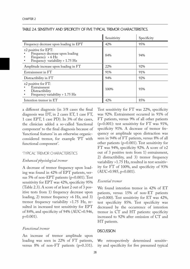
Background: Distinguishing between different tremor disorders can be challenging. Some tremor disorders are thought to have typical tremor characteristics: the current study aims to provide sensitivity and specificity for five ‘typical’ tremor phenomena. Methods: Retrospectively, we examined 210 tremor patients referred for electrophysiological recordings in the period January 2008-January 2014. The final clinical diagnosis was used as gold standard. The first step was to determine whether patients met the neurophysiologi-cal criteria for their type of tremor. Once established, we focused on ‘typical’ characteristics: tremor frequency decrease upon loading (enhanced physiological tremor (EPT)), amplitude increase upon loading, distractibility and entrainment (functional tremor (FT)), and inten-tion tremor (essential tremor (ET)). The prevalence of these phenomena in the ‘typical’ group was compared with the whole group. results: Most patients (87%) concurred with all core clinical neurophysiological criteria for their tremor type. We found a frequency decrease upon loading to be a specific (95%), but not sensitive (42%) test for EPT. Distractibility and entrainment both scored high on sensitivity (92%, 91%) and specificity (94%, 91%) in FT, whereas a tremor amplitude increase was spe-cific (92%), but not sensitive (22%). Intention tremor was a specific finding in ET (85%), but not a sensitive test (45%). Combination of characteristics improved sensitivity.discussion: In this study, we retrospectively determined sensitivity and specificity for five ‘typical’ tremor characteristics. The characteristics proved specific, but few were sensitive. These data on tremor phenomenology will help practicing neurologists to better distinguish between different tremor disorders.

sensitiVity anD sPecificity of fiVe tremor Phenomena
23
2
introDuction
Although tremors are the most common movement disorders, distinguishing between different tremor disorders can be challeng-ing (1, 2). The phenomenology of tremor is complex, involving a broad variety of signs and symptoms. Some tremor disorders seem to have a typical tremor characteristic that points to the diagnosis, but if sensitivity and specificity of these presumed hallmarks are unknown, their significance remains uncer-tain. In the present study we establish how well the clinical tremor diagnosis met the clinical neurophysiological criteria. Fur-thermore, we aim to provide sensitivity and specificity numbers for five ‘typical’ tremor characteristics.
Firstly, a frequency decrease after loading or weighing of the tremulous hand is found in enhanced physiological tremor (EPT). This phenomenon has been long known (3) and is also reported in normal subjects (4). The frequency shift is thought to appear because EPT is considered to be caused partly by mechanical reflex oscillation. This oscillation is dependent of the hand’s resonant frequen-cy and therefore changes with increased in-ertial loading (5). The frequencies of tremor disorders that are considered to be generated by a central oscillator are supposed to be in-variable upon loading (6). However, no stud-ies on the sensitivity and specificity of this phenomenon exist.
Secondly, we aim to investigate three phe-nomena that seem typical of functional tremor (FT): an amplitude increase after loading of the tremulous hand (7), entrain-ment (7-9) and distractibility (10, 11). These characteristics have been described in previ-ous small studies, and are considered to be positive symptoms for the diagnosis of FT. On the other hand, it is known from clini-cal experience that these features occasion-
ally occur in ‘organic’ tremor patients, which raises the question how specific these charac-teristics really are (12).
Lastly, intention tremor, which is tremor increasing during goal-directed movement, is known to occur in essential tremor (ET) (13), but is atypical in most other tremors. A recent study reported intention tremor in 28% of ET patients versus only 4% of Par-kinson’s disease patients (14). We would like to extend these numbers to the general trem-or population.
In this study, we retrospectively determine sensitivity and specificity for typical tremor phenomena, to extend the available data on clinical tremor phenomenology and aid cli-nicians in their neurological examinations and diagnostic process.
methoDs
suBjects
We searched the database of the department of Clinical Neurophysiology of the Univer-sity Medical Center Groningen, a tertiary re-ferral centre, for patients who had undergone a polymyography as part of the diagnostic work-up for upper limb tremor. All subjects had to be >18 years old. The search started at January 1st, 2014, and continued until the three groups of which we intended to test specific tremor characteristics (EPT/ET/FT) each contained 50 subjects ( January, 2008). Patients with other tremor diagnoses were also included to attain a diverse general tremor population as a control group.
clinical diagnosis
As a starting point, we took the most recent clinical diagnosis by the attending neurolo-gist as the gold standard: the final diagnosis after polymyography and possibly imaging or laboratory testing. Patients were not in-

chaPter 2
24
cluded if the neurologist had considerable doubt about the diagnosis: in case of a cur-rent impossibility to differentiate between two disorders. Another exclusion criterion was lack of a final clinical diagnosis, if corre-spondence was unavailable. For each subject, we recorded from their clinical records: age,
sex, primary diagnosis pre-polymyography, and the final clinical diagnosis.
clinical neurophysiology testing
In our centre’s tremor-specific polymyo-graphy recording, tremor is assessed during rest, posture and specific tasks. All our data
table 2.1. clinical neuroPhysiological guiDeline.
Criteria for electrophysiological diagnosis at our hospital Prevalence*
EPT
Core criteria: • Unstable tremor frequency: change >1 Hz upon change of posture
or loading• Predominantly distal tremorSupportive criterion: • High frequency (>7 Hz)
90%
78%
78%
ET
Core criteria: • Bilateral tremor during posture/action• Stable tremor frequency: <2 Hz variation throughout registrationSupportive criterion: • Intention tremor
96%94%
42%
FT
Core criterion: • Unstable tremor frequency: >1 Hz variation or temporal tremor
suppression upon change of posture, mental distraction or entrainment
Supportive criteria: • Increase in tremor amplitude upon loading
94%
22%
PT
Core criteria:• Tremor at rest• Stable tremor frequency: <2 Hz variation throughout registration Supportive criteria: • Increase in tremor amplitude during mental tasks• Frequency between 4 and 7 Hz
95%100%
39%95%
DT
Core criteria:• Signs of dystonia, co-contraction between agonists and antagonists,
overflowSupportive criteria: • Irregular tremor• Proximal tremor• Influence of sensory stimuli
33%
50%n.a.n.a.
CTCore criteria: • Tremor predominantly during action• Intention tremor
100%100%
HT
Core criteria:• Tremor present at rest, posture and action• Low frequency (<4Hz) Supportive criterion: • Intention tremor
100%100%
100%
EPT: enhanced physiological tremor, ET: essential tremor, FT: functional tremor, PT: Parkinsonian tremor, DT: dystonic tremor, CT: cerebellar tremor, HT: Holmes tremor. *: prevalence in the study group with a corresponding final clinical diagnosis; for group information see Results section and Table 2, n.a.: not available, these criteria were not consistently reported.

sensitiVity anD sPecificity of fiVe tremor Phenomena
25
2
is derived from reports of these standard-ized electrophysiological recordings, written by two experienced clinical neurophysiolo-gists ( JWE, JvdH). They based their reports on continuous recordings of accelerometry, EMG, and video. EMG was recorded with Ag/AgCl surface electrodes placed over wrist and elbow flexors and extensors. Accelerom-eters were placed on the dorsal side of both hands. All frequency analyses were based on accelerometry. Data was recorded using BrainRT software (OSG BVBA, Rumst, Belgium). In Table 2.1 we have summarized the criteria used in our clinic for the clinical neurophysi-ological diagnosis (15-17). For each group, we calculated how many patients met these criteria. To assess the influence of polymyography on diagnosis, we compared the clinical pre-pol-ymyography diagnosis, the neurophysiologi-cal diagnosis derived from polymyography, and the final clinical post-polymyography diagnosis to determine how the outcome of the neurophysiological testing affected the diagnosis. In case of a change in diagnosis, we noted the nature of the conversion.
‘typical’ treMor phenoMena
We will describe the five specific tremor char-acteristics of which we aimed to test sensitiv-ity and specificity in more detail. These are routinely assessed: results could be derived from the clinical neurophysiology reports.Loading of the arm was realized by attach-ing one or two 500 g weights, depending on the patient’s strength, to the patient’s wrist. We recorded whether there was a decrease of tremor frequency (>1Hz) upon loading, and/or an increase of tremor amplitude compared to the unloaded condition, as reported by the neurophysiologist.Entrainment was investigated while the most-affected hand was held in the position that evoked maximal tremor. Patients were
instructed to imitate tapping motions with their least-affected hand at the same speed as the laboratory technician, who would vary the frequency between ±1-4 Hz. A positive entrainment test result was scored in case of a notable tremor frequency shift (decrease>1Hz) of the contralateral hand, or temporary tremor suppression. Distractibility was assessed formally with hands held in the position that evoked maxi-mal tremor. Patients were instructed to seri-ally subtract seven from a hundred out loud (100, 93, 86, etc.). Moreover, distractibility was investigated informally during conver-sation and instruction of tasks. We chose to combine these assessments because it is our impression that not all patients are sufficiently distracted by formal yet simple tasks: assess-ment during the rest of the consultation is of equal importance. Distractibility was defined as notable frequency shift (decrease>1Hz) or temporary tremor suppression during formal or informal mental distraction. Intention tremor was assessed with finger-to-nose manoeuvres, where patients were instructed to move the index finger of their outstretched arm to the tip of their nose. If tremor amplitude increased as the patient’s finger approached the nose this was scored as a positive test result.
statistical analysis
Patient and tremor characteristics were com-pared between groups using Chi-square tests for gender and Kruskal-Wallis tests for all continuous, not-normally distributed data in SPSS 20 (SPSS, Chicago, IL). In case of differences between groups, post-hoc testing was performed using Mann-Whitney tests. We compared the frequency of positive test results for each tremor characteristic with Fisher’s exact tests, and calculated sensitivity and specificity for each test. We considered results significant if p<0.05. To place the phenomena in a broader per-

chaPter 2
26
spective and improve discriminative value, we combined tests (presence of tremor phe-nomena) with tremor frequency and fre-quency variability. In case of multiple sig-nificantly different tests for one diagnosis versus all others we investigated combina-tions. Cut-off values for tremor frequency and variability were first estimated based on visual inspection, and we calculated ROC-curves for frequencies between 6.0-7.0 Hz and frequency variability between 1.25-2.0 Hz at 0.25 Hz intervals: the combinations with the largest area under the ROC-curve (AUC), reflecting the highest discriminative value, are reported.
results
patient characteristics
Two hundred-ten patients were included in this study (Table 2.2). Patients had a diag-nosis of EPT (n=50), ET (n=50), FT (n=50), Parkinsonian tremor (PT, n=41), dystonic tremor (n=7), cerebellar tremor (CT, mostly MS-related, n=8) or Holmes or rubral tremor (HT, n=4). Gender distribution did not dif-
fer between groups. There was an age differ-ence (p<0.001): EPT patients were younger than ET, FT and PT patients (all: p<0.001). Moreover, ET patients were older than FT (p<0.001) and PT patients (p=0.006).
clinical neurophysiology
The final clinical diagnosis met with all (87%) or at least one (92%) of our core neu-rophysiological criteria in most cases. The supportive criteria were met less frequently (see Table 2.1). Median tremor frequency was 8.2 Hz in EPT patients, 5.8 Hz in ET patients, 5.3 Hz in FT patients and 5.4 Hz in PT patients (Table 2.2, Figure 2.1). There was a difference between patient groups (p<0.001): tremor frequency was higher in EPT compared to ET, FT and PT (all: p<0.001). Frequency variability was different between groups (p<0.001): frequency vari-ability was higher in EPT (2.5 Hz) and FT (2.3) compared to ET (1.0) and PT patients (0.9) patients (all: p<0.001).
table 2.2. Patient characteristics
N M/F Age Mean frequency Frequency variability
EPT 50 30/20 44 (38)* 8.2 (2.0)* 2.5 (1.4)*
ET 50 29/21 71 (11)* 5.8 (0.8) 1.0 (0.4)
FT 50 27/23 60 (16) 5.3 (1.4) 2.3 (1.4)*
PT 41 24/17 59 (18) 5.4 (1.3) 0.9 (0.3)
DT 7 3/3 51 (37) 5.7 (4.4) 2.0 (1.3)
CT 8 4/4 43 (13) 5.0 (1.9) 1.0 (0.8)
HT 4 1/3 66 (42) 3.3 (0.6) 0.8 (1.0)
All values except gender are displayed as median (interquartile range). EPT: enhanced physiological tremor, ET: essential tremor, FT: functional tremor, PT: parkinsonian tremor, DT: dystonic tremor, CT: cerebellar tremor, HT: Holmes tremor. M/F: Male/Female. *Significant difference, direct post-hoc comparison between EPT, ET, FT and/or PT (see text).

sensitiVity anD sPecificity of fiVe tremor Phenomena
27
2
influence of polyMyography on clinical diagnosis
The diagnosis that topped the differential diagnosis pre-polymyography was con-firmed by the polymyography in 70% of all cases. Contrarily, in 22%, the initial diagnosis changed. In those 45 cases, the incorrect pre-polymyography diagnosis was ET (n=21), EPT (n=10), PT (n=8), DT (n=2), tremor
due to a structural lesion (n=2), neuropathic tremor (n=1), or myoclonus (n=1). These 45 incorrect diagnoses turned into a final clini-cal diagnosis, after polymyography and oc-casionally other testing, of FT (n=18), EPT (n=13), ET (n=8), PT (n=5), and HT (n=1)(Table 2.3). In a small number of patients (5%), the initial pre-polymyography diag-nosis did not change, although the conclu-sion of the polymyography report suggested
Patient no.���������������
Mea
n tr
emor
freq
uenc
y (H
z)
��.�
��.�
�.�
�.�
�.�
�.�
.�
HTCTDTPDFTETEPT
Group
Patient no.���������������
Freq
uenc
y var
iabi
lity (
Hz)
�.�
�.�
�.�
�.�
�.�
�.�
.�
HTCTDTPDFTETEPT
Group
Figure 2.1. Mean tremor frequency (left) and tremor frequency variability (right) in Hz. ET: essential tremor, FT: functional tremor, PT: parkinsonian tremor, DT: dystonic tremor, CT: cerebellar tremor, HT: Holmes tremor.
table 2.3. changes in Diagnosis (n=45)
Final Pre EPT ET PT FT HT Total
EPT - 6 0 4 0 10
ET 11 - 3 6 1 21
PT 2 2 - 4 0 8
DT 0 0 1 1 0 2
Structural lesion 0 0 0 2 0 2
Neuropathic 0 0 1 0 0 1Myoclonus 0 0 0 1 0 1Total 13 8 5 18 1
Cases in which there was a difference between pre-polymyography diagnosis (‘Pre’: rows) and final diagnosis (‘Final’: columns).
chaPter 2
28
a different diagnosis (in 3/8 cases the final diagnosis was DT, in 2 cases ET, 1 case FT, 1 case EPT, 1 case PD). In 3% of the cases, the clinician added a so-called ‘functional component’ to the final diagnosis because of ‘functional features’ in an otherwise organic-considered tremor, for example ‘PT with functional component’.
‘typical’ treMor characteristics
Enhanced physiological tremor
A decrease of tremor frequency upon load-ing was found in 42% of EPT patients, ver-sus 5% of non-EPT patients (p<0.001). Test sensitivity for EPT was 42%, specificity 95% (Table 2.3). A score of at least 2 out of 3 pos-itive tests from 1) frequency decrease upon loading, 2) tremor frequency >6 Hz, and 3) tremor frequency variability >1.75 Hz, re-sulted in increased test sensitivity for EPT of 84%, and specificity of 94% (AUC=0.946, p<0.001).
Functional tremor
An increase of tremor amplitude upon loading was seen in 22% of FT patients, versus 8% of non-FT patients (p=0.331).
Test sensitivity for FT was 22%, specificity was 92%. Entrainment occurred in 91% of FT patients, versus 9% of all other patients (p<0.001): test sensitivity for FT was 91%, specificity 91%. A decrease of tremor fre-quency or amplitude upon distraction was seen in 94% of FT patients, versus 8% of all other patients (p<0.001). Test sensitivity for FT was 94%, specificity 92%. A score of ≥2 out of 3 positive tests from 1) entrainment, 2) distractibility, and 3) tremor frequency variability >1.75 Hz, resulted in test sensitiv-ity for FT of 100%, and specificity of 93% (AUC=0.985, p<0.001).
Essential tremor
We found intention tremor in 42% of ET patients, versus 15% of non-ET patients (p=0.000). Test sensitivity for ET was 42%, test specificity 85%. Test specificity was decreased by the occurrence of intention tremor in CT and HT patients: specificity increased to 92% after omission of CT and HT patients.
Discussion
We retrospectively determined sensitiv-ity and specificity for five presumed typical
table 2.4. sensitiVity anD sPecificity of fiVe tyPical tremor characteristics.
Sensitivity Specificity
Frequency decrease upon loading in EPT 42% 95%≥2 positive for EPT: • Frequency decrease upon loading• Frequency > 6 Hz• Frequency variability > 1.75 Hz
84% 94%
Amplitude increase upon loading in FT 22% 92%Entrainment in FT 91% 91%Distractibility in FT 94% 92%≥2 positive for FT: • Entrainment• Distractibility• Frequency variability > 1.75 Hz
100% 93%
Intention tremor in ET 42% 85%

sensitiVity anD sPecificity of fiVe tremor Phenomena
29
2
tremor characteristics, by comparing preva-lence of each phenomenon in 50 patients from the relevant tremor disorders versus patients from a diverse, general tremor popu-lation. First, we detected that in 87% of our patients the final clinical diagnosis concurred with all our core clinical neurophysiological crite-ria. Supportive criteria for different tremor types were met less frequently, underpinning their role as secondary criteria. As some of the used clinical neurophysiological criteria are consensus-based (15), we are pleased to reinforce these parameters here.
The polymyography diagnosis supported the pre-registration clinical tremor diagnosis in the majority of cases, whereas the diagno-sis changed in 22%. It is noteworthy to see what changes in diagnosis were made under the influence of the tremor-specific poly-myograpy. In nearly half the cases where the diagnosis changed the initial diagnosis was ET. Apparently, we are quick to think of ET, which is fitting with ET’s image as an over-diagnosed disorder (1, 2). Another point of interest is that FT was never an incorrect top differential, whereas of the incorrect di-agnoses, 18 out of 45 changed into FT. We conclude that in our tertiary referral centre neurologists are conservative in diagnosing tremor as functional. This is understandable, but also dangerous, as a positive, unambigu-ous diagnosis is key in the treatment of func-tional disorders (21).
Regarding the ‘typical’ tremor phenomena, our findings reveal that a frequency decrease upon loading of the tremulous arm is specific for EPT (95%). However, it is not a sensitive test (42%): lack of a change in frequency is therefore not informative, but if the tremor frequency decreases this points to EPT. To our knowledge, this is the first study to re-port sensitivity and specificity numbers for this test. Sensitivity increases to 84% when
the effect of loading is combined with tremor frequency (>6 Hz) and frequency variability (>1.75 Hz). These results suggest that a scor-ing system of at least 2 positive tests out of 3 for EPT may be diagnostically useful.
Of the phenomena we investigated that are believed to be typical for FT, testing for distractibility was most useful. A noticeable frequency decrease or temporary tremor sup-pression upon distraction occurred in almost all FT patients, making this a very sensi-tive feature (94%), while at the same time the phenomenon was specific for FT (92%). Tremor distractibility has been described before in FT (10) and one study reported a sensitivity for mental distraction by means of a simple calculation task (“serial subtrac-tions of 7”) of 58.3% (11). We report a much higher sensitivity in the current study, prob-ably because we assessed distractibility both formally with the same calculation task and informally throughout the registration. The test for entrainment resulted in similar high sensitivity (91%) and specificity (91%), and is therefore also informative. Again, we report higher numbers than previous studies (7, 8) probably because we applied less formal testing: either true entrainment, a noticeable frequency shift, or temporary tremor sup-pression scored as entrainment. We consider these extended definitions of distractibility and entrainment appropriate because they represent what neurologists want to assess clinically: the influence of mental or motor tasks on the tremor. Finally, testing for tremor amplitude increase upon loading was the least useful test for FT. Overall, the phenomenon was uncommon, and statistically, it did not occur significantly more often in FT than in other tremor dis-orders. Test sensitivity was very low (22%), although specificity was high (92%). Al-though a previous study (7) used a quantified accelerometry measure instead of our visual assessment of video/EMG/accelerometry

chaPter 2
30
recordings, their results for sensitivity and specificity were highly similar: 33% and 92%. In general, we would like to point out that although all FT-tests have a high specificity, none reached 100%. As is known from previ-ous work (7-9,11), ‘functional’ characteristics can occur in otherwise ‘organic’ tremor. In this study, we confirm that distractibility, en-trainment and an increase of tremor ampli-tude after loading can all be seen in organic tremor. It is of course possible that an exist-ing organic tremor is worsened by functional tremor. This was sometimes acknowledged by the neurologist, by adding ‘plus a func-tional component’ to their final diagnosis. Overall, a combination of entrainment, dis-tractibility and tremor frequency variability (>1.75 Hz) was most suited to classify FT patients. Scoring ≥2 positive test results out of 3 resulted in a test sensitivity of 100% and specificity of 93%, increasing the feasibility of diagnosing FT on positive findings in-stead of per exclusionem. This fits well with the current clinical approach of counting the positive rather than the negative symptoms in functional movement disorders (12).
Our data further reveal that intention trem-or occurs in two out of five ET patients, which is in accordance with previous stud-ies (13,18). We extended previous work on prevalence of intention tremor in ET versus PT patients (14) to the general tremor popu-lation, and found that intention tremor oc-curs in 15% of non-ET tremor patients. The feature was most common in CT and HT patients, which is to be expected as intention tremor is a sign of cerebellar disease, and in these disorders the cerebellum or cerebellar outflow-tract is affected (19,20). Omission of CT and HT patients increased test speci-ficity to 92%. Therefore, a positive finger-to-nose test is informative in distinguishing ET from EPT, PT, DT, and FT, but not CT and HT.
There are two potential weaknesses that re-late to our ‘gold standard’: the most recent clinical diagnosis. As the clinical diagnosis is partly based on features of which we set out to test sensitivity and specificity, there is a risk of a circular argument: patients are included in the EPT group because their tremor frequency decreases upon loading, and then we investigate loading as a diag-nostic test for EPT. To test the extent of this potential problem, we performed a sub-analysis on the 70% of patients in whom the primary differential diagnosis was confirmed by the polymyography report, thus excluding changes in diagnosis due to the polymyogra-phy-findings. As sensitivity and specificity of the five characteristics hardly changed in this subgroup, we concluded that the diagnosis circular argument does not play a major role in our findings. Note that the final diagno-sis did not rely solely on the characteristics we investigated, but also takes into account history taking, examination and imaging. Another weakness is that the clinical diag-nosis may not have been correct in all cases. However, as most patients were seen by ex-perienced movement disorders specialists, all underwent a tremor-specific polymyography, and MR- and PET-imaging were performed when indicated, we are confident that the vast majority of cases was assigned to the ap-propriate group. Two final limitations that need to be noted are that patients with an inconclusive diag-nosis were excluded. Finally, distractibility was investigated both informally and for-mally. This increases the sensitivity but may also increase bias.
A strength of this study is that characteristics were tested in a general tremor population, and not only in isolated groups such as ET vs PD. This makes it possible to relate the results to the actual clinical setting of a pa-tient presenting with tremor. These data on tremor phenomenology will help practicing

sensitiVity anD sPecificity of fiVe tremor Phenomena
31
2
neurologists to better distinguish between different tremor disorders.
referenceS1. Jain S, Lo SE, Louis ED. Common misdiagno-sis of a common neurological disorder: How are we misdiagnosing essential tremor? Arch Neurol. 2006 Aug;63(8):1100-4.
2. Schrag A, Munchau A, Bhatia KP, Quinn NP, Marsden CD. Essential tremor: An overdiagnosed condition? J Neurol. 2000 Dec;247(12):955-9.
3. Elble RJ, Randall JE. Mechanistic components of normal hand tremor. Electroencephalogr Clin Neuro-physiol. 1978 Jan;44(1):72-82.
4. Raethjen J, Pawlas F, Lindemann M, Wenzel-burger R, Deuschl G. Determinants of physiologic tremor in a large normal population. Clin Neuro-physiol. 2000 Oct;111(10):1825-37.
5. Elble RJ. Central mechanisms of tremor. J Clin Neurophysiol. 1996 Mar;13(2):133-44.
6. Gironell A, Kulisevsky J, Pascual-Sedano B, Barbanoj M. Routine neurophysiologic tremor analysis as a diagnostic tool for essential tremor: A prospective study. J Clin Neurophysiol. 2004 Nov-Dec;21(6):446-50.
7. Schwingenschuh P, Katschnig P, Seiler S, Saifee TA, Aguirregomozcorta M, Cordivari C, et al. Moving toward “laboratory-supported” criteria for psychogen-ic tremor. Mov Disord. 2011 Dec;26(14):2509-15.
8. McAuley J, Rothwell J. Identification of psy-chogenic, dystonic, and other organic tremors by a coherence entrainment test. Mov Disord. 2004 Mar;19(3):253-67.
9. Zeuner KE, Shoge RO, Goldstein SR, Dambrosia JM, Hallett M. Accelerometry to distinguish psycho-genic from essential or parkinsonian tremor. Neurol-ogy. 2003 Aug 26;61(4):548-50.
10. Koller W, Lang A, Vetere-Overfield B, Findley L, Cleeves L, Factor S, et al. Psychogenic tremors. Neu-rology. 1989 Aug;39(8):1094-9.
11. Kenney C, Diamond A, Mejia N, Davidson A, Hunter C, Jankovic J. Distinguishing psycho-
genic and essential tremor. J Neurol Sci. 2007 Dec 15;263(1-2):94-9.
12. Stone J, Edwards M. Trick or treat? show-ing patients with functional (psychogenic) motor symptoms their physical signs. Neurology. 2012 Jul 17;79(3):282-4.
13. Louis ED, Frucht SJ, Rios E. Intention tremor in essential tremor: Prevalence and association with disease duration. Mov Disord. 2009 Mar 15;24(4):626-7.
14. Sternberg EJ, Alcalay RN, Levy OA, Louis ED. Postural and intention tremors: A detailed clinical study of essential tremor vs. parkinson’s disease. Front Neurol. 2013 May 10;4:51.
15. Deuschl G, Bain P, Brin M. Consensus state-ment of the movement disorder society on tremor. ad hoc scientific committee. Mov Disord. 1998;13 Suppl 3:2-23.
16. Milanov I. Electromyographic differentiation of tremors. Clin Neurophysiol. 2001 Sep;112(9):1626-32.
17. O’Suilleabhain PE, Matsumoto JY. Time-fre-quency analysis of tremors. Brain. 1998 Nov;121 ( Pt 11)(Pt 11):2127-34.
18. Deuschl G, Wenzelburger R, Loffler K, Raethjen J, Stolze H. Essential tremor and cerebellar dysfunc-tion clinical and kinematic analysis of intention tremor. Brain. 2000 Aug;123 ( Pt 8)(Pt 8):1568-80.
19. Koch M, Mostert J, Heersema D, De Key-ser J. Tremor in multiple sclerosis. J Neurol. 2007 Feb;254(2):133-45.
20. Gajos A, Bogucki A, Schinwelski M, Soltan W, Rudzinska M, Budrewicz S, et al. The clinical and neuroimaging studies in holmes tremor. Acta Neurol Scand. 2010 Nov;122(5):360-6.
21. Gelauff JM, Dreissen YE, Tijssen MA, Stone J. Treatment of functional motor disorders. Curr Treat Options Neurol. 2014 Apr;16(4):286,014-0286-5.

1. Department of Neurology and Clinical Neurophysiology, University Medical Center Groningen, Groningen, The Netherlands.
2. Department of Biomedical Engineering, Strathclyde University, Glasgow, United Kingdom.
Clinical Neurophysiology, 2014doi: 10.1016/j.clinph.2014.10.157
A.m.m. vAn der Stouwe1, B.A. conwAy2, J.w. eLtInG1, m.A.J. tIJSSen1, n.m. mAurItS1
can We Differentiate Postural tremor using intermuscular coherence anD cumulant
analysis?
cHAPter 3
chaPter 3
34
abstract
oBjective: To investigate the potential value of two advanced EMG measures as additional diagnostic measures in the polymyographic assessment of postural upper-limb tremor. Methods: We investigated coherence as a measure of dependency between two EMG signals, and cumulant analysis to reveal patterns of synchronicity in EMG activity in muscle pairs. Eighty datasets were analysed retrospectively, obtained from four groups: essential tremor (ET), Parkinson’s disease (PD), enhanced physiological tremor (EPT), and functional tremor (FT). We used strict diagnostic inclusion criteria combining clinical, neurophysiological and imaging informationresults: Intermuscular coherence was highest in the PD group (0.58), intermediate in FT (0.43) and ET (0.40), and weakest in EPT (0.16) (p=0.002). EPT patients could be distin-guished by low coherence: coherence<0.18 in the wrist + elbow extensors differentiates EPT in this sample with a sensitivity of 86% and specificity of 84%. Cumulant analysis showed predominantly alternating activity between wrist and elbow extensor in ET patients, while a more synchronous pattern was predominant in PD, EPT and FT (p=0.008). EMG activity in wrist and elbow flexors tended to be more synchronous in PD (p=0.059).conclusion: Our results suggest that coherence and cumulant analysis may be of additional value in the diagnostic work-up of postural tremor.significance: These additional measures may be helpful in diagnosing difficult tremor cases.

intermuscular coherence anD cumulant analysis in Postural tremor
35
3
introDuction
Although tremors are the most common movement disorders, distinguishing one type of postural tremor from another can be chal-lenging (1). History taking and clinical ex-amination by a movement disorders special-ist are of primary importance. Additionally, a clinician can request polymyography. Unfor-tunately, this general work-up of choice does not always lead to a conclusive diagnosis (2).
In the outpatient clinic, accurate diagnosis can be challenged by the fact that not all patients have a classic presentation. Trem-ors that present as a postural tremor could mainly be essential, enhanced physiological, dystonic, parkinsonian, or functional. How-ever, not all parkinsonian tremors start of as a typical pill-rolling rest tremor, and not all essential tremor is symmetrical, action-induced and with a slight intention compo-nent (3). Neither does every essential tremor patient have a positive family history or a positive response to alcohol (4, 5). Organic tremor patients can present with a story that seems ‘functional’, whereas functional tremor patients might be hard to distract from their symptoms, making their tremor appear or-ganic.
In more difficult cases, a clinician can request polymyography to help establish a diagnosis (6). These tests are usually of great value: for instance, a prospective study by Gironell and colleagues proposed a set of six neurophysi-ological criteria for essential tremor with very high sensitivity and good specificity for this type of tremor (7). However, this has not been done for all types of tremor, and inter-pretation is not always straightforward. For example, although tremor frequency can be of help, the typical frequencies of different types of tremor overlap and as a result fre-quency is not always a distinguishing feature (8). Other tremor characteristics, such as fre-
quency change at loading in enhanced physi-ological tremor, or entrainment in functional tremor (9) are not present in all patients and sensitivity and specificity are generally poorly known.
In the current study, we sought to add to the diagnostic power of routine polymyography, by investigating the potential additional diagnostic value of two advanced EMG measures: coherence and cumulant analysis. Coherence analysis is a method to detect a common input for the generation of two sig-nals, and is therefore relevant for the study of relationships between the activities of trem-ulous muscles (10). Coherence is a normal-ized measure, which takes on a value of 1 in case of absolute dependence, and 0 in case of complete independence between two signals. Applied to two tremulous EMG signals, this implies that high coherence indicates a common drive from a generator mechanism. While coherence analysis provides a measure in the frequency domain, cumulant analysis is informative of the relationship between two signals over time. Applied to two tremu-lous EMG signals, cumulant analysis can be used to assess the timing relations between EMG bursts in pairs of muscles, in a more objective way than by visual inspection of the EMG signals.
Previous studies have investigated intermus-cular coherence (11-13) or muscle activity patterns (11, 14, 15), but generally without direct comparison between commonly en-countered tremor types. In this study, we compared four groups of carefully selected patients with essential tremor (ET), parkin-sonian tremor (PD), enhanced physiological tremor (EPT) and functional tremor (FT). Our aim was to examine whether intermus-cular coherence and cumulant analysis might be of help as additional diagnostic measures in polymyographic assessment of postural tremor.
chaPter 3
36
methoDs
suBjects
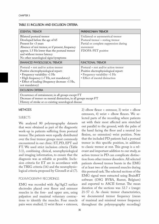
We analyzed 80 polymyography datasets that were obtained as part of the diagnostic work-up in patients suffering from postural tremor. The patients were equally distributed over the four tremor groups most commonly encountered in our clinic: ET, PD, EPT and FT. We used strict inclusion criteria (Table 3.1), combining clinical, neurophysiological and imaging information, to ensure that the diagnosis was as reliable as possible. Inclu-sion criteria for ET are in accordance with the TRIG criteria (16) and the neurophysio-logical criteria proposed by Gironell et al (7).
polyMyography recordings
EMG was recorded with Ag/AgCl surface electrodes placed over flexor and extensor muscles in the fore- and upper arm, using palpation and maximal voluntary contrac-tions to identify the muscles. Four muscle pairs were studied; 1) wrist flexor + extensor,
2) elbow flexor + extensor, 3) wrist + elbow extensors, 4) wrist + elbow flexors. We se-lected parts of the recording where patients sat with their most affected arm stretched out parallel to the ground, with the palm of the hand facing the floor and a neutral (no flexion, no extension) wrist position. Note that the included PD patients had a postural tremor in this specific position, in addition to classic tremor at rest. This group is a rel-evant and important addition to our study, as action tremor in PD can complicate distinc-tion from other tremor disorders. All selected patients showed tremor bursts in the EMG of at least two of the assessed muscles during this postural task. The selected sections of the EMG signal were extracted using BrainRT software (OSG BVBA, Rumst, Belgium) and exported to ASCII format. The mean duration of the sections was 32.7 s (range: 21-57 s). As classic tremor characteristics, we documented tremor frequency (mean of maximal and minimal tremor frequency throughout the polymyography recording)
table 3.1. inclusion anD exclusion criteria.
essential tremor Parkinsonian tremor
Bilateral postural tremorDeveloped before the age of 65Present for >3 yearsAbsence of rest tremor, or if present, frequency approx. 1.5 Hz lower than the postural tremor and without tremor latencyNo other neurological signs/symptoms
Unilateral or asymmetrical tremorPostural tremor + resting tremorPartial or complete suppression during movementFDOPA-PET positive
enhanceD Physiological tremor functional tremor
Postural + rest and/or action tremorPositive electrophysiological report: •Frequencyvariability>1Hz •Highfrequency (>7 Hz, not mandatory) •Effectofloading(frequencydecrease>1Hz,not mandatory)
Postural + rest and/or action tremor Positive electrophysiological report:•Frequency variability >1 Hz•Effect of mental distraction
exclusion criteria
Occurrence of entrainment, in all groups except FTDecrease of tremor on mental distraction, in all groups except FTHistory of stroke or co-existing neurological disease

intermuscular coherence anD cumulant analysis in Postural tremor
37
3
and frequency variability (maximal – mini-mal tremor frequency) for each patient from their clinical neurophysiology reports.
coherence and cuMulant analysis
Data was analyzed in MATLAB R2007a (MathWorks, Natick, MA, USA), using the signal processing toolbox and NeuroSpec 2.0 (www.neurospec.org) to calculate power spectra, coherence, phase and cumulant den-sity for each patient and each of the muscle pairs (17) (Figure 3.1 A-E). Our script is available as supplementary material online.Sample frequency was 1 kHz. A Butterworth 10 Hz highpass filter was used to remove drift and movement artefacts. Next, the data was full-wave rectified, thereby enhancing the firing rate information of the signal (17). Segments were selected to contain 210 data-points (1.024 sec). We did not apply smooth-ing or tapering. The quality of the EMG signals was judged based on the 95% confidence interval of the
individual power spectra for each muscle; signals with a dominant (first) tremor peak smaller than the confidence interval were excluded. Moreover, signals where the am-plitude of the dominant tremor peak was smaller than were excluded because this was assumed to reflect poor signal-to-noise ratio. For each patient and muscle pair, we registered the presence or absence of signifi-cant coherence (as reflected in at least one coherence value above the confidence limit), and coherence values at the dominant tremor peak were used for statistical analysis.Cumulant density plots were classified man-ually for each patient and muscle pair. Plots were classified as 1) a broad positive peak around zero, indicating tremor burst activity that was more in-phase and synchronous, 2) a broad negative peak around zero, indicating tremor burst activity that was more out-of-phase and alternating, and 3) a narrow cen-tral peak close to zero, indicating short-term synchronization with tremor bursts consist-ent with a common presynaptic drive (18-
Figure 3.1. Example of output of the coherence and cumulant analysis for an ET patient. Note the appearance of two peaks in the coherence spectrum (C), at the dominant tremor frequency and its first harmonic. The cumulant (E) shows a broad negative peak around zero for muscle pair 3: wrist + elbow extensors and indicates an alternating pattern of muscle activity, as can be verified in the EMG (F and G).
0 5 10 15 20 25 302.4
2.6
2.8
3
3.2
3.4
Frequency (Hz)
A) FREQUENCY SPECTRUM ELBOW EXTENSOR MUSCLE
0 5 10 15 20 25 304
4.5
5
5.5
Frequency (Hz)
B) FREQUENCY SPECTRUM WRIST EXTENSOR MUSCLE
0 5 10 15 20 25 300
0.5
1
Frequency (Hz)
C) COHERENCE SPECTRUM
0 5 10 15 20 25 30
-2
0
2
Frequency (Hz)
D) PHASE
-500 0 500-400
-200
0
200
400
Lag (ms)
E) CUMULANT DENSITY
21 21.5 22 22.5 23time (s)
F) SAMPLE OF (RECTIFIED) ELBOW EXTENSOR EMG
G) SAMPLE OF (RECTIFIED) WRIST EXTENSOR EMG
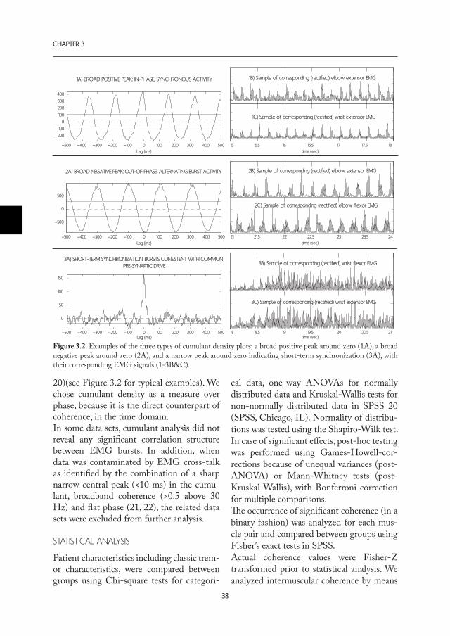
chaPter 3
38
20)(see Figure 3.2 for typical examples). We chose cumulant density as a measure over phase, because it is the direct counterpart of coherence, in the time domain. In some data sets, cumulant analysis did not reveal any significant correlation structure between EMG bursts. In addition, when data was contaminated by EMG cross-talk as identified by the combination of a sharp narrow central peak (<10 ms) in the cumu-lant, broadband coherence (>0.5 above 30 Hz) and flat phase (21, 22), the related data sets were excluded from further analysis.
statistical analysis
Patient characteristics including classic trem-or characteristics, were compared between groups using Chi-square tests for categori-
cal data, one-way ANOVAs for normally distributed data and Kruskal-Wallis tests for non-normally distributed data in SPSS 20 (SPSS, Chicago, IL). Normality of distribu-tions was tested using the Shapiro-Wilk test. In case of significant effects, post-hoc testing was performed using Games-Howell-cor-rections because of unequal variances (post-ANOVA) or Mann-Whitney tests (post-Kruskal-Wallis), with Bonferroni correction for multiple comparisons.The occurrence of significant coherence (in a binary fashion) was analyzed for each mus-cle pair and compared between groups using Fisher’s exact tests in SPSS.Actual coherence values were Fisher-Z transformed prior to statistical analysis. We analyzed intermuscular coherence by means
1A) BROAD POSITIVE PEAK: IN-PHASE, SYNCHRONOUS ACTIVITY
2A) BROAD NEGATIVE PEAK: OUT-OF-PHASE, ALTERNATING BURST ACTIVITY
3A) SHORT-TERM SYNCHRONIZATION: BURSTS CONSISTENT WITH COMMON PRE-SYNAPTIC DRIVE
−500 −400 −300 −200 −100 0 100 200 300 400 500
0
50
100
150
Lag (ms)
−500 −400 −300 −200 −100 0 100 200 300 400 500
−500
0
500
Lag (ms)
−500 −400 −300 −200 −100 0 100 200 300 400 500
−200
−100
0
100
200
300
400
Lag (ms)
2B) Sample of corresponding (rectified) elbow extensor EMG
2C) Sample of corresponding (rectified) elbow flexor EMG
2423.52322.52221.521time (sec)
1B) Sample of corresponding (rectified) elbow extensor EMG
1C) Sample of corresponding (rectified) wrist extensor EMG
1817.51716.51615.515time (sec)
3B) Sample of corresponding (rectified) wrist flexor EMG
3C) Sample of corresponding (rectified) wrist extensor EMG
2120.52019.51918.518time (sec)
Figure 3.2. Examples of the three types of cumulant density plots; a broad positive peak around zero (1A), a broad negative peak around zero (2A), and a narrow peak around zero indicating short-term synchronization (3A), with their corresponding EMG signals (1-3B&C).

intermuscular coherence anD cumulant analysis in Postural tremor
39
3
of a linear mixed effects model with muscle pair and tremor group as factors, instead of using a repeated-measures ANOVA, because mixed-effects models are more robust to missing data (23). Analysis was performed with the lmer and pvals.fnc functions in the lme4 library, as well as the pamer.fnc func-tion in the LMERConvenienceFunction library for the statistical software R (www.r-project.org, version 3.0.1). Statistical analysis of the cumulant density classifications was done by means of Fisher’s exact tests.
results
patient characteristics
No difference in gender distribution was de-tected between groups (Table 3.2). There was a difference in age across groups (p=0.003); ET patients were older than PD (p=0.01), EPT (p=0.008) and FT patients (p=0.005).
classic eMg treMor characteristics
Mean tremor frequency was 5.54 Hz in ET patients, 5.54 Hz in PD patients, 7.84 Hz in EPT patients and 5.42 Hz in FT patients (Table 3.2). There was a difference between patient groups (p=0.001): tremor frequency was higher in the EPT group (p=0.000). Frequency variability, defined as maximal tremor frequency – minimal tremor frequen-cy throughout the polymyography, was on average 1.0 Hz in ET patients, 0.8 Hz in PD
patients, 2.3 Hz in EPT patients and 2.1 Hz in FT patients. There was a significant dif-ference between groups (p=0.000); post-hoc analyses revealed that frequency variability was higher in EPT and FT than in ET and PD patients (p=0.000). We found no differ-ence in tremor amplitude on EMG (defined as mean absolute EMG of the lower extensor in µV) between groups (p=0.104).
advanced eMg Measures: coherence & cuMulant
Overall, significant intermuscular coherence was found in 98% of PD measurements, 97% of FT measurements, 80% of ET measure-ments, and 72% of EPT measurements. Statistically, a significant difference between groups was found in the occurrence of sig-nificant coherence for muscle pair 3: wrist + elbow extensors. For this muscle pair, in-termuscular coherence occurred in all PD patients, 94% of FT patients, 70% of ET patients, and only in 50% of EPT patients (p=0.001). Median intermuscular coherence was 0.58 (IQR 0.37) in the PD group, 0.43 (0.34) in the FT group, 0.40 (0.32) in the ET group, and 0.16 (0.14) in the EPT group (Figure 3.3). Coherence differed between groups (p=0.002). Coherence was higher in PD pa-tients than in ET patients (p=0.049), as well as in EPT patients (p<0.001). Moreover, co-herence was higher in ET patients compared to EPT patients (p=0.041). Finally, coher-
table 3.2. Patient characteristics.
M/F Age Mean freq. Freq. var. Amplitude
ET 11/9 69 (10)* † 5.54 (0.93) 1.0 (0.7)* 33.4 (34.2)*PD 9/10 54 (10)* 5.54 (0.70) 0.8 (0.9)* 32.3 (38.1)*EPT 13/6 43 (36)* 7.84 (1.36) † 2.3 (2.4)* † 25.2 (47.8)*FT 11/10 57 (18)* 5.42 (0.56) 2.1 (2.0)* † 49.1 (59.1)*
ET: essential tremor, PD: parkinsonian tremor, EPT: enhanced physiological tremor, FT: functional tremor. M/F: male/female. Mean frequency in Hz, frequency variability in Hz, amplitude in µV. Mean (standard deviation). *Median (interquartile range). †Significant difference at p<0.05.

chaPter 3
40
ence was larger in FT patients than in EPT patients (p=0.004). To summarize: the rela-tionship between groups for intermuscular coherence was PD > ET > EPT, FT > EPT.
As coherence was highest in PD patients, and lowest in EPT patients, we investi-gated cut-off values for coherence measures in these tremor types, using ROC-curves. Unfortunately, we did not find useful (i.e. with a sensitivity and specificity > 80%) cut-off values for PD. For EPT, we were able to establish two useful cut-off values. First, in the muscle pair wrist flexor + extensor, a co-herence value below 0.35 distinguishes EPT from the other tremors with a sensitivity of 89% and a specificity of 80% (positive pre-dictive value 0.53, negative predictive value 0.97, odds ratio 40). Secondly, in the mus-cle pair wrist + elbow extensors, a coherence value below 0.18 differentiates EPT with a sensitivity of 86% and a specificity of 84% (positive predictive value 0.60, negative pre-dictive value 0.96, odds ratio 32). Moreover, we directly compared ET versus EPT, as this is a clinically relevant com-parison; distinction is frequently asked for in polymyography requests and can be difficult. Cut-off values were useful in the same two muscle pairs. In the muscle pair wrist flexor + extensor, a coherence value above 0.27 in-dicates ET with a sensitivity of 91% and a specificity of 78% (positive predictive value 0.88, negative predictive value 0.83, odds ra-tio 35), and in the muscle pair wrist + elbow extensors a coherence value above 0.18 dif-ferentiates ET from EPT with a sensitivity of 71% and a specificity of 86% (positive pre-dictive value 0.71, negative predictive value 0.86, odds ratio 45).
In the cumulant analysis, results differed be-tween groups for muscle pair 3: wrist + elbow extensors. In ET patients, the cumulant for this muscle pair showed more broad nega-tive peaks, indicating more alternating ac-
tivity. Contrarily, PD, FT and EPT showed more broad positive peaks, signifying more synchronous activity (p=0.008, test sensitiv-ity: 91%, specificity 64%, positive predictive value 0.5, negative predictive value 0.95, odds ratio 6). Moreover, results trended towards significance in muscle pair 4: wrist + elbow flexors in all PD patients showed broad posi-tive peaks, whereas a broad negative peak was found in about half of the other tremors (p=0.059, test sensitivity: 100%, specificity 43%, positive predictive value 0.41, negative predictive value 1.0; odds ratio incalculable). There were no differences between groups for muscles pairs 1 (p=0.379), or 2 (p=0.327).
Discussion
In this study, our aim was to examine the po-tential value of intermuscular coherence and cumulant analysis as additional diagnostic measures in the polymyographic assessment of postural tremor.
Firstly, differences in coherence were found between patient groups. PD patients scored highest (median coherence 0.58), EPT pa-tients lowest (0.16) and FT and ET patients scored intermediate (0.43 and 0.40 respec-tively). We determined cut-off values that may be diagnostically useful for the compari-son of EPT versus other tremors, and EPT versus ET specifically. Moreover, absence of significant coherence was also found sig-nificantly more often in EPT patients. These features may be of additional clinical value, most of all for diagnosis of EPT.
With regard to what is known about tremor pathophysiology, our coherence results are in broad agreement with expectations. As high coherence between different muscles at the tremor frequency indicates a common drive and generator mechanism (10) we expected high coherence in PD, which we indeed found. ET is also considered to be of cen-

intermuscular coherence anD cumulant analysis in Postural tremor
41
3
tral origin (11) and our results also support this. In FT, coherence has only been stud-ied between limbs so far: coherence between two tremulous limbs seems to be a sign of functional origin as it occurs in about half of FT patients but rarely in tremor of organic origin (9, 11, 12). The strong intermuscular, intralimb coherence we found in the current study is a new finding, and points towards a highly organized common drive most likely of central origin in FT. The strong coherence in PD, ET and FT is in contrast with the weak coherence found in EPT. EPT is regarded as a composite of 1) a peripheral mechanical reflex oscillation, which is dependent of the hand’s resonant frequency and therefore changes with in-creased inertial loading, and 2) a centrally driven component in the 7-14 Hz range (10). Under normal circumstances, coher-ence does not occur as a result of mechani-cal reflex oscillation; this only happens under high load conditions (24). The low coherence in our EPT patients corresponds well with a relatively strong role for the peripheral mechanism in EPT, compared to other types of tremor.
Cumulant analysis, the second advanced EMG measure we investigated, resulted in typical characteristics for ET and for PD. First, EMG activity in the wrist and elbow extensors was almost exclusively predomi-nantly alternating in ET patients, whereas a more synchronous, co-contracting pattern was predominant in other tremors. This fea-ture of ET has not been reported previously; it is informative on the phenomenology of ET, regarding the muscular expression of this movement disorder. The high sensitivity of this measure (91%) means that the find-ing of a synchronous instead of an alternat-ing pattern of muscle activity between wrist and elbow extensors in a patient decreases the likelihood of a postural tremor being ET.Secondly, a trend was found regarding the
relation between muscle activity in the wrist and elbow flexors: EMG burst activity was more synchronous between these muscle groups in all PD patients, but a more alter-nating burst activity pattern was found in about half of the other tremors (sensitiv-ity 100%, specificity 43%). This feature was described before in a group of PD patients that showed synchronous muscle activity patterns for the wrist and elbow flexors, and wrist and elbow extensors, as well (11). Those patients were performing a slightly different postural task, where they sat with the hand out-stretched and the arm supported by an armrest. Our findings are in accord with this previous study, and additionally the present study extends these results by comparing muscle activity patterns between tremor groups.
A limitation of this study is that the popula-tion studied was a carefully selected cohort. Diagnosis in all patients was maximally reli-able. This is ideal for an initial study, but sen-sitivity and specificity should ultimately be tested prospectively in an unselected group of patients with postural tremor, before the definite diagnostic value of the advanced EMG measures can be determined. A sec-ond limitation is that we used EMG power as an approximation for tremor amplitude: the optimal approach would be to derive tremor amplitude from accelerometry. However, in this paper, the focus lies on EMG analysis.
For those who are interested in implement-ing coherence and cumulant analysis in their neurophysiology clinic the Matlab script that we used is provided as supplementary mate-rial on the journal’s website. It can be used freely, together with the NeuroSpec library (open source, www.neurospec.org, (17))
In this carefully selected cohort, we inves-tigated whether intermuscular coherence and cumulant analysis might be of help as

chaPter 3
42
additional diagnostic measures in polymyo-graphic assessment of postural tremor. We have shown that intermuscular coherence differs between tremor groups, and were able to identify EPT patients with high specific-ity and sensitivity by low coherence in our study population. Cumulant analysis helped to distinguish ET or PD from other tremors. We conclude that coherence and cumulant analysis may be of additional value in the di-agnostic work-up of postural tremor.
referenceS1. Jain S, Lo SE, Louis ED. Common misdiagno-sis of a common neurological disorder: How are we misdiagnosing essential tremor? Arch Neurol. 2006 Aug;63(8):1100-4.
2. Schrag A, Munchau A, Bhatia KP, Quinn NP, Marsden CD. Essential tremor: An overdiagnosed condition? J Neurol. 2000 Dec;247(12):955-9.
3. Sternberg EJ, Alcalay RN, Levy OA, Louis ED. Postural and intention tremors: A detailed clinical study of essential tremor vs. parkinson’s disease. Front Neurol. 2013 May 10;4:51.
4. Lou JS, Jankovic J. Essential tremor: Clinical cor-relates in 350 patients. Neurology. 1991 Feb;41(2 ( Pt 1)):234-8.
5. Whaley NR, Putzke JD, Baba Y, Wszolek ZK, Uitti RJ. Essential tremor: Phenotypic expression in a clinical cohort. Parkinsonism Relat Disord. 2007 Aug;13(6):333-9.
6. Deuschl G, Krack P, Lauk M, Timmer J. Clinical neurophysiology of tremor. J Clin Neurophysiol. 1996 Mar;13(2):110-21.
7. Gironell A, Kulisevsky J, Pascual-Sedano B, Barbanoj M. Routine neurophysiologic tremor analysis as a diagnostic tool for essential tremor: A prospective study. J Clin Neurophysiol. 2004 Nov-Dec;21(6):446-50.
8. Burne JA, Hayes MW, Fung VS, Yiannikas C, Boljevac D. The contribution of tremor studies to diagnosis of parkinsonian and essential tremor: A statistical evaluation. J Clin Neurosci. 2002
May;9(3):237-42.
9. Schwingenschuh P, Katschnig P, Seiler S, Saifee TA, Aguirregomozcorta M, Cordivari C, et al. Moving toward “laboratory-supported” criteria for psychogen-ic tremor. Mov Disord. 2011 Dec;26(14):2509-15.
10. Elble RJ. Central mechanisms of tremor. J Clin Neurophysiol. 1996 Mar;13(2):133-44.
11. Raethjen J, Lindemann M, Schmaljohann H, Wenzelburger R, Pfister G, Deuschl G. Multiple os-cillators are causing parkinsonian and essential trem-or. Mov Disord. 2000 Jan;15(1):84-94.
12. Raethjen J, Kopper F, Govindan RB, Volkmann J, Deuschl G. Two different pathogenetic mecha-nisms in psychogenic tremor. Neurology. 2004 Sep 14;63(5):812-5.
13. van Rootselaar AF, Maurits NM, Koelman JH, van der Hoeven JH, Bour LJ, Leenders KL, et al. Co-herence analysis differentiates between cortical myo-clonic tremor and essential tremor. Mov Disord. 2006 Feb;21(2):215-22.
14. Milanov I. Electromyographic differentiation of tremors. Clin Neurophysiol. 2001 Sep;112(9):1626-32.
15. Nistico R, Pirritano D, Salsone M, Novellino F, Del Giudice F, Morelli M, et al. Synchronous pattern distinguishes resting tremor associated with essential tremor from rest tremor of parkinson’s disease. Par-kinsonism Relat Disord. 2011 Jan;17(1):30-3.
16. Bain P, Brin M, Deuschl G, Elble R, Jankovic J, Findley L, et al. Criteria for the diagnosis of essential tremor. Neurology. 2000;54(11 Suppl 4):S7.
17. Halliday DM, Rosenberg JR, Amjad AM, Breeze P, Conway BA, Farmer SF. A framework for the analysis of mixed time series/point process data--the-ory and application to the study of physiological trem-or, single motor unit discharges and electromyograms. Prog Biophys Mol Biol. 1995;64(2-3):237-78.
18. Sears TA, Stagg D. Short-term synchronization of intercostal motoneurone activity. J Physiol. 1976 Dec;263(3):357-81.
19. Hansen NL, Hansen S, Christensen LO, Petersen NT, Nielsen JB. Synchronization of lower limb mo-

intermuscular coherence anD cumulant analysis in Postural tremor
43
3
tor unit activity during walking in human subjects. J Neurophysiol. 2001 Sep;86(3):1266-76.
20. Halliday DM, Conway BA, Christensen LO, Hansen NL, Petersen NP, Nielsen JB. Functional coupling of motor units is modulated during walk-ing in human subjects. J Neurophysiol. 2003 Feb;89(2):960-8.
21. Farmer SF, Bremner FD, Halliday DM, Rosen-berg JR, Stephens JA. The frequency content of com-mon synaptic inputs to motoneurones studied during voluntary isometric contraction in man. J Physiol. 1993 Oct;470:127-55.
22. Hansen NL, Conway BA, Halliday DM, Hans-en S, Pyndt HS, Biering-Sorensen F, et al. Reduction of common synaptic drive to ankle dorsiflexor moto-neurons during walking in patients with spinal cord lesion. J Neurophysiol. 2005 Aug;94(2):934-42.
23. Baayen RH. 7.1 modeling data with fixed and random effects. In: Analyzing Linguistic Data: a practical introduction to statistics using R. . 1st ed. Cambridge University Press; 2008.
24. Matthews PB, Muir RB. Comparison of electro-myogram spectra with force spectra during human elbow tremor. J Physiol. 1980 May;302:427-41.


1. Department of Neurology, University Medical Center Groningen, University of Groningen, The Netherlands.
2. Department of Neurology and Clinical Neurophysiology, Academic Medical Center, University of Amsterdam, The Netherlands.
3. Department of Radiology, Academic Medical Center, University of Amsterdam, The Netherlands.
*Shared first authors
Submitted
A.m.m. vAn der Stouwe1*, m.BroerSmA1*, A.w.G. BuIJInK2*, B.m. de JonG1, P.f.c. Groot3, J.d. SPeeLmAn2, m.A.J. tIJSSen1, A.f. vAn rootSeLAAr2, n.m. mAurItS1
bilateral cerebellar actiVation in unilaterally challengeD essential tremor:
an emg-fmri stuDy
cHAPter 4

chaPter 4
46
abstract
oBjective: Essential tremor (ET) is the most common hyperkinetic movement disorder. Pre-vious research into the pathophysiology of ET suggested underlying cerebellar abnormali-ties. In this study, we added electromyography as an index of tremor severity to functional Magnetic Resonance Imaging (EMG-fMRI) to study a homogeneous group of ET patients.Methods: We included 21 propranolol sensitive patients with a definite diagnosis of ET defined by the Tremor Investigation Group. Simultaneous EMG-fMRI recordings were per-formed while patients were off tremor medication. Patients performed unilateral right hand and arm extension, inducing tremor, alternated with relaxation (rest). 21 healthy, age- and gender-matched participants mimicked tremor during right arm extension. EMG power variability at the individual tremor frequency was used as a regressor mathematically inde-pendent of the motor task in the general linear model used for fMRI analysis, to find specific tremor-related activations. results: Task-related activations were found in the classical upper-limb motor network, both for ET patients and healthy participants in motor, premotor and supplementary motor ar-eas. In ET patients, we found tremor-related activations bilaterally in the cerebellum: in left lobules VI and V, and in right lobules V, VI, VIIIa and b, and in the brainstem. In healthy controls we found simulated tremor-related activations in the right cerebellum lobule V. conclusions: Our results expand on previous findings of bilateral cerebellar involvement in et: we have identified specific areas in the bilateral somatomotor regions of the cerebellum. We hypothesize that the cerebellar cortex is disorganized in et, consequently leading to aber-rant cerebellar activity.

essential tremor: an emg-fmri stuDy
47
4
introDuction
Although essential tremor (ET) is the most common hyperkinetic movement disor-der (1), the underlying disease mechanism is poorly understood. ET has long been considered a benign disorder, but recently opinions about the disabling nature of ET changed (2).
Previous work investigating the pathophysi-ology of ET can be divided into pathology and neuroimaging studies (3-5). Post-mor-tem studies have provided interesting but conflicting results, with cerebellar degenera-tion reported in some (6, 7) but not all stud-ies (8). Neuroimaging results in ET are also incongruent, but do provide support for cere-bellar involvement. In structural imaging the most frequent result is cerebellar abnormality in ET although not consistently reported (9). PET experiments and an fMRI study exam-ining motor tasks showed abnormalities in the (bilateral) cerebellum and in some cases in the red nucleus, thalamus and inferior ol-ive (10-13). Although many of the results point towards the cerebellum, overall studies are inconclu-sive. One cause contributing to this diversity in findings may be that ‘ET’ used to be the label for ‘tremor not otherwise specified’, re-sulting in a heterogeneous group with high variability in clinical presentation, response to therapeutic intervention and on etiologic level. In this study, we have attempted to de-fine a homogeneous group of ET patients with a clear diagnosis and a positive response to propranolol (14). Moreover, we wish to improve functional imaging in ET by com-bining EMG and fMRI. This novel approach allows recording tremor simultaneously with brain activity. As cerebellar involvement is a common finding in previous studies, we particularly expect to find cerebellar abnor-malities.
methoDs
suBjects
This study was conducted in two academic hospitals in the Netherlands: the University Medical Center Groningen (UMCG) and the Academic Medical Center in Amster-dam (AMC). Patients who had a definite diagnosis of ET according to criteria defined by the Tremor Investigation Group were in-cluded (15). All patients had bilateral upper limb tremor, an age at onset <65 years, and a disease duration >5 years. A positive fam-ily history was present in most patients (see Table 4.1) but not required for inclusion. Pa-tients had to report a positive subjective re-sponse to propranolol. Patients and healthy controls (age- and gender matched) were all right-handed as assessed by the Annett Handedness scale (16). Another inclusion criterion was a score >25 on the Mini Mental State Examination to ensure proper under-standing of the task. Exclusion criteria were neurological comorbidity (for patients: other than ET), age < 18 and the use of medica-tion (other than for ET) affecting the central nervous system. The study was approved by the medical ethical committees of the Uni-versity Medical Center Groningen and the Academic Medical Center Amsterdam. This study was conducted according to the decla-ration of Helsinki (Seoul, 2008) and all par-ticipants gave written informed consent.
study set-up
Patients quit their medication for a mini-mum of three days before participating in the study. Tremor was assessed off medica-tion using the Fahn-Tolosa-Marin Tremor Rating Scale (TRS)(17) and a visual analog scale (VAS). In all patients, propranolol was washed in again at the end of the study, ac-cording to a personalized schedule.

chaPter 4
48
Motor tasks during eMg-fMri
An fMRI scan was performed, while EMG was recorded simultaneously, off-medication. During scanning patients executed a motor task in which they were instructed to alter-nate 21 periods of 30 seconds rest with 20 periods of 30 seconds right hand and arm extension without supporting the hand and arm. An additional silent reading task was presented during half of all action blocks.
Only the action blocks without the silent reading tasks were analysed in this study, i.e. 10 blocks of 30 seconds. Healthy controls only participated in this part of the study and mimicked a tremor during right arm ex-tension by self-paced wrist flexion extension. Before scanning, participants were instructed and practiced the task outside the scanner. All subjects received visual task instruction using slides.
table 4.1. Patients anD healthy ParticiPants characterists
Patients Age Gender Mean Tremor
frequency (Hz)
Age at onset
(years)
Dura-tion
(years)
Family history
Propranolol use (mg)
VAS-score off
medication
1 21 Male 10 10 11 + 40 5.42 22 Male 7 12 10 - 20 5.23 27 Male 7.5 0 27 - 160 8.74 30 Female 8 15 15 + 20 2.95 32 Female 7 3 29 + 40 66 35 Male 8 7 28 + 80 7.87 46 Male 7.5 5 41 + 80 4.48 47 Male 7 15 32 + 40 69 48 Female 7 10 38 + 120 5.410 53 Female 7.5 28 25 + 30 7.811 53 Male 8 16 37 + 50 8.612 57 Female 7 22 40 + 10 413 62 Female 8.5 5 57 + 100 8.514 63 Male 7 43 20 + 40 3.415 63 Female 7.5 39 24 + 80 7.416 64 Male 6.5 12 52 + 20 417 65 Female 7.5 60 5 + 80 2.718 69 Male 7.5 40 29 + 40 9.219 72 Male 6 10 62 + 320 9.220 74 Male 9 50 24 - 80 6.621 80 Female 6 60 20 + 80 6.9
Mean (SD)
51.6 (17.8)
M: 12F: 9
7.5(0.9)
22 (18.9)
29.8 (15)
72.9(67.8)
6.2(2.1)
VAS: Visual Analogue Scale, range 0-10. SD: Standard deviation. HP: healthy participants.

essential tremor: an emg-fmri stuDy
49
4
table 4.1. (continueD)
HP Age Gender Mean Tremor frequency
(Hz)
1 20 Male 52 22 Male 3.53 27 Male 54 30 Female 55 33 Female 3.56 36 Male 7.57 47 Male 68 49 Male 49 52 Male 6.510 52 Male 411 56 Male 3.512 57 Female 613 59 Female 514 59 Female 415 60 Male 5.516 60 Female 417 62 Male 4.518 68 Male 5.519 68 Male 5.520 72 Female 721 74 Male 6
Mean (SD)
50.6 (16.4)
M: 14F: 7
5.1(1.2)
eMg-fMri acquisition
Images were acquired on a Philips 3-T MR scanner (UMCG: Intera, AMC: Intera and Achieva, Philips, Best, The Netherlands) with SENSE-32 channel (UMCG) and SENSE-16 channel (AMC) head coils. In both centres, T2*-weighted, 3D functional images were obtained using multislice echo planar imaging (EPI) with an echo time (TE) of 30 ms and a repetition time (TR)
of 2000 ms. Per TR, 39 axial slices, with a field of view (FOV) of 224 mm, flip angle of 5° with a 64 X 64 matrix and isotropic voxel size of 3.5 x 3.5 x 3.5 mm were acquired. To provide anatomical information, additional T1-weighted 3D anatomical scans with an axial orientation and a matrix size of 256 x 256 mm were obtained (isotropic voxel size 1 X 1 X 1 mm). EMG was recorded simul-taneously (BrainProducts GmbH, Munich, Germany (UMCG) and MicroMed, Italy (AMC)) from five right arm muscles. To verify absence of left arm movement during the tasks, EMG was recorded from three left arm muscles as well (see Supplementary Ma-terials for more details).
eMg-fMri analysis
EMG data were corrected for scanning ar-tefacts using the MR correction algorithms embedded in Brain Vision Analyser (Im-aging Artefact Reduction method (18); UMCG data) and FARM (fMRI artefact reduction for motion (19); AMC data). Af-ter correction, data was further analyzed in Matlab (Matlab R2007a, Mathworks, Nat-rick, USA) using custom-made scripts. We calculated the frequency spectrum and total spectral power in a 5Hz symmetrical band around the individual (mimicked) tremor peak frequency for every two seconds of EMG data and exported these values as a vector for each right arm muscle. The vectors of the three muscles with the highest total power were averaged. This procedure result-ed in an EMG power vector with one entry for every 2 second scan. This vector was or-thogonalised, element-wise multiplied with the task vector, convolved with the canoni-cal haemodynamic response function (HRF) and scaled by their respective SDs (20). This vector is referred to as residual EMG or r-EMG vector and was used as a regressor in the fMRI design matrix in addition to the task regressor. It represents tremor variation

chaPter 4
50
over time during the motor task, indepen-dently of this motor task. See Supplementary Materials for more details about the analysis.
fMRI data was analysed using SPM8 (Well-come Trust Centre for Neuroimaging, UCL, London, UK; http://www.fil.ion.ucl.ac.uk/spm). Preprocessing consisted of standard re-alignment and coregistration steps. A group-specific anatomic template was created using DARTEL (diffeomorphic anatomical reg-istration through exponentiated lie algebra) for a more precise inter-subject alignment to take age-related changes in anatomy into account (21). Individual functional data was normalized and smoothed using the DAR-TEL template and an 8-mm full-width half maximum (FWHM) Gaussian kernel. To re-duce movement artefacts, the six movement parameters derived from realignment cor-rections were entered as covariates in each individual analysis. Inspection of the EMG was used to correct the task regressor for ac-tual on- and offsets of the motor task. Each single-subject first-level model thus con-sisted of a regressor for the motor task (task-related activations), a residual-EMG regres-sor (r-EMG, tremor related activations) and the six movement regressors. By constructing the design matrix in this manner, variation in activation due to ‘pure’ task execution will be mostly explained by the task regressor, whereas variation in activation due to tremor will be mostly explained by the r-EMG re-gressor, thereby overcoming the problem in traditional designs, where task- and tremor-related activations are mixed.Second level within-group comparisons for the task and r-EMG contrasts, and between-group comparisons for each individual con-trast were made on whole brain level. Ac-tivations were considered significant at a threshold of p<0.05 (FWE corrected) and an extent threshold (k) of 20 voxels. As we hypothesized cerebellar involvement
in ET, we additionally performed an analysis focused on the cerebellum using the Spatial-ly Unbiased Infratentorial Template (SUIT) toolbox (22) (see Supplementary materials). Contrasts were thresholded at voxel level p<0.001, uncorrected, applying a cluster size of 20 voxels.
results
suBject characteristics
A total of 40 ET patients were initially in-cluded in this study. Data of twenty-one ET patients and twenty-one age- and gender-matched healthy controls were analysed. Reasons for exclusion of patients from fur-ther analysis were either too much head-movement during scanning (one patient), insufficient tremor during fMRI data collec-tion (16 patients), failure of equipment dur-ing scanning (one patient) or incorrect nor-malisation to the DARTEL template (one patient). Analyzed ET patients (12 male) had a mean age of 51.6 (SD 17.8) years and mean disease duration of 29.8 (SD 15) years. See Table 4.1 for characteristics of patients and healthy controls. Healthy controls (14 male) had a mean age of 50.6 (SD 16.4) years. Age and gender did indeed not differ between the analysed groups (p=0.86 and p=0.35, respectively). Patients had a mean TRS score of 25.7 (SD 10.8) and a mean VAS score of 6.2 (SD 2.1) off medication. No left arm movement was seen in the EMG signal. Head movement during scanning did not differ between ET patients and healthy controls (see Supplementary Material).
task-related activations, whole Brain
Within-group results
For ET patients, task-related activations (task regressor) were found in the left mo-tor- and premotor cortex, the supplementary motor area (SMA) and the right cerebellum lobules IV and V. Additional activations were

essential tremor: an emg-fmri stuDy
51
4
table 4.2. results for task-relateD actiVations (task regressor)
contrast Voxels (k) area right/left t-Value xa ya za
ET patients1
Cerebrum 48 Parietal sup L 7.54 -26 -44 6628 Primary somatosensory
cortexR 7.30 20 -30 60
3156 Premotor cortex L 11.21 -30 -14 54sc SMA M 10.76 -6 -12 54sc Frontal sup R 10.71 18 0 60636 Supramarginal gyrus R 9.14 56 -36 40sc Supramarginal gyrus R 9.12 54 -24 32sc Supramarginal gyrus R 7.33 48 -38 44340 Frontal mid R 9.12 38 26 34sc Frontal mid R 7.80 32 46 22sc Frontal mid R 7.40 34 42 30493 Frontal inf, oper R 10.28 56 12 2459 Thalamus R 7.86 20 -14 20
Cerebellum 288 Cerebellum IV, V R 9.65 20 -46 -22Healthy participants1
Cerebrum 4290 Premotor cortex L 15.97 -24 -12 56sc SMA L 15.16 -4 -10 56sc Medial cingulate gyrus L 14.20 -4 -4 4845 Parietal inf L 8.44 -52 -22 4051 Medial cingulate gyrus R 10.18 16 -28 381546 Supramarginal gyrus R 11.15 52 -28 34sc Primary somatosensory
cortexR 11.01 36 -34 50
sc Supramarginal gyrus R 10.53 56 -22 24162 Rolandic oper R 9.04 56 4 18sc Frontal inf, oper R 8.98 54 10 26sc Premotor cortex R 8.80 56 4 34111 Supramarginal gyrus L 8.27 -66 -26 18sc Supramarginal gyrus L 7.98 -52 -26 20sc Supramarginal gyrus L 7.64 -64 -28 28
Cerebellum 1831 Vermis VII R 17.33 4 -64 -24sc Cerebellum IV, V R 15.47 18 -50 -22sc Vermis IV, V R 14.27 6 -62 -12

chaPter 4
52
found in right supramarginal gyrus, frontal areas, primary somatosensory cortex,superior parietal cortex and right thalamus (T>6.49, p<0.05 FWE, k=20, see Table 4.2). In healthy controls, we found task-related activations (task regressor) in the left motor cortex and bilateral premotor cortex, the SMA, and the right cerebellum lobules IV, V, VI and VIII. In addition, activations were observed in the left cerebellum lobule VI, left supramarginal gyrus, the inferior parietal cortex and fron-tal regions (T>6.95, p<0.05 FWE k=20, see Table 4.2).
Between group comparisons
No significant increased activations were de-tected in ET patients when compared with healthy controls. Healthy controls had in-creased activations in the right cerebellum lobule VI and left sensori-motor cortex com-pared to ET patients (both T>5.34, p<0.05 FWE, k=20, see Table 4.2).
treMor-related activations, whole Brain
Within-group results
For ET patients, tremor-related activations (r-EMG regressor) were detected in left cer-ebellum lobule VI and the left motor-, pre-motor and somatosensory cortex. Additional activations were found in the bilateral visual cortex, the middle part of the cingulate gyrus and the right motor cortex (T>6.74, p<0.05 FWE, k=20, see Table 4.3). In healthy con-trols, no significant activations were seen in relation with mimicked tremor (T>7.05, p<0.05 FWE, k=20). Between-group comparisons
Compared to healthy controls, ET patients showed increased activations in the right mo-tor cortex, middle part of the cingulate gyrus and the left somatosensory cortex (T>5.45, p<0.05 FWE, k=20 see Table 4.3). The re-verse contrast (healthy controls > ET pa-tients) was not further investigated because we found no significant mimicked tremor-related activations in healthy controls.
contrast Voxels (k) area right/left t-Value xa ya za
21 Cerebellum VI L 7.62 -28 -58 -26207 Cerebellum VIII R 12.53 24 -58 -50sc Cerebellum VIII R 7.20 14 -70 -50
ET patients> Healthy participants2
No significiant resultsHealthy participants> ET patients2 Cerebrum 37 Primary somatosensory
cortexL 7.33 -38 -30 56
Cerebellum 133 Cerebellum VI R 7.33 18 -54 -22 ET: essential tremor; sc: same cluster; R: right; L: left; M: midline; a: MNI. 1 Initial voxel-height thresh-old p<0.05 (FWE corrected, extend threshold k=10 voxels). Coordinates refer to the voxels of maxi-mum activation within clusters of significant activation (p<0.05, FWE whole brain cluster-level corrected). 2 Initial voxel-height threshold p<0.001 (uncorrected, extend threshold k=20 voxels). Coordinates refer to the voxels of maximum activation within clusters of significant activation (p<0.05, FWE whole brain cluster-level corrected).
table 4.2. (continueD)

essential tremor: an emg-fmri stuDy
53
4
task-related activations, cereBelluM (suit analysis)
Task-related activations in the cerebellum of ET patients were found in the right lobule V, VI and VIIIa (T>3.58, p<0.001 uncorrected,
see Table 4.4 and Figure 4.1A). Healthy con-trols showed a large cluster of task-related activations in the right lobules V, VI, VIIIa and b (T>3.61, p<0.001 uncorrected, see Ta-ble 4.4 and Figure 4.1CA). ET patients had no increased activations compared to healthy
table 4.3. results for tremor-relateD actiVations (r-emg regressor)
contrast Voxels (k) area r/l t-Value xa ya za
ET patients1
Cerebrum 669 Premotor cortex L 8.38 -30 -20 68sc Supramarginal gyrus L 8.28 -52 -22 36sc Premotor cortex L 8.13 -26 -26 56104 Precuneus R 7.63 6 -40 58sc Primary motor cortex R 7.34 14 -40 50sc Primary motor cortex R 7.25 12 -32 54119 Medial cingulate gyrus L 8.71 -12 -34 46100 Medial cingulate gyrus L 7.72 -2 -4 42sc Medial cingulate gyrus L 7.48 -10 -8 40106 Primary somatosensory cortex L 9.81 -64 -20 16sc Supramarginal gyrus L 6.85 -52 -24 18103 Medial temporal gyrus L 8.38 -30 -28 12794 Primary visual cortex R 8.80 10 -62 6sc Primary visual cortex R 8.58 10 -78 6sc Primary visual cortex L 7.99 -6 -70 674 Associative visual cortex L 9.16 -40 -86 023 Associative visual cortex R 7.77 30 -84 -16
Cerebellum 318 Cerebellum VI L 9.47 -32 -54 -20sc Fusiform gyrus L 8.93 -32 -60 -14sc Cerebellum VI L 7.94 -18 -74 -14
Healthy participants1 No significant resultsET patients> Healthy participants1
Cerebrum 92 Primary motor cortex R 6.47 10 -32 52123 Medial cingulate gyrus L 6.35 -10 -40 5224 Primary somatosensory cortex L 6.00 -48 -24 52
Healthy controls> ET patients1 No significant results
ET: essential tremor; sc: same cluster; R: right; L: left; M: midline; a: MNI . 1 Initial voxel-height threshold p<0.001 (uncorrected, extend threshold k=20 voxels). Coordinates refer to the voxels of maximum activation within clusters of significant activation (p<0.05, FWE whole brain cluster-level corrected).

chaPter 4
54
table 4.4. results for suit-analysis, cerebellum
contrast Voxels (k) area r/l t-Value xa ya za
Task-related1
ET patients 1579 Lobule V R 8.57 20 -46 -21sc Lobule V R 7.80 4 -62 -23sc Lobule VI R 6.80 28 -48 -2922 Lobule IX L 4.83 -4 -50 -2962 Lobule VIIIa R 5.23 28 -48 -47sc Lobule VIIIa R 4.80 22 -60 -51
Healthy participants 5187 Vermis VI R 17.69 6 -66 -27sc Lobule V R 13.45 16 -54 -1731 Lobule VIIIa L 5.47 -28 -40 -4322 Lobule VIIIa L 5.60 -30 -54 -5343 Lobule VIIIb L 4.62 -12 -48 -57
ET patients> Healthy participants No significant resultsHealthy participants> ET patients
448 Lobule V R 6.74 18 -52 -25sc Vermis VI R 5.38 4 -66 -21sc Lobule V R 4.44 10 -54 -13
Tremor related2 ET patients 1903 Lobule V L 8.48 -26 -46 -15
sc Lobule VI L 7.67 -20 -62 -11sc Brainstem L 6.99 -8 -36 -351071 Lobule VI R 7.23 26 -54 -17sc Lobule VI R 6.41 12 -68 -9sc Lobule VI R 6.28 28 -48 -23602 Crus II R 5.30 4 -76 -37sc Lobule IX L 5.27 -16 -50 -47sc Lobule VIIb L 4.92 -6 -76 -51113 Lobule VIIIa R 4.77 22 -60 -49sc Lobule VIIIb R 4,52 20 -50 -5176 Brainstem M 5.45 8 -16 3sc Brainstem M 4.99 8 -26 -7sc Brainstem M 3.78 4 -34 1303 Brainstem M 5.21 -10 -28 -5sc Brainstem M 4.82 -20 -28 -5sc Brainstem M 4.52 -12 -24 3
Healthy participants 75 Lobule V R 6.29 2 -74 -7

essential tremor: an emg-fmri stuDy
55
4
contrast Voxels (k) area siDe t-Value xa ya za
sc Lobule V R 4.81 0 -62 -1415 Lobule V R 8.32 4 -62 -21sc Lobule V R 5.27 16 -54 -21sc Lobule V R 5.17 12 -56 -11
ET patients> Healthy participants
22 Lobule V L 3.73 -16 -54 -944 Lobule VIIIb L 4.16 -16 -52 -4765 Brainstem M 4.21 -8 -28 -11sc Brainstem M 3.72 -6 -18 -542 Brainstem M 4.22 -8 -34 -27
Healthy participants> ET patients
10 Crus II R 3.81 42 -76 -47
ET: essential tremor; sc: same cluster; R: right; L: left; M: midline; a: MNI 1 p<0.05 (FWE corrected, extend thresh-old k=10 voxels). 2 p<0.001 (uncorrected, extend threshold k=20 voxels)
controls in the cerebellum. Healthy controls showed increased activations in the right lobules V, and VI and in Vermis VI com-pared to ET patients (both T>3.32, p<0.001 uncorrected, see Table 4.4).
treMor-related activations, cereBelluM (suit analysis)
Tremor-related activations in the cerebellum were found in left lobules V, VI,VIIb and IX, right lobules V, VI, VIIIa and b and in the brainstem in ET patients (T>3.58, p<0.001 uncorrected, see Table 4.4 and Figure 4.1B). Healthy controls showed mimicked tremor-related activations in the right cerebellum in lobule V (T>3.61, p<0.001 uncorrected, see Table 4.4 and Figure 4.1D). Increased acti-vations were detected in ET patients when compared with healthy controls in the brain-stem and left lobules V and VIIIb. Healthy controls showed increased activation com-pared to ET patients in the right crus II (T=3.32, p<0.001 uncorrected, see Table 4.4).
Discussion
Using a combination of EMG and fMRI we identified specific, explicable areas in the bi-lateral somatomotor regions of the cerebel-lum associated with tremor. The technique employed here has been used successfully before in a small sample of ET patients, in patients with cortical myoclonic tremor and in Parkinson’s tremor (23-25). By carefully selecting patients with a clear diagnosis of ET, we aimed to identify brain areas correlat-ing specifically with ET. To our knowledge this is the first controlled EMG-fMRI study investigating a large homogeneous group of ET patients.
task-related Brain activations
The participants performed a unilateral (right) arm extension task and, not to our surprise, the classical motor network was activated in both patients and healthy con-trols. These motor network activations were stronger in healthy participants compared to patients, probably because the tremor simulating movement made by the healthy
table 4.4. (continueD)

chaPter 4
56
controls was deliberate and had an observed larger amplitude than the trembling in ET patients.
treMor-related Brain activations
In ET patients, tremor-related activations were found bilaterally in the cerebellum: in left and right lobules VI and V, and ad-ditionally in right lobules VIIIa and b, and in the brainstem. These results expand on earlier findings that the bilateral cerebellum is involved in ET (13,26). Indeed, with our EMG-fMRI approach, we discovered spe-cific, well-defined areas within the cerebel-lum, thus adding detailed information to the
more diffuse localisations that have previ-ously been described. We identified two distinct tremor related ac-tivations in lobules V-VI and in lobule VIII of the right cerebellum, ipsilateral to the right hand and thus particularly implicated in left-hemisphere functions. This particular cerebellar location indeed accurately fits with a previous study on functional connectivity of the cerebral motor hand region which re-vealed somatomotor regions of the cerebel-lum (27). In this study representation in the cerebellum was cross-lateralised and had a double representation, with a strong pri-mary somatomotor representation in lobules V and VI, and a slightly weaker secondary
Figure 3.2. Increased cerebellar activations in essential tremor patients related to the within group comparisons for the task contrast, p<0.05 (FWE corrected, extent k = 10) (A: task-related activations), and activations related to the within group comparisons for the r-EMG contrast, p<0.001 (uncorrected, extent k = 20) (B: tremor-related activations). Results are projected on the SUIT-template (ref ). The color coded bars at the bottom of the figure indicate SPM T-map intensities. The z-coordinates indicate the position of the transversal planes relative to the anterior commisure-posterior commisure plane. L: left hemisphere, R: right hemisphere.
A. ET : TASK-RELATED ACTIVATIONS B. ET: TREMOR-RELATED ACTIVATIONS
y=-46 y=-60
ANATOMY: LEGENDSLobule IV
Lobule V
Lobule VI
Crus I
Crus II
Lobule VIIb
Lobule VIIIa
Lobule VIIIb
Lobule IX
Vermis X
Vermis VI
0 4 8 0 4 8
R
C. HC: TASK-RELATED ACTIVATIONS D. HC: TREMOR-RELATED ACTIVATIONS
z=-17
z=-51
L
z=-17
z=-51
0 7.5 15
y=-46
z=-17
z=-51
y=-60
z=-17
z=-510 3 6

essential tremor: an emg-fmri stuDy
57
4
representation in lobule VIIIb. However, in addition to the cerebellar activity ipsilat-eral to the tremulous hand, activations were also observed in the contralateral cerebellar hemisphere at the same locations as in the ipsilateral cerebellar hemisphere. Activations were evident in the left somatomotor areas, lobules V and VI, and at a lower threshold we found activations in the left lobule VIII as well. Thus, we found increased activations in specific somatomotor areas of the bilateral cerebellum. We like to point out, in this re-spect, that these activations were specifically tremor- rather than movement related, as the brain activation in these areas covaried with tremor intensity over time independently of movement task performance. In healthy con-trols activations covarying with simulated tremor intensity were found in the ipsilateral cerebellum, right lobule V. This corresponds to earlier findings in an EMG-fMRI study examining similar motor tasks in healthy participants (20). Signs of neurodegeneration such as Purkinje cell loss and torpedoes have been reported particularly in the cerebellar cortex in ET (6, 7) with simultaneous remodelling of the cerebellar cortex (28). Also, GABAergic dys-function within the cerebellum has been ob-served, with increased 11C-flunazenil bind-ing to GABA-receptors in the cerebellar cortex, increasing with tremor severity (29). In this light, our findings could be explained by hypothesizing that the cerebellar cor-tex is disorganized, consequently leading to aberrant cerebellar activity. The fact that we found increased instead of decreased cerebel-lar activations may seem counter-intuitive at first, but could be explained by suggest-ing that if the affected cells are deficient and disorganized, they are less efficient, and this inefficiency leads to increased activations.
Activation of the right cerebellum is congru-ent with the right hand and arm extension task and the activated motor cortex in the left
hemisphere. Left cerebellar activation points at functional coherence with cortical regions of the right hemisphere, thus opposite to the executive motor cortex for right arm move-ment. In this respect, it is noteworthy that, at a lower threshold, we indeed found increased activations in the right cerebral cortex in ET patients compared to healthy participants. These activations were located in the anterior parietal and premotor cortex. Together, these areas are known to play a major role in sen-sorimotor transformations underlying e.g. task-related visuomotor control (30) and the organization of stereotypic movement (31). Increased coupling between left cerebellum and right parietal cortex was recently dem-onstrated by functional imaging investigat-ing multisensory processing (32), indepen-dently of right or left arm involvement. One might therefore speculate that ET patients encounter more difficulties in maintaining a steady raised-arm position, which is im-aginable because of their tremor, and that the increased activations in the functionally coherent areas of left cerebellar and right anterior parietal and premotor cortex reflect increased higher-order somatosensory pro-cessing implicated in motor tuning during posture maintenance.
Methodological considerations
In this study, the use of propranolol was one of the inclusion criteria we applied to define a homogeneous group of ET patients. This is one of the many variables that can be cho-sen for patient selection. The advantage of choosing this variable is the future option to compare the current propranolol group with other ET patient groups using different medication. This is a first step at an attempt, as far as we know, to differentiate medica-tion-based subtypes of ET. A common difficulty in fMRI research lies in selecting a suitable task for healthy con-trols that corresponds well with the patients’
chaPter 4
58
task. In this study, a mimicked tremor was used. Consequently, the two groups were ac-tually performing a different task: we asked the tremor patients to maintain their right arm in a postured position, while the healthy controls had to deliberately move their hand. These tasks were chosen to allow optimal dis-tinction of brain networks involved in invol-untary tremor as opposed to compensation or afferent feedback by deliberate, mimicked tremor movements. The mimicked tremor movement overall had a slightly lower peak frequency and had a larger flexion–exten-sion movement of the right wrist compared to the tremor in ET patients. The effect of this behavioural difference can be seen in the task-related activations: the healthy controls showed a more widespread and a higher ac-tivation signal in comparison with the ET patients.
conclusions
In the current study, we used EMG-fMRI to identify brain activations specifically as-sociated with variations in tremor severity in essential tremor patients. By including a homogeneous patient group we were able to identify specific bilateral areas in the cerebel-lum involved in essential tremor: lobules V, VI and VIII.
referenceS1. Louis ED, Ottman R, Hauser WA. How common is the most common adult movement disorder? esti-mates of the prevalence of essential tremor throughout the world. Mov Disord 1998;13:5-10.
2. Louis ED, Okun MS. It is time to remove the ‘be-nign’ from the essential tremor label. Parkinsonism Relat Disord 2011;17:516-520.
3. Raethjen J, Deuschl G. The oscillating central network of essential tremor. Clin Neurophysiol 2012;123:61-64.
4. Gironell A. The GABA hypothesis in essential
tremor: Lights and shadows. Tremor Other Hyperki-net Mov (N Y) 2014;4:254.
5. Louis ED. Re-thinking the biology of essential tremor: From models to morphology. Parkinsonism Relat Disord 2014;20 Suppl 1:S88-93.
6. Louis ED, Faust PL, Vonsattel JP, et al. Neuro-pathological changes in essential tremor: 33 cases com-pared with 21 controls. Brain 2007;130:3297-3307.
7. Shill HA, Adler CH, Sabbagh MN, Connor DJ, Caviness JN, Hentz JG, Beach TG. Pathologic find-ings in prospectively ascertained essential tremor sub-jects. Neurology 2008;70:1452-1455.
8. Rajput AH, Robinson CA, Rajput ML, Robinson SL, Rajput A. Essential tremor is not dependent upon cerebellar purkinje cell loss. Parkinsonism Relat Dis-ord 2012;18:626-628.
9. Sharifi S, Nederveen AJ, Booij J, van Rootselaar AF. Neuroimaging essentials in essential tremor: A systematic review. Neuroimage Clin 2014;5:217-231.
10. Colebatch JG, Findley LJ, Frackowiak RS, Marsden CD, Brooks DJ. Preliminary report: Acti-vation of the cerebellum in essential tremor. Lancet 1990;336:1028-1030.
11. Jenkins IH, Bain PG, Colebatch JG, et al. A posi-tron emission tomography study of essential tremor: Evidence for overactivity of cerebellar connections. Ann Neurol 1993;34:82-90.
12. Wills AJ, Jenkins IH, Thompson PD, Findley LJ, Brooks DJ. Red nuclear and cerebellar but no olivary activation associated with essential tremor: A positron emission tomographic study. Ann Neurol 1994;36:636-642.
13. Bucher SF, Seelos KC, Dodel RC, Reiser M, Oer-tel WH. Activation mapping in essential tremor with functional magnetic resonance imaging. Ann Neurol 1997;41:32-40.
14. Zesiewicz TA, Elble R, Louis ED, et al. Practice parameter: Therapies for essential tremor: Report of the quality standards subcommittee of the american academy of neurology. Neurology 2005;64:2008-2020.

essential tremor: an emg-fmri stuDy
59
4
15. Bain P, Brin M, Deuschl G, et al. Criteria for the diagnosis of essential tremor. Neurology 2000;54:S7.
16. Annett M. A classification of hand preference by association analysis. Br J Psychol 1970;61:303-321.
17. Fahn S. Clinical rating scale for tremor. Parkin-son’s Disease and Movement Disorders 1993:271-280.
18. Allen PJ, Josephs O, Turner R. A method for removing imaging artifact from continuous EEG recorded during functional MRI. Neuroimage 2000;12:230-239.
19. van der Meer JN, Tijssen MA, Bour LJ, van Rootselaar AF, Nederveen AJ. Robust EMG-fMRI artifact reduction for motion (FARM). Clin Neuro-physiol 2010;121:766-776.
20. van Rootselaar AF, Renken R, de Jong BM, Hoogduin JM, Tijssen MA, Maurits NM. fMRI analysis for motor paradigms using EMG-based designs: A validation study. Hum Brain Mapp 2007;28:1117-1127.
21. Ashburner J. A fast diffeomorphic image registra-tion algorithm. Neuroimage 2007;38:95-113.
22. Diedrichsen J. A spatially unbiased atlas template of the human cerebellum. Neuroimage 2006;33:127-138.
23. van Rootselaar AF, Maurits NM, Renken R, Koelman JH, Hoogduin JM, Leenders KL, Tijssen MA. Simultaneous EMG-functional MRI record-ings can directly relate hyperkinetic movements to brain activity. Hum Brain Mapp 2008;29:1430-1441.
24. Helmich RC, Janssen MJ, Oyen WJ, Bloem BR, Toni I. Pallidal dysfunction drives a cerebellotha-lamic circuit into parkinson tremor. Ann Neurol 2011;69:269-281.
25. Contarino MF, Groot PF, van der Meer JN, et al. Is there a role for combined EMG-fMRI in ex-ploring the pathophysiology of essential tremor and improving functional neurosurgery? PLoS One 2012;7:e46234.
26. Buijink AWG, Broersma M, van der Stouwe AMM, et al. Rhythmic finger tapping reveals cerebel-
lar dysfunction in essential tremor
parkinsonism & related disorders. Parkinsonism & related disorders Accepted.
27. Buckner RL, Krienen FM, Castellanos A, Diaz JC, Yeo BT. The organization of the human cerebel-lum estimated by intrinsic functional connectivity. J Neurophysiol 2011;106:2322-2345.
28. Lin CY, Louis ED, Faust PL, Koeppen AH, Von-sattel JP, Kuo SH. Abnormal climbing fibre-purkinje cell synaptic connections in the essential tremor cer-ebellum. Brain 2014;137:3149-3159.
29. Gironell A, Figueiras FP, Pagonabarraga J, Her-ance JR, Pascual-Sedano B, Trampal C, Gispert JD. Gaba and serotonin molecular neuroimaging in es-sential tremor: A clinical correlation study. Parkin-sonism Relat Disord 2012;18:876-880.
30. Grafton ST, Fagg AH, Woods RP, Arbib MA. Functional anatomy of pointing and grasping in hu-mans. Cereb Cortex 1996;6:226-237.
31. de Jong BM, Leenders KL, Paans AM. Right parieto-premotor activation related to limb-in-dependent antiphase movement. Cereb Cortex 2002;12:1213-1217.
32. Hagura N, Oouchida Y, Aramaki Y, Okada T, Matsumura M, Sadato N, Naito E. Visuokinesthetic perception of hand movement is mediated by cerebro-cerebellar interaction between the left cerebellum and right parietal cortex. Cereb Cortex 2009;19:176-186.

chaPter 4
60
suPPlementary materials
1. Study set-up2. EMG-fMRI acquisition3. EMG-fMRI analysis4. SUIT analysis5. Head motion
1. study set-up
Patients quit their medication for a mini-mum of three days before participating in the study. Tremor was assessed off medica-tion using the Fahn-Tolosa-Marin Tremor Rating Scale (TRS) and a visual analog scale (VAS). The TRS is composed of three parts. Part A consists of assessment of tremor am-plitude during rest, posture, movement and finger-to-nose manoeuvres. Part B consists of tremor-inducing tasks, including writ-ing, two standardized Archimedes spirals, a line-drawing task and a water pouring task. In part C the patients rate the limitations they experience in daily life due to tremor. Parts A and B were performed and vide-otaped for both hands, separately. An experi-enced movement disorders specialist ( J.D.S.) blindly determined TRS scores for part A and B. The range of the total TRS (part A, B and C) is 0-88. The VAS subjectively rated tremor severity, with patients marking a 10 cm line ranging from 0 to 10, 0 mean-ing no tremor at all and 10 meaning intoler-able tremor. In all patients, propranolol was washed in again at the end of the study, ac-cording to a personalized schedule.
2. eMg-fMri acquisition
EMG was recorded simultaneously with BrainProducts GmbH, Munich, Germany (UMCG) and MicroMed, Italy (AMC) while scanning. EMG was recorded from five right arm muscles: extensor carpi ulnaris, flexor carpi radialis, extensor carpi radialis longus, flexor carpi ulnaris and first dorsal interosseus. To verify
the absence of left arm movement during the tasks, EMG was recorded from three left arm muscles as well: extensor carpi ulnaris, flexor carpi radialis and first dorsal interos-seus. Pairs of sintered silver/silver-chloride MR-compatible surface EMG electrodes were placed bilaterally above the mentioned muscles. A ground electrode was placed on the left wrist joint. Further EMG recording procedures were similar to the methodology developed in our previous studies.2, 3
3. eMg-fMri analysis
EMG data were corrected for echo planar imaging artefacts using the MR correction algorithms embedded in Brain Vision Ana-lyser (Imaging Artefact Reduction method4; UMCG data) and FARM (fMRI artefact reduction for motion5; AMC data). Af-ter correction, data was further analyzed in Matlab (Matlab R2007a, Mathworks, Nat-rick, USA) using custom-made scripts. For each segment of 2 seconds, corresponding to one scan, the frequency spectrum was calcu-lated using the default fast Fourier transform in Matlab (FFT). The individual frequency at the dominant tremor peak (tremor, mim-icked-tremor) was determined for each pa-tient and healthy control by visual inspection of the segments. Patients without a clear and regular tremor frequency in the EMG dur-ing the task segments were excluded from further analysis. Total spectral power in a 5Hz symmetrical band around the individ-ual (mimicked) tremor peak frequency was exported for each segment and each right arm muscle, resulting in five vectors of the length of the number of scans/segments. The vectors of the three muscles with the high-est total power were averaged. This procedure resulted in an EMG power vector with one entry for every scan. Next, this vector was or-thogonalised with respect to the motor task using Gram-Schmidt orthogonalisation, to subtract the information that is already pre-

essential tremor: an emg-fmri stuDy
61
4
sent in the task vector.3 The orthogonalised EMG vector (referred to as residual EMG or r-EMG vector) now provides a measure of additional EMG relative to the mean EMG value across the task. It represents the varia-tion in tremor severity over time, indepen-dently of movement task. Subsequently, the
r-EMG vector was element-wise multiplied with the task vector to obtain a vector that only has nonzeroes for the r-EMG during task, and zeroes otherwise. Finally, this vec-tor was convolved with the canonical HRF, scaled by its SD and used as a regressor in the fMRI design matrix in addition to the task
EMG
FFT individual peak frequency
Mean EMG (3 muscles) Total spectral power 5Hz band
Gram-Schmidt orthogonalization
Multiply with task vector
Convolve with HRF
Scanner artefact correction
Scale with SD
fMRI
Realignment & coregistration
DARTEL normalization & smooting
Task vector
Adjust onset of task based on EMG
Single subject rst level model Task regressor | rEMG regressor | Movement parameters
Figure S1. Flowchart of the EMG-fMRI Analysis. fMRI: functional magnetic resonance imaging; EMG: electromyography; DARTEL: Diffeomorphic Anatomical Registration using Exponentiated Lie algebra; FFT: fast Fourier transform; HRF: Haemodynamic response function; SD standard deviation.

chaPter 4
62
regressor. 3 See Figure S1 for a flowchart of the EMG analysis.
4. suit analysis
As we hypothesized cerebellar involvement in ET, we additionally performed an analysis focused on the cerebellum using the Spatial-ly Unbiased Infratentorial Template (SUIT) toolbox.6 This toolbox isolates the cerebel-lum and creates a mask. The individual T1 image of the cerebellum was normalized to the SUIT template using nonlinear deforma-tions. The contrast images resulting from the first-level whole-brain analysis were masked with the created cerebellum mask, normal-ized into SUIT atlas space and smoothed with a Gaussian filter of 4-mm FWHM.
5. head Motions during scanning
Given the tremor, it is plausible that the ET patients made more head movements than healthy controls when executing the motor task. To test this we used the scan-by-scan realignment parameters calculated during fMRI preprocessing. We calculated the to-tal range of head motion for translation (x, y
and z direction) and rotation (pitch, roll and yaw) across each session per participant. This showed that head movements during scan-ning did not differ between ET patients and healthy controls, see Table S1.
suppleMentary references
1. Fahn S. Clinical rating scale for tremor. Parkinson’s Disease and Movement Disorders 1993:271-280.
2. van Rootselaar AF, Maurits NM, Renken R, Koelman JH, Hoogduin JM, Leenders KL, Tijssen MA. Simultaneous EMG-functional MRI record-ings can directly relate hyperkinetic movements to brain activity. Hum Brain Mapp 2008;29:1430-1441.
3. van Rootselaar AF, Renken R, de Jong BM, Hoog-duin JM, Tijssen MA, Maurits NM. fMRI analysis for motor paradigms using EMG-based designs: A validation study. Hum Brain Mapp 2007;28:1117-1127.
4. Allen PJ, Josephs O, Turner R. A method for remov-ing imaging artifact from continuous EEG recorded during functional MRI. Neuroimage 2000;12:230-239.
5. van der Meer JN, Tijssen MA, Bour LJ, van Rootselaar AF, Nederveen AJ. Robust EMG-fMRI artifact reduction for motion (FARM). Clin Neuro-physiol 2010;121:766-776.
6. Diedrichsen J. A spatially unbiased atlas template of the human cerebellum. Neuroimage 2006;33:127-138.
table s1. test statistics for heaD motion.
ET mean (SD)
HC mean (SD)
t-test results
Translation x (in mm)
0.82 (0.54)
0.85 (0.43)
t(40)=0.001 p=0.87
Translation y (in mm)
1.09 (0.45)
1.09 (0.76)
t(40)=0.27 p=0.97
Translation z (in mm)
2.19 (1.30)
2.20 (0.84)
t(40)=1.26 p=0.99
Rotation - pitch (in degrees)
0.05 (0.03)
0.04 (0.03)
t(40)=1.11 p=0.60
Rotation - roll (in degrees)
0.02 (0.01)
0.02 (0.02)
t(40)=0.08 p=0.96
Rotation - yaw (in degrees)
0.02 (0.01)
0.02 (0.01)
t(40)=0.16 p=0.61
ET: essential tremor; HC: healthy controls

essential tremor: an emg-fmri stuDy
63
4


cHAPter 5
Accepted for publication in Brain
1. Department of Neurology, University Medical Center Groningen, University of Groningen, The Netherlands.
2. Department of Neurology and Clinical Neurophysiology, Academic Medical Center, University of Amsterdam, The Netherlands.
3. Department of Radiology, Academic Medical Center, University of Amsterdam, The Netherlands.
*Shared first authors
A.m.m. vAn der Stouwe1*, A.w.G. BuIJInK2*, m. BroerSmA1*, S. SHArIfI2, P.f.c. Groot3, J.d. SPeeLmAn2, n.m. mAurItS1, A.f. vAn rootSeLAAr2
motor netWork DisruPtion in essential tremor:
a functional anD effectiVe connectiVity stuDy

chaPter 5
66
abstract
Background: Although involvement of the cerebello-thalamo-cortical network, has often been suggested in essential tremor, the source of oscillatory activity remains largely unknown. To elucidate mechanisms of tremor generation, it is of crucial importance to study the dy-namics within the cerebello-thalamo-cortical network. Using a combination of electromyo-graphy and functional Magnetic Resonance Imaging, it is possible to record the peripheral manifestation of tremor simultaneously with brain activity related to tremor generation. Our first aim is to study the intrinsic activity of regions within the cerebello-thalamo-cortical network using Dynamic Causal Modelling to estimate effective connectivity driven by the concurrently recorded tremor signal. Our second aim is to objectify how the functional integ-rity of the cerebello-thalamo-cortical network is affected in essential tremor. Methods: We investigated the functional connectivity between cerebellar and cortical mo-tor regions showing activations during a motor task. Twenty-two essential tremor patients and 22 healthy controls were analysed. For the effective connectivity analysis, a network of tremor-signal related regions was constructed, consisting of the left primary motor cortex, premotor cortex, supplementary motor area, left thalamus, and right cerebellar motor regions lobule V and lobule VIII. A measure of variation in tremor severity over time, derived from the electromyogram, was included as modulatory input on intrinsic connections and on the extrinsic cerebello-thalamic connections, giving a total of 128 models. Bayesian Model Selec-tion and Random effects Bayesian Model Averaging were used. Separate seed-based func-tional connectivity analyses for the left primary motor cortex, left supplementary motor area and right cerebellar lobules IV, V, VI and VIII were performed. results: We report two novel findings that support an important role for the cerebellar system in the pathophysiology of essential tremor. First, in the effective connectivity analysis, tremor variation during the motor task has an excitatory effect on both the extrinsic connection from cerebellar lobule V to the thalamus, and the intrinsic activity of cerebellar lobule V and thala-mus. Second, the functional integrity of the motor network is affected in essential tremor, with a decrease in functional connectivity between cortical and cerebellar motor regions. This decrease in functional connectivity, related to the motor task, correlates with an increase in clinical tremor severity. Interestingly, increased functional connectivity between right cerebel-lar lobules I-IV and the left thalamus correlates with an increase in clinical tremor severity. conclusion: our findings suggest that cerebello-dentato-thalamic activity and cerebello-cortical connectivity is disturbed in essential tremor, supporting previous evidence of func-tional cerebellar changes in essential tremor.

essential tremor: a functional anD effectiVe connectiVity stuDy
67
5
introDuction
Essential tremor is one of the most common neurological disorders, and is characterised by a progressive postural and kinetic tremor (1). Evidence of alleviation of tremor follow-ing thalamic deep brain stimulation, and after stroke anywhere in the cerebello-thalamo-cortical network, prompted the hypothesis of essential tremor as an ‘oscillating network’ disorder (2, 3). Evidence is accumulating that the cerebellum plays an important role in the pathophysiology of essential tremor (4-6). An important supportive feature is the posi-tive effect of alcohol on essential tremor (7). Furthermore, emerging clinical features such as ataxic gait (8, 9, 10), eye movement abnor-malities (11-13) and intention tremor (14, 15) all point to cerebellar changes in essential tremor. Whether these abnormalities relate to structural or functional cerebellar changes is under debate. Pathology studies show an incongruent picture, but provide evidence for neurodegeneration of the cerebellum (16). There is evidence for morphometric changes and possibly loss of Purkinje cells (17-20). Moreover, changes in the dentate nucleus have been established, with decreased num-bers of GABA receptors reported in essential tremor cases (21). On the other hand, imag-ing studies show a striking lack of convinc-ing structural involvement, but do provide evidence for functional abnormalities of the cerebellum (see (22) for a review). Although the notional involvement of the cerebello-thalamo-cortical network, and of the cerebellum in particular, is becoming increasingly evident, the source of oscil-latory activity in essential tremor remains largely unknown (23, 24). To elucidate the mechanisms of tremor generation it is of crucial importance to study network dynam-ics within the cerebello-thalamo-cortical network. Using a combination of EMG and functional MRI (EMG-fMRI), we can re-cord the peripheral manifestation of tremor
simultaneously with brain activity related to tremor generation. Previous studies by our group and others have proven that EMG-fMRI allows identification of brain areas involved in the generation of tremor (25-28). In a recent EMG-fMRI study, we have demonstrated tremor-related increases in ac-tivations in specific somatomotor regions of the bilateral cerebellum in essential tremor, as described in Chapter 4. In the current, complementary study, we study effective and functional connectivity within the tremor network, incorporating information from the concurrently recorded EMG signals to provide better insight into changes within the cerebello-thalamo-cortical network in essential tremor. While functional connec-tivity describes simple correlations between spatially segregated neuronal events, effective connectivity tries to estimate the underlying, direct, causal connections, which is of crucial importance in the investigation of the under-lying biological network (29).Our first aim is to study intrinsic activity of regions within the cerebello-thalamo-corti-cal network by using an effective connectiv-ity analysis called Dynamic Causal Model-ling (DCM). DCM explores how observed brain activations are generated by estimat-ing the effective connectivity between and within specified regions of interest (30). For instance, DCM has been shown to be able to identify the correct neural driver behind epileptic seizures by including the occurrence of spike-and-wave-discharges obtained from concurrently recorded EEG signals into the model (31). We hypothesise that internal cerebellar feedback is altered in essential tremor. The cerebellum is thought to have multiple somatotopic representations (32). However, until now these have not been studied nor discussed separately in essential tremor. Hence, we will look specifically at intrinsic feedback changes within the ante-rior motor regions, composed of cerebellar lobules I to V, and posterior motor regions,

chaPter 5
68
mainly composed of cerebellar lobule VIII, of the cerebellum (32).Our second aim is to objectify how the func-tional integrity of the cerebello-thalamo-cortical network is affected by any cerebellar changes in essential tremor, by means of a functional connectivity analysis, investigat-ing the functional connections between cer-ebellar and cortical motor regions using a seed-based correlation approach (32, 26). As suggested in a previous study, due to altered cerebellar functioning, we expect to find con-sequential alterations to functional connec-tivity between cerebellar and cortical motor regions in essential tremor (33). Advancing insights strongly suggest that essential tremor patients form a widely het-erogeneous group, possibly giving rise to conflicting results between essential tremor studies (34). In this study, we have defined a homogeneous group of essential tremor pa-tients, with a clear diagnosis according to the criteria defined by the Tremor Investigation Group (35) and a positive effect of proprano-lol, a drug with level A evidence for treat-ment of essential tremor (36).
methoDs
participants
In total, forty patients and twenty-two healthy controls were included. This study was conducted in two academic hospitals in the Netherlands: the Academic Medical Center in Amsterdam and the University Medical Center Groningen. Patients with a definite diagnosis of essential tremor accord-ing to criteria defined by the Tremor Inves-tigation Group were selected if they fulfilled the following criteria (35): bilateral upper limb tremor, an age at onset <65 years, and a disease duration >5 years. Furthermore, pa-tients had to be right handed and report a positive effect of propranolol on the tremor. Healthy controls, matched for age, gender and handedness, were selected. Exclusion
criteria were: a score <26 on the Mini Men-tal State Examination, neurological disorders (for patients: other than essential tremor), age < 18 years, the use of medication affect-ing the central nervous system and MR-re-lated contra-indications. Tremor severity was assessed off medication by an experienced movement disorders neurologist ( JDS) using the Fahn-Tolosa-Marin Tremor Rating Scale (TRS) parts A and B (37). Medication was discontinued at least 3 days before the study. Item A on the TRS represents tremor sever-ity of the arms in rest, posture and during ac-tion. Item B represents clinical assessment of tremor severity during tremor-inducing task performance. Finally, tremor severity was as-sessed using a visual analogue scale (VAS). The study was approved by the local medical ethical committees and conducted according to the Declaration of Helsinki (Seoul, 2008). All participants gave written informed con-sent.
functional Mri task
An fMRI scan was performed, while EMG was recorded simultaneously, off-medication. Participants executed a motor task in which they were instructed to alternate 21 peri-ods of 30 seconds rest with 20 periods of 30 seconds performing the task. Before scan-ning, subjects were first carefully instructed about the motor task and then practised it outside the scanner to ascertain correct task performance. ET patients performed right hand and arm extension, the aim being to induce action tremor. Healthy controls were instructed to mimic a tremor during all task blocks by extending the right arm and per-forming self-paced wrist flexion-extension. Since essential tremor is known to aggra-vate during mental tasks, an additional silent reading task was presented during half of all action blocks, with the aim to evoke more variation in tremor amplitude (38). During the other half of action blocks, a visual task

essential tremor: a functional anD effectiVe connectiVity stuDy
69
5
instruction “stretch out your arm” was pre-sented during scanning, which elicited trem-or as well. All instructions were presented using slides projected onto a screen located outside the scanner bore and visible by way of a mirror. Correct task performance was as-sessed by visual inspection during scanning.
data acquisition and pre-processing
For full details of fMRI and EMG acqui-sition and pre-processing see supplemen-tary material. Images were acquired using a Philips 3T Magnetic Resonance (MR) scanner at both sites. T2*-weighted, 3D functional images were obtained using mul-tislice echo planar imaging (EPI) with an echo time (TE) of 30 ms and a repetition time (TR) of 2000 ms. EMG was recorded simultaneously (BrainProducts GmbH, Mu-nich, Germany (UMCG) and MicroMed, Italy (AMC)) from five right arm muscles. EMG data were corrected for MR artefacts using the MR-artefact correction algorithms (Imaging Artefact Reduction method (39); UMCG data) embedded in the BrainVision Analyzer software (BrainProducts GmbH, Munich, Germany) and FARM (fMRI arte-fact reduction for motion (40); AMC data). FMRI data was analysed using SPM12 (Wellcome Trust Centre for Neuroimaging, UCL, London, UK; http://www.fil.ion.ucl.ac.uk/spm, v6225, DCM version 12), and in-cluded standard pre-processing (supplemen-tary material). Inspection of the EMG was used to correct the block design regressor for actual on- and offsets of the motor task. For each subject, scan-by-scan EMG power was calculated in a 5-Hz band around the peak tremor frequency. Finally, this EMG ‘tremor’ vector was orthogonalised with respect to the block regressor, scaled to the maximum value per subject to ensure that the variance was similar between subjects, convolved with the canonical hemodynamic response function and used as a regressor (residual-EMG) in
the General Linear Model (27). As motion-related and other non-neuronal signal chang-es are effectively reduced by global signal re-gression, tissue-based signals and their first derivative were also used as nuisance regres-sors and were calculated as the average signal across all voxels within the whole-brain mask (41). Each single-subject first-level model thus consisted of two block regressors for the motor task, a residual-EMG regressor, six movement regressors and two global signal regressors. For the functional connectivity analysis, the residual-EMG regressor was excluded from the first-level models since the objective of this analysis was to primarily look at the integrity of the motor network without concurrently assessing tremor se-verity. Brain activations during motor task execution and tremor-related (EMG-based) activations are reported in more detail in Chapter 4. In short, motor task-related ac-tivations were found in the well-known up-per-limb motor network, i.e. both for essen-tial tremor patients and healthy controls in motor, premotor and supplementary motor areas. In essential tremor patients, we found tremor-related (EMG-based) activations in the left primary motor cortex, supplemen-tary motor area, premotor cortex and thala-mus, and bilaterally in the cerebellum: in left lobules VI and V, and in right lobules V, VI, and VIII, and in the brainstem. Ipsilateral cerebellar activity was related to mimicked-tremor in healthy participants. Tremor based activations are used in the effective connec-tivity analysis; motor-task-based activations are used in the functional connectivity analy-sis. Finally, the amount of head movement during scanning was estimated by calculating the summed Euclidean distance between the first and last scan per individual subject for translation (i.e. x, y and z direction) and rota-tion (i.e. pitch, roll, yaw) separately, and com-pared between patients and healthy controls using two-sample two-tailed t-tests (26).

chaPter 5
70
effective connectivity - dynaMic causal Modelling
DCM models how neural activity within a network of brain regions is driven by external perturbations that result from experimentally controlled manipulations (30). These pertur-bations are described by means of external inputs u that can enter the model in one of two ways (30). First, they can elicit responses through direct influences on specific regions and can be described as “driving” inputs or “stimulus-bound perturbations”. An example would be the command to stretch out your arm. Second, they can change the strength of coupling among or within regions, and can be described as “modulatory” inputs or “contextual perturbations”. For example fluc-tuations in tremor severity over time could change the intrinsic activity within regions of the cerebello-thalamo-cortical network. An important concept in DCM is that regions contain self-inhibitory properties, mediated by self-connections (“intrinsic” or within-re-gion connections), preventing runaway out-bursts of neural activity. The left primary mo-tor cortex (M1), left premotor cortex (PMC), left supplementary motor area (SMA), left ventral lateral nucleus of the thalamus, right cerebellar lobule V/VI and right cerebel-lar lobule VIII were included in our models since these regions have been associated with tremor previously using fMRI (42, 33) and showed tremor-related (EMG-based) acti-vations in the patient group, as mentioned previously (Chapter 4). Regions were defined for each patient individually, based on acti-vations associated with the residual-EMG regressor, and centred at the location of the local maxima with a 4 mm radius, within 10 mm of the group maximum (MNI coordi-nates: M1 x -36 y -22 z 61; PMC x -28 y -22 z 54; SMA x -2 y -14 z 55; thalamus x -12 y -24 z -1 ;cerebellar lobule V/VI x 34 y -50 z -25; cerebellar lobule VIII x 21 y -52 z -56). We assumed full endogenous connectivity
between regions, with the exemption of con-nections between cerebellar regions and the thalamus (only unidirectional from cerebel-lum to thalamus) and between cortical and cerebellar regions (only unidirectional from cortical to cerebellar regions) based on neu-ronal tracing studies in macaque monkeys (Fig. 1) (43), leaving 28 endogenous con-nections. We furthermore assumed a direct effect of the motor task on the activity of all premotor regions (left SMA, left PMC) (44, 45). The task regressor was divided into two separate regressors to compare the direct ef-fects of the motor task and the motor plus silent reading task to each other.The residual EMG regressor, which repre-sents variations in tremor amplitude over time, was included as a modulatory input on the intrinsic connections of all regions (Fig. 2A). In this manner, the residual EMG re-gressor functions as a modifier of the ‘state’ a region is in depending on the intensity of tremor. Since the dentate nucleus is an im-portant region within the tremor network, but not included as a node in our network, additional interest was focused on the cer-ebello-thalamic connections. These connec-tions represent the net effect of the cerebello-dentatal output onto the thalamus. Therefore, modulatory input of tremor onto the cere-bello-thalamic connections was added to the model space (Fig. 1). This gave a total of 27 = 128 models. Figure 1 gives an overview of the DCM framework for this study; a list of models and their modulatory inputs is pro-vided in the supplementary material. Models were compared using Bayesian model selec-tion (BMS) on group level (46-47). Subse-quently, a post hoc BMS family analysis was used to evaluate the exceedance probabilities of a modulatory effect on each region or con-nection. The exceedance probability (F) cor-responds to the belief that a model or family is more likely than any other, given the data from all subjects (46).We then used random effects Bayesian mod-

essential tremor: a functional anD effectiVe connectiVity stuDy
71
5
el averaging (BMA) on the winning halve of model space, in which parameter estimates are weighted by the model evidence to com-pare resulting coupling parameters (47,48). This method is convenient when many mod-els are compared and when there is no obvi-ous winning model. The posterior densities
of the parameters are calculated across sub-jects and across the winning halve of mod-els. More weight is given to the models with the highest posterior probability according to Bayes’ rule (46). The resulting coupling parameters represent connection strengths (30). The posterior distributions are calcu-
Figure 5.1. Overview of the model space A. Anatomy of the tremor network as derived from (Helmich et al., 2013). B. Simplified model derived from the anatomical tremor network to be used for the DCM analysis. Task input (the command to stretch out the arm) entered the model on SMA and PMC. C. The residual EMG regressor, or ‘tremor variability’, entered the model as modulatory input, affecting the intrinsic connections within regions and affecting the extrinsic cerebello-thalamic connections. 128 models in total were set up. M1 = left primary motor cortex; SMA = left supplementary motor area; PMC = left premotor cortex; CB lob V = right cerebellar lobule V; CB lob VIII = right cerebellar lobule VIII.

chaPter 5
72
lated using a Gibbs sampling approach by drawing samples from a multinomial distri-bution of posterior beliefs for the included models (46). Subsequently, posterior means and standard deviations of parameters were
obtained and tested for significance using one-sample two-tailed t-tests. Because we tested forty parameters of interest (28 en-dogenous, 8 modulatory and 4 task inputs) we have adjusted the significance threshold
table 5.1. Patients’ characteristics.
Age
Gen
der
Trem
or
frequ
ency
(H
z)
TRS
A+B
Dur
atio
n (y
ears
)
Fam
ily
histo
ry
Prop
rano
lol
use (
mg)
VAS-
scor
e off
med
icatio
n
Alco
hol
sens
itivi
ty
1 21 Male 10 8 11 + 40 5.4 +2 22 Male 7 6 10 - 20 5.2 +3 27 Male 7.5 16 27 - 160 8.7 +4 30 Female 8 7 15 + 20 2.9 ?5 35 Male 8 11 28 + 80 7.8 ?6 46 Male 7.5 10 41 + 80 4.4 +7 47 Male 7 10 32 + 40 6.0 +8 48 Female 7 27 38 + 120 5.4 -9 53 Female 7.5 22 25 + 30 7.8 +10 62 Female 8.5 5 57 + 100 8.5 ?11 63 Male 7 11 20 + 40 3.4 +12 63 Female 7.5 21 24 + 80 7.4 +13 64 Male 6.5 7 52 + 20 4.0 +14 65 Female 7.5 4 5 + 80 2.7 ?15 69 Male 7.5 8 29 + 40 9.2 -16 73 Female 5 21 55 + 80 2.6 +17 74 Male 9 23 24 - 80 6.6 ?18 80 Female 6 29 20 + 80 6.9 +Excluded from the effective connectivity analysis:19 32 Female 7 10 29 + 40 6.0 ?20 53 Male 8 15 37 + 50 8.6 +21 57 Female 7 17 40 + 10 4.0 ?22 72 Male 6 31 62 + 320 9.2 +Median (range)
59.5 (21-80)
M: 12 F: 10
7.5 (5-10)
11 (4-31)
30.5 (5-62)
+: 19 -: 3
65 (20-320)
6.0 (2.6 – 9.2)
+: 13 -: 2 ?: 7
Four patients were excluded for the effective connectivity analysis due to absent significant tremor-related activations at an uncorrected threshold of p < 0.001. VAS: Visual Analogue Scale, range 0-10. TRS: tremor rating scale (off medication). TRS A + B scores were assessed while off medication. + = positive; - = negative; ? = unknown.

essential tremor: a functional anD effectiVe connectiVity stuDy
73
5
using the Bonferroni method (a = 1-(1- a)1/40) = 0.001282). Positive coupling pa-rameters suggest a facilitation of neural ac-tivity, whereas negative coupling parameters can be interpreted as inhibition of neural activity. Coupling parameters are reported in Hz, reflecting the amount of activity that ‘flows’ from one region to another per sec-ond. For the effective connectivity analysis, we chose to include only essential tremor pa-tients and not to include a group comparison as the two “tasks” performed by both groups (mimicking tremor vs. real tremor) are quali-tatively different.
functional connectivity – seed Based correlation analysis
To assess the functional integrity of the motor network in essential tremor, we per-formed separate seed-based functional con-nectivity analyses between six areas showing the strongest response relating to the motor task in essential tremor patients and healthy controls: left M1, left SMA and right cer-ebellar hemisphere lobules IV, V, VI and VIII (supplementary material). We chose to look at activations related to the motor task because this allowed us to compare essential tremor patients to healthy controls, and be-cause functional coupling between cerebellar and cortical motor regions is most specific during motor tasks (42). Time-courses of all regions were obtained by extracting the first eigenvariates with SPM12, adjusted for ef-fects of interest, for significant voxels using a threshold of P < 0.001 (uncorrected) (32, 29, 26). Regions were defined for each subject, individually centred at the location of the lo-cal maxima with a 4 mm radius, within 10 mm of the group maximum (MNI coordi-nates: M1 x -28, y -28, z 53; SMA x -2, y -8, z 57; cerebellar lobule I-IV x 4 y -64 z -21, cerebellar lobule V x 14 y -50 z -19, cerebel-lar lobule VI x 22 y -50 z -25, cerebellar lob-ule VIII x 24 y -58 z -49). For each subject
and each region, we then entered this time-course as a regressor in a multiple regression analysis together with the task regressor and nuisance regressors. The task regressor was added to exclude activations related to the motor task. For the second-level between group comparisons, nonparametric permuta-tion tests were performed; this is preferred over parametric methods as this does not require that the data is normally distributed (49)(Statistical non-Parametric Mapping 13b, http://www.sph.umich.edu/ni-stat/SnPM/ (50), 10,000 permutations). Con-trasts were built to test (i) for significant between group differences in functional con-nectivity, and (ii) for significant correlations of functional connectivity within the patient group with clinically assessed tremor sever-ity (TRS A+B), subjectively assessed tremor severity (VAS) and disease duration. Cor-relations between objective (i.e. TRS A+B) and subjective (i.e. VAS) measures of tremor severity are known to be limited (51; Chap-ter 7). We expect TRS A+B to give the best representation of tremor amplitude, whereas VAS scores entail several entities such as tremor severity, psychological and social fac-tors (51; Chapter 7). A cluster-wise infer-ence was used (P < 0.05 (FWE corrected), cluster-forming threshold P < 0.001). To test specifically for changes in cerebellar-cortical correlations, seed-based correlations were masked with either the whole cerebel-lum (for the M1 and SMA seed) (52) or a cerebral motor mask including left M1, left PMC, left SMA and left thalamus (for the cerebellar seeds) (52). The probabilistic atlas of the cerebellar cortex and the AAL toolbox were used to define anatomical locations of activations (52, 53).
results
participants
Eighteen patients and one healthy control were excluded for further analysis. Reasons

chaPter 5
74
for exclusion of datasets were sudden ex-cessive head movements during scanning causing striping artefacts (one patient, one
healthy control), insufficient tremor during fMRI data collection (16 patients) or failure of equipment during scanning (one patient).
table 5.2. local maxima of grouP Differences in cerebello-cortical functional
connectiVity
region Voxels (k) r/l t-Value x y z
Controls > ET - left M1 seedCerebellar lobule V 86 R 4.68 14 -50 -13Cerebellar lobule V sc R 3.95 22 -46 -23Cerebellar lobules VI sc R 3.93 18 -54 -21Cerebellar lobules VI 43 R 4.36 30 -64 -27Controls > ET - left SMA seed Cerebellar vermis VI 29 R 4.49 2 -62 -27Cerebellar lobules VI 128 R 4.41 24 -50 -25Cerebellar lobule V sc R 4.24 16 -46 -25Cerebellar lobule V 102 R 4.40 12 -48 -13Cerebellar lobule V sc R 4.03 12 -60 -13Cerebellar lobules VI 38 L 4.35 -12 -74 -25Controls > ET - right cerebellar lobule IV seedPrimary motor cortex 85 L 6.46 -32 -22 49Supplementary motor area 24 L 4.33 -2 2 59Controls > ET - right cerebellar lobule V seed
Supplementary motor area 26 L 4.89 0 4 61
Primary motor cortex 34 L 4.68 -32 22 45
Controls > ET - right cerebellar lobule VI seedPrimary motor cortex 49 L 5.59 -34 -24 49Supplementary motor area 72 L 4.92 -2 2 61Supplementary motor area sc L 3.81 0 4 51Primary motor cortex 29 L 4.58 -44 -4 49Controls > ET - right cerebellar lobule VIII seedSupplementary motor area 27 L 4.02 -2 2 59Supplementary motor area sc L 3.68 0 10 55Primary motor cortex 23 L 3.80 -50 -5 51Primary motor cortex L 3.67 -44 -2 53
Stereotactic coordinates of local maxima of cerebello-cortical functional connectivity in essential tremor patients compared to controls (p > 0.05, FWE corrected, cluster-defining threshold of p < 0.001), coordinates in MNI space. ET: essential tremor, SMA: supplementary motor cortex, sc: same cluster.

essential tremor: a functional anD effectiVe connectiVity stuDy
75
5
Healthy controls (14 male) had a median age of 56.5 years (range 20-72). For the effective connectivity analysis, four additional patients were excluded because they did not show significant tremor-related activations at an uncorrected threshold of p < 0.001, a prereq-uisite for the DCM analysis, thus 18 patients were included in the effective connectivity analysis. See table 1 for a full overview of included essential tremor patients. Included patients and healthy controls exhibited simi-lar amounts of head movement during scan-ning (mean translation parameters; patients: 2.64 mm (SD 1.36), healthy controls: mean
translation parameters 2.68 mm (SD 0.97), t[42] = 0.2720, p = 0.92 & mean rotation pa-rameters patients 0.056 degrees (SD 0.03), healthy controls 0.052 degrees (SD 0.03), t[42] = 0.43, p = 0.67).
effective connectivity - Bayesian Model selection
Figure 2B gives an example of observed and predicted BOLD time-courses of one sub-ject, based on the DCM estimation. Model 124 showed the highest posterior exceedance probability (F = 0.0128), but is closely fol-lowed by several other models. Based on the
EXAMPLE OF THE OBSERVED AND PREDICTED TIME-SERIES OF ALL INCLUDED REGIONS
A RESIDUAL EMG REGRESSOR OR ‘TREMOR VARIABILITY’ INPUT
B
Primary motor cortex
Supplementary motor area
Premotor cortex
�alamus
Cerebellar lobule V
Cerebellar lobule VIII
Tremor variability(orthogonalized withrespect to the mean EMG during all movement blocks)
Observed Predicted
Figure 5.2. Example of the included residual EMG regressor and observed and predicted BOLD time-courses based on DCM. (A) Scaled residual EMG regressor or ‘tremor variability’ input displayed as a function of time, representing changes in EMG power over scans of one subject. Grey bars represent the motor task during which subjects had to stretch out their arm. (B) Example of model fit of the same subject; observed and predicted BOLD time-courses of all regions included in the model based on the DCM estimation. Green = observed, blue = predicted.

chaPter 5
76
BMS there was no obvious winning model (Fig. 3A). The post-hoc family analysis, where models are grouped by the presence of modulatory effects on the six tremor regions and cerebello-dentato-thalamic pathway, showed quite convincingly that modulatory input on the cerebello-thalamic connections (F > 99) was more likely than no input on the cerebello-thalamic connections (Fig. 3B). The thalamus (F = 0.74), cerebellar lobule V (F = 0.71), SMA (F = 0.74) and PMC (F = 0.63) were also more likely to be modulated by tremor variation (Fig. 3B). The primary motor cortex (F = 0.52) and cerebellar lobule VIII (F = 0.45) showed no clear preference for models with or without modulatory input of tremor variation.
effective connectivity - Bayesian Model averaging
Modulatory inputs on the six intrinsic and
two extrinsic, cerebello-dentato-thalamic, connections were extracted. Modulatory input of tremor variation exhibited a sig-nificant excitatory influence on the intrinsic thalamic (mean 1.26, SD 0.42, p < 0.0000) and cerebellar lobule V (mean 0.32, SD 0.32, p = 0.0006) connections, and on the extrinsic connection from cerebellar lobule V to the thalamus (mean 0.82, SD 0.89, p = 0.00128). Modulatory input of tremor variation exhib-ited a significant inhibitory influence on M1 (mean -0.30, SD 0.25, p < 0.0000), SMA (mean -0.91, SD 0.27, p < 0.0000), PMC (mean -0.55, SD 0.29, p < 0.0000) and cer-ebellar lobule VIII (mean -0.28, SD 0.27, p = 0.0003). Results are summarized in Figure 3.There was a significant driving force of task on SMA and PMC (see supplementary ma-terial for full details of coupling parameters). Furthermore, there was a difference in driv-ing force on the SMA between the motor
table 5.3. local maxima of cerebello-cortical functional connectiVity correlateD
With tremor seVerity
region Voxels (k) r/l t-Value x y z
ET correlated negatively with TRS A+B - left M1 seedCerebellar lobule crus II 645 L 5.98 -28 -78 -51Cerebellar lobule VIIb sc L 5.29 -6 -74 -39Cerebellar lobule crus II sc R 5.09 4 -80 -35Cerebellar vermis VI 38 M 4.51 0 -70 -23Cerebellar lobule VI sc R 3.66 10 -72 -29Cerebellar lobule VI 34 R 4.38 18 -56 -27ET correlated negatively with TRS A+B - right cerebellar lobule VIII seed
Primary motor cortex 41 L 5.37 -6 -22 73Primary motor cortex sc L 4.66 -4 -14 71ET correlated positively with TRS A+B - right cerebellar vermis seedThalamus 23 L 5.40 -10 -20 11Thalamus sc L 3.78 -10 -28 9
Stereotactic coordinates of local maxima of cerebello-cortical functional connectivity in essential tremor patients correlated with tremor severity (p > 0.05, FWE corrected, cluster-defining threshold of p < 0.001), coordinates in MNI space. M1: primary motor cortex, ET: essential tremor, TRS: tremor rating scale, sc: same cluster.

essential tremor: a functional anD effectiVe connectiVity stuDy
77
5
task with reading versus without reading (t[34] = 10.79, p < 0.0000). There was no dif-ference in driving force between tasks on the PMC (t[34] = 0.13, p = 0.39).
functional connectivity results in es-sential treMor and healthy controls
In essential tremor patients, the M1 and SMA seeds showed reduced functional con-nectivity with right cerebellar lobules V and VI compared to healthy controls (Fig. 4A, Table 2). Right cerebellar lobules I-IV, V, VI
and VIII seeds all showed reduced functional connectivity with M1 and SMA compared to healthy controls (Table 2). For the M1 seed, functional connectivity with right cerebellar lobules VI, crus II, vermis VI and lobule VIII, and left cerebellar lobule VIIb, crus II and lobule VIII, correlated neg-atively with tremor severity (TRS A+B). For the cerebellar lobule VIII seed, functional connectivity with the primary motor cortex correlated negatively with tremor severity (TRS A+B) (Fig. 4B, Table 3). M1 and cer-ebellar lobule VIII thus show a reciprocally
A BAYESIAN MODEL SELECTION (RANDOM EFFECTS) B MODEL FAMILY ANALYSES
C SIGNIFICANT MODULATORY INPUT OF TREMOR VARIATION Models
Mod
el e
xcee
danc
e pr
obab
ility
ModulationNo modulation
Figure 5.3. Results of Bayesian model selection and Bayesian model averaging in essential tremor patients. (A) Exceedance probabilities of all 128 models. Models 1-64 have no modulatory input on the cerebello-thalamic con-nections, models 65-128 have modulatory input on the cerebello-thalamic connections. (B) Post hoc family analysis identified a preference for models with a modulatory effect on the cerebellar-thalamic connections (F > 99%) (C) Graphical representation of the significant estimated connectivity parameters resulting from Bayesian Model Aver-aging in essential tremor. For clarity reasons only modulatory influences are depicted. Coupling parameter strength is depicted in red (excitatory effect) and blue (inhibitory effect). Significant modulatory input is depicted in Hz. M1 = left primary motor cortex; SMA = left supplementary motor area; PMC = left premotor cortex; Thal = left thalamus; CB lob V = right cerebellar lobule V; CB lob VIII = right cerebellar lobule VIII. For full coupling parameter details see supplementary material.
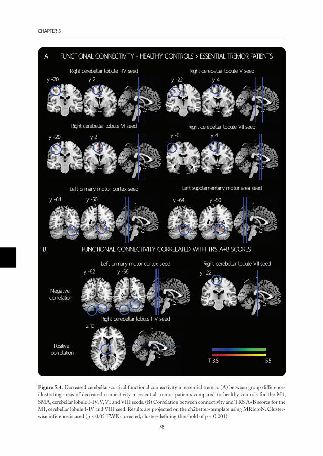
chaPter 5
78
Right cerebellar lobule I-IV seed
Right cerebellar lobule I-IV seed
Right cerebellar lobule V seed
Right cerebellar lobule VI seed
Left primary motor cortex seed
Left primary motor cortex seed
Left supplementary motor area seed
Right cerebellar lobule VIII seed
Right cerebellar lobule VIII seed
T 3.5 5.5
A FUNCTIONAL CONNECTIVITY - HEALTHY CONTROLS > ESSENTIAL TREMOR PATIENTS
B FUNCTIONAL CONNECTIVITY CORRELATED WITH TRS A+B SCORES
y -20 y 2
y -20 y 2
y -22 y 4
y -6 y 4
y -64 y -50y -64 y -50
y -62 y -56 y -22
z 10
Negative correlation
Positive correlation
Figure 5.4. Decreased cerebellar-cortical functional connectivity in essential tremor. (A) between group differences illustrating areas of decreased connectivity in essential tremor patients compared to healthy controls for the M1, SMA, cerebellar lobule I-IV, V, VI and VIII seeds. (B) Correlation between connectivity and TRS A+B scores for the M1, cerebellar lobule I-IV and VIII seed. Results are projected on the ch2better-template using MRIcroN. Cluster-wise inference is used (p < 0.05 FWE corrected, cluster-defining threshold of p < 0.001).

essential tremor: a functional anD effectiVe connectiVity stuDy
79
5
observed functional disconnection correlated to increasing tremor severity. For the right cerebellar lobule I-IV seed, functional con-nectivity with the left thalamus correlated positively with tremor severity (TRS A+B) (Fig. 4B, Table 3). None of the seed regions’ functional connectivities correlated with VAS scores or disease duration.
Discussion
This study provides two novel findings that support an important role for the cerebellum, the thalamus, and the cerebello-dentato-tha-lamic tracts in the pathophysiology of essen-tial tremor. First, the effective connectivity analysis demonstrated a significant excitatory modulating effect of tremor variation on the extrinsic cerebello-dentato-thalamic connec-tion and on intrinsic thalamic and cerebellar lobule V activity. Furthermore, we have rep-licated and expanded findings of decreased cerebello-cortical functional connectivity, related to a motor task, between the motor cerebellum and cortical motor areas in es-sential tremor patients compared to controls (33). More importantly, decreased functional coupling between the primary motor cortex and posterior cerebellum was associated with an increase in clinically assessed tremor se-verity during the motor task. Additionally, an increase in clinically assessed tremor sever-ity was associated with increased functional connectivity between cerebellar lobule I-IV and the motor thalamus in patients with es-sential tremor.
altered cereBellar output
Our findings advocate that modulatory tremor input is associated with activity with-in the cerebello-dentato-thalamic network. During the motor task, inducing action tremor, all included motor regions exhibited self-inhibiting properties. When incorporat-ing tremor variation during the motor task, intrinsic inhibitory activity of the cortical
motor regions and cerebellar lobule VIII increased. However, tremor modulation ex-hibited an excitatory modulating effect on the cerebello-dentato-thalamic tract, leading from cerebellar lobule V to the thalamus, and intrinsic cerebellar lobule V and thalamic ac-tivity. Our results do not give a direct answer as to whether this excitation would give rise to tremor. It is important to note that this ex-citation does not directly represent a neuro-physiological correlate, but is modelled based on the fMRI and EMG signals. Our results do indicate that cerebello-dentato-thalamic activity is perturbed in essential tremor, which can be placed in a broader framework of evidence regarding the pathophysiology of essential tremor. Previously, GABAer-gic neurotransmission dysfunction within the cerebellum has been observed, with in-creased 11C-flunazenil binding to GABA-receptors in the cerebellar cortex, increasing with tremor severity (54). Pathology studies also show evidence for cerebellar changes, with Purkinje cell loss and axonal swelling (19, 20, 55), and simultaneous remodelling of the cerebellar cortex (17, 18, 56). Purkinje cells form the sole output channel from the cerebellar cortex, and lead to the deep cer-ebellar nuclei, including the dentate nucleus. GABAergic Purkinje cell synapses constitute the majority of all synapses in the dentate nucleus, with their action strongly regulating the intrinsic activity of the dentate nucleus (57). Besides pathological changes in the cer-ebellar cortex, altered dentate nucleus func-tion has been postulated in essential tremor (58, 4, 21). Whether the cerebellar cortical pathology is secondary to changes in the dentate nucleus, or vice versa, remains con-troversial. Altered 11C-flunazenil binding to GABA-receptors (58) and a decrease in the number of GABA receptors in the dentate nucleus in essential tremor patients (21) both suggest abnormal functionality of GABA re-ceptors within the dentate nucleus. Electro-physiology data indicate that neurons within
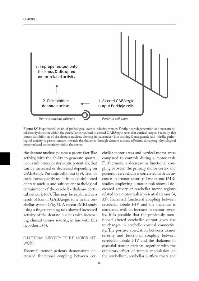
chaPter 5
80
the dentate nucleus possess a pacemaker-like activity, with the ability to generate sponta-neous inhibitory postsynaptic potentials, that can be increased or decreased depending on GABAergic Purkinje cell input (59). Tremor could consequently result from a disinhibited dentate nucleus and subsequent pathological entrainment of the cerebello-thalamo-corti-cal network (60). This may be explained as a result of loss of GABAergic tone in the cer-ebellar system (Fig. 5). A recent fMRI study using a finger-tapping task showed increased activity of the dentate nucleus with increas-ing clinical tremor severity, in line with this hypothesis (4).
functional integrity of the Motor net-work
Essential tremor patients demonstrate de-creased functional coupling between cer-
ebellar motor areas and cortical motor areas compared to controls during a motor task. Furthermore, a decrease in functional cou-pling between the primary motor cortex and posterior cerebellum is correlated with an in-crease in tremor severity. Two recent fMRI studies employing a motor task showed de-creased activity of cerebellar motor regions related to a motor task in essential tremor (4, 33). Increased functional coupling between cerebellar lobule I-IV and the thalamus is correlated with an increase in tremor sever-ity. It is possible that the previously men-tioned altered cerebellar output gives rise to changes in cerebello-cortical connectiv-ity. The positive correlation between tremor severity and functional coupling between cerebellar lobule I-IV and the thalamus in essential tremor patients, together with the excitatory effect of tremor modulation on the cerebellum, cerebellar outflow tracts and
Figure 5.5 Hypothetical chain of pathological events inducing tremor. Firstly, neurodegeneration and neurotrans-mission dysfunction within the cerebellar cortex lead to altered GABAergic cerebellar cortical output. Secondly, this causes disinhibition of the dentate nucleus, altering its pacemaker-like activity. Consequently and thirdly, patho-logical activity is passed onward towards the thalamus through dentate nucleus efferents, disrupting physiological motor-related connectivity within the cortex.

essential tremor: a functional anD effectiVe connectiVity stuDy
81
5
the thalamus during the motor task as ob-served in the effective connectivity analysis, support the idea of pathological entrainment within the cerebellar-thalamic system. In the case of tremor interference, and tremor oscil-lations throughout the motor network, one would also expect increased cerebello-corti-cal coupling due to entrainment of the cer-ebello-thalamo-cortical network. However, an EEG-EMG coherence study has shown that cortical involvement in tremor is only intermittent, and therefore does not seem to be a crucial player within the tremor net-work (61). Alternatively, perturbed cerebel-lar output could generate improper thalamic activity and consequently disrupt physiologi-cal motor-related connectivity with the mo-tor cortex (59, 60). Our results support the hypothesis that increasing tremor sever-ity proportionally disrupts cerebello-cortical connectivity. Moreover, continuous increased input from the dentate nucleus via the thala-mus could cause amplification of inhibitory mechanisms within the cerebral cortex. In-hibitory circuits within the motor cortex are reported to be aberrant and less modifiable in essential tremor (62). In addition, increased 11C-flunazenil binding to GABA-receptors has also been found in the ventrolateral thal-amus and lateral premotor cortex in essential tremor (58).
differential involveMent of the anterior and posterior cereBelluM in essential treMor
The anterior cerebellum is formed by lobules I to V/VI, and is divided by the primary fis-sure from the posterior cerebellum, formed by lobules VI/VII to X (63, 64). Interestingly, to our knowledge, the anterior and posterior cerebellum, although both involved in mo-tor control, are not discussed separately in essential tremor research, even though the physiological, developmental and genetic properties of each are quite different (63-
65). Our functional and effective connectiv-ity results suggest that both the anterior and posterior cerebellum are involved in essential tremor. There is however a discrepancy in re-duced functional connectivity between M1 and the posterior cerebellum associated with increasing tremor severity, and an apparent lack of this reduced functional connectivity between M1 and regions within the anterior cerebellum. On the other hand, an excitatory modulatory effect of tremor was observed in cerebellar lobule V (anterior cerebellum) and on the connections between cerebellar lobule V and the thalamus. We currently have no clear explanation for this observed difference. Although this discrepancy could be due to insufficient sample size, for future pathology studies, it would be of interest to divorce the involvements of the anterior and posterior cerebellum by assessing them separately.
Methodological considerations
A known and persistent problem with fMRI studies is their limited temporal resolution. This makes the identification of a tremor generator challenging. However, it is a useful technique for studying properties of regions within the cerebello-thalamo-cortical net-work, especially when combined with EMG recordings. This is the first time EMG sig-nals were incorporated in a DCM analysis. It needs to be stressed that the residual EMG regressor is not the EMG signal as recorded from the muscle. It is a reflection of the wax-ing and waning EMG signal with respect to the task, i.e., the involuntary movements, and does not necessarily say something about clinical severity. Further studies employing electrophysiological techniques may be re-quired to provide deeper insights into the synaptic mechanisms involved. Although our results appear robust, they will need to be replicated in the future. For this study, the parameters characterizing the cerebello-thalamic connections were chosen as indirect

chaPter 5
82
measures to assess the possible involvement of the dentate nucleus and the cerebello-dentato-thalamic tract in essential tremor. These connections represent the net effect of the cerebello-dentato-thalamic tracts. No tremor-related activity was observed in the dentate nucleus in individual subjects, pos-sibly due to the high iron-content of the dentate nucleus and resulting low signal-to-noise ratio of its BOLD signal (66). To be able to include the dentate nucleus in future models, studies with a higher spatial and temporal resolution are warranted to repro-duce our observed excitatory effect on the cerebello-dentato-thalamic pathway.A common difficulty in functional imaging studies lies in selecting a suitable task for healthy controls that corresponds well with the patients’ task. For this study, a mimicked tremor was chosen. Consequently, the two groups were actually performing a qualita-tively different task. These tasks were cho-sen to allow optimal distinction of brain networks for the functional connectivity analysis, involved in involuntary tremor as opposed to compensation or afferent feed-back by deliberate, mimicked tremor move-ments. However, due to this qualitative dif-ference, for the effective connectivity analysis the patient group was not compared with a healthy control group. Future studies could circumvent this problem by employing other techniques such as enforcing passive wrist oscillations as an additional control condi-tion, as has been used previously by Bucher and colleagues (42). One could then addi-tionally assess whether there are differences within the tremor circuitry in excitatory and inhibitory connections between patients and healthy controls.Finally, as mentioned in the methods section, a silent reading task was offered during half of the task blocks, which may have influ-enced activity within the motor network and could therefore have affected our effective connectivity results. There was a significant
difference in driving effect of the two tasks on the SMA and not on the PMC, as ob-served in the effective connectivity analysis. However, the motor task with silent reading had merely an additional excitatory effect compared to the motor task in which only the command to stretch the right arm was given. We expect that this will not have af-fected the final conclusions of the effective connectivity analysis.
In conclusion, our findings suggest that cere-bello-dentato-thalamic activity and cerebel-lar-cortical connectivity are perturbed in es-sential tremor, supporting previous evidence of cerebellar pathology in essential tremor. This perturbed cerebello-dentato-thalamic activity could subsequently affect the rest of the cerebello-thalamo-cortical network, leading to tremor on the one hand and possi-bly less effective physiological output on the other hand. Investigating effective connec-tivity changes in essential tremor represents a new avenue of study that may shed light on its underlying pathophysiology.
acknowledgMents
We are grateful to all study participants for their contributions, to Aart Nederveen for support, to Alexander Munts for help with the inclusion of patients and to Marina Ti-jssen for her valuable comments. We thank Louise Marshall for grammatical corrections.
referenceS1. Louis ED, Ferreira JJ. How common is the most common adult movement disorder? Update on the worldwide prevalence of essential tremor. Mov Dis-ord. 2010; 25: 534–541.
2. Dupuis MJ-M, Evrard FLA, Jacquerye PG, Pic-ard GT, Lermen OG. Disappearance of Essential Tremor after Stroke. Mov. Disord. 2010; 25: 2884–2887.
3. Raethjen J, Deuschl G. The oscillating central net-

essential tremor: a functional anD effectiVe connectiVity stuDy
83
5
work of Essential tremor. Clin. Neurophysiol. 2012; 123: 61–4.
4. Buijink AWG, Broersma M, van der Stouwe AMM, van Wingen GA, Groot PFC, Speelman JD, et al. Rhythmic finger tapping reveals cerebellar dysfunction in essential tremor. Parkinsonism Relat. Disord. 2015: 9–14.
5. Grimaldi G, Manto M. Is essential tremor a Purkinjopathy? The role of the cerebellar cortex in its pathogenesis. Mov. Disord. 2013; 00: 1–3.
6. Passamonti L, Novellino F, Cerasa A, Chiriaco C, Rocca F, Matina MS, et al. Altered cortical-cerebellar circuits during verbal working memory in essential tremor. Brain 2011; 134: 2274–86.
7. Haubenberger D, McCrossin G, Lungu C, Con-sidine E, Toro C, Nahab FB, et al. Octanoic acid in alcohol-responsive essential tremor: A randomized controlled study. Neurology 2013
8. Fasano A, Herzog J, Raethjen J, Rose FEM, Mu-thuraman M, Volkmann J, et al. Gait ataxia in es-sential tremor is differentially modulated by thalamic stimulation. Brain 2010; 133: 3635–48.
9. Hoskovcová M, Ulmanová O, Sprdlík O, Sieger T, Nováková J, Jech R, et al. Disorders of balance and gait in essential tremor are associated with midline tremor and age. Cerebellum 2013; 12: 27–34.
10. Stolze H, Petersen G, Raethjen J, Wenzelburger R, Deuschl G. The gait disorder of advanced essential tremor. Brain 2001; 124: 2278–2286.
11. Gitchel GT, Wetzel PA, Baron MS. Slowed Sac-cades and Increased Square Wave Jerks in Essential Tremor. Tremor and Other Hyperkinetic Movements 2013; 3: 1–7.
12. Helmchen C, Hagenow A, Miesner J, Sprenger A, Rambold H, Wenzelburger R, et al. Eye move-ment abnormalities in essential tremor may indicate cerebellar dysfunction. Brain 2003; 126: 1319–1332.
13. Kronenbuerger M, Gerwig M, Brol B, Block F, Timmann D. Eyeblink conditioning is impaired in subjects with essential tremor. Brain 2007; 130: 1538–51.
14. Deuschl G, Wenzelburger R, Lo K, Raethjen J,
Stolze H. Essential tremor and cerebellar dysfunction Clinical and kinematic analysis of intention tremor. Brain 2000; 123: 1568–1580.
15. Louis ED, Frucht SJ, Rios E. Intention Tremor in Essential Tremor: Prevalence and Association with Disease Duration. Mov. Disord. 2009; 24: 617–8.
16. Louis ED. Re-thinking the biology of essential tremor: from models to morphology. Parkinsonism Relat. Disord. 2014; 20 Suppl 1: S88–93.
17. Babij R, Lee M, Cortés E, Vonsattel J-PG, Faust PL, Louis ED. Purkinje cell axonal anatomy: quan-tifying morphometric changes in essential tremor ver-sus control brains. Brain 2013; 136: 3051–61.
18. Lin C-Y, Louis ED, Faust PL, Koeppen AH, Vonsattel J-PG, Kuo S-H. Abnormal climbing fibre-Purkinje cell synaptic connections in the essential tremor cerebellum. Brain 2014: 1–11.
19. Louis ED, Faust PL, Vonsattel J-PGP, Honig LS, Rajput AA, Robinson CA, et al. Neuropathological changes in essential tremor: 33 cases compared with 21 controls. Brain 2007; 130: 3297–307.
20. Rajput AH, Robinson CA, Rajput ML, Robin-son SL, Rajput A. Essential tremor is not dependent upon cerebellar Purkinje cell loss. Parkinsonism Relat. Disord. 2012; 18: 626–8.
21. Paris-Robidas S, Brochu E, Sintes M, Emond V, Bousquet M, Vandal M, et al. Defective dentate nu-cleus GABA receptors in essential tremor. Brain 2011; 135: 105–116.
22. Sharifi S, Nederveen AJ, Booij J, van Rootselaar A-F. Neuroimaging essentials in essential tremor: A systematic review. NeuroImage. Clin. 2014; 5: 217–31.
23. Muthuraman M, Heute U, Arning K, Anwar a R, Elble R, Deuschl G, et al. Oscillating central mo-tor networks in pathological tremors and voluntary movements. What makes the difference? Neuroimage 2012; 60: 1331–9.
24. Schnitzler A, Münks C, Butz M, Timmermann L, Gross J. Synchronized brain network associated with essential tremor as revealed by magnetoencepha-lography. Mov. Disord. 2009; 24: 1629–35.

chaPter 5
84
25. Contarino MF, Groot PFC, van der Meer JN, Bour LJ, Speelman JD, Nederveen AJ, et al. Is there a role for combined EMG-fMRI in exploring the pathophysiology of essential tremor and improving functional neurosurgery? PLoS One 2012; 7: e46234.
26, Helmich RC, Janssen MJR, Oyen WJG, Bloem BR, Toni I. Pallidal dysfunction drives a cerebell-othalamic circuit into Parkinson tremor. Ann. Neurol. 2011; 69: 269–81.
27. Van Rootselaar A-FF, Renken R, de Jong BM, Hoogduin JM, Tijssen MAJ, Maurits NM. fMRI analysis for motor paradigms using EMG-based de-signs: a validation study. Hum.Brain Mapp. 2007; 28: 1117–1127.
28. Van Rootselaar A-F, Maurits NM, Renken R, Koelman JHTM, Hoogduin JM, Leenders KL, et al. Simultaneous EMG-functional MRI recordings can directly relate hyperkinetic movements to brain activ-ity. Hum. Brain Mapp. 2008; 29: 1430–41.
29. Friston KJ. Functional and effective connectiv-ity in neuroimaging: A synthesis. Hum. Brain Mapp. 1994; 2: 56–78.
30. Friston KJ, Harrison L, Penny W. Dynamic causal modelling. Neuroimage 2003; 19: 1273–1302.
31. David O, Guillemain I, Saillet S, Reyt S, Deran-sart C, Segebarth C, et al. Identifying neural drivers with functional MRI: an electrophysiological valida-tion. PLoS Biol. 2008; 6: 2683–97.
32. Buckner RL, Krienen FM, Castellanos A, Diaz JC, Yeo BTT. The organization of the human cerebel-lum estimated by intrinsic functional connectivity. J. Neurophysiol. 2011; 106: 2322–45.
33. Neely KA, Kurani AS, Shukla P, Planetta PJ, Wa-gle Shukla A, Goldman JG, et al. Functional Brain Activity Relates to 0-3 and 3-8 Hz Force Oscillations in Essential Tremor. Cereb. Cortex 2014
34. Louis ED. The Essential Tremors : A Family of Neurodegenerative Disorders? Arch.Neurol. 2009; 66: 1202–1208.
35. Bain PG, Brin M, Deuschl G, Elble RJ, Jankovic J, Findley LJ, et al. Criteria for the diagnosis of es-sential tremor. Neurology 2000; 54: S7.
36. Deuschl G, Raethjen J, Hellriegel H, Elble R. Treatment of patients with essential tremor. Lancet Neurol. 2011; 10: 148–61.
37. Fahn S. Clinical Rating Scale for Tremor. Pafkinson’s Dis. Mov. Disord. 1993: 271–280.
38. Koller WC, Biary NM. Volitional control of in-voluntary movements. Mov. Disord. 1989; 4: 153–156.
39. Allen PJ, Josephs O, Turner R. A method for re-moving imaging artifact from continuous EEG re-corded during functional MRI. Neuroimage 2000; 12: 230–9.
40. Van der Meer JN, Tijssen M a J, Bour LJ, van Rootselaar a F, Nederveen a J. Robust EMG-fMRI artifact reduction for motion (FARM). Clin. Neuro-physiol. 2010; 121: 766–76.
41. Power JD, Mitra A, Laumann TO, Snyder AZ, Schlaggar BL, Petersen SE. Methods to detect, char-acterize, and remove motion artifact in resting state fMRI. Neuroimage 2014; 84: 320–41.
42. Bucher SF, Seelos KC, Dodel RC, Reiser M, Oer-tel WH. Activation mapping in essential tremor with functional magnetic resonance imaging. Ann. Neurol. 1997; 41: 32–40.
43. Middleton F a., Strick PL. Basal ganglia and cerebellar loops: Motor and cognitive circuits. Brain Res. Rev. 2000; 31: 236–250.
44. Goldman-Rakic PS, Bates JF, Chafee M V. The prefrontal cortex and internally generated motor acts. Curr. Opin. Neurobiol. 1992; 2: 830–835.
45. Wang LE, Fink GR, Diekhoff S, Rehme AK, Eickhoff SB, Grefkes C. Noradrenergic enhancement improves motor network connectivity in stroke pa-tients. Ann. Neurol. 2011; 69: 375–388.
46. Penny WD, Stephan KE, Daunizeau J, Rosa MJ, Friston KJ, Schofield TM, et al. Comparing families of dynamic causal models. PLoS Comput. Biol. 2010; 6: e1000709.
47. Stephan KE, Penny WD, Moran RJ, den Ouden HEM, Daunizeau J, Friston KJ. Ten simple rules for dynamic causal modeling. Neuroimage 2010; 49: 3099–109.

essential tremor: a functional anD effectiVe connectiVity stuDy
85
5
48. Torrisi SJ, Lieberman MD, Bookheimer SY, Alt-shuler LL. Advancing understanding of affect la-beling with dynamic causal modeling. Neuroimage 2013; 82: 481–488.
49. Thirion B, Pinel P, Mériaux S, Roche A, Dehaene S, Poline J-B. Analysis of a large fMRI cohort: Sta-tistical and methodological issues for group analyses. Neuroimage 2007; 35: 105–20.
50. Holmes AP, Blair RC, Watson JD, Ford I. Non-parametric analysis of statistic images from function-al mapping experiments. J. Cereb. Blood Flow Metab. 1996; 16: 7–22.
51. Van der Stouwe AMM, Broersma M, Buijink AWG, van Rootselaar AF, Maurits NM. Limited correlations between clinician-based and patient-based measures of essential tremor severity. Parkin-sonism Relat. Disord. 2015: 3–6.
52. Diedrichsen J. A spatially unbiased atlas tem-plate of the human cerebellum. Neuroimage 2006; 33: 127–38.
53. Eickhoff SB, Stephan KE, Mohlberg H, Grefkes C, Fink GR, Amunts K, et al. A new SPM toolbox for combining probabilistic cytoarchitectonic maps and functional imaging data. Neuroimage 2005; 25: 1325–35.
54. Gironell A, Figueiras FP, Pagonabarraga J, Her-ance JR, Pascual-Sedano B, Trampal C, et al. Gaba and serotonin molecular neuroimaging in essential tremor: a clinical correlation study. Park. Disord. 2012; 18: 876–880.
55. Symanski C, Shill HA, Dugger B, Hentz JG, Adler CH, Jacobson SA, et al. Essential tremor is not associated with cerebellar Purkinje cell loss. Mov. Disord. 2014; 00: 1–5.
56. Erickson-Davis CR, Faust PL, Vonsattel J-PG, Gupta S, Honig LS, Louis ED. ‘Hairy baskets’ as-sociated with degenerative Purkinje cell changes in essential tremor. J. Neuropathol. Exp. Neurol. 2010; 69: 262–71.
57. Uusisaari M, Knöpfel T. GABAergic synaptic communication in the GABAergic and non-GABAe-rgic cells in the deep cerebellar nuclei. Neuroscience 2008; 156: 537–49.
58. Boecker H, Weindl A, Brooks DJ, Ceballos-Bau-mann AO, Liedtke C, Miederer M, et al. GABAergic dysfunction in essential tremor: an 11C-flumazenil PET study. J. Nucl. Med. 2010; 51: 1030–5.
59. Jahnsen H. Extracellular activation and mem-brane conductances of neurones in the guinea-pig deep cerebellar nuclei in vitro. J. Physiol. Paris 1986; 372: 149–168.
60. Pinault D, Deschênes M. The origin of rhythmic fast subthreshold depolarizations in thalamic relay cells of rats under urethane anaesthesia. Brain Res. 1992; 595: 295–300.
61. Raethjen J, Govindan RB, Kopper F, Muthura-man M, Deuschl G. Cortical involvement in the gen-eration of essential tremor. J. Neurophysiol. 2007; 97: 3219–3228.
62. Chuang W-L, Huang Y-Z, Lu C-S, Chen R-S. Reduced cortical plasticity and GABAergic modula-tion in essential tremor. Mov. Disord. 2014; 00: 1–7.
63. Eisenman LM. Antero-posterior boundaries and compartments in the cerebellum: evidence from se-lected neurological mutants. Prog. Brain Res. 2000; 124: 23–30.
64. Herrup K, Kuemerle B. The compartementaliza-tion of the cerebellum. Ann. Rev. Neurosci. 1997; 20: 61–90.
65. Witt ST, Meyerand ME, Laird AR. Functional neuroimaging correlates of finger tapping task varia-tions: An ALE meta-analysis. Neuroimage 2009; 42: 343–356.
66. Diedr ichsen J, Maderwald S, Küper M, Thürling M, Rabe K, Gizewski ER, et al. Imaging the deep cerebellar nuclei: a probabilistic atlas and normaliza-tion procedure. Neuroimage 2011; 54: 1786–94.

chaPter 5
86
Mod
el n
o.
Cer
ebell
ar o
f
SMA
PMC
Thala
mus
CB
V
CB
VII
I
M1
Mod
el n
o.
Cer
ebell
ar o
f
SMA
PMC
Thala
mus
CB
V
CB
VII
I
M1
Mod
el n
o.
Cer
ebell
ar 0
f
SMA
PMC
Thala
mus
CB
V
CB
VII
I
M1
1 0 0 0 0 0 0 0 45 0 1 0 1 1 0 0 89 1 0 1 1 0 0 02 0 0 0 0 0 0 1 46 0 1 0 1 1 0 1 90 1 0 1 1 0 0 13 0 0 0 0 0 1 0 47 0 1 0 1 1 1 0 91 1 0 1 1 0 1 04 0 0 0 0 0 1 1 48 0 1 0 1 1 1 1 92 1 0 1 1 0 1 15 0 0 0 0 1 0 0 49 0 1 1 0 0 0 0 93 1 0 1 1 1 0 06 0 0 0 0 1 0 1 50 0 1 1 0 0 0 1 94 1 0 1 1 1 0 17 0 0 0 0 1 1 0 51 0 1 1 0 0 1 0 95 1 0 1 1 1 1 08 0 0 0 0 1 1 1 52 0 1 1 0 0 1 1 96 1 0 1 1 1 1 19 0 0 0 1 0 0 0 53 0 1 1 0 1 0 0 97 1 1 0 0 0 0 010 0 0 0 1 0 0 1 54 0 1 1 0 1 0 1 98 1 1 0 0 0 0 111 0 0 0 1 0 1 0 55 0 1 1 0 1 1 0 99 1 1 0 0 0 1 012 0 0 0 1 0 1 1 56 0 1 1 0 1 1 1 100 1 1 0 0 0 1 113 0 0 0 1 1 0 0 57 0 1 1 1 0 0 0 101 1 1 0 0 1 0 014 0 0 0 1 1 0 1 58 0 1 1 1 0 0 1 102 1 1 0 0 1 0 115 0 0 0 1 1 1 0 59 0 1 1 1 0 1 0 103 1 1 0 0 1 1 016 0 0 0 1 1 1 1 60 0 1 1 1 0 1 1 104 1 1 0 0 1 1 117 0 0 1 0 0 0 0 61 0 1 1 1 1 0 0 105 1 1 0 1 0 0 018 0 0 1 0 0 0 1 62 0 1 1 1 1 0 1 106 1 1 0 1 0 0 119 0 0 1 0 0 1 0 63 0 1 1 1 1 1 0 107 1 1 0 1 0 1 020 0 0 1 0 0 1 1 64 0 1 1 1 1 1 1 108 1 1 0 1 0 1 1
21 0 0 1 0 1 0 0 65 1 0 0 0 0 0 0 109 1 1 0 1 1 0 0
22 0 0 1 0 1 0 1 66 1 0 0 0 0 0 1 110 1 1 0 1 1 0 1
23 0 0 1 0 1 1 0 67 1 0 0 0 0 1 0 111 1 1 0 1 1 1 0
24 0 0 1 0 1 1 1 68 1 0 0 0 0 1 1 112 1 1 0 1 1 1 125 0 0 1 1 0 0 0 69 1 0 0 0 1 0 0 113 1 1 1 0 0 0 026 0 0 1 1 0 0 1 70 1 0 0 0 1 0 1 114 1 1 1 0 0 0 127 0 0 1 1 0 1 0 71 1 0 0 0 1 1 0 115 1 1 1 0 0 1 028 0 0 1 1 0 1 1 72 1 0 0 0 1 1 1 116 1 1 1 0 0 1 129 0 0 1 1 1 0 0 73 1 0 0 1 0 0 0 117 1 1 1 0 1 0 030 0 0 1 1 1 0 1 74 1 0 0 1 0 0 1 118 1 1 1 0 1 0 131 0 0 1 1 1 1 0 75 1 0 0 1 0 1 0 119 1 1 1 0 1 1 032 0 0 1 1 1 1 1 76 1 0 0 1 0 1 1 120 1 1 1 0 1 1 133 0 1 0 0 0 0 0 77 1 0 0 1 1 0 0 121 1 1 1 1 0 0 034 0 1 0 0 0 0 1 78 1 0 0 1 1 0 1 122 1 1 1 1 0 0 1
35 0 1 0 0 0 1 0 79 1 0 0 1 1 1 0 123 1 1 1 1 0 1 0
36 0 1 0 0 0 1 1 80 1 0 0 1 1 1 1 124 1 1 1 1 0 1 1
37 0 1 0 0 1 0 0 81 1 0 1 0 0 0 0 125 1 1 1 1 1 0 038 0 1 0 0 1 0 1 82 1 0 1 0 0 0 1 126 1 1 1 1 1 0 139 0 1 0 0 1 1 0 83 1 0 1 0 0 1 0 127 1 1 1 1 1 1 040 0 1 0 0 1 1 1 84 1 0 1 0 0 1 1 128 1 1 1 1 1 1 141 0 1 0 1 0 0 0 85 1 0 1 0 1 0 042 0 1 0 1 0 0 1 86 1 0 1 0 1 0 143 0 1 0 1 0 1 0 87 1 0 1 0 1 1 044 0 1 0 1 0 1 1 88 1 0 1 0 1 1 1
suPPlementary table 5.1. list of moDels anD moDulatory inPuts
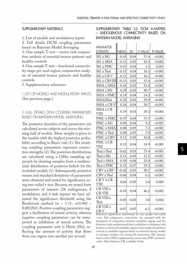
essential tremor: a functional anD effectiVe connectiVity stuDy
87
5
suPPlementary materials
1. List of models and modulatory inputs2. Full details DCM coupling parameters based on Bayesian Model Averaging3. One sample T-test – motor task conjunc-tion analysis of essential tremor patients and healthy controls4. One sample T-test – functional connectiv-ity maps per seed region, conjunction analy-sis of essential tremor patients and healthy controls5. Supplementary references
1. list of Models and Modulatory inputs (See previous page.)
2. full details dcM coupling paraMeters Based on Bayesian Model averaging
The posterior densities of the parameters are calculated across subjects and across the win-ning half of models. More weight is given to the models with the highest posterior proba-bility according to Bayes’ rule (1). The result-ing coupling parameters represent connec-tion strengths (2). The posterior distributions are calculated using a Gibbs sampling ap-proach by drawing samples from a multino-mial distribution of posterior beliefs for the included models (1). Subsequently, posterior means and standard deviations of parameters were obtained and tested for significance us-ing two-tailed t-test. Because we tested forty parameters of interest (28 endogenous, 8 modulatory and 4 task inputs) we have ad-justed the significance threshold using the Bonferroni method (a = 1-(1- a)1/40) = 0.001282). Positive coupling parameters sug-gest a facilitation of neural activity, whereas negative coupling parameters can be inter-preted as inhibition of neural activity. The coupling parameter unit is Hertz (Hz), re-flecting the amount of activity that flows from one region into another per second.
suPPlementary table 5.2. Dcm a-matrix – enDogenous connectiVity baseD on bayesian moDel aVeraging
PArAmeter eStImAte: meAn Sd t-vALue P-vALue
M1 à M1 -0.45 0.04 51.4 <0.001M1 à SMA -0.13 0.05 10.5 <0.001M1 à PMC -0.03 0.04 3.5 0.003M1 à Thal -0.15 0.04 16.1 <0.001M1 à CB V -0.13 0.03 16.3 <0.001M1 à CBVIII -0.13 0.03 15.4 <0.001SMA à SMA -0.43 0.03 53.8 <0.001SMA à M1 0.29 0.03 40.7 <0.001SMA à PMC 0.14 0.04 14.3 <0.001SMAàThal 0.25 0.03 29.9 <0.001SMA à CB V 0.26 0.04 30.7 <0.001SMA à CB VIII 0.19 0.03 29.2 <0.001
PMC à PMC -0.47 0.04 53.5 <0.001PMC à M1 0.09 0.04 9.2 <0.001PMC à SMA 0.08 0.05 7.1 <0.001PMC à Thal 0.08 0.05 7.0 <0.001PMC à CBV 0.11 0.05 10.0 <0.001PMC à CB VIII 0.15 0.04 14.9 <0.001
Thal à Thal -0.62 0.03 75.4 <0.001Thal à M1 0.13 0.05 12.1 <0.001Thal à SMA 0.50 0.08 25.0 <0.001Thal à PMC 0.31 0.07 19.3 <0.001CBV à CBV -0.42 0.03 50.1 <0.001CBV à Thal -0.06 0.04 6.2 <0.001CB V à CB VIII 0.04 0.05 3.0 0.009
CB VIII à CB VIII -0.43 0.04 46.5 <0.001
CB VIII à Thal -0.05 0.05 3.8 0.002
CB VIII à CB V 0.07 0.05 6.1 <0.001Statistical significance determined by one sample two-tailed t-test. Full endogenous connectivity was assumed with the exemption of connections between cerebellar regions and the thalamus (only unidirectional from cerebellum to thalamus) and between cortical and cerebellar regions (only unidirectional from cortical to cerebellar regions) based on neuronal tracing studies in macaque monkeys (3), leaving 28 connections. M1: primary motor cortex, SMA: supplementary motor area, PMC: premotor cortex. Thal: thalamus, CB: cerebellar lobule.

chaPter 5
88
suPPlementary table 5.3. Dcm b-matrix – moDulatory tremor Variation inPut
moduLAtory InPut on: meAn Sd t-vALue P-vALue
Primary motor cortex (intrinsic) -0.30 0.25 5.1 <0.001Supplementary motor area (intrinsic) -0.91 0.27 14.3 <0.001Premotor cortex (intrinsic) -0.53 0.29 7.7 <0.001Thalamus (intrinsic) 1.26 0.42 12.6 <0.001Cerebellar lobule V (intrinsic) 0.31 0.32 4.2 <0.001Cerebellar lobule VIII (intrinsic) -0.28 0.27 4.5 <0.001Cerebellar lobule V to thalamus (extrinsic) 0.82 0.89 3.9 0.001Cerebellar lobule VIII to thalamus (extrinsic) 0.60 1.38 1.8 0.08
Posterior means and standard deviations of the estimated modulatory effect of tremor variation on all regions and cerebello-dentato-thalamic tracts. Tested for significance using two-tailed t-tests. Bonferroni-corrected significant p-values in bold (p > 0.00128). Results are summarized graphically in Figure 3 within the main text.
suPPlementary table 5.4. Dcm c-matrix – Direct (task) inPut
PArAmeter eStImAte: meAn Sd t-vALue P-vALue
Task input SMA (with silent reading task) 0.10 0.01 43.3 <0.001Task input SMA (without silent reading task) 0.07 0.01 33.4 <0.001Task input PMC (with silent reading task) 0.04 0.01 18.6 <0.001Task input PMC (without silent reading task) 0.05 0.01 22.5 <0.001
Mean influence of task input. statistical significance determined by one sample two-tailed t-test. Bonferroni-corrected significant parameters in bold (p > 0.00128). SMA = supplementary motor area. PMC = premotor cortex.

essential tremor: a functional anD effectiVe connectiVity stuDy
89
5
3. one saMple t-test – Motor task conjunction analysis of essential treMor patients and healthy controls
reGIon voXeLS r/L t-vALue X y Z
Cerebellar lobule IV 3841 Right 14.9 4 54 -21Cerebellar lobule V Right 14.1 14 -50 -19Cerebellar lobule VI Right 13.9 22 -50 -25Primary motor cortex 5048 Left 14.5 -28 -28 53Supplementary motor area Left 14.0 -2 -8 57
Primary motor cortex Left 13.5 -36 -32 61
Task-related activity - results of a one sample T-test – conjunction analysis of essential tremor patients and healthy controls. The six most significant peak-voxels are listed. Cerebellar lobule VIII is located within the most significant cluster with peak-region cerebellar lobule IV.
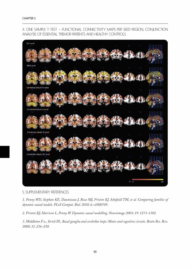
chaPter 5
90
4. one saMple t-test – functional connectivity Maps per seed region, conjunction analysis of essential treMor patients and healthy controls
5. suppleMentary references
1. Penny WD, Stephan KE, Daunizeau J, Rosa MJ, Friston KJ, Schofield TM, et al. Comparing families of dynamic causal models. PLoS Comput. Biol. 2010; 6: e1000709.
2. Friston KJ, Harrison L, Penny W. Dynamic causal modelling. Neuroimage 2003; 19: 1273–1302.
3. Middleton F a., Strick PL. Basal ganglia and cerebellar loops: Motor and cognitive circuits. Brain Res. Rev. 2000; 31: 236–250.

essential tremor: a functional anD effectiVe connectiVity stuDy
91
5

1. Department of Neurology and Clinical Neurophysiology, University Medical Center Groningen, Groningen, The Netherlands.
2. Neuro-imaging Centre, Groningen, The Netherlands
A.m.m. vAn der Stouwe1, m. BroerSmA1, J.B.c. mArSmAn2, A.f. vAn rootSeLAAr1, m.A.J. tIJSSen1, n.m. mAurItS1
increaseD cerebellar actiVations During goal-DirecteD moVement in
essential tremor: an fmri stuDy
cHAPter 6

chaPter 6
94
abstract
introduction: Abnormalities in goal-directed movement are an important symptom of es-sential tremor (ET), and are clinically related to cerebellar disease. The current study aims to examine how these movements are associated with abnormal brain activity in ET patients.Methods: Nineteen ET patients and seventeen healthy participants performed a goal-di-rected movement task using an MR-compatible wrist device. Spatial coordinates of partici-pants’ movements were recorded during scanning and used to calculate reaction times and variability in movement paths. The target stimuli that prompted participants to move were incorporated in an event-related fMRI design, to correlate the goal-directed movements with cerebral activity. Disease severity, disease duration, intention tremor (assessed clinically with finger-to-nose manoeuvres) were used as clinical covariates in separate analyses. results: ET patients were affected mildly (off-medication median Tremor Rating Scale score 22 (range 13-45)). We found no significant difference in reaction time or variability in move-ment paths between patients and healthy participants. We found increased activations in cerebellar vermis 4/5/6 in ET patients (T=5.11, coordinates in MNI space x=3, y=-63, z=-5), when comparing back-to-centre movements versus baseline. There was no correlation be-tween brain activation during the goal-directed movement task and any of the clinical covari-ates in ET patients. discussion: We report increased cerebellar activation in mildly affected ET patients during goal-directed movements, in the absence of differences in task performance compared to healthy participants. These results extend on earlier reports of increased cerebellar activation during postural tasks, and lend further support to the notion of underlying cerebellar abnor-malities in ET.

goal-DirecteD moVement in essential tremor: an fmri stuDy
95
6
introDuction
Essential tremor (ET) is the most common tremor disorder, and is characterized by bi-lateral action tremor of the hands and fore-arms (1, 2). The tremor occurs during action and posturing, and apart from the hands and arms, the voice, head and more infrequently jaw, tongue, legs and feet can also be affected (3). ET often runs in families, and half of the patients claim their tremor is reduced by the intake of one or two glasses of alcohol. A feature that is common in ET, but atypical in most other tremor disorders, is intention tremor: tremor worsening during goal-di-rected movement (4-6)(see also Chapter 2). Intention tremor is considered to be a sign of cerebellar disease (4), and apart from ET, it is also seen in multiple sclerosis (7) and Holmes (rubral) tremor (8): diseases that affect the cerebellum or cerebellar outflow-tract. Therefore, the occurrence of intention tremor in ET fits well with the emerging pathophysiological view of ET as a cerebel-lar disease (9, 10). Likewise, in support of the cerebellar hypothesis, there is increasing acknowledgement of ataxia in ET. Several studies have shown signs of mild ataxia in ET patients, such as mild gait ataxia (11-13), hypermetria (4, 14), and drawing ataxia (15). These clinical signs and symptoms lend further support to the notion of underlying cerebellar abnormalities. Neuroimaging studies also provide evidence for cerebellar involvement in ET (16). Two published motor fMRI-studies both dem-onstrated abnormal cerebellar activity in ET (17)(see also Chapter 4). However, the movement tasks investigated in these stud-ies did not extend beyond a comparison be-tween posturing of the tremulous arm versus rest. As abnormalities in intentional move-ment are an important clinical symptom of ET, the current study aims to examine how goal-directed movement is associated with abnormal brain activity in the cerebel-
lum and other brain regions in ET patients. Goal-directed movements can be executed and measured in the MRI scanner by em-ploying a centre-out steptracking task and an MR-compatible wrist device (18). Recently, attention has been paid to the effect of deep brain stimulation (DBS) on reach-to-grasp movement in severely affected ET patients (19, 20). DBS of the subthalamic area re-duced ataxia especially, returning upon su-prathreshold stimulation, leading the authors to hypothesize on the roles of different cer-ebellar networks in goal-directed movement in ET. We set out to extend on this work by using a goal-directed movement task suit-able for fMRI-scanning, thus enabling a link between behavioral and cerebral or cerebellar abnormalities.
methoDs
participants
The Medical Ethics Committee of the Uni-versity Medical Center Groningen approved the neuroimaging study of which this study is a subpart. Participants took part after providing informed consent in accordance with the declaration of Helsinki (21). We included patients who had a definite diag-nosis of ET according to the TRIG criteria (1): all patients had bilateral upper limb ac-tion tremor in the absence of other neuro-logical signs, and disease duration had to be >5 years. Additionally, age at onset was required to be <65 years, thereby excluding late-onset ‘senile’ ET, which may have a dif-ferent pathophysiology (2). Patients who took medication for their tremor quit their medication minimally three days before test-ing. Patients and healthy, age- and gender-matched participants were all right-handed as assessed by the Annett Handedness scale (22). All participants had to score >25 on the Mini Mental State Examination (23) to ensure proper understanding of the task. Exclusion criteria were neurological comor-

chaPter 6
96
bidity (for patients: other than ET), the use of medication affecting the central nervous system, and MR-related contra-indications such as claustrophobia, ferromagnetic im-plants or pregnancy.
clinical assessMent of treMor
Tremor was assessed off-medication using the Fahn-Tolosa-Marin Tremor Rating Scale (TRS) (24) parts A, (assessment of tremor amplitude during standardized postures and movements), B (tremor-inducing tasks), and C (a structured interview about limitations in daily life due to tremor). The examination was recorded on video and supplemented with the corresponding spiral drawings. An experienced movement disorders specialist (Dr. J.D. Speelman, AMC) scored TRS parts A and B based on this material. Part C was assessed by vd Stouwe for all patients. Sub-jective tremor severity was documented using 10 cm visual analogue scales (VAS) ranging from 0: ‘no tremor at all’ to 10: ‘worst tremor imaginable’, on which patients marked their scores. Intention tremor was assessed clini-cally by scoring presence or absence of in-tention tremor during finger-to-nose ma-noeuvres, and this score was used as a clinical covariate in the fMRI analysis later on. Scor-ing of intention tremor was done clinically because the rapid wrist movements within the employed step-tracking task are too brief
to detect any tremor, making it impossible to assess intention tremor during task perfor-mance.
experiMental set-up
Participants performed the task with the right hand, using an MR-compatible ma-nipulandum: a joystick-like device that can rotate in two planes allowing all combina-tions of wrist flexion-extension and ulnar-radial deviation (Figure 6.1A-C). To provide visual feedback on task performance, angu-lar displacement was measured in both (X and Y) planes by potentiometers mounted in-line with the axes of the manipulandum rings and displayed as a cursor (a 5 x 5 mm square) following digitization using a Power 1401 analog-to-digital converter controlled using Spike 2 (Cambridge Electronic De-sign (CED), Cambridge, UK). Data were re-corded simultaneously at a sampling rate of 100 Hz. During scanning, performance was visually monitored on a second computer in the MR control room.
step-tracking task
Participants were asked to place their cursor in a centre box (3 x 1.5 cm open rectangle) at the start of the experiment. After two seconds, the centre box disappeared and a target stimulus (3 x 1.5 cm open rectangle)
90
45
0
315
270
225
135
180
A B C D
Figure 6.1. MR-compatible wrist device is shown with A) hand held in neutral position, B) wrist extension and C) radial deviation. D) Schematic overview of movement task consisting of 8 different out-of-centre movements. Out-of-centre stimuli appear randomly, and are always followed by a back-to-centre movement.

goal-DirecteD moVement in essential tremor: an fmri stuDy
97
6
appeared at one of eight possible positions (Figure 6.1D). Participants were instructed to move towards the target as quickly as possible, and subsequently hold the cursor in the target box until it disappeared (three seconds after appearance), after which they returned to the reappeared centre box. Each entire (outward-inward) step-track trial lasted 5 seconds. After every 10 step-track trials, there was a short break of 4 seconds. One step-track block consisted of 40 out-of-centre and 40 back-to-centre stimuli, 5 for each of the 8 different directions presented in fixed randomised order (randomised but in the same order for every participant). The time intervals between appearances of the stimuli were randomised (jitter: 0.8 s +/- 0.4 s). The entire task consisted of four blocks, totalling 320 stimuli, with 30 seconds of rest in-between blocks.
analysis of reaction tiMe
Kinematic data was further analysed using Matlab (Matlab R2013, Mathworks, Nat-rick, USA). In-house developed software was used to determine reaction time (RT) for each movement in a semi-automatic way. Movement onset was defined as the first time point where the derivative of the smoothed distance vector exceeded 0.02. Initial auto-matic definitions were checked manually and corrected, if necessary. When no appar-ent movement was present in the traces, the corresponding stimulus was not used as an event in the fMRI-analysis. Mean reaction time per movement direction per participant was calculated and exported to be analysed in SPSS.
analysis of variaBility in MoveMent paths
As differences in task execution between groups may induce differences in BOLD ac-tivation, we tested for differences in variabil-ity in movement execution by applying an approach similar to an analysis employed in
earlier studies (19, 20). Since in healthy peo-ple recurrent movements are characterized by low variability in movement execution, the deviation from a participants’ own aver-age movement trajectory while performing the goal-directed movement task was chosen as a measure to quantify movement execu-tion variability. To do so, we first computed the mean movement trajectory for each par-ticipant per type of movement, resulting in 8 mean out-of-centre movement trajecto-ries, and 8 mean back-to-centre movement trajectories. Subsequently, we calculated the distance participants deviated from their personal mean trajectory per trial, and de-rived the mean deviation for each of the 16 types of movements. Note that this assess-ment of movement paths may capture ataxic performance, referring to abnormal, uncoor-dinated movement, but not to hypermetria (overshoot) per se.
statistics
Baseline participant characteristics were compared between groups using Chi-square tests for categorical data (gender), and Kruskal-Wallis tests for non-normally dis-tributed data (age) in SPSS 22 (IBM, Chi-cago, IL). Normality of distributions was tested using the Shapiro-Wilk test. When log transformation resulted in a normal distribution, mixed design ANOVAs were employed to assess whether RT and variabil-ity in movement execution differed between groups.
Mri characteristics
fMRI data acquisition was performed us-ing a 3 Tesla Magnetic Resonance Sys-tem (Philips, Best, The Netherlands) with a 32-channel head coil. T2*-weighted, 3D functional images were obtained using mul-tislice echo planar imaging (EPI) with an echo time (TE) of 30 ms and a repetition time (TR) of 2000 ms. Per TR 39 axial slices,

chaPter 6
98
with a field of view (FOV) of 224 mm, flip angle of 5° with a 64 X 64 matrix and iso-tropic voxel size of 3.5 x 3.5 x 3.5 mm were acquired. To provide anatomical informa-tion, additional T1-weighted 3D anatomical scans with an axial orientation, a matrix size of 256 x 256 mm and isotropic voxel size of 1 x 1 x 1 mm were obtained.
fMri analysis
fMRI data was analysed using SPM8 (Well-come Department of Cognitive Neurology, London, UK; http://www.fil.ion.uck.ac.uk/spm). Pre-processing consisted of realign-ment to correct for individual participant movement, and coregistration to align all functional data to each participant’s ana-tomical scan. A group-specific anatomic template was created (for patients and healthy participants together) with DAR-TEL (Diffeomorphic Anatomical Regis-tration Through Exponentiated Lie algebra (25)) to take age-related changes in anatomy into account, and achieve a more precise in-ter-subject alignment. Individual functional data were normalized and smoothed using the DARTEL template and a 4-mm full-width half maximum (FWHM) Gaussian kernel. We chose a 4 mm kernel because we were particularly interested in cerebellar (i.e. subcortical) activations, where the effect of a larger 8 mm kernel can be unfavourable (26). To reduce movement artefacts, the six move-ment parameters derived from realignment corrections were entered as covariates in each participant’s analysis. The stimulus onsets derived from the Spike log-file were added to the design as events. This was done separately for out-of-centre and back-to-centre stimuli, because we speculated that differences might exist between these two types of movement, due to the predictable location of the back-to-centre stimuli. Each single-participant first-level model thus consisted of a regressor representing out-of-centre events, a regres-
sor representing back-to-centre events, and the six movement regressors. Subsequently, second level within- and between group comparisons were made on whole brain lev-el. Contrasts were initially thresholded at a voxel level of p<0.001, uncorrected, applying a cluster size of 20 voxels. Activations were then considered significant at a cluster cor-rected p-level <0.05, FWE-corrected. To test whether brain activations correlated with clinical characteristics in our patient group, we employed Statistical non-Para-metric Mapping (version 13b, http://www.sph.umich.edu/ni-stat/SnPM/, 10.000 per-mutations). We built separate contrasts with presence of intention tremor (scored clinical-ly as either absent or present), disease dura-tion and disease severity (total TRS score) as covariates. Cluster-wise inference was used (p<0.05 FWE-corrected, cluster-forming threshold p<0.05 FWE-corrected).
results
participant characteristics
Nineteen ET patients and seventeen healthy participants participated in this study (see Table 6.1 for patient characteristics). Gender and age distribution did not differ between groups (p=1.000 and p=0.733, respectively). ET patients had a mean age of 55 years, healthy participants of 56 years. Mean dis-ease duration was 31 years. 89% of patients had a positive family history for tremor, whereas 53% reported a decrease of tremor upon alcohol intake. Sixteen patients were treated with propranolol, with a median dose of 60 mg daily, ranging from 10 tot 160 mgs. One patient was treated with primidon, and two patients did not take any medication for their tremor. Clinically, disease severity varied from mild to severe, with a median TRS score of 22 ranging from 13 to 45 off-medication. Patient-perceived tremor sever-ity, measured with visual analogue scales, also varied from mild to severe with off-medi-

goal-DirecteD moVement in essential tremor: an fmri stuDy
99
6
cation scores ranging from 2.1 to 9.4 on a 10-point scale. None of the patients showed tremor at rest, whereas 9 out of 19 patients had intention tremor.
task perforMance
Mean RT was 410 (sd 68) ms in ET patients, and 398 (sd 73) ms in healthy participants.
There was no difference between groups (p=0.596). Regarding variability in move-ment paths, during out-of-centre move-ments, the median distance to mean move-ment trajectory was 1.4 cm (interquartile range (IQR) 0.39) in ET patients, and 1.4 cm (IQR 0.36) in healthy participants. For back-to-centre movements, median distance
table 6.1. Patient characteristicsN
o.
Age
Gen
der
Age
at o
nset
Dise
ase
dura
tion
Fam
ily h
istor
y
Res
pons
e to
alcoh
ol
Med
icatio
n
Dos
e (m
g)
VAS
(off
)
TRS
(off
)
IT Ata
xia (
cm)
1 21 M 10 11 + + Propranolol 40 5.4 35 - 2.372 27 M 0 27 - + Propranolol 160 8.7 22 + 1.253 30 F 15 15 + ? Propranolol 20 2.9 17 - 1.724 32 F 3 29 + + Propranolol 40 6.0 22 - 1.665 35 M 7 28 + ? Propranolol 80 7.8 17 + 1.306 46 M 5 41 + + Propranolol 80 4.4 19 + 1.107 50 M 35 15 + + Propranolol 80 6.8 22 + 1.458 53 M 15 38 + + Propranolol 20 7.2 14 - 1.079 57 M 18 39 + ? Propranolol 10 2.8 25 - 2.2110 57 F 22 35 + ? Propranolol 10 4.0 23 + 1.29
11 62 M 22 40 + ? None - 9.4 22 - 1.2312 64 M 12 52 + + Propranolol 20 4.0 17 - 1.0013 65 M 30 35 + + Propranolol 40 4.4 20 - 1.2814 69 M 40 29 + - Propranolol 40 9.2 45 - 1.4815 70 F 30 40 + ? Propranolol 80 2.1 13 - 1.8116 74 M 50 24 - ? Propranolol 80 6.6 32 + 2.9217 74 M 20 54 + + Primidon 125 2.7 15 + 1.2818 77 M 50 27 + ? Propranolol 80 5.4 23 + 1.2519 80 F 60 20 + + None - 9.2 42 + 2.19
Gro
up
55±18 15:5 23
±1733±12 17:2 10:
1:8 17:2:140 iqr60
5.7±2.4
22iqr8
9:10
1.3 iqr 0.55
VAS: visual analogue scale, scored off-medication (range 0-10), TRS: Fahn-Tolosa-Marin Tremor Rating Scale, scored off-medication (parts A-C, range 0-88), IT: intention tremor, iqr: interquartile range. Ataxia: mean value in cm across all movements. Group values: mean ± sd, unless otherwise indicated.

chaPter 6
100
to mean movement trajectory was 1.3 cm (interquartile range (IQR) 0.63) in ET pa-tients, and 1.3 cm (IQR 0.66) in healthy par-ticipants. There were no differences between groups (p=0.189)(Figure 6.2).
within-group fMri results
All fMRI results can be found in Table 6.2. For the contrast ‘all movements versus base-line’, we found cerebral activations in healthy participants in the left premotor cortex, mo-tor cortex, somatosensory cortex and subcen-tral gyrus. Cerebellar activations were found in the right lobules 4, 5 and 6, and left lobule 6. In ET patients, we found cerebral and cer-ebellar activations for the contrast ‘all move-ments versus base-line’ in the same areas as in the healthy participants, except for the subcentral gyrus and left cerebellar lobule 6.
To check whether differences between the two types of movement exist, we compared brain activations related to out-of-centre movements versus back-to-centre move-
ments directly. In healthy participants, we found increased activations in the left dor-solateral prefrontal cortex, parietal cortex, somatosensory cortex, and anterior cingulate cortex, and in the right secondary visual cor-tex (V2). For the same contrast (‘out-of-cen-tre movement > back-to-centre movement’) in ET patients, we found increased activa-tions in the left anterior cingulate and fron-tal eye fields, and in the right visual cortices (V1-V5), posterior cingulate, occipitotempo-ral cortex and dorsolateral prefrontal cortex. We found no increased activations for either of the groups for the reverse contrast, ‘back-to-centre movements versus out-of-centre movements’.
Between-group fMri results
We found no differences between groups for the contrasts ‘all movements versus baseline’ and ‘out-of-centre movements versus base-line’. For the contrast ‘back-to-centre move-ments versus baseline’, we found increased activations in the cerebellum in vermis 6 ex-
0
0.5
1
1.5
2
2.5
1 2 3 4 5 6 7 8 9 10 11 12 13 14 15 16
Ata
xia (c
m)
ET HP
Figure 6.2. Mean variability in movement paths, defined as mean deviation from participants’ mean movement trajectory in cm, is depicted for each movement direction. Numbers 1-8 indicate out-of-centre movements; numbers 9-16 indicate back-to-centre movements. ET: essential tremor, HP: healthy participants.

goal-DirecteD moVement in essential tremor: an fmri stuDy
101
6
table 6.2. significant fmri results for et Patients anD healthy ParticiPants
comPArISon LocAtIoncLuSter SIZe (voXeLS) t-vALue X y Z
All movements: Healthy participants
Motor cortex (BA4), extending into somatosensory cortex (BA3) 146 12.6 -34 -18 52Premotor cortex (BA6) 1040 9.68 29 -15 58Parietal cortex (BA40/BA5) 1799 10.1 33 -37 46Fusiform gyrus (BA37) 339 6.23 51 -67 0Subcentral gyrus (BA43) 134 6.15 60 -15 36Cerebellum: lobule 6, extending into lobules 4 and 5 2157 9.28 21 -51 -21Cerebellum: lobule 6 215 7.32 -22 -51 -24
All movements: ET patients
Premotor cortex (BA6), extending into motor cortex (BA4) 7465 10.56 -2 -10 52Somatosensory cortex (BA2/3) 38 8.54 -39 -25 49Parietal cortex (BA40/BA3) 456 5.55 36 -36 49Fusiform gyrus (BA37) 91 5.07 50 -63 -3Cerebellum: lobules 4/5 1650 10.97 15 -51 -21
Out-of-centre movements > back-to-centre movements: HP
Parietal cortex (BA40) 469 7.29 -52 -49 43Dorsolateral prefrontal cortex (BA9) 232 7.17 -24 27 37Somatosensory cortex (BA3) 95 7.08 -52 -12 34Anterior cingulate (BA24) 879 7.49 0 21 37Secondary visual cortex (BA18) 180 8.21 20 -67 -8
Out-of-centre movements > back-to-centre movements: ET patients
Anterior cingulate (BA32) 118 6.59 -6 18 42Motor cortex (BA4) 95 6.04 -54 6 28Dorsolateral prefrontal cortex (BA9) 103 5.85 -26 9 55Posterior cingulate (BA23) 128 4.44 8 -28 27Occipitotemporal cortex (BA37) 99 5.84 42 -63 6Parietal cortex (BA48/40/42/22) 117 6.64 51 -48 28Primary visual cortex (BA17) 902 10.08 10 -81 16Secondary visual cortex (BA18) 951 8.48 12 -75 -6Associative visual cortex (BA19) 272 6.07 26 -72 25
Back-to-centre movements: ET > HP
Cerebellum: vermis 6, extending into vermis 4/5. 118 5.11 4 -66 -8
ET: essential tremor. HP: healthy participants. BA: broadmann area. Voxel-peak level was initially set at p<0.001, uncorrected, k = 30 voxels. Reported activations correspond with the voxel of maximum activation within clusters of significant activation at p<0.05, FWE-corrected. X-, y- and z-coordinates refer to MNI (Montreal Neurology Institute and Hospital) space.

chaPter 6
102
tending into vermis 4 and 5, in ET patients.
clinical covariate fMri results
There were no correlations between brain activations in ET patients and presence of intention tremor, disease duration or disease severity.
Discussion
In the current study, we found increased cerebellar activations during goal-directed movement in ET patients compared to healthy participants. These differences in cerebellar activation were found despite the absence of differences in ataxic performance. We suppose our ET patients performed normally because they were relatively mildly affected: all patients took medication or had no treatment for their tremor, as opposed to the more severely affected cases in other studies in which, for instance, all patients were treated with DBS (19, 20). On that account, we believe our patients represent an earlier disease stage, where patients cannot be distinguished from healthy participants in terms of ataxia. None-theless, the fact that we found a difference
in brain activation despite the similarities in movement execution makes the cerebral dif-ference even more interesting: the change in brain function may even precede related dis-ease features such as ataxia, and, rather than being related to task performance, may be inherent to ET as a disorder.
In terms of anatomical location, the identi-fied abnormal brain activation is located in a region that is suspected to play a major role in ET pathophysiology (9, 10). At first, it may seem unanticipated that we found in-creased instead of decreased cerebellar acti-vations, because neurodegeneration, marked by Purkinje cell loss and torpedoes (27, 28), has been reported particularly in the cer-ebellum including the vermis specifically (29) in ET by some groups. Yet, we may be able to explain our findings by hypothesiz-ing that the cells that are affected in ET are less efficient - being deficient and disorgan-ized – and that this inefficiency may result in increased activations. Another hypothesis would be that the increase in cerebellar activ-ity represents a compensational mechanism for decreased functionality, as our patients performed the task as well as the healthy participants. Moreover, increased cerebellar activations have been reported before dur-
L L
Figure 6.3. Between-group fMRI results: increased activations (SPM T-maps) in ET patients compared to healthy participants in cerebellar vermis lobules 4/5/6 are shown in yellow, for the comparison back-to-centre movements versus base-line. Activations are shown above a clusterwise threshold of p<0.05, FWE-corrected. Coordinates of the positions of the three figures are x = 3, y = -63, z = -5 relative to the anterior commissure-posterior commissure plane. L = left hemisphere.
goal-DirecteD moVement in essential tremor: an fmri stuDy
103
6
ing simple motor tasks in ET (17)(see also Chapter 4). Why the increased activation is located in the vermal part of lobule 4/5/6 (motor cerebellum) is slightly puzzling, al-though it has been reported earlier that ET patients have 2-4 times higher torpedo counts in their vermis than healthy subjects (29), indicating that ET involves the spino- as well as the ponto-cerebellum. In the same study, the vermal torpedo count correlated positively with cerebellar hemispheric torpe-do count, and with ET cases with head, voice and jaw tremor. Contrarily, in our popula-tion, only 1/19 patients had a head tremor, whereas 12/19 patients scored 1/4 points on the TRS’ speech item: our patients do not score high on cranial tremor.
It is noteworthy that the difference in brain activation between patients and healthy participants was found specifically for the back-to-centre movements, rather than for out-of-centre or all movements. We specu-late this may be because of our task set-up. The peripheral, out-of-centre targets were located quite far from the central target, meaning that participants hardly had to terminate their out-of-centre movement: the movement was terminated because the wrist/device would not allow further out-ward movement. Contrarily, on the back-to-centre movements, participants had to steer and stop more actively, which might be more demanding as a cerebellar task. However, when comparing back-to-centre to out-of-centre movements within each group, we did not find any increases in activation, including the cerebellum. For the reverse comparison, out-of-centre versus back-to-centre movements, we found increased ac-tivations in the visual cortices, dorsolateral prefrontal cortex, and parietal cortex in both groups. This is probably caused by the fact that the appearance of the next back-to-centre target is predictable (always back to the centre), whereas the up-coming location
of the out-of-centre target is unpredictable. The areas where activation was increased can therefore be explained as playing a role in in-creased visuospatial attention and planning (30-34) for the unpredictable out-of-centre movements.
liMitations and strengths
The fact that our population was in a rela-tively mild disease stage, where most patients take medication but none would immediately consider deep brain stimulation, can be con-sidered as a limitation to the applicability of our results. It would be interesting to repeat this study in patients in a more advanced dis-ease stage, to see whether the found abnor-malities are enhanced. As mentioned above, it may be considered suboptimal that in our task set-up, we set our out-of-centre targets (too) far from the central target, decreasing the need to accu-rately terminate movement at these targets. However, this weakness was compensated by the other half of the performed movements, namely the back-to-centre movements, in which there was a need to actively terminate movements. A strength of our study is the simultane-ous measurement of task performance dur-ing scanning. By using an MR-compatible device, we were able to study goal-directed movements and their cerebral correlates at the same time. Moreover, many scientific studies have fo-cused on more severely affected ET patients. Our results can thus be seen as a valuable complimentary effort. The fact that we found cerebellar differences in a less-affected, less-progressed population makes these abnor-malities even more distinctive.
conclusion
To conclude, we found increased cerebellar activations related to goal-directed move-ments in mildly affected ET patients com-

chaPter 6
104
pared to healthy participants, in the absence of differences in movement execution.
referenceS
1. Bain P, Brin M, Deuschl G, Elble R, Jankovic J, Findley L, et al. Criteria for the diagnosis of essential tremor. Neurology. 2000;54(11 Suppl 4):S7.
2. Deuschl G, Elble R. Essential tremor--neuro-degenerative or nondegenerative disease towards a working definition of ET. Mov Disord. 2009 Oct 30;24(14):2033-41.
3. Elble RJ. What is essential tremor? Curr Neurol Neurosci Rep. 2013 Jun;13(6):353,013-0353-4.
4. Deuschl G, Wenzelburger R, Loffler K, Raethjen J, Stolze H. Essential tremor and cerebellar dysfunction clinical and kinematic analysis of intention tremor. Brain. 2000 Aug;123 ( Pt 8)(Pt 8):1568-80.
5. Louis ED, Frucht SJ, Rios E. Intention tremor in essential tremor: Prevalence and association with disease duration. Mov Disord. 2009 Mar 15;24(4):626-7.
6. Sternberg EJ, Alcalay RN, Levy OA, Louis ED. Postural and intention tremors: A detailed clinical study of essential tremor vs. parkinson’s disease. Front Neurol. 2013 May 10;4:51.
7. Koch M, Mostert J, Heersema D, De Key-ser J. Tremor in multiple sclerosis. J Neurol. 2007 Feb;254(2):133-45.
8. Gajos A, Bogucki A, Schinwelski M, Soltan W, Rudzinska M, Budrewicz S, et al. The clinical and neuroimaging studies in holmes tremor. Acta Neurol Scand. 2010 Nov;122(5):360-6.
9. Helmich RC, Toni I, Deuschl G, Bloem BR. The pathophysiology of essential tremor and par-kinson’s tremor. Curr Neurol Neurosci Rep. 2013 Sep;13(9):378,013-0378-8.
10. Louis ED. Essential tremor: Evolving clinico-pathological concepts in an era of intensive post-mor-tem enquiry. Lancet Neurol. 2010 Jun;9(6):613-22.
11. Stolze H, Petersen G, Raethjen J, Wenzelburger R, Deuschl G. The gait disorder of advanced essential
tremor. Brain. 2001 Nov;124(Pt 11):2278-86.
12. Louis ED, Rios E, Rao AK. Tandem gait per-formance in essential tremor: Clinical correlates and association with midline tremors. Mov Disord. 2010 Aug 15;25(11):1633-8.
13. Hoskovcova M, Ulmanova O, Sprdlik O, Sieger T, Novakova J, Jech R, et al. Disorders of balance and gait in essential tremor are associated = with midline tremor and age. Cerebellum. 2013 Feb;12(1):27-34.
14. Britton TC, Thompson PD, Day BL, Rothwell JC, Findley LJ, Marsden CD. Rapid wrist move-ments in patients with essential tremor. the critical role of the second agonist burst. Brain. 1994 Feb;117 ( Pt 1)(Pt 1):39-47.
15. Louis ED, Gillman A, Boschung S, Hess CW, Yu Q, Pullman SL. High width variability dur-ing spiral drawing: Further evidence of cerebellar dysfunction in essential tremor. Cerebellum. 2012 Dec;11(4):872-9.
16. Passamonti L, Cerasa A, Quattrone A. Neuroim-aging of essential tremor: What is the evidence for cer-ebellar involvement? Tremor Other Hyperkinet Mov (N Y). 2012;2:02,67-421-3. Epub 2012 Sep 17.
17. Bucher SF, Seelos KC, Dodel RC, Reiser M, Oer-tel WH. Activation mapping in essential tremor with functional magnetic resonance imaging. Ann Neurol. 1997 Jan;41(1):32-40.
18. Toxopeus CM, de Jong BM, Valsan G, Conway BA, Leenders KL, Maurits NM. Direction of move-ment is encoded in the human primary motor cortex. PLoS One. 2011;6(11):e27838.
19. Herzog J, Hamel W, Wenzelburger R, Potter M, Pinsker MO, Bartussek J, et al. Kinematic analysis of thalamic versus subthalamic neurostimulation in pos-tural and intention tremor. Brain. 2007 Jun;130(Pt 6):1608-25.
20. Groppa S, Herzog J, Falk D, Riedel C, Deuschl G, Volkmann J. Physiological and anatomical decom-position of subthalamic neurostimulation effects in es-sential tremor. Brain. 2014 Jan;137(Pt 1):109-21.
21. World Medical Association. World medical asso-ciation declaration of helsinki: Ethical principles for medical research involving human subjects. JAMA.

goal-DirecteD moVement in essential tremor: an fmri stuDy
105
6
2013 Nov 27;310(20):2191-4.
22. Annett M. A classification of hand prefer-ence by association analysis. Br J Psychol. 1970 Aug;61(3):303-21.
23. Cockrell JR, Folstein MF. Mini-mental state examination (MMSE). Psychopharmacol Bull. 1988;24(4):689-92.
24. Clinical rating scale for tremor. In: Fahn S, To-losa E, Marin C, editors. Parkinson’s disease and movement disorders. Second edition ed. Baltimore, MD: Williams & Wilkins; 1993. p. 225-234.
25. Ashburner J. A fast diffeomorphic image registra-tion algorithm. Neuroimage. 2007 Oct 15;38(1):95-113.
26. Stelzer J, Lohmann G, Mueller K, Buschmann T, Turner R. Deficient approaches to human neuroim-aging. Front Hum Neurosci. 2014 Jul 1;8:462.
27. Louis ED, Faust PL, Vonsattel JP, Honig LS, Rajput A, Robinson CA, et al. Neuropathological changes in essential tremor: 33 cases compared with 21 controls. Brain. 2007 Dec;130(Pt 12):3297-307.
28. Shill HA, Adler CH, Sabbagh MN, Connor DJ, Caviness JN, Hentz JG, et al. Pathologic findings in prospectively ascertained essential tremor subjects. Neurology. 2008 Apr 15;70(16 Pt 2):1452-5.
29. Louis ED, Faust PL, Ma KJ, Yu M, Cortes E, Vonsattel JP. Torpedoes in the cerebellar vermis in essential tremor cases vs. controls. Cerebellum. 2011 Jun 8.
30. Thakral PP, Slotnick SD. The role of parietal cor-tex during sustained visual spatial attention. Brain Res. 2009 Dec 11;1302:157-66.
31. Malhotra P, Coulthard EJ, Husain M. Role of right posterior parietal cortex in maintaining at-tention to spatial locations over time. Brain. 2009 Mar;132(Pt 3):645-60.
32. de Jong BM, van der Graaf FH, Paans AM. Brain activation related to the representations of ex-ternal space and body scheme in visuomotor control. Neuroimage. 2001 Nov;14(5):1128-35.
33. Binkofski F, Buccino G, Posse S, Seitz RJ, Riz-zolatti G, Freund H. A fronto-parietal circuit for ob-
ject manipulation in man: Evidence from an fMRI-study. Eur J Neurosci. 1999 Sep;11(9):3276-86.
34. Frith CD, Friston K, Liddle PF, Frackow-iak RS. Willed action and the prefrontal cortex in man: A study with PET. Proc Biol Sci. 1991 Jun 22;244(1311):241-6.


1. Department of Neurology and Clinical Neurophysiology, University Medical Center Groningen, Groningen, The Netherlands.
2. Department of Neurology and Clinical Neurophysiology, Academic Medical Center, Amsterdam, The Netherlands
Parkinsonism & Related Disorders, 2015doi: 10.1016/j.parkreldis.2015.03.004
A.m.m. vAn der Stouwe1, m. BroerSmA1, A.w.G. BuIJInK2, A.f. vAn rootSeLAAr2, n.m. mAurItS1
limiteD correlations betWeen clinician-baseD anD Patient-baseD measures of
essential tremor seVerity
cHAPter 7

chaPter 7
108
abstract
introduction We investigated the relation between changes in clinician-based and patient-based measures of tremor severity, within the Fahn-Tolosa-Marin Tremor Rating Scale (TRS) and Visual Analogue Scale (VAS) in essential tremor patients.Methods: Thirty-seven patients were assessed twice: on- and off-medication. Clinician-based, objective TRS assessments, consisting of part A (postures/movements) and part B (tremor-inducing tasks) were conducted by a blinded assessor using video-tapes. Patients completed TRS part C (limitations in activities of daily life) and indicated subjective tremor severity using VAS.results: Patients’ total TRS and VAS scores improved on-medication (both p<0.001). Mean improvement was 6.3 (sd 5.4) points on the total TRS and 2.3 (sd 2.3) points on the VAS score. Within the TRS, we found moderate correlations between changes in clinician-based TRS-B and patient-based TRS-C scores (ρ=0.387, p=0.011), but not between changes in clinician-based TRS-A and TRS-C scores (ρ=0.128, p=0.232). Moreover, changes in subjec-tive VAS scores correlated with changes in total TRS (ρ=0.422, p=0.007), changes in TRS-C scores (ρ=0.367, p=0.015) and, more weakly, with changes in TRS-B scores (ρ=0.281, p=0.049), but again: not with changes in TRS-A scores (ρ=-0.008, p=0.482). discussion: We found no correlation between changes in clinician-based TRS-A, and pa-tient-based measures TRS-C or VAS scores, and a weak correlation between clinician-based TRS-B and VAS scores. The limited correlations between changes in clinician-based and patient-based measures of tremor severity suggest that the different scales measure different aspects of tremor severity and support the additional use of subjective patient-based assess-ments in clinical practice and clinical trials.

clinician-baseD anD Patient-baseD measures of essential tremor seVerity
109
7
introDuction
The Fahn-Tolosa-Marin Tremor Rating Scale (TRS)(1) is well known and widely used as a tool to assess tremor severity in clinical trials (2-4). The TRS includes both clinician-based ratings in parts A and B, and patient-based ratings in part C (see Table 7.1). Part A comprises clinical assessment of tremor severity based on observation of tremor amplitude during rest, posture, move-ment and finger-to-nose manoeuvres. Part B entails clinical assessment of severity during tremor-inducing task performance, includ-ing writing, standardized Archimedes spi-rals, a line-drawing task and a water-pouring task. Together, parts A and B express tremor severity from a clinical point of view: an objective (impartial and unprejudiced) rat-ing. Recently, a Movement Disorders Soci-ety task force recommended the use of the Fahn-Tolosa-Marin TRS, although it was expressed that parts A and B have been in-vestigated more thoroughly, and that part C requires additional clinimetric study (5). Part C is patient-based, and consists of a structured interview where patients rate the limitations they experience in daily life due to tremor. This interview is rather time consuming, and is not always used in clini-cal studies. Therefore, some clinical trials rely
quite heavily on the clinician-based parts of the TRS.
Here, we investigate how well neurologist-based, objective assessments of changes in tremor severity (TRS-A and TRS-B) cor-relate with patient-based, subjective assess-ments of changes (TRS-C and VAS) upon taking medication. Intuitively, one would suspect these to correlate well; however, these intuitions have not been tested and may be deceiving. Correlations between some parts of the TRS and quality of life have been in-vestigated before (6), but never direct cor-relations between changes in objectively and subjectively assessed tremor severity.
methoDs
Thirty-seven essential tremor (ET) patients, who were participating in a neuroimaging study in the University Medical Center Gro-ningen and the Academic Medical Center in Amsterdam, were assessed. The Medical Ethical Committees of both sites approved the study. Subjects participated after provid-ing informed consent in accordance with the declaration of Helsinki (7). We includ-ed patients who had a definite diagnosis of ET according to the TRIG criteria (8): all had bilateral upper limb action tremor in
table 7.1. Different measures of tremor seVerity on/off meDication: clinician-baseD anD Patient-baseD
trs–a trs-b trs-c Vas
test Details
Clinician-based assessment of standardized postures and movements
Clinician-based assessment of writing and task performance
Patient-based assessment of limitations in daily life
Patient-based assessment of tremor severity
scoring range
0-24 0-32 0-32 0-10
TRS: tremor rating scale (Fahn-Tolosa-Marin), VAS: visual analogue scale.

chaPter 7
110
the absence of other neurological signs, and in addition disease duration had to be >5 years. Age at onset was <65 years, thereby excluding late-onset ET, which may have a different pathophysiology (9). The other sup-portive TRIG criteria, which are a positive family history and positive response to alco-hol, were present in most patients but were not required for inclusion. Patients were all right-handed as assessed by the Annett Handedness scale (10). All subjects scored >25 on the Mini Mental State Examination ensuring proper understanding of tasks and questions (11). Exclusion criteria were neu-rological comorbidity, the use of medication affecting the central nervous system, and, because of the related imaging study, MR-related contra-indications.
Patients were assessed twice: on- and off-medication. Patients had quit their medica-tion minimally three days before off-medi-cation testing. TRS-A and -B were recorded on video, and supplemented with the draw-ings (two standardized Archimedes spirals, three straight lines, a written standard sen-tence, signature and date). An experienced movement disorders specialist (Dr. J.D. Speelman, AMC), who was blinded to medi-cation status, determined TRS scores based on this material. AWGB scored TRS-C for all patients from Amsterdam, and AMMS for all patients from Groningen, while they were aware of medication status. Patients in-dicated VAS scores on each visit by marking a 10 cm line ranging from ‘no tremor at all’ (0) to ‘worst tremor imaginable’ (10). First, differences in tremor severity meas-ures on/off-medication were assessed using paired samples-t tests for normally distrib-uted data, as tested using the Shapiro-Wilk test. Correlations between changes in tremor severity measures were assessed using Pear-son’s correlation coefficient (ρ). We used one-tailed testing because we hypothesized
that larger changes in TRS scores would be related to larger changes in VAS scores. Note that a sample size of 37 patients has a power of 0.8045 to detect correlations of 0.4 for a=0.05 (one-tailed) (12).
results
Thirty-seven ET patients participated in this study. Patients had a median age of 62 years (interquartile range 21, range 21-80) and a median age at onset of 22 years (interquar-tile range 34). The mean disease duration was 28 (sd 16) years. 92% of patients had a posi-tive family history for tremor, whereas 43% reported a decrease of tremor upon alcohol-intake. Thirty-five patients were treated with propranolol, with a median dose of 80 mg daily (interquartile range 55), and two patients were treated with primidon. TRS scores varied, with a mean total TRS score of 25 (sd 9) off-medication, improving to a mean TRS score of 19 (sd 9) on-medication (p<0.001). The mean change in TRS score was 6 (sd 5, range from -4 to 20). Patient-perceived tremor severity, as measured with VAS scores, also varied from mild to severe with a mean off-medication score of 6.2 (sd 2), ranging from 2.1 to 9.5 on a 10-point scale. VAS scores improved on-medication to a mean score of 3.9 (sd 2.2, p<0.001). The mean change in VAS score was 2.3 (sd 2.3, range from -2.0 to 7.5).
Correlations are depicted in Figure 7.1. We found moderate correlations within the TRS between changes in clinician-based TRS-B and patient-based TRS-C scores, but not between changes in clinician-based TRS-A and TRS-C scores. Moreover, changes in subjective VAS scores correlated with chang-es in total TRS scores, changes in TRS-C and (weakly, <0.3) with changes in TRS-B scores, but again: not with changes in TRS-A scores.

clinician-baseD anD Patient-baseD measures of essential tremor seVerity
111
7
Discussion
Overall, there is a moderate correlation be-tween changes in tremor severity as rated in the combined three parts of the Fahn-Tolo-
sa-Marin Tremor Rating Scale and subjec-tively experienced changes in tremor severity as expressed in VAS scoring.However, when zooming in on our results, we find correlations between objective and
806040200-20
20
15
10
5
0
-5
R 2 Linear = 0.178
Change in VAS score-2 0 2 4 6 8
-5
20
15
10
5
0 ρ = 0.422p = 0.007*
806040200-20
5
4
3
2
1
0
-1
Change in VAS score-2 0 2 4 6 8
5
-1
4
3
2
1
0 ρ = -0.008p = 0.482
806040200-20
8
5
3
0
-3
R 2 Linear = 0.079
Change in VAS score-2 0 2 4 6 8
-3
0
3
5
8
ρ = 0.281p = 0.049*
806040200-20
10
8
5
3
0
R 2 Linear = 0.135
Change in VAS score-2 0 2 4 6 8
10
0
3
5
8
ρ = 0.367p = 0.015*
1086420-2
5
4
3
2
1
0
-1
Change in TRS-C score-2 0 2 4 6 8 10
5
-1
4
3
2
1
0 ρ = 0.128p = 0.232
1086420-2
8
5
3
0
-3
R 2 Linear = 0.150
Change in TRS-C score-2 0 2 4 6 8 10
-3
0
3
5
8
ρ = 0.387p = 0.011*
Cha
nge i
n T
RS-A
scor
e
Cha
nge i
n to
tal T
RS sc
ore (
A+B
+C)
Cha
nge i
n T
RS-B
scor
e
Cha
nge i
n T
RS-A
scor
eC
hang
e in
TRS
-B sc
ore
Cha
nge i
n T
RS-C
scor
e
Figure 7.1. Scatterplots of the correlations between changes in clinician-based and patient-based measures of tremor severity. TRS: tremor rating scale (Fahn-Tolosa-Marin), VAS: visual analogue scale, ρ: Pearson’s correlation coef-ficient, *: p<0.05. Linear regression lines are provided for significant correlations.

chaPter 7
112
subjective measures of improvement in trem-or severity to be limited. When using the TRS and VAS as measures for changes on/off-medication, we found no correlation be-tween changes in part A and C scores within the TRS, or between changes in TRS-A and VAS scores. This indicates that patients ap-praise medication-related changes in their tremor quite differently than clinicians when performing standard tests including posture and finger to nose manoeuvres. This may be because TRS-A is known to be a crude measure (13), and because patients may base their impression of changes in tremor se-verity more heavily on abilities in daily life, rather than simply on tremor amplitude dur-ing standardized postures/movements. It is necessary to consider this result for clinical trials where tremor severity scores are used as outcome measures: our results show that a patient-based measure such as TRS-C or VAS adds important information on medi-cation-related changes in tremor severity, in addition to clinician-based measures.
TRS-A and TRS-B include those tests that are typically done by most neurologists in the examination room to assess tremor. Our results suggest that although assessment of tremor during different postures/movements is key to tremor diagnosis (14), it is useful to recognize that changes in this assessment do not relate to patient-perceived improvement.
Regarding TRS-C, it is interesting to inter-pret our findings in relation to the scale that is recommended for quality-of-life assess-ment (QUEST)(5). QUEST was made spe-cifically for ET (15), and assesses slightly dif-ferent aspects of tremor impact than TRS-C: quality of life versus limitations in activities of daily life. QUEST was found to correlate with TRS-A/B in single measurements (6, 16), however, whether these correlations re-main when assessing changes in tremor sever-ity needs to be established. Some QUEST-
subparts were also found to correlate with a subjective tremor severity measure that is comparable to VAS scoring (15, 17), similar to the correlation we found between TRS-C and VAS. Whether QUEST and TRS-C correlate is also unknown: TRS-C was not assessed in the mentioned studies because of the overlap between both measures. Overall, TRS-C and QUEST seem to share some characteristics, but it is impossible to say which scale is more useful as they have never been compared directly.
As a limitation, we would like to note that although TRS-A and –B were rated blindly; TRS-C and VAS scoring were performed while assessors were not blind to medica-tion status. We cannot verify whether this induced bias. An optimal way to control for any bias would be to use a placebo-controlled, double-blind design. A strength of this study is that the same subscales were assessed by the same rater in each patient, avoiding the problem of inter-rater variability.
To summarize, we found no correlation be-tween clinician-based TRS-A and patient-based assessments of change of tremor sever-ity on/off-medication, and a weak correlation between changes in TRS-C and VAS scores. These findings carry implications for the use of patient-based assessments, in clinical practice and particularly in clinical studies: our results underline the importance of using subjective patient-based assessments along-side objective assessments.
acknoWleDgements
We would like to thank Dr. J.D. Speelman for the assessment of the videotapes of TRS-A and B, and Prof. Dr. M.A.J. Tijssen for her valuable suggestions for revision of the manuscript.

clinician-baseD anD Patient-baseD measures of essential tremor seVerity
113
7
referenceS1. Clinical rating scale for tremor. In: Fahn S, Tolosa E, Marin C, editors. Parkinson’s disease and move-ment disorders. Second edition ed. Baltimore, MD: Williams & Wilkins; 1993. p. 225-234.
2. Elble RJ, Lyons KE, Pahwa R. Levetiracetam is not effective for essential tremor. Clin Neuropharma-col. 2007 Nov-Dec;30(6):350-6.
3. Ondo WG, Jankovic J, Connor GS, Pahwa R, El-ble R, Stacy MA, et al. Topiramate in essential trem-or: A double-blind, placebo-controlled trial. Neurol-ogy. 2006 Mar 14;66(5):672-7.
4. Blomstedt P, Sandvik U, Hariz MI, Fytagoridis A, Forsgren L, Hariz GM, et al. Influence of age, gender and severity of tremor on outcome after thalamic and subthalamic DBS for essential tremor. Parkinsonism Relat Disord. 2011 Jun 13.
5. Elble R, Bain P, Joao Forjaz M, Haubenberg-er D, Testa C, Goetz CG, et al. Task force report: Scales for screening and evaluating tremor: Cri-tique and recommendations. Mov Disord. 2013 Nov;28(13):1793-800.
6. Chandran V, Pal PK. Quality of life and its de-terminants in essential tremor. Parkinsonism Relat Disord. 2013 Jan;19(1):62-5.
7. World Medical Association. World medical asso-ciation declaration of helsinki: Ethical principles for medical research involving human subjects. JAMA. 2013 Nov 27;310(20):2191-4.
8. Bain P, Brin M, Deuschl G, Elble R, Jankovic J, Findley L, et al. Criteria for the diagnosis of essential tremor. Neurology. 2000;54(11 Suppl 4):S7.
9. Deuschl G, Elble R. Essential tremor--neuro-degenerative or nondegenerative disease towards a working definition of ET. Mov Disord. 2009 Oct 30;24(14):2033-41.
10. Annett M. A classification of hand prefer-ence by association analysis. Br J Psychol. 1970 Aug;61(3):303-21.
11. Cockrell JR, Folstein MF. Mini-mental state examination (MMSE). Psychopharmacol Bull. 1988;24(4):689-92.
12. Howitt D, Cramer D. Introduction to research methods in psychology. Second edition ed. Pearson Education Limited; 2008. p. 63.
13. Elble RJ, Pullman SL, Matsumoto JY, Raethjen J, Deuschl G, Tintner R, et al. Tremor amplitude is logarithmically related to 4- and 5-point tremor rat-ing scales. Brain. 2006 Oct;129(Pt 10):2660-6.
14. Buijink AW, Contarino MF, Koelman JH, Speel-man JD, van Rootselaar AF. How to tackle tremor - systematic review of the literature and diagnostic work-up. Front Neurol. 2012 Oct 23;3:146.
15. Troster AI, Pahwa R, Fields JA, Tanner CM, Lyons KE. Quality of life in essential tremor ques-tionnaire (QUEST): Development and initial validation. Parkinsonism Relat Disord. 2005 Sep;11(6):367-73.
16. Cullinane PW, Browne PJ, Leahy TK, McGov-ern EM, Counihan TJ. Tremor severity is a poor predictor of social disability in patients with essential tremor. Parkinsonism Relat Disord. 2014 Sep 16.
17. Martinez-Martin P, Jimenez-Jimenez FJ, Car-roza Garcia E, Alonso-Navarro H, Rubio L, Calleja P, et al. Most of the quality of life in essential tremor questionnaire (QUEST) psychometric properties re-sulted in satisfactory values. J Clin Epidemiol. 2010 Jul;63(7):767-73.


Discussion
cHAPter 8
chaPter 8
116
In this thesis, we addressed two aims: to im-prove on diagnosis of tremor, and to investi-gate the pathophysiology of essential tremor by means of functional neuroimaging. In the following paragraphs, important findings re-lated to these aims will be discussed. Next, I will share some general considerations con-nected to this work. Moreover, I will indicate future directions for follow-up research, and provide general conclusions.
Diagnosing tremor correctly
distinguishing treMor disorders By typi-cal treMor phenoMena
It was a valuable endeavour to determine the sensitivity and specificity of tremor charac-teristics that are thought to be typical for a certain tremor disorder, as we did in Chapter 2. We demonstrated that the five phenom-ena that we investigated were indeed spe-cific: a decrease in tremor frequency upon loading of the arm does point to enhanced physiological tremor (EPT), while signs of distraction, entrainment or an increase in tremor amplitude upon loading indicate a diagnosis of functional tremor (FT), and intention tremor suggests essential tremor (ET). Contrarily, it should be noted that not all characteristics were sensitive measures to identify their corresponding tremor disorder. This was the case for the measures related to changes in tremor appearance as a result of loading in both EPT and FT, and intention tremor in ET. Therefore, absence of these phenomena is not informative, and clinicians need to be aware of this fact when looking for these characteristics in the examination room. We would also like to point out that although specificity of all characteristics was high (85-95%) it never reached a hundred percent. An absolute one-to-one translation from phenomenon to tremor diagnosis is thus impossible. This is particularly impor-tant regarding FT: the reality is that ‘func-tional’ characteristics can occur in tremor
that is ultimately diagnosed as ‘organic’. This has been reported before (3-6), and illus-trates that functional symptoms can occur in organic tremor (or other neurological disease (10)). Although the tremor phenomena have been discussed in separate studies (1-9), we are the first group to establish the prevalence of these phenomena in such a large and di-verse tremor population, and we believe the information we presented in Chapter 2 will help clinicians to better distinguish different types of tremor.
clinical neurophysiology testing for treMor diagnosis
The frequency-based phenomena that are mentioned in Chapter 2 and in the previ-ous paragraph are best assessed by means of clinical neurophysiology testing. These tests are generally of great value (11, 12), as we also confirmed in Chapter 2. In Chapter 3, we explored the potential value of inter-muscular coherence and cumulant analysis as additional diagnostic measures in the poly-myographic assessment of postural tremor. This proved a valuable first exploration: co-herence values differed between groups, and we were able to distinguish EPT patients by their low coherence, and we found charac-teristic muscle activity patterns in ET and PD. Apart from contributing to diagnosis, our results are also input to the discourse on pathophysiology in terms of contributions of central oscillators to tremor in ET, PT and FT, versus a larger role for a peripheral ori-gin of tremor in EPT (13-15). Overall, our results do not directly translate to medical practice as of yet, as this was a retrospective study performed in 4 groups of 20 strictly selected patients. However, our investigation does provide indications for potentially fruit-ful follow-up studies, for instance in distin-guishing ET and EPT.

Discussion
117
8
PathoPhysiology of essential tremor
functional cereBellar aBnorMalities in et
In our functional MRI studies, we found in-creased cerebellar activations in ET patients compared to healthy participants. Firstly, in Chapter 4, we demonstrated that increases in tremor severity over the course of an fMRI session correlate with increases in cerebellar activation in ET patients. We were able to identify specific bilateral areas in the cerebel-lum: in the left lobules V and VI, and right lobules V, VI, VIIIa and b. An early fMRI study, without simultaneous EMG record-ing, has reported bilateral cerebellar activa-tion before (16), and our findings reinforce the results in this report as well as go beyond. Rather than diffuse bilateral activations (16), our tremor-related activations are specifically located in the somatomotor regions of the cerebellum (17, 18). It should be noted that these activations were specifically tremor-related, rather than movement related, as we correlated the brain activation in these ar-eas with fluctuations in tremor severity over time independently of the movement task of holding the arm at a raised posture. This demonstrates the additional value of simul-taneously recorded EMG, when it is math-ematically manipulated to represent fluctua-tions in tremor independent from the task. In Chapter 6, we again demonstrated in-creased cerebellar activations in ET patients compared to healthy participants while performing goal-directed movements. We added this task to investigate the brain ac-tivations in relation to the intentional com-ponent of essential tremor, in addition to the postural component studied in Chapter 4, to examine whether abnormal brain activa-tion may be found in similar areas. Together, the results from these two complementary fMRI studies lend further support to the notion of underlying cerebellar pathology in ET, fitting with some of the evidence from
neuropathology studies (19-22), as well PET imaging (23-25), and early MRI studies (16, 26). At first, the fact that we found increased rather than decreased cerebellar activations may seem counter-intuitive in the light of the neurodegeneration hypothesis, because signs of neurodegeneration such as Purkinje cell loss and axonal swellings (torpedoes) have been reported in the cerebellar cortex in ET by some groups (19, 20), which seems more compatible with decreased than in-creased activation. However, we can explain our consistent finding of increased activation in ET by hypothesizing that the affected cells are deficient and disorganized, making them less efficient, and that this inefficiency leads to increased activations. The idea of cerebellar pathology is further advanced by the results from our connectiv-ity analyses. Regarding effective connectiv-ity, tremor variation during the motor task has an excitatory effect on both the extrinsic connection from cerebellar lobule V to the thalamus, and the instrinsic activitiy of cer-ebellar lobule V and the thalamus. In add-tion, we found that functional connectivity between the cortical and cerebellar motor regions. This decrease in functional connec-tivity correlates with an increase in clinical tremor severity. Overall, in this thesis, we report evidence of increased cerebellar activations related to fluctuations in postural tremor, increased cerebellar activations during goal-directed movement, excitatory intrinsic cerebellar ac-tivity when incorporating tremor variation during the motor task as modulator of in-trinsic activity, and decreased functional con-nectivity between primary motor cortex and cerebellum, which is partly correlated with clinically assessed tremor severity. Com-bined, these results impart a major role of the cerebellum in ET.
A strength of the imaging studies is that we

chaPter 8
118
measured aspects of task performance dur-ing scanning. In Chapter 4, we used EMG to derive tremor severity fluctuations over time, and used this measure in Chapter 5 as well. In Chapter 6, we used kinematic data to determine how participants performed goal-directed movement while being scanned. This way, we could directly correlate behav-ioural performance to cerebral (or cerebel-lar) activations, and were able to compare functioning of our patients and healthy par-ticipants. This matters for the interpretation of results: differences in brain function may be attributed to difference in performance, rather than interpreted as cerebral changes ‘in itself ’. In Chapter 6, we found that our patients did not perform different during the goal-directed movement task from their healthy counterparts. We expected a differ-ent performance based on current essential tremor studies where mild ataxia has been described. The lack of ataxia in our patients might be due to the limited duration and se-verity of their disease, although it can also be speculated that our method was not sensitive enough. In previous studies, more advanced patients were included (27, 28)). The fact that we found a difference in brain activation despite these similarities in behaviour makes the cerebellar difference even more interest-ing. This suggests that this change in brain function may even precede related disease features such as ataxia, and that it may be in-herent to ET as a disorder.
general consiDerations
selecting the ‘right’ patients
In this thesis, we have used several differ-ent methods for patient inclusion, because to best answer our research questions, dif-ferent inclusion methods were called for. In Chapter 2, we wanted to test sensitivity and specificity of tremor phenomena in ‘the real world’, which we felt meant in a varied tremor population, not only in the text book
cases where there is zero doubt about diag-nosis. Therefore, we put no constraints on in-clusion, other than that patients had to have had a tremor-specific polymyography, and based our selection on the final clinical diag-nosis made by the neurologist. Contrarily, in Chapter 3, we wanted to explore whether we could differentiate different types of tremor using coherence and cumulant analysis. Be-cause of the explorative nature of this study we ascertained that diagnosis in all patients was maximally reliable, using clinical, neuro-physiological and imaging inclusion criteria: ideal for an initial study. In the second part of this thesis, we aimed to select definite ET patients to study pathophysiology with func-tional imaging. All our patients met the core TRIG criteria (29) and additionally they met at least two supportive criteria: disease dura-tion >5 years, and a positive family history and/or alcohol responsiveness. Moreover, we decided that age at onset had to be <65 years, thereby excluding ‘senile’ ET, which some consider to have a different pathophysiology (30). Despite the fact that ET is supposed to be such a common movement disorder (31, 32) it was difficult to include definite ET pa-tients. Despite several methods used to find patients, most patients who contacted us did not meet clinical criteria for ET. This illus-trates the fact that, traditionally, the label ‘ET’ has been used and misused as a ‘contain-er’ diagnosis for all types of tremor that did not fit any particular diagnosis. This situation has improved over the last two decades with the establishment of successive clinical cri-teria (29, 33), but has not dissolved entirely.
liMitations of fMri analysis
It is necessary to consider limitations of fMRI analysis to appreciate fMRI results. Here, I focus on interpretation issues concerning group analysis, because this applies to the work on ET presented in this thesis. An (im-

Discussion
119
8
plicit) assumption in the analysis of groups of participants is the assumption of univer-sality: the idea that spatio-temporal dynam-ics of brain functions have a high degree of uniformity within a population (34). The most commonly used group-analysis meth-ods treat the overlapping activations shared across subjects within the group as true acti-vation (35). Activations that are not shared, and occur only in one participant or a subset of participants are thus considered as noise. Therefore, the significant supra-threshold re-sults depicted in figures and tables may be incomplete, missing activations that occur in a subset of the group, i.e.: may represent false negatives. Contrarily, it has been shown that if only a part of the population shares a cer-tain activation, but with a very strong effect size, such activation may reach a significant level, and is therefore ascribed to the entire group. As such, this method of group-level inference generates false positive findings, as well (34). In the fMRI studies described in this thesis, we were much aware of these issues. The chance of underlying differences in brain activation between ET patients is even more likely than in a group of healthy participants, given the heterogeneity of the disorder (discussed in more detail later on). As a consequence, we paid ample attention to the single-subject (first level) results in our ET patients, particularly in Chapter 4. Macroscopically, we did not find different patterns of activations in our ET patients, although we did see that the effect sizes of the described activations differed between subjects. Conjunction analysis may be of ad-ditional value in future analyses (36), as it al-lows the description of activation on group level while at the same time providing infor-mation about how frequently the activations occurred within the group.
future Directions
future directions in research aiMed at treMor diagnosis & phenoMenology
In terms of continuing our own research, the retrospective study we describe in Chapter 2 should be repeated in a prospective and blind study. A complication in organizing such a study is that this is not how everyday clinical practice works: the clinical neurophysiolo-gists are not blind to the request that was sent in, and this helps them focus their polymyo-graphy report. To achieve a prospective blind study, we would need to change our clinical practice for approximately five years to get to the same number of patients. A multi-centre approach would reduce this period. A prospective, blinded follow-up coherence and cumulant study should be conducted. Such a prospective study is useful; interest-ing questions to answer are whether the cut-off values for coherence remain sensitive and specific, particularly in distinguishing EPT, and whether the cumulant density functions we found in ET and PD remain typical.
Related to this topic of tremor diagnosis is the definition of ET that remains problem-atic. Over the past years, a debate has evolved whether it is possible to define ET as a single disease entity (37, 38), or whether ET is bet-ter understood as a family of diseases (39-41). The variation and complexity of signs and symptoms present in ET appears to be larger than was previously believed, with recent at-tention for age at onset (42), rate of progres-sion (43, 44), alcohol responsiveness (45), head tremor (44, 46), resting tremor (47, 48), intention tremor (7-9), gait ataxia (49-51), limb ataxia (27, 52), eye movement abnor-malities (53, 54), dystonia (55), and non-mo-tor symptoms (56). It has been hypothesized that the heterogeneity in ET phenomenol-ogy has led to heterogeneic findings, and that the lack of adequately defined ET subtypes detains scientific advancements regarding

chaPter 8
120
disease mechanism(s) and treatment. As a follow-up to our imaging study, we have recently started to map phenomenology in families related to the patients who partici-pated previously. The aim is to investigate the level of diversity within families, to examine which phenomena are familial, and whether patterns of disease progression can be found within families that may differ between fam-ilies. Ultimately, such phenotypical charac-terization could lead to a better definition of disease subtypes, and a potential gateway to improved, more powerful neuroimaging and particularly genetic studies.
future directions in neuroiMaging re-search of essential treMor
Apart from the suggestions regarding the definition and comparison of ET subtypes, functional neuroimaging will benefit from investigation of different tasks. In relation to our own work, a logical next step is to in-vestigate a task where ET patients raise their left arm instead of their right arm, to inves-tigate lateralisation effects in such a bilateral tremor disorder. Moreover, we are currently investigating a different postural task that may maximize postural tremor. Another suggestion, regarding the investigation of ataxia, is to see what abnormalities in brain activations can be found in more advanced ET patients, during goal-directed movement or other ataxia-related tasks such as diado-chokinesis.
conclusions
To conclude, we advanced the proper diag-nosis of tremor in the clinical and clinical neurophysiological setting, by establishing sensitivity and specificity for typical tremor phenomena and exploring the additional val-ue of intermuscular coherence and cumulant analysis. Secondly, we added to the debate on the pathophysiology of ET, with our results of increased cerebellar activations related to
tremor and goal-directed movement, and changes in cerebellar connectivity, which lend important new support to the notion of underlying cerebellar abnormalities in ET.
referenceS1. Elble RJ. Central mechanisms of tremor. J Clin Neurophysiol. 1996 Mar;13(2):133-44.
2. Raethjen J, Pawlas F, Lindemann M, Wenzel-burger R, Deuschl G. Determinants of physiologic tremor in a large normal population. Clin Neuro-physiol. 2000 Oct;111(10):1825-37.
3. Kenney C, Diamond A, Mejia N, Davidson A, Hunter C, Jankovic J. Distinguishing psycho-genic and essential tremor. J Neurol Sci. 2007 Dec 15;263(1-2):94-9.
4. McAuley J, Rothwell J. Identification of psy-chogenic, dystonic, and other organic tremors by a coherence entrainment test. Mov Disord. 2004 Mar;19(3):253-67.
5. Schwingenschuh P, Katschnig P, Seiler S, Saifee TA, Aguirregomozcorta M, Cordivari C, et al. Moving toward “laboratory-supported” criteria for psychogen-ic tremor. Mov Disord. 2011 Dec;26(14):2509-15.
6. Zeuner KE, Shoge RO, Goldstein SR, Dambrosia JM, Hallett M. Accelerometry to distinguish psycho-genic from essential or parkinsonian tremor. Neurol-ogy. 2003 Aug 26;61(4):548-50.
7. Deuschl G, Wenzelburger R, Loffler K, Raethjen J, Stolze H. Essential tremor and cerebellar dysfunction clinical and kinematic analysis of intention tremor. Brain. 2000 Aug;123 ( Pt 8)(Pt 8):1568-80.
8. Louis ED, Frucht SJ, Rios E. Intention tremor in essential tremor: Prevalence and association with disease duration. Mov Disord. 2009 Mar 15;24(4):626-7.
9. Sternberg EJ, Alcalay RN, Levy OA, Louis ED. Postural and intention tremors: A detailed clinical study of essential tremor vs. parkinson’s disease. Front Neurol. 2013 May 10;4:51.
10. Stone J, Carson A, Duncan R, Roberts R, Cole-man R, Warlow C, et al. Which neurological dis-
Discussion
121
8
eases are most likely to be associated with “symptoms unexplained by organic disease”. J Neurol. 2012 Jan;259(1):33-8.
11. Deuschl G, Krack P, Lauk M, Timmer J. Clinical neurophysiology of tremor. J Clin Neurophysiol. 1996 Mar;13(2):110-21.
12. Gironell A, Kulisevsky J, Pascual-Sedano B, Barbanoj M. Routine neurophysiologic tremor analysis as a diagnostic tool for essential tremor: A prospective study. J Clin Neurophysiol. 2004 Nov-Dec;21(6):446-50.
13. Raethjen J, Lindemann M, Schmaljohann H, Wenzelburger R, Pfister G, Deuschl G. Multiple os-cillators are causing parkinsonian and essential trem-or. Mov Disord. 2000 Jan;15(1):84-94.
14. Raethjen J, Kopper F, Govindan RB, Volkmann J, Deuschl G. Two different pathogenetic mecha-nisms in psychogenic tremor. Neurology. 2004 Sep 14;63(5):812-5.
15. Deuschl G, Raethjen J, Lindemann M, Krack P. The pathophysiology of tremor. Muscle Nerve. 2001 Jun;24(6):716-35.
16. Bucher SF, Seelos KC, Dodel RC, Reiser M, Oer-tel WH. Activation mapping in essential tremor with functional magnetic resonance imaging. Ann Neurol. 1997 Jan;41(1):32-40.
17. Buckner RL, Krienen FM, Castellanos A, Diaz JC, Yeo BT. The organization of the human cerebel-lum estimated by intrinsic functional connectivity. J Neurophysiol. 2011 Nov;106(5):2322-45.
18. Stoodley CJ, Schmahmann JD. Functional to-pography in the human cerebellum: A meta-analysis of neuroimaging studies. Neuroimage. 2009 Jan 15;44(2):489-501.
19. Louis ED, Faust PL, Vonsattel JP, Honig LS, Rajput A, Robinson CA, et al. Neuropathological changes in essential tremor: 33 cases compared with 21 controls. Brain. 2007 Dec;130(Pt 12):3297-307.
20. Shill HA, Adler CH, Sabbagh MN, Connor DJ, Caviness JN, Hentz JG, et al. Pathologic findings in prospectively ascertained essential tremor subjects. Neurology. 2008 Apr 15;70(16 Pt 2):1452-5.
21. Rajput AH, Robinson CA, Rajput ML, Robin-son SL, Rajput A. Essential tremor is not dependent upon cerebellar purkinje cell loss. Parkinsonism Relat Disord. 2012 Jun;18(5):626-8.
22. Louis ED, Faust PL, Ma KJ, Yu M, Cortes E, Vonsattel JP. Torpedoes in the cerebellar vermis in essential tremor cases vs. controls. Cerebellum. 2011 Jun 8.
23. Wills AJ, Jenkins IH, Thompson PD, Findley LJ, Brooks DJ. Red nuclear and cerebellar but no olivary activation associated with essential tremor: A posi-tron emission tomographic study. Ann Neurol. 1994 Oct;36(4):636-42.
24. Colebatch JG, Findley LJ, Frackowiak RS, Marsden CD, Brooks DJ. Preliminary report: Acti-vation of the cerebellum in essential tremor. Lancet. 1990 Oct 27;336(8722):1028-30.
25. Jenkins IH, Bain PG, Colebatch JG, Thompson PD, Findley LJ, Frackowiak RS, et al. A positron emission tomography study of essential tremor: Evi-dence for overactivity of cerebellar connections. Ann Neurol. 1993 Jul;34(1):82-90.
26. Passamonti L, Cerasa A, Quattrone A. Neuroim-aging of essential tremor: What is the evidence for cer-ebellar involvement? Tremor Other Hyperkinet Mov (N Y). 2012;2:02,67-421-3. Epub 2012 Sep 17.
27. Louis ED. Essential tremor: Evolving clinico-pathological concepts in an era of intensive post-mor-tem enquiry. Lancet Neurol. 2010 Jun;9(6):613-22.
28. Herzog J, Hamel W, Wenzelburger R, Potter M, Pinsker MO, Bartussek J, et al. Kinematic analysis of thalamic versus subthalamic neurostimulation in pos-tural and intention tremor. Brain. 2007 Jun;130(Pt 6):1608-25.
29. Groppa S, Herzog J, Falk D, Riedel C, Deuschl G, Volkmann J. Physiological and anatomical decom-position of subthalamic neurostimulation effects in es-sential tremor. Brain. 2014 Jan;137(Pt 1):109-21.
30. Bain P, Brin M, Deuschl G, Elble R, Jankovic J, Findley L, et al. Criteria for the diagnosis of essential tremor. Neurology. 2000;54(11 Suppl 4):S7.
31. Deuschl G, Elble R. Essential tremor--neuro-degenerative or nondegenerative disease towards a

chaPter 8
122
working definition of ET. Mov Disord. 2009 Oct 30;24(14):2033-41.
32. Louis ED, Ferreira JJ. How common is the most common adult movement disorder? update on the worldwide prevalence of essential tremor. Mov Dis-ord. 2010 Apr 15;25(5):534-41.
33. Jain S, Lo SE, Louis ED. Common misdiagno-sis of a common neurological disorder: How are we misdiagnosing essential tremor? Arch Neurol. 2006 Aug;63(8):1100-4.
34. Deuschl G, Bain P, Brin M. Consensus state-ment of the movement disorder society on tremor. ad hoc scientific committee. Mov Disord. 1998;13 Suppl 3:2-23.
35. Stelzer J, Lohmann G, Mueller K, Buschmann T, Turner R. Deficient approaches to human neuroim-aging. Front Hum Neurosci. 2014 Jul 1;8:462.
36. Friston KJ, Holmes AP, Worsley KJ, Poline JP, Frith CD, Frackowiak RSJ. Statistical parametric maps in functional imaging: A general linear ap-proach. Human Brain Mapping. 1994(2):189,1990-210.
37. Heller R, Golland Y, Malach R, Benjamini Y. Conjunction group analysis: An alternative to mixed/random effect analysis. Neuroimage. 2007 Oct 1;37(4):1178-85.
38. Elble RJ. Essential tremor is a monosymptomatic disorder. Mov Disord. 2002 Jul;17(4):633-7.
39. Jankovic J. Essential tremor: A heterogenous dis-order. Mov Disord. 2002 Jul;17(4):638-44.
40. Louis ED. Essential tremors: A family of neu-rodegenerative disorders? Arch Neurol. 2009 Oct;66(10):1202-8.
41. Elble RJ. What is essential tremor? Curr Neurol Neurosci Rep. 2013 Jun;13(6):353,013-0353-4.
42. Louis ED. ‘Essential tremor’ or ‘the essential tremors’: Is this one disease or a family of diseases? Neuroepidemiology. 2014;42(2):81-9.
43. Louis ED, Hernandez N, Rabinowitz D, Ott-man R, Clark LN. Predicting age of onset in familial essential tremor: How much does age of onset run in families? Neuroepidemiology. 2013;40(4):269-73.
44. Louis ED, Hernandez N, Ionita-Laza I, Ott-man R, Clark LN. Does rate of progression run in es-sential tremor families? slower vs. faster progressors. Parkinsonism Relat Disord. 2013 Mar;19(3):363-6.
45. Whaley NR, Putzke JD, Baba Y, Wszolek ZK, Uitti RJ. Essential tremor: Phenotypic expression in a clinical cohort. Parkinsonism Relat Disord. 2007 Aug;13(6):333-9.
46. Knudsen K, Lorenz D, Deuschl G. A clinical test for the alcohol sensitivity of essential tremor. Mov Disord. 2011 Oct;26(12):2291-5.
47. Louis ED. When do essential tremor patients develop head tremor? influences of age and duration and evidence of a biological clock. Neuroepidemiology. 2013 Jul 11;41(2):110-5.
48. Cohen O, Pullman S, Jurewicz E, Watner D, Louis ED. Rest tremor in patients with essential tremor: Prevalence, clinical correlates, and elec-trophysiologic characteristics. Arch Neurol. 2003 Mar;60(3):405-10.
49. Papengut F, Raethjen J, Binder A, Deuschl G. Rest tremor suppression may separate essential from parkinsonian rest tremor. Parkinsonism Relat Dis-ord. 2013 Apr 25.
50. Stolze H, Petersen G, Raethjen J, Wenzelburger R, Deuschl G. The gait disorder of advanced essential tremor. Brain. 2001 Nov;124(Pt 11):2278-86.
51. Fasano A, Herzog J, Raethjen J, Rose FE, Mu-thuraman M, Volkmann J, et al. Gait ataxia in es-sential tremor is differentially modulated by thalamic stimulation. Brain. 2010 Dec;133(Pt 12):3635-48.
52. Hoskovcova M, Ulmanova O, Sprdlik O, Sieger T, Novakova J, Jech R, et al. Disorders of balance and gait in essential tremor are associated = with midline tremor and age. Cerebellum. 2013 Feb;12(1):27-34.
53. Louis ED, Gillman A, Boschung S, Hess CW, Yu Q, Pullman SL. High width variability dur-ing spiral drawing: Further evidence of cerebellar dysfunction in essential tremor. Cerebellum. 2012 Dec;11(4):872-9.
54. Helmchen C, Hagenow A, Miesner J, Sprenger A, Rambold H, Wenzelburger R, et al. Eye move-ment abnormalities in essential tremor may indi-

Discussion
123
8
cate cerebellar dysfunction. Brain. 2003 Jun;126(Pt 6):1319-32.
55. Gitchel GT, Wetzel PA, Baron MS. Slowed sac-cades and increased square wave jerks in essential tremor. Tremor Other Hyperkinet Mov (N Y). 2013 Sep 3;3:tre,03-178-4116-2. eCollection 2013.
56. Louis ED, Hernandez N, Alcalay RN, Tirri DJ, Ottman R, Clark LN. Prevalence and features of unreported dystonia in a family study of “pure” essential tremor. Parkinsonism Relat Disord. 2013 Mar;19(3):359-62.
57. Chandran V, Pal PK. Essential tremor: Beyond the motor features. Parkinsonism Relat Disord. 2012 Jun;18(5):407-13.
58. Testa CM. Key issues in essential tremor genetics research: Where are we now and how can we move forward? Tremor Other Hyperkinet Mov (N Y). 2013;3:tre,03-105-1843-1. Epub 2013 Jan 22.
59. Kuhlenbaumer G, Hopfner F, Deuschl G. Genet-ics of essential tremor: Meta-analysis and review. Neurology. 2014 Mar 18;82(11):1000-7.
60. Troster AI, Pahwa R, Fields JA, Tanner CM, Lyons KE. Quality of life in essential tremor ques-tionnaire (QUEST): Development and initial validation. Parkinsonism Relat Disord. 2005 Sep;11(6):367-73.
61. Chandran V, Pal PK. Quality of life and its de-terminants in essential tremor. Parkinsonism Relat Disord. 2013 Jan;19(1):62-5.
62. Cullinane PW, Browne PJ, Leahy TK, McGov-ern EM, Counihan TJ. Tremor severity is a poor predictor of social disability in patients with essential tremor. Parkinsonism Relat Disord. 2014 Sep 16.
63. Elble R, Bain P, Joao Forjaz M, Haubenberg-er D, Testa C, Goetz CG, et al. Task force report: Scales for screening and evaluating tremor: Cri-tique and recommendations. Mov Disord. 2013 Nov;28(13):1793-800.
64. Clinical rating scale for tremor. In: Fahn S, To-losa E, Marin C, editors. Parkinson’s disease and movement disorders. Second edition ed. Baltimore, MD: Williams & Wilkins; 1993. p. 225-234.


SummAry In dutcH
neDerlanDse samenVatting: dIAGnoStIeK & BeeLdvormInG vAn eSSentIëLe
en Andere tremoren

samenVatting
126
Tremor, het ongewild trillen van de handen, is de meest voorkomende bewegingsstoornis bij volwassenen. De meeste patiënten die zich met een tremor presenteren hebben dit symptoom in het kader van de ziekte van Parkinson (PT), dan wel een essentiële trem-or (ET), een versterkt fysiologische (VFT) tremor of een functionele tremor (FT). Het type tremor dat bij het algemene publiek het meest bekend is, is PT: zodoende zijn veel patiënten in eerste instantie bezorgd dat ze aan de ziekte van Parkinson lijden. Het is van belang om de ene vorm van tremor van de andere te onderscheiden, aangezien dit consequenties heeft voor de prognose en de behandeling. De prognose varieert van een relatief milde, mono-symptomatische en niet-progressieve aandoening zoals bij VFT, tot een gecompliceerde, progressieve en le-vensbekortende aandoening als de ziekte van Parkinson. Ook de behandeling van verschil-lende tremoren loopt sterk uiteen.
Bij het stellen van de juiste diagnose is de anamnese en het lichamelijk onderzoek door een neuroloog van primair belang. Hier-bij moet echter worden opgemerkt dat de klinische diagnose soms lastig kan zijn door-dat niet alle patiënten een klassieke pre-sentatie laten zien. De fenomenologie van tremor is complex, en bestaat uit een variëteit aan tekenen en symptomen. Sommige tre morsyndromen zouden één of meer typ- ische kenmerken hebben die op de diagnose zouden wijzen: in Hoofdstuk 2 beschri-jven we het onderzoek naar de sensitiviteit en specificiteit van vijf van deze ‘typis-che’ tremorfenomenen. Dit deden we door retrospectief 210 patiënten te onderzoeken die tussen januari 2008 en januari 2014 werden verwezen voor elektrofysiologisch onderzoek ter diagnostiek van hun tremor. Hierbij gebruikten we de uiteindelijke kli-nische diagnose, na klinisch neurofysiolo-gisch en overig aanvullend onderzoek, als gouden standaard. De vijf fenomenen die
werden onderzocht waren het zich voor-doen van een afname in tremorfrequen-tie na het verzwaren van de trillende hand (typisch voor VFT), een toename van de tremor amplitude na verzwaren, het voorkomen van afleidbaarheid en het voorkomen van entrainment (typisch voor FT), en het zich voordoen van een inten-tietremor (typisch voor ET). De prevalen-tie van deze feno-menen werd vergeleken tussen de ‘typische’ groep en de hele groep. Frequentie-afname na verzwaren bij VFT, amplitudo-toename na verzwaren bij FT en het voorkomen van een in-tentietremor bij ET bleken alle drie fenomenen die specifiek waren voor het bij- behorende tremortype, maar niet erg sensitief. Het voorkomen van afleidbaarheid en entrainment bleken zowel specifiek als sensitief voor FT. Zodoende kunnen we stellen dat met name de áánwezigheid van typische tremorkarakteristieken van grote w aarde is bij het onderscheiden van verschil-lende tremorsyndromen. Daarnaast is het goed te realiseren dat hoewel voor de feno-menen afleidbaarheid, entrainment en een toename van de tremoramplitude na ver-zwaren geldt dat ze een hoge specificiteit kennen voor een functionele origine, deze specificiteit niet 100% is: functionele kenmerken kunnen voorkomen bij ‘organische’ tremorsyndromen. Deze informatie over tremorfenomenologie kan praktiserend neurologen helpen om een betere klinische diagnose te stellen.
In Hoofdstuk 3 wordt verder ingegaan op de diagnostiek van tremoren. Naast het stel-len van de diagnose op basis van anamnese en neurologisch onderzoek kan desgewenst aanvullend onderzoek gedaan worden. Een belangrijke rol is hierbij weggelegd voor klinische neurofysiologisch onderzoek middels oppervlakte-EMG. In Hoofdstuk 3 onderzochten we of twee complexe EMG-maten van toegevoegde waarde zouden
samenVatting
127
kunnen zijn voor de polymyografis-che beoordeling van houdingstremor. De onderzochte maten waren intermusculaire coherentie, een maat voor de afhankelijk-heid tussen twee signalen, en intermusculaire cumulant-analyse, waarmee iets gezegd kan worden over de patronen van synchroniciteit tussen twee spieren in EMG-activiteit. We maakten een vergelijking tussen groepen patiënten met ET, PT, VFT en FT, de vier tremortypen die in het UMCG het meest gezien worden, en vonden dat de intermus-culaire coherentie het hoogste was in de groep patiënten met PT, gemiddeld bij ET en FT, en zwak bij VFT. De VFT-patiën-ten konden op basis van hun lage coheren-tiewaarden worden onderscheiden van de andere patienten, met redelijke sensitiviteit en specificiteit. De lage waarden zijn pas-send voor dit type tremor, aangezien bij VFT een perifere component een relatief grote rol speelt, wat de coherentie van activiteit tus-sen verschillende spieren verlaagt. Wat de intermusculaire cumulant-analyse betreft werd gevonden dat er in het spierpaar van pols- en elleboog-extensoren in ET-patiën-ten met name alternerende activiteit werd gevonden, terwijl PT-patiënten vooral een synchroon patroon lieten zien. Deze resultaten suggereren dat coherentie- en cumulant-analyse van toegevoegde waarde kunnen zijn in de diagnostiek van houdingstremor, waarschijnlijk vooral bij patiënten bij wie na onderzoek met conventionele maten onze-kerheid blijft bestaan.
Na Hoofdstuk 3 verschuift de focus van dit proefschrift naar de diagnostiek van verschil-lende soorten tremor, naar een bepaald type tremor, namelijk essentiële tremor (ET). Eerder, onder andere pathologisch onder-zoek naar de pathofysiologie van ET sug-gereert dat er sprake zou zijn van cerebellaire afwijkingen. In Hoofdstuk 4 gebruikten we een innovatieve techniek om de pathofysi-ologie van ET in vivo te onderzoeken: we
voegden EMG als maat voor fluctuaties in tremor toe aan functionele MRI (EMG-fMRI). Op deze manier waren we in staat tegelijkertijd de perifere manifestatie van tremor als de centrale aansturing te onder-zoeken. We bekeken een homogene groep ET-patiënten die voor het onderzoek tijdelijk hun tremormedicatie lieten staan. Liggend in de MRI-scanner voerden patiënten een bewegingstaak uit, waarbij ze hun rechterarm en –hand optilden en extendeerden, waardoor houdingstremor geïnduceerd werd, afgewisseld met rust. Een zelfde aantal op leeftijd en geslacht gematchte gezonde deelnemers voerde een zelfde taak uit, alleen simuleerden zij daarbij het trillen tijdens het optillen van de arm. Gedurende deze taak werd zowel EMG als fMRI opgenomen. De vari-abiliteit in EMG-power op de individuele tremorfrequentie werd als regressor gebruikt in de fMRI-analyse, daarbij wiskundig onafhankelijk gemaakt van de bewegingstaak zelf, om zo specifieke tremor-gerelateerde hersenactiviteit vast te stellen. We vonden bewegingstaak-gerelateerde hersenacti-viteit in het klassieke motornetwerk van de bovenste extremiteit, zowel in ET-patiënten als in gezonde deelnemers. Wat betreft de specifieke tremor-gerelateerde activiteit, die vonden we bij ET-patiënten vooral bilateraal in het cerebellum: in de linker lobuli V en VI, en in de rechter lobuli V, VI en VIIIa en b, en in de hersenstam. Bij de gezonde deelne-mers werd alleen activiteit gerelateerd aan de gesimuleerde tremor gevonden in het ipsi-laterale cerebellum in lobulus V. Deze resul-taten zijn een uitbreiding op eerdere bevin-dingen van cerebellaire betrokkenheid in ET: we vonden hier specifieke gebieden in de bilaterale somatomotore gebieden van het cerebellum. Onze hypothese is dat de cerebellaire cortex gedesorganiseerd is in ET, en dat dit leidt tot abnormale cerebellaire ac-tiviteit.

samenVatting
128
In Hoofdstuk 5 breiden we het onderzoek naar de pathofysiologie van ET verder uit: na het EMG-fMRI onderzoek in Hoofdstuk 4, wordt hier ingezoomd op de dynamiek in het cerebello-thalamo-corticale netwerk. Het eerste doel hierbij was om de intrin-sieke activiteit van de verschillende gebieden in dit netwerk te onderzoeken, waarbij we ‘Dynamic Causal Modelling’ (DCM) ge-bruikten om de door het tremor signaal (zoals gemeten middels EMG) gedreven effectieve connectiviteit te schatten. Het tweede doel was om te bepalen hoe de functionele connectiviteit van het cerebello-thalamo-corticale netwerk is aangedaan in ET. Dezelfde twee groepen uit Hoofdstuk 4 werden hier nader bestudeerd. Voor de effectieve connectiviteitsanalyse werd op basis van de gevonden activiteit in de be-wegingstaak een netwerk geconstrueerd, bestaand uit de linker primaire motorcortex, premotorcortex, supplementaire motorcor-tex, linker thalamus en rechter cerebellaire motor gebieden in lobulus V en lobulus VIII. Dezelfde maat voor tremorvariabiliteit als in Hoofdstuk 4 werd hierbij gebruikt als modulerende input. De resulteerde 128 mo-gelijke modellen werden middels technieken gebaseerd op Bayesiaanse statistiek onder-zocht. We vonden dat tremorvariabiliteit gedurende de bewegingstaak een excitatoir ef-fect heeft op zowel de extrinsieke connectie van cerebellaire lobulus V naar de thalamus, als de intrinsieke activiteit van diezelfde lobu-lus V en de thalamus. Voor de functionele connectiviteitsanalyse werden separate seed-based analyses gedaan voor de linker primaire motor cortex, supplementaire motorcortex en rechter lobuli IV, V, VI en VIII. Hieruit kwam naar voren dat de functionele inte-griteit van het motornetwerk aangedaan is in ET, met een afgenomen functionele con-nectiviteit tussen de corticale en cerebellaire motorgebieden. Deze afname in functionele connectiviteit correleert met de klinisch beoordeelde ernst van de tremor. Daarnaast
correleerde een toename van functionele connectiviteit tussen ipsilaterale cerebellaire lobuli I-IV en de contralaterale thalamus ook met de ernst van de tremor. Deze resultaten laten zien dat naast de cerebellaire activiteit, ook de cerebello-corticale connectiviteit ver-stoord is in ET.
In Hoofdstuk 6 wordt extra aandacht besteed aan de aansturing van doelgerichte bewegingen in ET. In Hoofdstuk 2 hebben we gevonden dat intentietremor bij 1 op de 3 ET-patiënten voorkomt, en zo zijn er meer bijzonderheden beschreven bij doel-gerichte bewegingen in ET. Klinisch worden dergelijke symptomen gerelateerd aan cerebellaire aandoeningen. In Hoofdstuk 6 was het ons doel om te onderzoeken hoe doelgerichte bewegingen geassocieerd zijn met abnormale herseninactiviteit in ET, door patiënten en gezonde deelnemers een doel-gerichte-bewegingstaak te laten uitvoeren met een MR-compatibel polsdevice. We registreerden de coördinaten van de bewegingen gedurende het scannen en kon den zo de reactietijden en variabiliteit van de bewegingstrajecten bepalen. De doel- stimuli werden vervolgens in de fMRI-analyse gebruikt als ‘events’, om op die manier doelgerichte bewegingen te correleren met hersenactiviteit. De deelnemende ET-patiënten waren mild aangedaan. We vonden geen verschillen in de uitvoer van de bewegingen tussen de ET-patiënten en de gezonde deelnemers. Desondanks vonden we toch toegenomen hersenactiviteit in cerebellaire vermis 4-6 in ET- patiënten. Deze resultaten bevestigen eerdere bevindingen van toegenomen cerebellaire activiteit gedurende houdingstaken, en ondersteunen de gedachte van onderliggen-de cerebellaire veranderingen in ET.
In de laatste publicatie van dit proefschrift, Hoofdstuk 7, verplaatsen we onze aandacht naar de beleving van de patiënt met ET. ET

samenVatting
129
wordt geregeld een ‘benigne aandoening’ ge-noemd, maar in gemiddelde tot gevorderde ziektestadia kan ET zowel fysiek als sociaal beperkend zijn. De Fahn-Tolosa-Marin Tremor Rating Scale (TRS) is een bekende en veel gebruikte maat om de ernst van ET te meten, bijvoorbeeld in klinische trials naar de effectiviteit van medicatie. De TRS bestaat uit zowel dokter-gebaseerde onderdelen in deel A (houdingen/bewegingen) en B (tremor-inducerende taken), als een patiënt-gebaseerde vragenlijst in deel C (beperkingen in het dagelijks leven). Dit laatste onderdeel is vrij tijdrovend en wordt niet altijd gedaan in klinische trials. Een andere patiënt- gebaseerde manier om de ernst van de tremor te meten is middels een Visueel Analoge Schaal (VAS). Intuïtief valt te verwachten dat dokter-gebaseerde en patiënt-gebaseerde maten goed met elkaar correleren: in Hoofdstuk 7 was het ons doel om te onderzoeken in hoeverre dit inderdaad het geval is. Bij de patiënten die meededen aan het EMG-fMRI-onderzoek beschre-ven in Hoofdstuk 4-6 werden tweemaal een volledige TRS en een VAS-score afgenomen, éénmaal zonder en éénmaal met medicatie: zo waren we in staat om veranderingen in ernst te meten met ver-schillende maten. TRS-A en TRS-B werden op video opgenomen en beoordeeld door een neuroloog die geblindeerd was voor medica-tiestatus. TRS-C werd door een arts-onder-zoeker gescoord. Allereerst stelden we vast dat de totale TRS en de VAS-scores verbeterden bij gebruik van medicatie, zoals verwacht mag worden. We vonden geen correlatie tussen verandering in dokter-gebaseerd onderdeel TRS-A en patiënt-gebaseerde maten TRS-C of VAS-scores, en een zwakke correlatie tussen dokter-gebaseerd onderdeel TRS-B en VAS-scores. Deze beperkte correlaties tussen maten van tremor ernst vanuit het perspectief van de dokter en het perspectief van de patiënt suggereren dat de verschil-lende schalen verschillende aspecten van
tremor ernst meten, waardoor het zeer aan te raden is een subjectieve, patiënt-gebaseerde beoordeling toe te voegen aan de dokter-ge-baseerde beoordeling in de klinische praktijk en in klinische trials.
Samenvattend worden in dit proefschrift enerzijds handvatten geboden voor de diagnostiek van verschillende soorten tremor, en wordt er anderzijds bewi-js geleverd voor verstoorde, verhoogde cerebellaire activiteit in ET, gerelateerd aan variabiliteit in tremor gedurende een houdingstaak en in het kader van doelgerichte bewegingen, alsmede een verstoring van de connectiviteit in het cerebello-thalamo-corticale netwerk. Deze kennis is aan de ene kant direct van toegepaste waarde in de dagelijkse klinische praktijk en draagt aan de andere kant bij aan het begrip van de pathofysiologie van essentiële tremor.


AcKnowLedGementS
DankWoorD

DankWoorD
132
Een promotietraject en het resulterende proefschrift is allerminst een solo-prestatie: achter elke PhD-kandidaat staat een team, waarzonder dit alles niet mogelijk was geweest. In mijn periode als MD/PhD-student heb ik uit allerlei hoeken hulp en ondersteuning mogen ervaren, waarvoor ik zeer dankbaar ben.
Marja, mijn naaste collega en partner-in-crime in het ET-project: ik denk dat geen promotor beledigd kan zijn dat ik eerst jou en dan pas hen bedank, aangezien onze samen-werking zo intensief is geweest. We hadden geen invloed op elkaars aanstelling en het mag een klein wonder genoemd worden dat we het zo goed met elkaar kunnen vinden. Ik ben heel blij dat ik mijn ervaringen van de af-gelopen jaren met jou heb kunnen delen. We zijn helemaal op elkaar ingespeeld geraakt en hebben ondertussen aan een half woord ge-noeg. De twee grootste hindernissen binnen ons project waren de patiënteninclusie en de EMG-fMRI-analyse; samen hebben we ons erop gestort totdat de zaken voor elkaar waren. Het was geen picknick en het heeft geleid tot een gedeelde allergie voor dat ene, zo onschuldig klinkende woordje ‘opnieuw’. Daarnaast was het een feest om met jou suc-cessen te delen: de laatste proefpersoon, de geslaagde analyse, het gepubliceerde stuk. Ik heb veel om je dankbaar voor het zijn, van je betrouwbaarheid tot je kwaliteiten als scan-ner en het feit dat je me naar mijn eerste voetbalwedstrijd meenam. Ik ben blij dat je mijn paranimf bent en op mijn promotiedag naast me zult staan samen met Myrthe, die andere vedette van kamer V4.131 met wie het zowel binnen als buiten het ziekenhuis zo gezellig is.
Prof. dr. ir. N.M. Maurits, beste Natasha, bedankt voor je hulp de afgelopen jaren. Samen met prof. Leenders begeleidde je mij als derdejaars JSM-student en het was meteen opvallend hoeveel tijd je voor me
vrijmaakte om samen aan analyses te besteden. Aangezien ik twee proefprojecten tegelijk deed op verschillende afdelingen was me direct duidelijk dat zo’n begeleiding niet overal vanzelfsprekend is. Ik ben je dankbaar voor je betrokkenheid en onovertroffen snelheid in het nakijken van stukken. Je hebt een groot en onvermoeibaar oog voor detail, waarmee je ook in gevor-derde versies kleine foutjes ontdekt. Daar-naast wil ik jou bedanken voor het feit dat je Marja en mij beiden voor dit project hebt aangenomen. Door de jaren heen heb je het steeds drukker gekregen; ik wens je toe dat je tijd blijft houden om dat te doen wat je volgens mij graag doet, namelijk met je studenten technische problemen oplossen, om zo door middel van innovatieve technieken klinisch-wetenschappelijke vragen te beant-woorden.
Prof. dr. M.A.J. de Koning-Tijssen, beste Marina, ik ben blij dat je bij dit project betrokken bent geraakt en dankbaar dat ik zodoende met je ben gaan samenwerken. Ik waardeer de manier waarop je van de onderzoeksgroep bewegingsstoornissen in minder dan geen tijd een hechte, bruisende groep hebt weten te maken, waarin ondanks de groepsomvang de lijntjes kort en de onderlinge activiteiten veelvuldig zijn. Je bent een enorme inspirator en ik kan bij jou altijd terecht met nieuwe ideeën, waaraan jij steevast waardevolle toevoegingen doet, zodat ik nog weer enthousiaster bij je vandaan kom. Aan de andere kant, als je ergens niet tevreden over bent ben je niet bang je mening te geven: meerdere van mijn papers zijn volstrekt over de kop gegaan en er stukken sterker uitgekomen dankzij jouw opstelling. Je bent een wetenschapper die met anderhalf been stevig in de kliniek staat en het belang van de patiënt voorop stelt. Het meedoen met je poli heb ik een fantastische ervaring gevonden: buitengewoon leerzaam, gezellig en motiverend. Kortom, van

DankWoorD
133
onschatbare waarde, heel hartelijk dank hiervoor.
Arthur Buijink en Fleur van Rootselaar, onze partners in het AMC, ook aan jullie ben ik dank verschuldigd. Het viel niet mee, een multi-center EMG-fMRI onderzoek, en op de vraag of ik zoiets aanraad zou ik niet zonder enige terughoudendheid kunnen reageren vanwege de technische en organisa-torische problemen die zo’n situatie oplevert. En toch: meer mensen weten meer, kunnen meer. Samen hebben we uiteindelijk bereikt wat we van plan waren: voldoende patiënten includeren en dusdanig onderzoeken dat meerdere voorliggende vragen over essen-tiële tremor te beantwoorden zijn, met al een aantal schitterende publicaties als resul-taat. Dit is het eerste van drie proefschriften binnen dit project en ik kijk zeer uit naar de volgende twee.
De proefpersonen die aan dit onderzoek hebben mee gedaan wil ik hartelijk bedank-en. Zonder enig persoonlijk voordeel hebben zij tijd en moeite geïnvesteerd om dit project mogelijk te maken. Met name voor patiënten vind ik dit bewonderenswaardig, aangezien hen gevraagd werd opdrachten uit te voeren die nu juist hun ‘handicap’ benadrukten en zodoende best confronterend kunnen zijn. Door dit onderzoek heb ik de mogelijkheid gehad met vele essentiële tremor patiënten te spreken, en het viel mij op hoe gering het publieke bewustzijn van deze aandoening is. Een aantal patiënten is momenteel bezig met de oprichting van een patiëntenvereniging, een stap die ik van harte toejuich en waarmee ik hen alle succes wil wensen.
Vanzelfsprekend ben ik ook de coauteurs die hebben bijgedragen aan de verschillende hoofdstukken dank verschuldigd. Een aantal coauteurs wil ik in het bijzonder noemen. Professor Conway, Bernie, I would like to thank you very much for your support rela-
ted to Chapter 3. You published some of the landmark articles in coherence and cumulant analysis and your expertise was indispensable for our analysis and the interpretation of our results. I cherish fond memories of Glasgow and in particular of ‘cullen skink’, which I will always relate to you. Dr. Elting, Jan Willem, jou wil ik bedanken voor de gesprekken over onze intuïties ten aanzien van de diagnostische waarde van verschillende tests. Via Marina’s poli zijn een aantal bijzondere patiënten voorbij gekomen, en je enthousiasme voor de klinische neuro-fysiologie in dat kader werkt aanstekelijk. Bedankt voor de hulp waarin dit alles result-eerde in met name Hoofdstuk 2. Dr. de Jong, Bauke, aan jou ben ik dank ver-schuldigd voor de inzichten in de werking van het cerebellum en daarnaast als beden-ker van de prachtige figuur in Hoofdstuk 4, waarop ik heel trots ben. Samenwerken deden we eigenlijk al langer, bij onderzoek dat buiten de scope van dit proefschrift valt, en ik bewonder je vanwege je uitgebreide kennis en je scherpe pen.Dr. Marsman, Jan Bernard, bedankt voor je hulp in Hoofdstuk 6. Samen zijn we nog aanvullende analyses aan het uitvoeren op het moment en ik ben heel benieuwd waar we uiteindelijk op uit zullen komen; zonder jou was dat niet gelukt.
I would like to thank my Reading Commit-tee. Professor Deuschl, I feel very privileged that you agreed to assess my thesis, and am pleased and honoured that you approve of its contents. Sadly, you are unable to at-tend the defense, but hopefully we will find another opportunity to discuss tremor- related research. Professor Kremer en profes-sor Van Hilten, ook u wil ik hartelijk danken voor de tijd en moeite die u in de beoordeling van dit proefschrift hebt gestoken.
Goede technische ondersteuning is ge-durende een promotieonderzoek natuurlijk

DankWoorD
134
onontbeerlijk. Daarom zou ik graag Remco Renken bedanken voor pittige discussies over fMRI-analyse-technieken, Anita voor de technische ondersteuning op het NIC, en Badmeester Hans van de KNF, omdat ook een gezellig praatje op z’n tijd niet onderge-waardeerd mag worden.
Gedurende dit traject heb ik verschillende studenten mogen begeleiden, wat ik als leuk en zinvol heb ervaren. Lisanne, jou wil ik bedanken voor de pilot die je deed voor Hoofdstuk 3. Lisette, met jou zijn we gestart met vervolgonderzoek naar erfelijkheid en genetica; bedankt voor je inzet hierbij.
Een woord van dank richt ik ook met veel plezier tot mijn collega-promovendi op de afdeling Neurologie. Ik kan me niet voor-stellen dat het iemand op V4 is ontgaan dat zich de afgelopen paar jaar een heel gezel-lige groep onderzoekers gevormd heeft. In het begin werd ik welkom ontvangen door mijn eerste kamergenoten Laura, Anne Marthe, Carolien en Anna. Later breidde de groep zich als een olievlek uit zodat we nu in een situatie zijn waarin uitgebreid wordt geluncht, gedineerd en geborreld, waarin Neuroklaas tot een traditie is geworden en er weekendjes weg worden georganiseerd. Dit alles heeft buitengewoon bijgedragen aan mijn plezier als MD/PhD-student, en uit dankbaarheid en omdat het zo leuk is om je eigen naam gedrukt te zien ga ik hier alle promovendi bedanken: Marja, Myrthe, Hans, Myrthe, Anouk, Wieke, Robbert, Jeannette, Rodi, Esther, Octavio, Marouska, Marenka, Marieke, Tinka, Sygrid, Arnoud, Jonathan, bedankt!
Ook mijn vrienden en familie wil ik bedank-en. Enerzijds omdat zij altijd geïnteresseerd zijn geweest in mijn belevenissen, moeili-jkheden (veel) en successen (sporadisch), maar anderzijds misschien nog wel meer omdat zij mij er aan herinneren dat er zich
een heel leven afspeelt buiten de muren van het ziekenhuis. Een deel van mijn vrienden deed of doet ook promotieonderzoek, waar-onder mijn zusje Elise, en het is altijd fijn om verhalen te delen. Mijn ouders wil ik hierbij speciaal noemen: bedankt voor jullie uitzonderlijke betrokkenheid en de hulp die jullie me altijd hebben geboden bij het maken van keuzes.
Tot slot wil ik graag Job bedanken. Tien jaar zijn we nu bij elkaar. Je was er dus dit hele promotietraject bij, van mijn sollicitatie des-tijds tot het ontwerp van dit boekje. Je bent breed geïnteresseerd, van nature en vanuit je instelling als filosoof, en ik ben dankbaar voor je belangstelling voor mijn onderzoek. Daarnaast was je bij tegenslagen mijn eerste steun en altijd in staat een probleem eens van een andere kant te belichten. Ik zou veel kunnen zeggen over de manieren waarop jij mijn leven mooier maakt, maar ik kan ook mijn mond houden en het laatste woord laten aan een dichter die daar daadwerkelijk talent voor had:
“Silence is the perfectest herault of joy. I were but little happy if I could say how much.”
Shakespeare.

DankWoorD
135