University of Groningen Diagnosis and imaging of essential ...

University of Groningen
Time for new imaging and therapeutic approaches in cardiac amyloidosisSlart, Riemer H J A; Glaudemans, Andor W J M; Noordzij, Walter; Bijzet, Johan; Hazenberg,Bouke P C; Nienhuis, Hans L APublished in:European Journal of Nuclear Medicine and Molecular Imaging
DOI:10.1007/s00259-019-04325-4
IMPORTANT NOTE: You are advised to consult the publisher's version (publisher's PDF) if you wish to cite fromit. Please check the document version below.
Document VersionFinal author's version (accepted by publisher, after peer review)
Publication date:2019
Link to publication in University of Groningen/UMCG research database
Citation for published version (APA):Slart, R. H. J. A., Glaudemans, A. W. J. M., Noordzij, W., Bijzet, J., Hazenberg, B. P. C., & Nienhuis, H. L.A. (2019). Time for new imaging and therapeutic approaches in cardiac amyloidosis. European Journal ofNuclear Medicine and Molecular Imaging, 46(7), 1402-1406. https://doi.org/10.1007/s00259-019-04325-4
CopyrightOther than for strictly personal use, it is not permitted to download or to forward/distribute the text or part of it without the consent of theauthor(s) and/or copyright holder(s), unless the work is under an open content license (like Creative Commons).
Take-down policyIf you believe that this document breaches copyright please contact us providing details, and we will remove access to the work immediatelyand investigate your claim.
Downloaded from the University of Groningen/UMCG research database (Pure): http://www.rug.nl/research/portal. For technical reasons thenumber of authors shown on this cover page is limited to 10 maximum.
Download date: 26-12-2020
brought to you by COREView metadata, citation and similar papers at core.ac.uk
provided by University of Groningen

European Journal of Nuclear Medicine and Molecular Imaging
Time for new imaging & therapeutic approaches in cardiac amyloidosis.--Manuscript Draft--
Manuscript Number:
Full Title: Time for new imaging & therapeutic approaches in cardiac amyloidosis.
Article Type: Editorial
Keywords: cardiac amyloidosis; molecular imaging; new targets
Corresponding Author: Riemer SlartUniversity Medical Center GroningenGroningen, Groningen NETHERLANDS
Corresponding Author SecondaryInformation:
Corresponding Author's Institution: University Medical Center Groningen
Corresponding Author's SecondaryInstitution:
University Medical Center Groningen
First Author: Riemer Slart
First Author Secondary Information:
Order of Authors: Riemer Slart
Andor Glaudemans
Walter Noordzij
Johan Bijzet
Bouke Hazenberg
Hans Nienhuis
Order of Authors Secondary Information:
Funding Information:
Abstract: None
Suggested Reviewers:
Opposed Reviewers:
Powered by Editorial Manager® and ProduXion Manager® from Aries Systems Corporation

1
EDITORIAL
Time for new imaging & therapeutic approaches in cardiac amyloidosis
Riemer H.J.A. Slart1,2,5, Andor W.J.M. Glaudemans1,5, Walter Noordzij1,5, Johan Bijzet3,5,
Bouke P.C. Hazenberg3,5, Hans L. A. Nienhuis4,5
1Medical Imaging Center, Department of Nuclear Medicine and Molecular Imaging,
3Department of Rheumatology & Clinical Immunology, 4Department of Internal Medicine,
5Amyloidosis Center of Expertise, University of Groningen, University Medical Center
Groningen, The Netherlands, 2Department of Biomedical Photonic Imaging, University of
Twente, TechMed Centre, Enschede, The Netherlands.
Cardiac amyloidosis (CA), commonly resulting from deposition of misfolded immunoglobulin
light chain (AL) or transthyretin (ATTR) protein, is an underestimated cause of heart failure
[1, 2]. ATTR has gained increasing attention in recent years and can be divided into a
hereditary type (ATTRv) and a wild-type (ATTRwt) [3]. Diagnosis of CA is frequently delayed
for several reasons [4]. Clinical manifestations are varied, serum cardiac biomarker elevation
is non-specific, awareness of CA is lacking, and noninvasive techniques for specific diagnosis
became only more recently available. In patients with heart failure with preserved ejection
fraction (HFpEF), moderate or severe interstitial amyloid deposition is present in 5-13% of
the cases, while mild interstitial and/or intramural coronary vascular deposition was present
in 12% [2, 5].
Selective treatment is delayed in a substantial proportion of the affected individuals
because of this late recognition. Accurate and early diagnosis of heart failure as a result of
CA has major implications on prognosis and treatment. CMR imaging with late gadolinium
enhancement and T1 mapping may be helpful but is not able to reliably differentiate
between cardiac amyloidosis due to ATTR or to other types of amyloidosis. However,
assessment of extracellular volume using quantitative T1 mapping (either native or before
and after application of contrast agent) has been reported as a promising biomarker for
Manuscript Click here to access/download;Manuscript;EJNMMIEDITORIAL FINAL Cardiac amyloidosis Slart et al 2019.docx
Click here to view linked References
1 2 3 4 5 6 7 8 9 10 11 12 13 14 15 16 17 18 19 20 21 22 23 24 25 26 27 28 29 30 31 32 33 34 35 36 37 38 39 40 41 42 43 44 45 46 47 48 49 50 51 52 53 54 55 56 57 58 59 60 61 62 63 64 65

2
moderate micro-morphological changes, which presumably are present in early stages of the
disease [6]. Molecular imaging with PET and SPECT nowadays play a critical role in the
diagnosis, identification and distinction between ATTR and AL type CA.
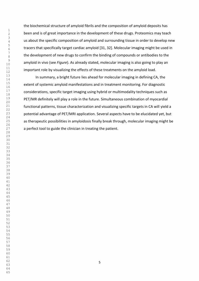
The figure shows the pathogenesis of amyloid deposition for the three major types,
i.e. AL, ATTR and AA type in the heart. Increased thickness of the ventricular walls and strain
differences (apical sparing) caused by extracellular deposition of amyloid can be detected by
echocardiography. Delayed enhancement of gadolinium using MRI reflects an increased
extravascular volume of the myocardial tissue. Serum amyloid P component (SAP) binds in a
calcium-dependent way to all types of amyloid in many vital organs, but when labeled with
123I, not to amyloid in the heart [7]. This may be caused by the lack of a fenestrated
endothelium in the myocardium, hindering access of the large 125 kDa tracer to the
extracellular space. Florbetapen is closely related to thioflavin T, a strong stain of amyloid
that binds to repetitive motifs at the surface of the fibrils [8]. Bisphosphonates and
pyrophosphate bind strongly to ATTR amyloid and weakly or not at all to AA and AL amyloid.
Although there may be a relation with microcalcifications [9], the bone tracer 18F-NaF does
not always show the same strong tracer retention in ATTR CA [10]. The specific binding is
probably not only to calcium, but technetium can bind to some metals [11] and/or to
sulfhydryl groups on ATTR amyloid [12, 13]. Aprotinin has been used in the past to detect CA
[14] and it may bind to repetitive motifs and/or electrostatically [15]. MIBG normally
accumulates in vesicles in sympathetic nerve endings close to myocardial cells and the
reduced uptake and increased loss probably reflects myocardial cell damage caused by
amyloid [16].
In the current study of Kircher and colleagues in this issue, the performance of 18F-
florbetaben-PET/CT in the detection of CA (AL, ATTR and AA) was examined in 22 patients (5
histologically proven and 17 clinically suspected) and compared to echocardiography, CMR
and 99mTc-DPD bone scintigraphy [17]. Additionally, the use of 18F-florbetaben-PET/CT for
quantification of amyloid burden, including myocardial tracer retention (MTR), and
monitoring of treatment response was assessed. Myocardial 18F-florbetaben retention was
found consistent with CA in 14/22 patients. Suspicion of CA was subsequently dropped in all
eight PET-negative patients. Amyloid subtypes showed characteristic retention patterns (AL
> AA > ATTR; all p<0.005). MTR correlated with morphologic and functional parameters, as
measured by CMR and echo (all r>0.47|, all p<0.05), but not with cardiac biomarkers.
1 2 3 4 5 6 7 8 9 10 11 12 13 14 15 16 17 18 19 20 21 22 23 24 25 26 27 28 29 30 31 32 33 34 35 36 37 38 39 40 41 42 43 44 45 46 47 48 49 50 51 52 53 54 55 56 57 58 59 60 61 62 63 64 65

3
Changes in MTR from baseline to follow-up corresponded well to treatment response, as
assessed by cardiac biomarkers and performance status.
The authors concluded that imaging of CA with 18F-florbetaben PET/CT is feasible and
might be useful in differentiating CA subtypes. Although this study consisted of a small,
heterogeneous patient cohort, including treatment-naïve as well as pre-treated patients, it
underlines the clinical value of applying new more specific PET imaging tracers in cardiac
imaging. To demonstrate, however, the potential of amyloid-directed PET as a non-invasive
instrument of (early) therapy monitoring, an understanding of the underlying biology is of
utmost importance. Histological proof was lacking in the current study, including additional
whole-body imaging to detect further sites of organ involvement in systemic amyloidosis,
and this should be addressed in future studies.
More specific PET-imaging tracers in amyloidosis selectively bind to β-amyloid
plaques and were originally designed as an aid to establish the clinical diagnosis of
Alzheimer’s disease. These specific radiopharmaceuticals are the benzothiazoles 11C–
Pittsburgh compound-B (11C-PiB) and 18F–florbetaben, while18F–florbetapir is a stilbene
derivative with a very similar structure.
11C-PiB as well as 18F-florbetapir have been used as tracers for cardiac amyloid in
patients with ATTR and AL cardiac amyloidosis [18, 19]. However, 11C-PiB is only available in
centers with an on-site cyclotron. Manwani et al. evaluated cardiac uptake with 18F-
florbetapir PET in patients with systemic AL amyloidosis and cardiac involvement before and
after treatment, as well as its serial utility in monitoring in 15 patients [20]. There was a
suggestion that treatment-naïve patients may have higher cardiac uptake. In addition,
correlation of myocardial 18F-florbetapir uptake with histological findings in 20 amyloidosis
patients (10 AL and 10 ATTR) versus 10 control subjects revealed significantly lower specific
18F-florbetapir binding in controls (p=0.002) [21]. Specific 18F-florbetapir binding in AL
samples was significantly higher than in ATTR samples (p=0.001). Furthermore, increase in
18F-florbetapir binding on autoradiography correlated well with increasing
echocardiography-derived LV wall mass, due to more advanced stages of the disease. More
importantly, 18F-florbetapir binding was already present in small amounts before LV wall
thickness increased.
A recent systematic review of the application of PET imaging with 11C-PiB, 18F-
florbetapir and 18F-florbetaben in 6 studies (n=98 subjects) demonstrated a sensitivity of
1 2 3 4 5 6 7 8 9 10 11 12 13 14 15 16 17 18 19 20 21 22 23 24 25 26 27 28 29 30 31 32 33 34 35 36 37 38 39 40 41 42 43 44 45 46 47 48 49 50 51 52 53 54 55 56 57 58 59 60 61 62 63 64 65

4
92% and a specificity of 83% for the detection of AL and ATTR CA [22]. Further, regular bone
scintigraphy has emerged as a reliable, non-invasive tool to diagnose cardiac amyloidosis
due to ATTR (either ATTRv or ATTRwt), with a sensitivity of 92.2% (95% CI 89-95%) and a
specificity of 95.4% [23, 24]. The use of iodine-123 labelled metaiodobenzylguanidine (123I-
MIBG), a chemical modified analogue of norepinephrine, is well established in patients with
heart failure and plays also an important role in the evaluation of sympathetic innervation
in cardiac amyloidosis [25].
Current treatments are targeted at reducing the production of or stabilisation of the
precursor protein of amyloid deposits and thereby aim to stop or slow down further
accumulation of amyloid. In AL amyloidosis treatment is directed against light chain-
producing plasma cells in order to normalize the light chain serum levels. Recently, gene-
silencing oligonucleotide drugs that inhibit hepatic synthesis of the precursor protein
transthyretin proved to be effective in hereditary ATTR amyloidosis [26, 27]. Progression of
peripheral polyneuropathy almost ceases entirely and progression of cardiac manifestations
may be also be halted, or even reversed [28]. Another treatment approach for ATTR CA is
stabilization of the TTR tetramer, thereby interfering with the supply of precursor protein
and resulting in slowing down of disease progression [29]. Molecular imaging should be able
to visualize regression of CA under these new treatment regimens, but data are lacking at
this moment. However, these new drugs probably do not inhibit synthesis of variant
transthyretin by the choroid plexus in the central nervous system (CNS) leading to
meningeal-vascular ATTRv amyloid deposition in the long-term, i.e. 10-15 years. This may
become a new challenge in ATTRv patients, as these drugs are expected to improve life
expectancy far beyond the current survival without treatment. Future studies with molecular
imaging are necessary to assess the extent of the CNS manifestations. 11C-Pib-PET scan
currently appears to be the best imaging technique for early detection of these CNS
manifestations, by showing - in a presymptomatic state - a pattern different from that seen
in Alzheimer’s disease [30]. Imaging with 18F-florbetaben-PET/CT is also promising as it may
visualize both intra-cerebral deposition of amyloid as well as deposition in other organs,
particularly the heart.
Several treatments aiming at promoting amyloid removal are currently being
investigated. These treatments, based on the use of monoclonal antibodies and small
compounds, employ immunological mechanisms to clear amyloid deposits. Understanding
1 2 3 4 5 6 7 8 9 10 11 12 13 14 15 16 17 18 19 20 21 22 23 24 25 26 27 28 29 30 31 32 33 34 35 36 37 38 39 40 41 42 43 44 45 46 47 48 49 50 51 52 53 54 55 56 57 58 59 60 61 62 63 64 65
5
the biochemical structure of amyloid fibrils and the composition of amyloid deposits has
been and is of great importance in the development of these drugs. Proteomics may teach
us about the specific composition of amyloid and surrounding tissue in order to develop new
tracers that specifically target cardiac amyloid [31, 32]. Molecular imaging might be used in
the development of new drugs to confirm the binding of compounds or antibodies to the
amyloid in vivo (see Figure). As already stated, molecular imaging is also going to play an
important role by visualizing the effects of these treatments on the amyloid load.
In summary, a bright future lies ahead for molecular imaging in defining CA, the
extent of systemic amyloid manifestations and in treatment monitoring. For diagnostic
considerations, specific target imaging using hybrid or multimodality techniques such as
PET/MR definitely will play a role in the future. Simultaneous combination of myocardial
functional patterns, tissue characterization and visualizing specific targets in CA will yield a
potential advantage of PET/MRI application. Several aspects have to be elucidated yet, but
as therapeutic possibilities in amyloidosis finally break through, molecular imaging might be
a perfect tool to guide the clinician in treating the patient.
1 2 3 4 5 6 7 8 9 10 11 12 13 14 15 16 17 18 19 20 21 22 23 24 25 26 27 28 29 30 31 32 33 34 35 36 37 38 39 40 41 42 43 44 45 46 47 48 49 50 51 52 53 54 55 56 57 58 59 60 61 62 63 64 65

6
References
1. Maurer MS, Elliott P, Comenzo R, Semigran M, Rapezzi C. Addressing common questions
encountered in the diagnosis and management of cardiac amyloidosis. Circulation 2017;
135:1357-1377.
2. Gonzalez-Lopez E, Gallego-Delgado M, Guzzo-Merello G, de Haro-Del Moral FJ, Cobo-
Marcos M, Robles C, et al. Wild-type transthyretin amyloidosis as a cause of heart failure
with preserved ejection fraction. Eur Heart J 2015; 36:2585–2594.
3. Benson MD, Buxbaum JN, Eisenberg DS, Merlini G, Saraiva MJM, Sekijima Y, et al.
Amyloid nomenclature 2018: recommendations by the International Society of
Amyloidosis (ISA) nomenclature committee. Amyloid 2018; 25:215-219.
4. Lousada I, Comenzo RL, Landau H, Guthrie S, Merlini G. Light Chain Amyloidosis: Patient
Experience Survey from the Amyloidosis. Research Consortium. Adv Ther 2015; 32:920-8.
5. Mohammed SF, Mirzoyev SA, Edwards WD, Dogan A, Grogan DR, Dunlay SM, et al. Left
ventricular amyloid deposition in patients with heart failure and preserved ejection
fraction. JACC Heart Fail 2014; 2:113–122.
6. Martinez-Naharro A, Treibel TA, Abdel-Gadir A, Bulluck H, Zumbo G, Knight DS, et al.
Magnetic resonance in transthyretin cardiac amyloidosis. J Am Coll Cardiol 2017; 70:466–
477.
7. Hazenberg BP, van Rijswijk MH, Piers DA, Lub-de Hooge MN, Vellenga E, Haagsma EB, et
al. Diagnostic performance of 123I-labeled serum amyloid P component scintigraphy in
patients with amyloidosis. Am J Med 2006; 119: 355.e15-24.
8. Biancalana M, Koide S. Molecular mechanism of Thioflavin-T binding to amyloid fibrils.
Biochim Biophys Acta 2010; 1804:1405-12.
9. Stats MA, Stone JR. Varying levels of small microcalcifications and macrophages in ATTR
and AL cardiac amyloidosis: implications for utilizing nuclear medicine studies to subtype
amyloidosis. Cardiovasc Pathol 2016; 25:413-7.
10. Ng QKT, Sethi P, Saunders TA, Pampaloni MH, Flavell RR. Discordant Findings on 18F-NaF
and 99mTc-HDP Bone Scans in a Patient With ATTR Cardiac Amyloidosis. Clin Nucl Med
2018; 43:e89-e92.
1 2 3 4 5 6 7 8 9 10 11 12 13 14 15 16 17 18 19 20 21 22 23 24 25 26 27 28 29 30 31 32 33 34 35 36 37 38 39 40 41 42 43 44 45 46 47 48 49 50 51 52 53 54 55 56 57 58 59 60 61 62 63 64 65

7
11. Susuki S, Ando Y, Sato T, Nishiyama M, Miyata M, Suico MA, et al. Multi-elemental
analysis of serum and amyloid fibrils in familial amyloid polyneuropathy patients.
Amyloid 2008; 15:108-16.
12. Morton KA. Extra-skeletal uptake of bone agents. J Nucl Med Technol 1999; 27:51-3.
13. Nakanishi T, Yoshioka M, Moriuchi K, Yamamoto D, Tsuji M, Takubo T. S-sulfonation of
transthyretin is an important trigger step in the formation of transthyretin-related
amyloid fibril. Biochim Biophys Acta 2010; 1804:1449-56.
14. Aprile C, Marinone G, Saponaro R, Bonino C, Merlini G. Cardiac and pleuropulmonary AL
amyloid imaging with technetium-99m labelled aprotinin. Eur J Nucl Med 1995; 22:1393-
401.
15. Cardoso I, Pereira PJ, Damas AM, Saraiva MJ. Aprotinin binding to amyloid fibrils. Eur J
Biochem 2000; 267:2307-11.
16. Jonker DL, Hazenberg BP, Nienhuis HL, Slart RH, Glaudemans AW, Noordzij W. Imaging
cardiac innervation in hereditary transthyretin (ATTRm) amyloidosis: a marker for
neuropathy or cardiomyopathy? J Nucl Cardiol 2019 [Epub ahead of print].
17. Kircher M, Ihne S, Brumberg J, Morbach C, Knop S, Kortüm KM, et al. Detection of
Cardiac Amyloidosis with 18F-Florbetaben-PET/CT in Comparison to Echocardiography,
Cardiac MRI and DPD-Scintigraphy. Eur J Nucl Med Mol Imag. 2019 Current issue.
18. Antoni G, Lubberink M, Estrada S, Axelsson J, Carlson K, Lindsjo L, et al. In vivo
visualization of amyloid deposits in the heart with 11C-PIB and PET. J Nucl Med. 2013;
54:213-20.
19. Dorbala S, Vangala D, Semer J, Strader C, Bruyere JR Jr, Di Carli MF, et al. Imaging cardiac
amyloidosis: A pilot study using 18Fflorbetapir positron emission tomography. Eur J Nucl
Med Mol Imaging. 2014; 41:1652-62.
20. Manwani R, Page J, Lane T, Burniston M, Skillen A, Lachmann HJ, et al. A pilot study
demonstrating cardiac uptake with 18F-florbetapir PET in AL amyloidosis patients with
cardiac involvement. Amyloid. 2018; 25:247-252.
21. Park MA, Padera RF, Belanger A, Dubey S, Hwang DH, Veeranna V, et al. 18F-Florbetapir
Binds Specifically to Myocardial Light Chain and Transthyretin Amyloid Deposits:
Autoradiography Study. Circ Cardiovasc Imaging. 2015 Aug;8(8).
22. Kim YJ, Ha S, Kim YI. Cardiac amyloidosis imaging with amyloid positron emission
tomography: A systematic review and meta-analysis. J Nucl Cardiol. 2018 Jul 18.
1 2 3 4 5 6 7 8 9 10 11 12 13 14 15 16 17 18 19 20 21 22 23 24 25 26 27 28 29 30 31 32 33 34 35 36 37 38 39 40 41 42 43 44 45 46 47 48 49 50 51 52 53 54 55 56 57 58 59 60 61 62 63 64 65

8
23. Glaudemans AW, van Rheenen RW, van den Berg MP, Noordzij W, Koole M, Blokzijl H, et
al. Bone scintigraphy with (99m)technetium-hydroxymethylene diphosphonate allows
early diagnosis of cardiac involvement in patients with transthyretin-derived systemic
amyloidosis. Amyloid 2014; 21:35–44.
24. Treglia G, Glaudemans AWJM, Bertagna F, Hazenberg BPC, Erba PA, Giubbini R, et al. Eur
J Nucl Med Mol Imaging. 2018 Oct; 45(11):1945-1955.
25. Slart RHJA, Glaudemans AWJM, Hazenberg BPC, Noordzij W. Imaging cardiac innervation
in amyloidosis. J Nucl Cardiol. 2019 Feb; 26(1):174-187.
26. Adams D, Gonzalez-Duarte A, O'Riordan WD, Yang CC, Ueda M, Kristen AV, et al.
Patisiran, an RNAi therapeutic for hereditary transthyretin amyloidosis. N Engl J Med
2018; 379:11-21.
27. Benson MD, Waddington-Cruz M, Berk JL, Polydefkis M, Dyck PJ, Wang AK, et al.
Inotersen treatment for patients with hereditary transthyretin amyloidosis. N Engl J Med
2018; 379:22-31.
28. Solomon SD, Adams D, Kristen A, Grogan M, González-Duarte A, Maurer MS, et al. Effects
of Patisiran, an RNA interference therapeutic, on cardiac parameters in patients with
hereditary transthyretin-mediated amyloidosis. Circulation. 2019; 139:431-443.
29. Maurer MS, Schwartz JH, Gundapaneni B, Elliott PM, Merlini G, Waddington-Cruz M, et
al. Tafamidis treatment for patients with transthyretin amyloid cardiomyopathy. N Engl J
Med 2018; 379:1007-16.
30. Sekijima Y, Yazaki M, Oguchi K, Ezawa N, Yoshinaga T, Yamada M, et al. Cerebral amyloid
angiopathy in posttransplant patients with hereditary ATTR amyloidosis. Neurology.
2016; 87:773-81
31. Brambilla F, Lavatelli F, Di Silvestre D, Valentini V, Palladini G, Merlini G, et al. Shotgun
protein profile of human adipose tissue and its changes in relation to systemic
amyloidoses. J Proteome Res. 2013; 12:5642-55.
32. Di Silvestre D, Brambilla F, Mauri PL. Multidimensional protein identification technology
for direct-tissue proteomics of heart. Methods Mol Biol 2013; 1005:25-38.
1 2 3 4 5 6 7 8 9 10 11 12 13 14 15 16 17 18 19 20 21 22 23 24 25 26 27 28 29 30 31 32 33 34 35 36 37 38 39 40 41 42 43 44 45 46 47 48 49 50 51 52 53 54 55 56 57 58 59 60 61 62 63 64 65
9
Figure legend
In the upper part the pathogenic pathways of AA, AL and ATTR, the major three types of
amyloidosis, are shown vertically. In AA, chronic inflammation stimulates the liver to
produce the acute phase protein SAA, that becomes cleaved and misfolded. In AL, clonal
plasma cells in the bone marrow produce immunoglobulin light chains, of which the variable
part becomes misfolded. In ATTR, the liver produces the tetrameric protein transthyretin,
that dissociates into monomers that become misfolded. The misfolded precursor proteins
aggregate, finally resulting in the formation of amyloid fibrils (AA shown in red, AL in blue,
and ATTR in green). Without specific immunohistochemical staining fibrils of the different
types are under the microscope indistinguishable from each other (shown in grey).
The lower part of the figure is focused on imaging of amyloid deposition in the extracellular
myocardial tissue. Echocardiography reveals increased wall thickness and stiffness, whereas
late gadolinium enhancement in MRI reflects an increased volume of the extracellular
myocardial tissue. Mechanisms used for molecular imaging of amyloid show specific binding
of tracers to amyloid-specific elements of fibrils and related extracellular matrix in which the
amyloid is anchored. Connected to amyloid in the extracellular matrix an increase of
molecules such as serum amyloid P component (SAP), heparan sulphate (HS), apolipoprotein
E (apoE), and laminin is found. Aprotinin and thioflavin-like agents (PiB, florbetapir and
florbetapen) bind directly to repetitive motifs on the exterior surface of the fibrils.
Technetium-labelled bone seeking agents (pyrophosphate and bisphosphonates) may bind
to calcium, but also to metals (Zn, Fe, Cu, Mn and Ba) and sulfhydryl (S-H) groups on the
ATTR amyloid fibril. Cardiac innervation imaging agents (e.g. 123I-MIBG, 11C-mHED)
accumulate less than expected in the nerve endings, as reflected in reduced cardiac tracer
uptake and enhanced wash-out.
1 2 3 4 5 6 7 8 9 10 11 12 13 14 15 16 17 18 19 20 21 22 23 24 25 26 27 28 29 30 31 32 33 34 35 36 37 38 39 40 41 42 43 44 45 46 47 48 49 50 51 52 53 54 55 56 57 58 59 60 61 62 63 64 65

Figure Click here to access/download;Figure;Editorial Slart et al Eur JNucl Med Mol Imag FINAL 2019.jpg