Typing of Pseudomonas aeruginosa from hemorrhagic ...
21
General rights Copyright and moral rights for the publications made accessible in the public portal are retained by the authors and/or other copyright owners and it is a condition of accessing publications that users recognise and abide by the legal requirements associated with these rights. Users may download and print one copy of any publication from the public portal for the purpose of private study or research. You may not further distribute the material or use it for any profit-making activity or commercial gain You may freely distribute the URL identifying the publication in the public portal If you believe that this document breaches copyright please contact us providing details, and we will remove access to the work immediately and investigate your claim. Downloaded from orbit.dtu.dk on: Jan 17, 2022 Typing of Pseudomonas aeruginosa from hemorrhagic pneumonia in mink (Neovison vison) Salomonsen, Charlotte Mark; Themudo, G. E.; Jelsbak, Lars; Molin, Søren; Høiby, N.; Hammer , Anne Sofie Published in: Veterinary Microbiology Link to article, DOI: 10.1016/j.vetmic.2012.12.003 Publication date: 2013 Link back to DTU Orbit Citation (APA): Salomonsen, C. M., Themudo, G. E., Jelsbak, L., Molin, S., Høiby, N., & Hammer , A. S. (2013). Typing of Pseudomonas aeruginosa from hemorrhagic pneumonia in mink (Neovison vison). Veterinary Microbiology, 163(1-2), 103-109. https://doi.org/10.1016/j.vetmic.2012.12.003
Transcript of Typing of Pseudomonas aeruginosa from hemorrhagic ...
General rights Copyright and moral rights for the publications made accessible in the public portal are retained by the authors and/or other copyright owners and it is a condition of accessing publications that users recognise and abide by the legal requirements associated with these rights.
Users may download and print one copy of any publication from the public portal for the purpose of private study or research.
You may not further distribute the material or use it for any profit-making activity or commercial gain
You may freely distribute the URL identifying the publication in the public portal If you believe that this document breaches copyright please contact us providing details, and we will remove access to the work immediately and investigate your claim.
Downloaded from orbit.dtu.dk on: Jan 17, 2022
Typing of Pseudomonas aeruginosa from hemorrhagic pneumonia in mink (Neovisonvison)
Salomonsen, Charlotte Mark; Themudo, G. E.; Jelsbak, Lars; Molin, Søren; Høiby, N.; Hammer , AnneSofie
Published in:Veterinary Microbiology
Link to article, DOI:10.1016/j.vetmic.2012.12.003
Publication date:2013
Link back to DTU Orbit
Citation (APA):Salomonsen, C. M., Themudo, G. E., Jelsbak, L., Molin, S., Høiby, N., & Hammer , A. S. (2013). Typing ofPseudomonas aeruginosa from hemorrhagic pneumonia in mink (Neovison vison). Veterinary Microbiology,163(1-2), 103-109. https://doi.org/10.1016/j.vetmic.2012.12.003
1
Typing of Pseudomonas aeruginosa from
hemorrhagic pneumonia in mink
(Neovison vison)
C. M. Salomonsen1, G. E. Themudo2, L. Jelsbak3, S. Molin3, N. Høiby4, A. S. Hammer1,5
1 National Veterinary Institute, Technical University of Denmark, Hangoevej 2, DK-8200 Aarhus N,
Denmark. E-mail: [email protected] (corresponding author).
2 National Institute of Public Health, University of Southern Denmark, Oester Farimagsgade 5A, 2, DK-
1353 Copenhagen, Denmark
3 Department of Systems Biology, Technical University of Denmark, Matematiktorvet, Building 301,
DK-2800 Lyngby, Denmark
4 Department of Clinical Microbiology, Rigshospitalet, Blegdamsvej 9, DK-2100 Copenhagen Ø,
Denmark
5 Present address: Department of Veterinary Disease Biology, Faculty of Health and Medical
Sciences, University of Copenhagen , Ridebanevej 3, DK-1870 Frederiksberg C, Denmark
2
Abstract
Hemorrhagic pneumonia in mink (Neovison vison) is caused by Pseudomonas aeruginosa and is an
acute and fatal disease in farmed mink. Earlier work has demonstrated that some outbreaks of
hemorrhagic pneumonia are caused by pathogenic strains while most outbreaks are caused by local
strains. The objective of this study was to determine the genetic and geographical relationship
among outbreaks of hemorrhagic pneumonia by pulsed field gel electrophoresis typing of P.
aeruginosa isolates. Furthermore, chosen isolates were typed by a commercial genotyping method
based on single nucleotide polymorphisms (SNPs) and compared to a larger dataset of human and
environmental origin. The bacterial isolates were obtained from diagnostic samples from 2002-2009
and contained 164 isolates from 95 outbreaks on 90 farms. Our results show that most outbreaks of
hemorrhagic pneumonia in mink are caused by distinct strains of P. aeruginosa. We also identified
related P. aeruginosa strains which, together with two prevalent but unrelated clones, caused one
third of the outbreaks of hemorrhagic pneumonia supporting the sparse literature on this subject.
None of the SNP typed strains were identified in a large dataset of human and environmental origin.
Keywords: Pseudomonas aeruginosa, mink, hemorrhagic pneumonia, pulsed-field gel
electrophoresis, single nucleotide polymorphism
Introduction
Pseudomonas aeruginosa has been described as a cause of hemorrhagic pneumonia in mink since
1953 (Knox, 1953). The disease is almost exclusively seen from September to early December in
Denmark and can cause an epizootic on the mink farm with mortalities ranging from 1% to 50%
(Knox, 1953; Honda et al., 1977). It is acute and characterized by sudden deaths among the mink
3
which are often found dead with blood around nostrils and mouth. No known underlying cause has
been identified preceding P. aeruginosa pneumonia in mink, which make mink the only species
known to be susceptible to contagious, acute and fatal lung infections with P. aeruginosa.
Only descriptive epidemiological research has been published on P. aeruginosa pneumonia in mink
(Knox, 1953; Shimizu et al., 1974; Long and Gorham, 1981; Hammer et al., 2003). P. aeruginosa is
widespread in both environment and various disease habitats. Earlier work has demonstrated that
specific clones only rarely are associated with certain habitats and that most P. aeruginosa possess
the genes required for establishing an infection (Römling et al., 1994; Alonso et al., 1999; Wolfgang
et al., 2003; Morales et al., 2004; Stewart et al., 2011)
To identify possible successful clones and explore the relationship of P. aeruginosa included in this
study, bacterial isolates were typed by pulsed-field gel electrophoresis (PFGE) which is considered
the “gold standard” for discriminative typing of P. aeruginosa (Grundmann et al., 1995; Tenover et
al., 1997; Johnson et al., 2007). To uncover the relationship of mink isolates with a larger dataset of
human and environmental isolates 18 PFGE-typed isolates were further characterized by an array
hybridization kit; the AT biochip (P. aeruginosa Genotyping Kit, Clondiag Chip Technologies,
Germany) as previously described (Morales et al., 2004; Wiehlmann et al., 2007). While PFGE is
based on mutations in restriction sites dispersed in the chromosome, the AT biochip recognizes SNPs
in the core genome and genetic markers for specific gene islets and islands in the accessory genome.
Materials and methods
Materials
One hundred and sixty-four isolates representing 95 outbreaks of hemorrhagic pneumonia on 90
mink farms were typed by PFGE. Only P. aeruginosa isolated from lungs of mink dying of hemorrhagic
pneumonia were included in the study. The mink were submitted for diagnostic investigations from
4
Danish mink farms during the period 2002-2009 and formed 85% of the recorded outbreaks of
hemorrhagic pneumonia in this period. P. aeruginosa were diagnosed on the basis of characteristic
colony morphology on blood agar and MacConkey agar, smell, Gram-stain and positive cytochrome
oxidase reaction. Serotyping was performed using polyclonal antisera (Difco™ polyclonal serotyping,
Detroit, MI, U.S.A).
The isolates were freeze-dried at the time of isolation and stored at room temperature before they
were revived by incubation in veal infusion broth (Difco™) for 24 hours at 37⁰C and plated on blood
agar plates.
Eighteen isolates were furthermore typed using the AT Biochip. The AT biochip recognizes 13 highly
conserved genetic regions and several regions in the accessory genome including genetic islands and
islets. The isolates selected for genotyping either belonged to a cluster which showed only few band
differences on the PFGE profile, or were identified as particularly prevalent or as originating from
repeated outbreaks on the same farm. In addition, two isolates were chosen only on behalf of
serotype to represent the serotypes 5 and 7/8 since the rest of the chosen isolates belonged to
serotype 6. The 18 PFGE types were responsible for 36 of the 95 outbreaks.
Methods
The PFGE procedure has been described elsewhere (Nauerby et al., 2000) and was followed with
some modifications. The agarose plugs were digested with 0.1 mg/ml proteinase K in lysis buffer (1M
Tris pH=8.0, 0.5M EDTA pH=8.0, 10 % N–lauroyl sarcosine) for 2 hours in a shaking water bath (56⁰C,
200 rpm). Thin slices cut from the plugs were digested with SpeI (BioLab, Ipswich, MA, USA) for 4
hours at 37⁰C. The restriction fragments were separated as previously described (Nauerby et al.,
2000). Lambda Ladder PFG markers (BioLab) were run with the samples on the gels. The gels were
stained with ethidium bromide (2 µg/ml) for 7 min., washed in distilled water for 15 min and
5
photographed under UV-light by GelDoc-It Imaging System (AH Diagnostics, Aarhus, Denmark) with
the software VisionWorks LS (UVP, Upland, CA, USA).
The resulting band profiles were analyzed using Bionumerics (Applied Maths, ver. 4.50) with Dice
band based comparison and a position tolerance of 1.7 % as suggested by Carriço et al. (2005)
The isolates were defined as belonging to the same strain if the isolates had indistinguishable PFGE
profiles. If the isolates differed by 1 to 5 bands (corresponding to similarities above 85 %) they were
regarded as belonging to a cluster of closely related strains.
Band profiles were exported from Bionumerics as binary data into Arlequin v.3.5.1.2 (Excoffier and
Lischer, 2010). Pairwise measures of genetic distance (FST) were calculated using Nei’s average
number of pairwise differences (Nei and Li, 1979). Geographical distances between farms were
calculated in ArcGIS (Redlands, CA, U.S.A.) based on their geographical coordinates. A Mantel test
was used for fitting regression models between the two matrices, consisting of the genetic distance
(response matrix) and the Euclidean geographic distance (explanatory matrix) (Mantel. 1967). The
Mantel test was performed using Arlequin.
The manufacturer’s protocol for the AT-Biochip was followed closely and has been described
elsewhere (Jelsbak et al., 2007; Wiehlmann et al., 2007).
Results
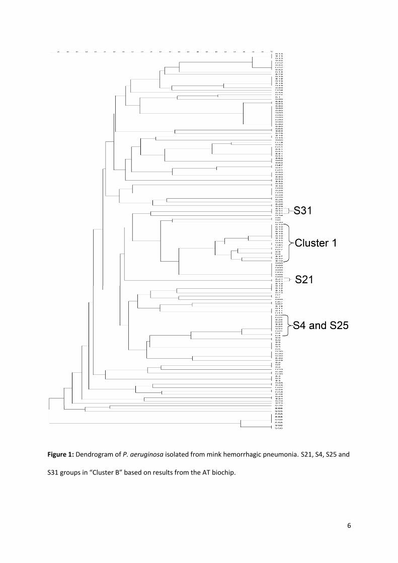
We determined the PFGE profiles of 164 isolates sampled from 95 outbreaks of hemorrhagic
pneumonia on 90 mink farms, and found 72 distinct PFGE patterns (Figure 1). Seventy-two per cent
of the outbreaks were caused by serotype 6 while serotype 5 was recovered in 22% of the outbreaks
and serotype 7/8 in the final six per cent. Isolates with similarities of their PFGE profiles of more than
80% invariably displayed the same serotype.
6
Figure 1: Dendrogram of P. aeruginosa isolated from mink hemorrhagic pneumonia. S21, S4, S25 and
S31 groups in “Cluster B” based on results from the AT biochip.
7
Isolates from two or more mink were typed in 47 (50%) of the outbreaks. In 41 (87%) of these
outbreaks the isolates showed indistinguishable PFGE profiles when recovered from the same
outbreak while two apparently unrelated PFGE profiles were discovered in six outbreaks represented
by two or more isolates. Six farms experienced hemorrhagic pneumonia outbreaks twice in the study
period with one to six years between the outbreaks. In three of these farms the outbreak was caused
by a P. aeruginosa strain with indistinguishable PFGE profile from the one causing the previous
outbreak (S3, S12, S19). On the other three farms, the PFGE profiles between the isolates causing
outbreak one and two showed similarities ranging from 33-52%. The serotypes of P. aeruginosa
causing these outbreaks belonged to the same serotype (6) in two out of three cases while the last
farm was infected with serotype 6 in the first outbreak and serotype 5 in the next.
Eleven strains were each recovered from two outbreaks on different farms, while four strains were
each recovered from three or more outbreaks on different farms. The outbreaks caused by the same
strains were sometimes located in the same geographic areas but also widely apart. Eight strains
responsible for two or three farm outbreaks occurred on farms sharing the same central food kitchen
while four strains causing two outbreaks did not share food kitchen. The remaining three strains
caused outbreaks on several farms of which some shared food kitchen and others did not (Table 1).
Seven strains (S10, S15, S45, S27, S8, S37 and S40) grouped in a cluster with similarities of above 85%
and PFGE profiles with 5 band differences or less among each other. This group was called “Cluster
1” (Figure 1). The members of this cluster caused nine outbreaks; they were geographically
widespread and occurred from 2002 to 2006.
The Mantel test between geographical distance and genetic difference (Fst) was not significant
(p=0.59), meaning there was no correlation between the outbreak locations and relationship among
the isolates. The dendrogram also revealed a large amount of differentiation among the PFGE types
(Figure 1). A larger version of the dendrogram can be viewed in supplementary material.
8
Eighteen isolates were further typed using the AT biochip. The isolates either belonged to Cluster 1
(S10, S15, S45, S27, S8, S37 and S40), were particularly prevalent (S25, S31, S66 and S80), showed
similarities of above 85% to one of the above mentioned strains (S4 to S25 and S83 to S80) or
occurred on a farm with previous outbreaks of hemorrhagic pneumonia (S73 and S84, together with
S66, S21, S25 and S37, which also belonged to Cluster 1 or were particularly prevalent). Two isolates
represented the serotypes 5 and 7/8 (S72 and S79).
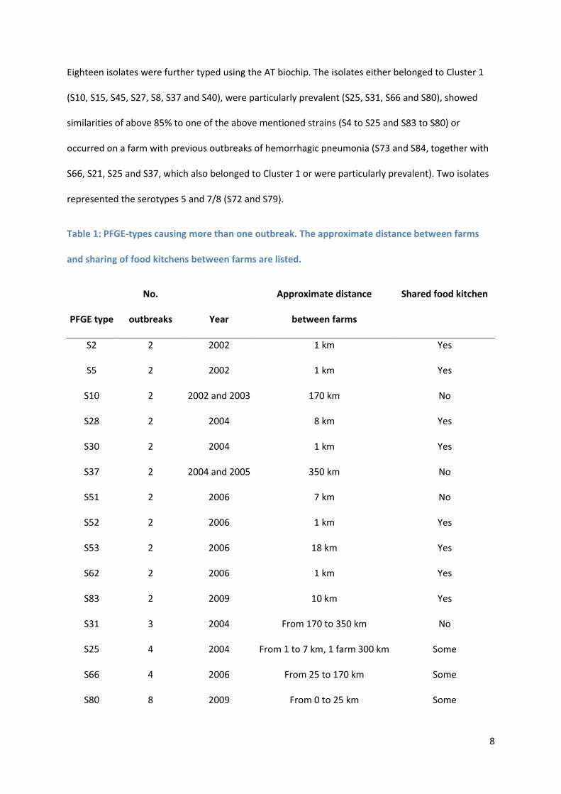
Table 1: PFGE-types causing more than one outbreak. The approximate distance between farms
and sharing of food kitchens between farms are listed.
PFGE type
No.
outbreaks Year
Approximate distance
between farms
Shared food kitchen
S2 2 2002 1 km Yes
S5 2 2002 1 km Yes
S10 2 2002 and 2003 170 km No
S28 2 2004 8 km Yes
S30 2 2004 1 km Yes
S37 2 2004 and 2005 350 km No
S51 2 2006 7 km No
S52 2 2006 1 km Yes
S53 2 2006 18 km Yes
S62 2 2006 1 km Yes
S83 2 2009 10 km Yes
S31 3 2004 From 170 to 350 km No
S25 4 2004 From 1 to 7 km, 1 farm 300 km Some
S66 4 2006 From 25 to 170 km Some
S80 8 2009 From 0 to 25 km Some
9
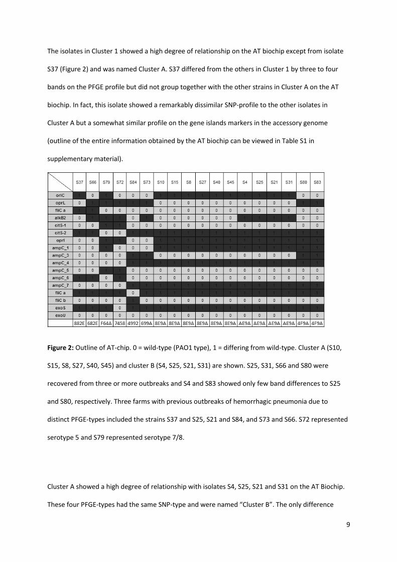
The isolates in Cluster 1 showed a high degree of relationship on the AT biochip except from isolate
S37 (Figure 2) and was named Cluster A. S37 differed from the others in Cluster 1 by three to four
bands on the PFGE profile but did not group together with the other strains in Cluster A on the AT
biochip. In fact, this isolate showed a remarkably dissimilar SNP-profile to the other isolates in
Cluster A but a somewhat similar profile on the gene islands markers in the accessory genome
(outline of the entire information obtained by the AT biochip can be viewed in Table S1 in
supplementary material).
Figure 2: Outline of AT-chip. 0 = wild-type (PAO1 type), 1 = differing from wild-type. Cluster A (S10,
S15, S8, S27, S40, S45) and cluster B (S4, S25, S21, S31) are shown. S25, S31, S66 and S80 were
recovered from three or more outbreaks and S4 and S83 showed only few band differences to S25
and S80, respectively. Three farms with previous outbreaks of hemorrhagic pneumonia due to
distinct PFGE-types included the strains S37 and S25, S21 and S84, and S73 and S66. S72 represented
serotype 5 and S79 represented serotype 7/8.
Cluster A showed a high degree of relationship with isolates S4, S25, S21 and S31 on the AT Biochip.
These four PFGE-types had the same SNP-type and were named “Cluster B”. The only difference
10
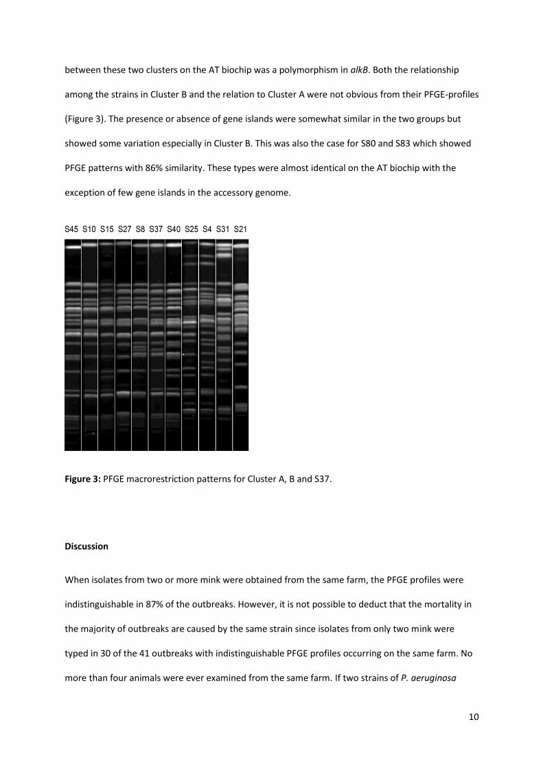
between these two clusters on the AT biochip was a polymorphism in alkB. Both the relationship
among the strains in Cluster B and the relation to Cluster A were not obvious from their PFGE-profiles
(Figure 3). The presence or absence of gene islands were somewhat similar in the two groups but
showed some variation especially in Cluster B. This was also the case for S80 and S83 which showed
PFGE patterns with 86% similarity. These types were almost identical on the AT biochip with the
exception of few gene islands in the accessory genome.
Figure 3: PFGE macrorestriction patterns for Cluster A, B and S37.
Discussion
When isolates from two or more mink were obtained from the same farm, the PFGE profiles were
indistinguishable in 87% of the outbreaks. However, it is not possible to deduct that the mortality in
the majority of outbreaks are caused by the same strain since isolates from only two mink were
typed in 30 of the 41 outbreaks with indistinguishable PFGE profiles occurring on the same farm. No
more than four animals were ever examined from the same farm. If two strains of P. aeruginosa
11
were causing an outbreak with the same frequency of disease attributed to each strain, one would
need to type at least 5 mink to be 95% certain of detecting both strains. The number of animals to be
examined is even higher if the prevalence of a different PFGE type is lower (Martin et al., 1987, pp.
37-38). Unfortunately typing of more isolates from each farm outbreak was not possible using the
present dataset.
Six farms experienced repeated outbreaks in the study period. On three of these farms
indistinguishable PFGE-types were found on the farm with one to three years between the outbreaks
which supports previous work (Hammer et al., 2003). This demonstrates that P. aeruginosa is able to
survive in the environment for at least three years and remain infectious for mink. No relationship
was found between the isolates found on the other three farms with two outbreaks where the time
between the two outbreaks ranged from one to six years. Since some of the farms probably
vaccinate against the disease after having experienced an outbreak, only little can be said of the
ability of P. aeruginosa to re-infect farms in succeeding years and how well it thrives in the
environment on the mink farm.
Differences in relationship among the strains were observed when comparing the results from PFGE
and the AT biochip. While the prevalent strain S80 was related to S83 both by PFGE typing and
especially by the AT biochip, S25, S31 and S21 (isolates from Cluster B) showed a relationship only
when typed by the AT biochip, while their PFGE profiles were quite dissimilar Figure 3). Similarly the
strain S37 was included in Cluster 1 based on the results from PFGE analysis but was unrelated to the
other members of this cluster when examined by the AT biochip. The reason for the observed
difference might lie in the fact that PFGE is considered more discriminative than the AT-biochip and
in some settings it may be too discriminating, making it hard to identify related isolates (Morales et
al., 2004; Johnson et al., 2007; Maatallah et al., 2011). Changes in the accessory genome may lead to
differences in the macro-restriction pattern, while the core genome is unchanged and shared among
related strains. This is more easily recognized by a SNP-based typing system like the AT biochip or
12
conventional multilocus sequence typing (MLST) (Kidd et al., 2011; Waters et al., 2012). Since
horizontal transfer and genetic recombination of the accessory genome are believed to be major
factors of evolution in P. aeruginosa (Römling et al., 1997; Kiewitz and Tümmler, 2000; Larbig et al.,
2002; Morales et al., 2004) few genetic events can cause significant changes in the PFGE pattern if
the bacterium gains or loses large pieces of genetic material. The observed differences in the
accessory genome might be the reason for the differences in PFGE pattern recovered in Cluster 1 and
in the isolates belonging to Cluster B and the types S80 and S83. Most of the isolates carried genes
for type A flagellin and ExoS while all isolates contained the PAGI-1 gene island which carries
regulatory genes and genes required for detoxification of reactive oxygen species (Kung et al., 2010).
ExoS is an effector of the type III secretion system. While the type III secretion system is recognized
as an important virulence factor, ExoU is regarded as being the most virulent effector of this system
(Shaver and Hauser, 2004). The biological significance of the presence or absence of certain genes in
the accessory genome is difficult to assess since the AT biochip was developed purely for typing and
does not reveal the level of expression of these genes. Furthermore nothing can be concluded on the
presence of these genes in the larger population of P. aeruginosa originating from hemorrhagic
pneumonia in mink since the isolates typed by the AT biochip in this dataset were not randomly
chosen.
When using PFGE as the typing method, the strains in Cluster 1 caused nine outbreaks of
hemorrhagic pneumonia which accounted for 9% of the recorded outbreaks. When using the AT
biochip as the typing method the strains in Cluster A and B caused 16 of the recorded outbreaks
(17%). Furthermore 12 outbreaks (13%) were caused by two unrelated but frequently prevalent
strains (S80 and S66). This means that two thirds of the recorded outbreaks of hemorrhagic
pneumonia in mink were caused by unique genotypes. This number may have been lower if a slightly
less discriminating typing method like MLST had been used since MLST is considered more likely to
reveal clonal relatedness (Kidd et al., 2011; Waters et al., 2012). Nonetheless, a large proportion of
outbreaks of hemorrhagic pneumonia are caused by distinct genotypes, which are probably
13
transmitted to the mink from the farm environment. Feed, water troughs, equipment, personnel, air
and feed manufacturers have been suggested as contamination sources (Knox. 1953; Shimizu et al.,
1974; Gierløff. 1980; Hammer et al., 2003).Since many outbreaks caused by indistinguishable P.
aeruginosa strains occurred on closely situated farms, it was not surprising that some of these farms
also shared food kitchen. However, in almost half of the outbreaks caused by indistinguishable PFGE-
types all farms infected with a particular strain did not share food kitchen. This point to local
environmental factors as being more important as contamination sources than the central food
kitchens. Investigations including genetic typing of P. aeruginosa in both animals and suspected
sources have never been carried out and hence no confirmed contaminating source has ever been
identified.
Roughly one third of the outbreaks could be attributed to infection with genotypes that for some
reason were more prevalent in the population of P. aeruginosa isolated from hemorrhagic
pneumonia in mink. This might be an underestimation due to the highly discriminating typing
technique used in this study. Having identified these more prevalent genotypes, we now have an
opportunity to further investigate their genomes and their virulence. The isolates in Cluster A and B
might share common traits making them more virulent to mink or they might be overrepresented in
the environment. When comparing the SNP-types of the 18 isolates typed by the AT biochip with 240
isolates from a previously published dataset (Wiehlmann et al., 2007), P. aeruginosa from mink
grouped with P. aeruginosa isolated from various infections (bacteremia, intensive care units, cystic
fibrosis) and even from water but the specific types as identified by their hexadecimal codes were
not present. This might imply that the mink strains are not overrepresented in the general
environment.
The relatively high numbers of distinct P. aeruginosa strains causing hemorrhagic pneumonia in mink
supports previous work in this field (Hammer et al., 2003). The results are to some degree
comparable to what has been found in human P. aeruginosa infections, where multiple distinct and a
14
number of highly clonal isolates are identified as the cause of various infections (Curran et al., 2004;
Scott and Pitt, 2004; Jelsbak et al., 2007; Tramper-Stranders et al., 2008). In contrast to what has
been found in human infections, the four most frequently isolated strains (S25, S31, S66 and S80)
(Table 1) appear to cause disease in one year only and hence cannot be considered as “widespread
successful clones”. This has been demonstrated for isolates from mink hemorrhagic pneumonia
before (Hammer et al., 2003). The relationship between genetic relatedness and geographical
distance was not significant implying that most often no particular successful strain is circulating
between mink farms in a geographical region. However, no records of the farms’ relationship with
each other are available and the investigation spans many years, so it is possible that some P.
aeruginosa strains have been spread vast distances by vehicles, people or trading of mink, which
would bias the test towards no relationship between geographic region and strain.
Conclusion
Clusters of highly related isolates were recognized using PFGE and the AT biochip. The related
clusters, together with two unrelated but frequently isolated strains, caused almost one third of the
outbreaks indicating that some clones have a higher virulence for mink or are more prevalent in the
environment. A comparison of the biochip typed mink isolates to 240 P. aeruginosa from various
sources showed that none of the mink isolates had been identified in this large dataset. Outbreaks
caused by the same strain of P. aeruginosa most often occurred in the same year with both local and
widespread geographical distribution. In almost half of these outbreaks the farms did not share food
kitchen. Two thirds of the recorded outbreaks could be attributed to distinct strains emphasizing that
environmental strains are likely the most frequent cause of hemorrhagic pneumonia in mink.

15
Acknowledgements
The technical assistance and guidance of Lis Nielsen, Herdis B. Johansen, Jane Andersen and Ulla R.
Johansen is gratefully acknowledged.
Nordvacc and Kopenhagen Fur are thanked for financial support of this study. The funding sources
were not involved in study design, data collection, analysis, interpretation, writing or decision to
publish the work presented in this communication.
Conflict of interest statement
The authors do not have any financial or personal conflicts of interest regarding the work presented
in this communication.
16
References
Alonso, A., Rojo, F., Martinez, J.L., 1999. Environmental and Clinical Isolates of Pseudomonas
aeruginosa show Pathogenic and Biodegradative Properties Irrespective of their Origin. Environ.
Microbiol. 1, 421-430.
Carriço, J.A., Pinto, F.R., Simas, C., Nunes, S., Sousa, N.G., Frazao, N., de Lencastre, H., Almeida, J.S.,
2005. Assessment of Band-Based Similarity Coefficients for Automatic Type and Subtype
Classification of Microbial Isolates Analyzed by Pulsed-Field Gel Electrophoresis. J. Clin.
Microbiol. 43, 5483-5490.
Curran, B., Jonas, D., Grundmann, H., Pitt, T., Dowson, C.G., 2004. Development of a Multilocus
Sequence Typing Scheme for the Opportunistic Pathogen Pseudomonas aeruginosa. J. Clin.
Microbiol. 42, 5644-5649.
Excoffier, L., Lischer, H.E.L., 2010. Arlequin Suite Ver 3.5: A New Series of Programs to Perform
Population Genetics Analyses Under Linux and Windows. Mol. Ecol. Resour. 10, 564-567.
Gierløff, B., 1980. Pseudomonas aeruginosa. IV. Pyocine Typing of Strains Isolated from the Blue Fox
(Alopex lagopus), Mink (Mustela vison), and Dog (Canis familiaris) and from their Environment.
Nord. Vet. Med. 32, 147-160.
Grundmann, H., Schneider, C., Hartung, D., Daschner, F.D., Pitt, T.L., 1995. Discriminatory Power of
Three DNA-Based Typing Techniques for Pseudomonas aeruginosa. J Clin Microbiol 33, 528-534.
Hammer, A.S., Pedersen, K., Andersen, T.H., Jorgensen, J.C., Dietz, H.H., 2003. Comparison of
Pseudomonas aeruginosa Isolates from Mink by Serotyping and Pulsed-Field Gel
Electrophoresis. Vet. Microbiol. 94, 237-243.
17
Honda, E., Homma, J.Y., Abe, C., Tanamoto, K., Noda, H., Yanagawa, R., 1977. Effects of the Common
Protective Antigen (OEP) and Toxoids of Protease and Elastase from Pseudomonas aeruginosa
on Protection Against Hemorrhagic Pneumonia in Mink. Zentralbl. Bakteriol. [Orig. A] 237, 297-
309.
Jelsbak, L., Johansen, H.K., Frost, A.L., Thogersen, R., Thomsen, L.E., Ciofu, O., Yang, L., Haagensen,
J.A., Hoiby, N., Molin, S., 2007. Molecular Epidemiology and Dynamics of Pseudomonas
aeruginosa Populations in Lungs of Cystic Fibrosis Patients. Infect. Immun. 75, 2214-2224.
Johnson, J.K., Arduino, A.M., Stine, O.C., Johnson, J.A., Harris, A.D., 2007. Multilocus Sequence Typing
Compared to Pulsed-Field Gel Electrophoresis for Molecular Typing of Pseudomonas aeruginosa.
J Clin Microbiol 45, 3707-3712.
Kidd, T.J., Grimwood, K., Ramsay, K.A., Rainey, P.B., Bell, S.C., 2011. Comparison of Three Molecular
Techniques for Typing Pseudomonas aeruginosa Isolates in Sputum Samples from Patients with
Cystic Fibrosis. J. Clin. Microbiol. 49, 263-268.
Kiewitz, C., Tümmler, B., 2000. Sequence Diversity of Pseudomonas aeruginosa: Impact on
Population Structure and Genome Evolution. J. Bacteriol. 182, 3125-3135.
Knox, B., 1953. Pseudomonas aeruginosa Som Årsag Til Enzootiske Infektioner Hos Mink (In Danish,
with English abstract). Nord. Vet. Med. 5, 731.
Kung, V.L., Ozer, E.A., Hauser, A.R., 2010. The Accessory Genome of Pseudomonas Aeruginosa.
Microbiol. Mol. Biol. Rev. 74, 621-641.
Larbig, K., Kiewitz, C., Tümmler, B., 2002. Pathogenicity Islands and PAI-Like Structures in
Pseudomonas Species. Curr. Top. Microbiol. Immunol. 264, 201-211.
18
Long, G.G., Gorham, J.R., 1981. Field Studies: Pseudomonas Pneumonia of Mink. Am. J. Vet. Res. 42,
2129-2133.
Maatallah, M., Cheriaa, J., Backhrouf, A., Iversen, A., Grundmann, H., Do, T., Lanotte, P., Mastouri,
M., Elghmati, M.S., Rojo, F., Mejdi, S., Giske, C.G., 2011. Population Structure of Pseudomonas
aeruginosa from Five Mediterranean Countries: Evidence for Frequent Recombination and
Epidemic Occurrence of CC235. PLoS One 6, e25617.
Mantel, N., 1967. The Detection of Disease Clustering and a Generalized Regression Approach.
Cancer res. 27, 209-220.
Martin, S.W., Meek, A.H., Willeberg, P., 1987. Sampling Methods. In: Martin, S.W., Meek, A.H.,
Willeberg, P., Veterinary Epidemiology. Iowa State University Press, Ames, IA, U.S.A, pp. 22-38.
Morales, G., Wiehlmann, L., Gudowius, P., van Delden, C., Tümmler, B., Martinez, J.L., Rojo, F., 2004.
Structure of Pseudomonas aeruginosa Populations Analyzed by Single Nucleotide Polymorphism
and Pulsed-Field Gel Electrophoresis Genotyping. J. Bacteriol. 186, 4228-4237.
Nauerby, B., Pedersen, K., Dietz, H.H., Madsen, M., 2000. Comparison of Danish Isolates of
Salmonella enterica serovar enteritidis PT9a and PT11 from Hedgehogs (Erinaceus europaeus)
and Humans by Plasmid Profiling and Pulsed-Field Gel Electrophoresis. J. Clin. Microbiol. 38,
3631-3635.
Nei, M., Li, W.H., 1979. Mathematical Model for Studying Genetic Variation in Terms of Restriction
Endonucleases. Proc. Natl. Acad. Sci. 76, 5269-5269.
Römling, U., Schmidt, K.D., Tümmler, B., 1997. Large Chromosomal Inversions Occur in Pseudomonas
aeruginosa Clone C Strains Isolated from Cystic Fibrosis Patients. FEMS Microbiol. Lett. 150, 149-
156.
19
Römling, U., Wingender, J., Muller, H., Tümmler, B., 1994. A Major Pseudomonas aeruginosa Clone
Common to Patients and Aquatic Habitats. Appl. Environ. Microbiol. 60, 1734-1738.
Scott, F.W., Pitt, T.L., 2004. Identification and Characterization of Transmissible Pseudomonas
aeruginosa Strains in Cystic Fibrosis Patients in England and Wales. J. Med. Microbiol. 53, 609-
615.
Shaver, C.M., Hauser, A.R., 2004. Relative Contributions of Pseudomonas Aeruginosa ExoU, ExoS, and
ExoT to Virulence in the Lung. Infect. Immun. 72, 6969-6977.
Shimizu, T., Homma, J.Y., Aoyama, T., Onodera, T., Noda, H., 1974. Virulence of Pseudomonas
aeruginosa and Spontaneous Spread of Pseudomonas Pneumonia in a Mink Ranch. Infect.
Immun. 10, 16-20.
Stewart, R.M.K., Wiehlmann, L., Ashelford, K.E., Preston, S.J., Frimmersdorf, E., Campbell, E.J., Neal,
T.J., Hall, N., Tuft, S., Kaye, S.B., Winstanley, C., 2011. Genetic Characterization Indicates that a
Specific Subpopulation of Pseudomonas aeruginosa Is Associated with Keratitis Infections. J.
Clin. Microbiol. 49, 993-1003.
Tenover, F.C., Arbeit, R.D., Goering, R.V., 1997. How to Select and Interpret Molecular Strain Typing
Methods for Epidemiological Studies of Bacterial Infections: A Review for Healthcare
Epidemiologists. Infect. Control Hosp. Epidmiol. 18, 426-439.
Tramper-Stranders, G.A., van der Ent, C.K., Wolfs, T.F., Kimpen, J.L., Fleer, A., Johansen, U., Johansen,
H.K., Hoiby, N., 2008. Pseudomonas aeruginosa Diversity in Distinct Paediatric Patient Groups.
Clin. Microbiol. Infect. 14, 935-941.
20
Waters, V., Zlosnik, J.E., Yau, Y.C., Speert, D.P., Aaron, S.D., Guttman, D.S., 2012. Comparison of Three
Typing Methods for Pseudomonas aeruginosa Isolates from Patients with Cystic Fibrosis. Eur. J.
Clin. Microbiol. Infect. Dis. 31, 3341-3350.
Wiehlmann, L., Wagner, G., Cramer, N., Siebert, B., Gudowius, P., Morales, G., Köhler, T., van Delden,
C., Weinel, C., Slickers, P., Tümmler, B., 2007. Population Structure of Pseudomonas aeruginosa .
PNAS 104, 8101-8106.
Wolfgang, M.C., Kulasekara, B.R., Liang, X., Boyd, D., Wu, K., Yang, Q., Miyada, C.G., Lory, S., 2003.
Conservation of Genome Content and Virulence Determinants among Clinical and
Environmental Isolates of Pseudomonas aeruginosa. Proc. Natl. Acad. Sci. U. S. A. 100, 8484-
8489.