TSDA Boot Camp September 11-14, 2014 Introduction to ...
50
TSDA Boot Camp September 11-14, 2014 Introduction to Aortic Valve Surgery George L. Hicks, Jr., MD
Transcript of TSDA Boot Camp September 11-14, 2014 Introduction to ...
TSDA Boot Camp
September 11-14, 2014
Introduction to
Aortic Valve Surgery
George L. Hicks, Jr., MD

Aortic Valve Pathology and
Treatment

Valvular Aortic Stenosis in Adults Average Course
(Post mortem data)
Circulation 38 (Suppl. 5) 61, 1968

Medically Treated Valvular Disease Average Course (Survival)
AI, MI, MS
AS
Rappaport E: Am J Cardiol 35:221, 1975

Valve Surgery
Represents a
growing public
health problem!
Growth
– over 10 years
– (1992-2001)

Valve Anatomy
4 Valves
Semilunar
Aortic
Pulmonic
Atrioventricular
Tricuspid
Mitral

Aortic Valve
Normal Valve
3 Leaflets
– Right Coronary ]
– Noncoronary ]
– Left Coronary ]
cross-sectional area of
about 3 cm sq
•
• •

Aortic Valve
•
• •
Conduction system
Anterior leaflet mitral
valve

Diagnostic Approach
Clinical findings – Exam
– Murmur
Chest X-Ray
ECG
Imaging – Echocardiography – Angiography
– MRI

As ventricular pressure ( ) increases and exceeds atrial pressure ( ) – atrioventricular valves close – causing the S1 heart sound
Cardiac Cycle

Aortic pressure ( ) exceeds Ventricular pressure ( ) – the semilunar valves close
causing the S2 heart sound
Cardiac Cycle

Transesophageal
Echo
LV short axis
Papillary view
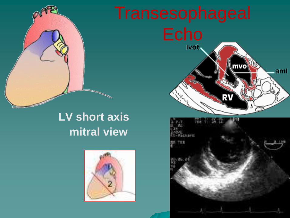
LV short axis
mitral view
Transesophageal
Echo

MRI
The Future
Aortic Regurgitation
Trileaflet Pulmonary Valve
Bileaflet Pulmonary Valve

Aortic Valve
Stenosis
Regurgitation

Preoperative Data: AVR
2%
28%
57%
13%
0%
20%
40%
60%
80%
100%
NYHA Class
% o
f P
ati
en
ts
I II III IV
Insufficiency
13%
Stenosis
65%
Valvular Lesion
Mixed
22%

Aortic Stenosis
Disease of the Elderly
Symptoms begin:
60-80 yrs. of age
Causes:
Scarring
Calcification
Rheumatic fever
Bicuspid valve

Aortic Stenosis
Pathophysiology
Increased resistance to blood flow from
the left ventricle to the aorta
– Increased work
– Left ventricular hypertrophy
Smaller ventricular cavity
– Increased oxygen demand
Can lead to ischemia
Heart failure
Aortic Stenosis Symptoms
Chest pain (angina)
– occurs during exertion – blood supply to the enlarged heart muscle is
inadequate
Heart failure develops – fatigue – shortness of breath during exertion
Syncope – Exertion leads to peripheral arterial vasodilatation – AS limits cardiac output preventing compensation
Sudden Death

Aortic Stenosis
Diagnosis
Physical Examination – Heart Murmur
Heard over aortic area
2nd right intercostal border
Midsystolic murmur
Radiates down left sternal border
ECG – Left ventricular hypertrophy

Aortic Stenosis
Chest X-ray
Prominent left ventricle

Aortic Stenosis
Diagnosis
Echocardiography Cardiac Cath >45 years
Short Axis
Long Axis

Aortic Stenosis
Treatment
Asymptomatic – Regular follow-up
Serial Echo
Symptomatic – Aortic valve area less than 1.2 cm2/M2
Critical Aortic Stenosis
–Valve area 1cm2
–Gradient >60 mm Hg (normal LV)
Medical Therapy – Diuretics
Surgical replacement – before irreversible damage

Aortic Stenosis
Survival
Symptomatic
Surgery
vs
Medical
NEJM, Carabello 346 (9): 677
February 28, 2002

Aortic Regurgitation
Increasing volume
and pressure in the
left ventricle
– Result: increased work
ventricles thicken
to compensate
hypertrophy
chambers dilate
Eventually CHF

Aortic Regurgitation
Rheumatic Fever – Historical cause
myxomatous degeneration
aortic aneurysms & dissection
bicuspid valve
infective endocarditis

Aortic Regurgitation
Symptoms
Mild – Asymptomatic heart murmur
Severe – Palpitations
LVH
– Congestive heart failure Shortness of breath
Fatigue
Angina
– Reduce SBP during ventricular diastole
– Wide pulse pressure

Aortic Regurgitation
Physical exam – Murmur
Heard with diaphragm
Midsystolic
ECG: – LVH
Chest X-ray – cardiomegaly

Aortic Regurgitation Echo
Cardiac Cath – 20% of people with aortic regurgitation also have
coronary artery disease

Aortic Regurgitation
Treatment Mild
– Medical management Digoxin, Diuretics
Calcium channel blocker
Ace
Moderate to severe: – Surgery
Cardiac Cath – 20% of people with aortic regurgitation also have
coronary artery disease

Aortic Valve Replacement
Possible Prostheses Mechanicals valves
Homografts
Engineered Tissue Valves
-Stented (porcine or bovine)
-Stentless (porcine)
Pulmonary Autograft
Aortic Remodeling

Mechanical Valve
Products
SJM Regent™
valve (bileaflet)
Carbomedics
Standard bileaflet
valve
Medtronic –
Hall™ tilting
disc valve

Tissue Valve Products
Stented Valve
Edwards Lifesciences
Carpentier - Edwards
Perimount™ valve
Stentless
Valve
St. Jude Medical
Toronto SPV®
valve
Homograft
LifeNet
Homograft

Stentless Aortic Valves
Modeled after native aortic valve
Eliminates residual stenosis caused by stents
Near-normal hemodynamics
Normalizes LV mass and performance

Toronto SPV® Valve Freestyle® Valve
Stentless Porcine Valves
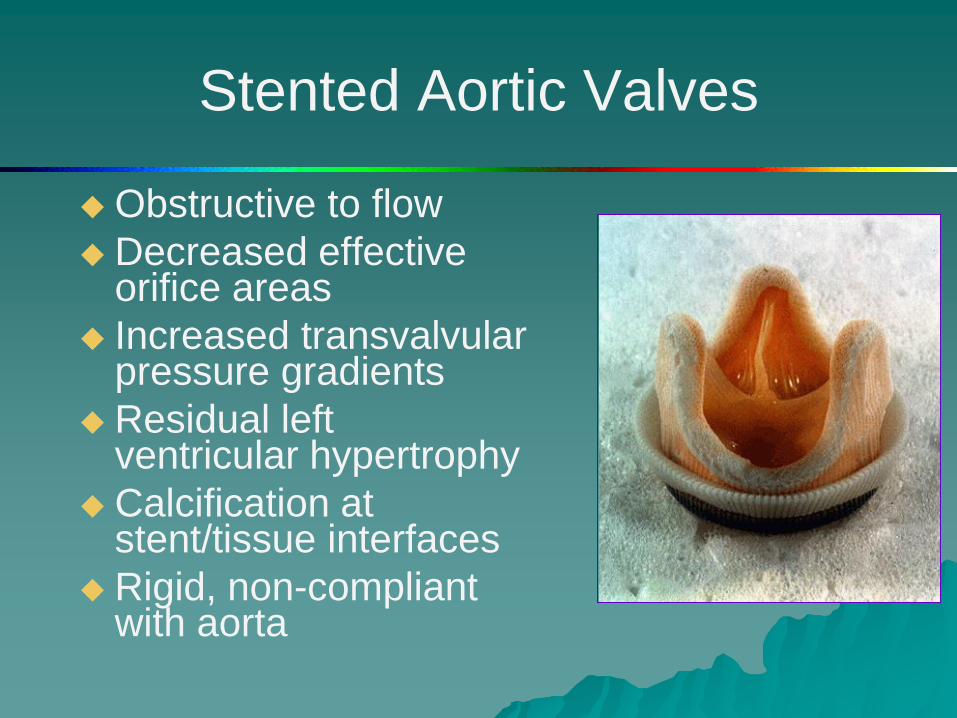
Stented Aortic Valves
Obstructive to flow
Decreased effective orifice areas
Increased transvalvular pressure gradients
Residual left ventricular hypertrophy
Calcification at stent/tissue interfaces
Rigid, non-compliant with aorta

Aortic Root Reconstruction
Aortic Root Replacement
Coronary Reimplantation

Aortic Root Remodeling
Aortic valve preservation
Remodeling of root geometry
Coronary Reimplantation

Echo
Stentless Freestyle Natural Valve
Bioprosthesis

Durability Anticoagulation
Valve Surgery Trends
Lim JTCVS 02 485 patients
composite TE Risk 7% per pt yr

Mechanical Valves
Thromboembolic event rate
– 1-4%/pt-year
Anticoagulant related hemorrhage rate
– 2-5%/pt-year

Risk Factors for
Thromboembolism
Mechanical valve implant
Atrial Fibrillation
Increased LV cavity size/LV dysfunction
Regional Wall Motion Abnormality
Depressed Ejection Fraction
Previous Thromboembolism
Hypercoagulability

St. Jude Medical, Training and Education
Anticoagulants - Coumarins
Development/clinical use began in the 1920s
Used for long-term anticoagulation therapy
– Oral anticoagulant
– Produces a functional deficiency of Vitamin K
several clotting factors on Vitamin K
– Takes 36 hours to achieve a therapeutic blood
level which will last 4 - 5 days
Blood levels can vary
– patients must be checked regularly

Minimally Invasive
Aortic Valve

Aortic Valve
Freedom from Valve Dysfunction

Aortic Valve Complications
Thromboembolism
Anti-coagulant related
hemorrhage
Valve
dysfunction/structural
failure
Para-valvular leak
Re-operation
Endocarditis
Hemolysis
Death

Aortic Valve Survival
Early (hospital) death - 3-6%
Time-related survival
· 5 years - 75%
· 10 years - 60%
· 15 years - 40%
Mode of death
· Early due to CHF, hemorrhage, infection, CVA
· Sudden - 20%
· Device related - 20%
CTSNet, Residents Section

Aortic Valve
Risk Factors for Survival after AVR · Advanced age · Functional status (NHYA class) · Depressed LV function (aortic incompetence) · Coronary artery disease · Presence of endocarditis · Aneurysm of ascending aorta · Mismatch of prosthesis and body size