The Size of It All Types of Microscopes. The Size of It All Remember that 1 inch = 2.54 cm and that...
23
The Size of It All Types of Microscopes
-
Upload
patricia-oconnor -
Category
Documents
-
view
213 -
download
0
Transcript of The Size of It All Types of Microscopes. The Size of It All Remember that 1 inch = 2.54 cm and that...

The Size of It All
Types of Microscopes

The Size of It All
Remember that 1 inch = 2.54 cm and that1 meter contains 1000000 micrometers (µm) or 1000000000 nanometers (nm)
Microorganisms are measured in µm or nm.

Question??
If a typical E.coli is 1 µm in length, how many inches would that be?
1 µm x 1m x 100 cm x 1 in. = ? 1000000µm 1 m 2.54cm
0.00003937 inches!!

Types of MicroscopesLight Microscopes:1. Compound Light Microscope (LM)2. Dissection or Stereoscope3. Darkfield Microscope4. Phase-Contrast Microscope5. Differential Interference Contrast (DIC)6. Fluorescence Microscopy7. Confocal Microscopy

Types of MicroscopesElectron Microscopes:1. Transmission Electron Microscope (TEM)2. Scanning Electron Microscope (SEM)
Scanned Probe Microscopes:3. Scanning Tunneling Microscope (STM)4. Atomic Force Microscope (AFM)

Compound Light Microscope• Uses visible light• Magnifies up to 2000x, but generally only
1000x• Good for: magnification resolution refractive index bright field illumination can examine live organisms

Darkfield Microscope• Good for examining live organisms
Phase-Contrast Microscope• Can be used to visualize internal organelles of
a cell• Uses one beam of light
Differential Interference Contrast• Similar to phase-contrast except it uses two
beams of light

Fluorescence Microscope• Used to rapidly detect and identify microbes
in tissues or clinical specimens
Technique: fluorescent antibody (FA) or immunofluorescence. Specific antibodies are treated so that they are fluorescent. The antibodies are then applied to the specimen, if the antigens are present the specimen will appear to fluoresce.

Confocal Microscopes• Similar to fluorescent microscopy except it uses laser light
Transmission Electron Microscope• An ultra thin section of specimen is placed on a copper
mesh grid where a beam of electrons pass through to an electromagnetic objective lens, which magnifies the image. The final image is seen as light and dark areas, referred to as a transmission electron micrograph.
• Disadvantages: 1. requires a very thin slice of the specimen 2. no 3-D aspect 3. specimens must be fixed, stained and placed under a vaccum which can cause distortion and shrinkage.

Scanning Electron Microscope• Provides a 3-D image, the stream of electrons
are provided by an electron “gun”• The image is referred to as a scanning electron
micrograph• Advantages: intact cells and viruses can be
seen• Disadvantages: surface structure only

Scanned Probe Microscopy• Uses a probe to map atomic and molecular
shapes (surface).• Can be used to determine magnetic, chemical
and temperature characteristics of the cell.
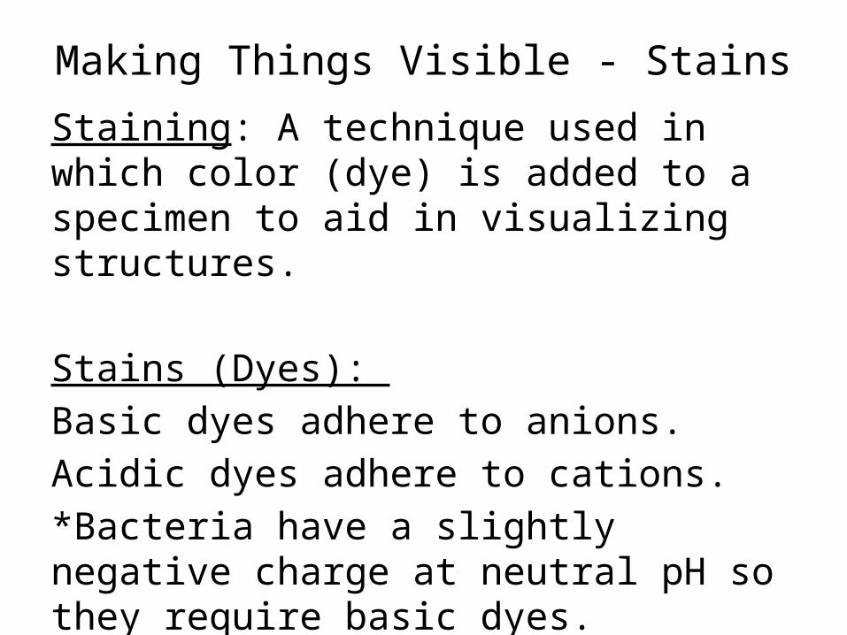
Making Things Visible - StainsStaining: A technique used in which color (dye) is added to a specimen to aid in visualizing structures.
Stains (Dyes): Basic dyes adhere to anions.Acidic dyes adhere to cations.*Bacteria have a slightly negative charge at neutral pH so they require basic dyes.

Simple Stains: A single stain is used to high light basic cellular shape and structure. (Mordants maybe required to intensify the stain.)
Differential Stains:A series of dyes are used to distinguish between various organisms and structures. Generally a primary stain is applied followed by a rinse or decolorizer and then finished with a counterstain to color structures that did not retain the primary stain.

Making A Slide• Label the slide• Apply the specimen to the slide• Fixing the specimen to the slide – Heat fixing– Spray fixative (generally alcohol based)– Wax setting– Frozen

The “How” of the Gram Stain1. Crystal Violet (primary stain) enters the cytoplasm of all bacteria.2. Gram’s Iodine (mordant) forms a large chemical complex with
the crystal violet.3. Decolorizer dehydrates the peptidoglycan layer of the gram
positive cells making it harder for the crystal violet-iodine complex to leave the cell. This will cause the gram positive bacteria to be seen as bluish-purple.
In gram negative cells the decolorizer dissolves the outer membrane leaving small holes that the crystal violet-iodine complex to wash out – leaving the cell “colorless”.4. Safranin (counterstain) enters the cytoplasm of the bacteria allowing the gram negative bacteria to be seen as reddish- pink.
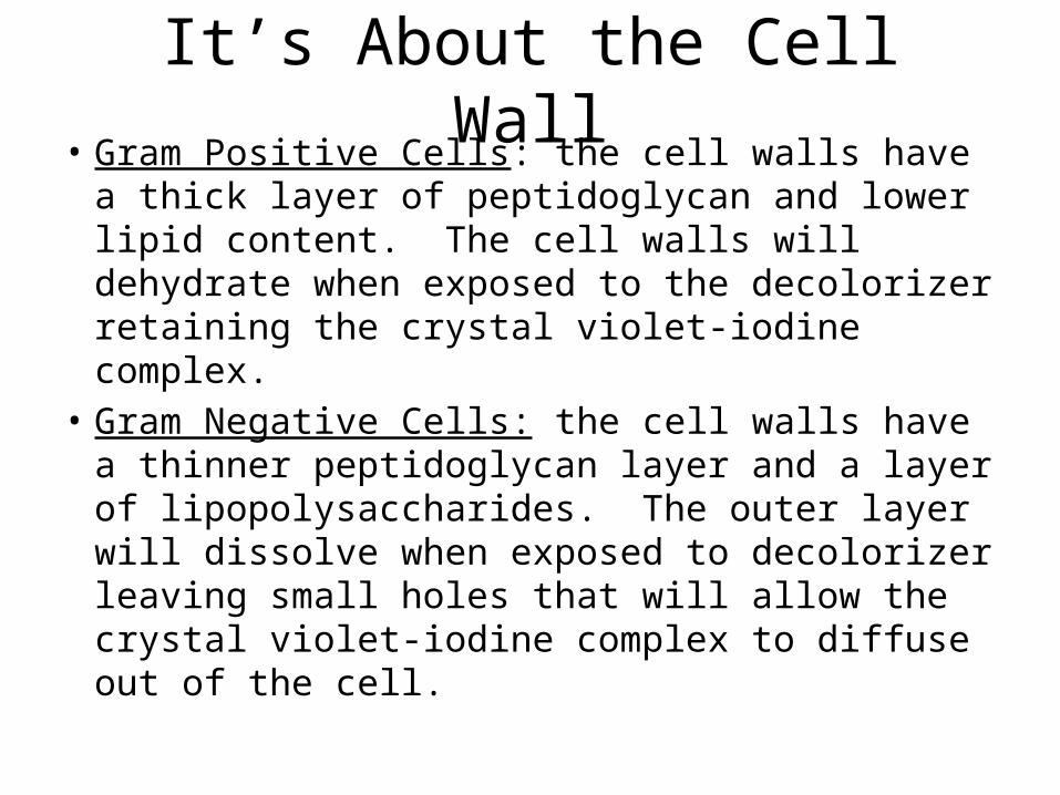
It’s About the Cell Wall• Gram Positive Cells: the cell walls have a thick
layer of peptidoglycan and lower lipid content. The cell walls will dehydrate when exposed to the decolorizer retaining the crystal violet-iodine complex.
• Gram Negative Cells: the cell walls have a thinner peptidoglycan layer and a layer of lipopolysaccharides. The outer layer will dissolve when exposed to decolorizer leaving small holes that will allow the crystal violet-iodine complex to diffuse out of the cell.

Types of Differential Stains• Gram’s Stain: (1884) Hans Christian Gram1. Crystal Violet (primary stain) – up to 1 minute2. Gram’s Iodine (mordant) – 30 seconds3. Decolorizer – 50/50 alcohol acetone – 10
seconds4. Safranin (counterstain) – 30 seconds**Remember which stain the cell retains depends on the cell wall structure.

• Gram Positive Cocci Gram Positive Rods
Gram Negative Cocci Gram Negative Rods

ACID FAST STAIN• Used for bacteria with a waxy coat, primarily
used for mycobacterium and nocardia.Technique:1. Carbol fuchsin (primary stain)2. Decolorizer3. Methylene Blue (counter stain)

Wright’s Stain• Also called Wright’s Giemsa Stain• Used for differential staining of blood cells.Technique:Methylene Blue (primary stain) - basicPhosphate buffer rinseEosin (counter stain) - acidic

Special StainsSpecial stains are used to visualize specific parts of the cell.1. Capsule stain – generally done as a negative
staining technique where the background is colored not the specimen.
India Ink is used to visualize the Capsules around the yeast.
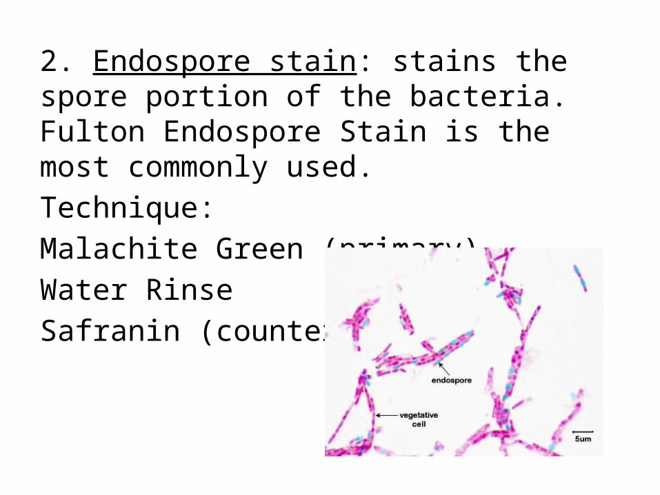
2. Endospore stain: stains the spore portion of the bacteria. Fulton Endospore Stain is the most commonly used.Technique:Malachite Green (primary)Water RinseSafranin (counter stain)

3. Flagella stain: staining technique developed to visualize the flagellum. It is a combination of carbol fuschin and a mordant.
4. Trichrome Stain: often used for the identification of parasites. It is a combination of acid fuschin and picric acid.