The Optic Syrinx: Cavernous Degeneration of the Optic ... · optic nerve has been repeatedly and...
4
John R. Jinkins 1 Received March 31 , 1986; accepted after revi· sion July 28, 1986. , Department of Rad iology, King Faisal Specialist Hospital and Research Centre, P.O. Box 3354, Riyadh 11211 , Saudi Arabia. Address reprint re- quests to H. R. Jinkins. AJNR 8:349-352, March/April 1987 0195-6108/87/0802-0349 © American Society of Neuroradiology The Optic Syrinx: Cavernous Degeneration of the Optic Nerve Demonstrated by lopamidol Optic Neurography 349 Cavernous degeneration of the optic nerve has been described predominantly in association with long-standing glaucoma, although less frequently it has been reported in other , disparate disease processes. The only descriptions to date have been in pathologic specimens in vitro. This is the first report to describe syrinx formation of the optic nerves in vivo in two patients with non glaucomatous conditions affecting the optic nerves. Histologic descriptions of cavernous degeneration of the optic nerve date from Schnabel's first account of a patient with glaucoma in 1885 [1]. Since then, associations between glaucoma and this degenerative process are commonplace [2-4]. More recently, many other disease processes have been linked with cavern- ous degeneration of the optic nerve, as revealed by postmortem histology [5-8] . This article describes in vivo syrinx formation in the first cranial nerve. Materials and Methods Two patients were studied in the manner described elsewhere [9] for optic neurography with the exception that a volume of 3-4 ml of 300 mg/ ml I iopamidol was in sti ll ed into the lumbar subarachnoid space. The dose was increased since the patients in this study were adults rather than children, as in the earlier report. The patients were scanned within 1 hr of contrast instillation. Axial and coronal high-resolution thin sections were obtained on a GE-9800 CT scanner. Neither patient encountered any adverse reaction either during or after the procedure. Results Case 1 The first patient was a 23-year-old woman who had infantile complete premature craniosynostosis, consequent long-standing increased intracranial pressure, and chronic papilledema. After an early, extensive craniectomy at the age of 6 years- in an attempt to open the sutures and relieve the elevated intracranial pressure (Fig . 1 A)-the patient's vision continued to deteriorate, accelerating over that of the previous year. An axial CT through the orbits showed enlarged optic nerve sheaths, while an optic cisternogram showed extreme atrophy of the optic nerves and chiasm (Fig . 18). The optic neurogram pointed out paradoxi cal prominence of the optic nerves within the dilated optic sheaths and relatively central neural accumulation of iopamidol contrast bilaterally in the retrobulbar region (Figs. 1 C- 1 F) . Case 2 The second patient was a 14-year-old girl with deteriorating vision, left worse
Transcript of The Optic Syrinx: Cavernous Degeneration of the Optic ... · optic nerve has been repeatedly and...

John R. Jinkins 1
Received March 31 , 1986; accepted after revi· sion July 28, 1986.
, Department of Radiology, King Faisal Specialist Hospital and Research Centre, P.O. Box 3354 , Riyadh 11211 , Saudi Arabia. Address reprint requests to H. R. Jinkins.
AJNR 8:349-352, March/April 1987 0195-6108/87/0802-0349 © American Society of Neuroradiology
The Optic Syrinx: Cavernous Degeneration of the Optic Nerve Demonstrated by lopamidol Optic Neurography
349
Cavernous degeneration of the optic nerve has been described predominantly in association with long-standing glaucoma, although less frequently it has been reported in other, disparate disease processes. The only descriptions to date have been in pathologic specimens in vitro. This is the first report to describe syrinx formation of the optic nerves in vivo in two patients with non glaucomatous conditions affecting the optic nerves.
Histologic descriptions of cavernous degeneration of the optic nerve date from Schnabel's first account of a patient with glaucoma in 1885 [1]. Since then, associations between glaucoma and this degenerative process are commonplace [2-4]. More recently, many other disease processes have been linked with cavernous degeneration of the optic nerve, as revealed by postmortem histology [5-8]. This article describes in vivo syrinx formation in the first cranial nerve.
Materials and Methods
Two patients were studied in the manner described elsewhere [9] for optic neurography with the exception that a volume of 3-4 ml of 300 mg/ml I iopamidol was instilled into the lumbar subarachnoid space.
The dose was increased since the patients in this study were adults rather than children, as in the earlier report . The patients were scanned within 1 hr of contrast instillation. Axial and coronal high-resolution thin sections were obtained on a GE-9800 CT scanner. Neither patient encountered any adverse reaction either during or after the procedure.
Results
Case 1
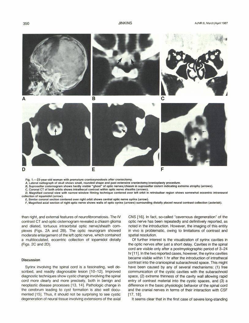
The first patient was a 23-year-old woman who had infantile complete premature craniosynostosis, consequent long-standing increased intracranial pressure, and chronic papilledema. After an early, extensive craniectomy at the age of 6 yearsin an attempt to open the sutures and relieve the elevated intracranial pressure (Fig. 1 A)-the patient's vision continued to deteriorate, accelerating over that of the previous year. An axial CT through the orbits showed enlarged optic nerve sheaths, while an optic cisternogram showed extreme atrophy of the optic nerves and chiasm (Fig . 18). The optic neurogram pointed out paradoxical prominence of the optic nerves within the dilated optic sheaths and relatively central neural accumulation of iopamidol contrast bilaterally in the retrobulbar region (Figs. 1 C-1 F).
Case 2
The second patient was a 14-year-old girl with deteriorating vision , left worse
350 JINKINS AJNR :8, March/April 1987
A 8 c
o E F
Fig. 1.-23-year-old woman with premature craniosynostosis after craniectomy. A, Lateral radiograph of skull shows small , rounded shape and past extensive craniectomy/cranioplasty procedure. B, Suprasellar cisternogram shows hardly visible "ghost" of optic nerves/chiasm in suprasellar cistern indicating extreme atrophy (arrows) . C, Coronal CT of both orbits shows intrathecal contrast within optic nerve sheaths (arrows). D, Magnified coronal view with narrow-window filming technique centered over left orbit in retrobulbar region shows somewhat eccentric intraneural
collection of iopamidol (arrow). E, Similar coronal section centered over right orbit shows central optic nerve syrinx (arrow). F, Magnified axial section of right optic nerve shows walls of optic syrinx (arrows) surrounding distally placed neural contrast collection (asterisk).
than right , and external features of neurofibromatosis. The IV contrast CT and optic cisternogram revealed a chiasm glioma and dilated, tortuous intraorbital optic nerve/sheath complexes (Figs. 2A and 28). The optic neurogram showed moderate enlargement of the left optic nerve, which contained a multiloculated, eccentric collection of iopamidol distally (Figs. 2C and 2D).
Discussion
Syrinx involving the spinal cord is a fascinating, well described , and readily diagnosable lesion [10-12]. Improved diagnostic techniques show cystic change involving the spinal cord more clearly and more precisely, both in benign and neoplastic disease processes [13, 14]. Pathologic change in the cerebrum leading to cyst formation is also well documented [15]. Thus, it should not be surprising to see cystic degeneration of neural tissue involving extensions of the axial
CNS [16]. In fact, so-called "cavernous degeneration" of the optic nerve has been repeatedly and definitively reported, as noted in the introduction. However, the imaging of this entity in vivo is problematic, owing to limitations of contrast and spatial resolution.
Of further interest is the visualization of syrinx cavities in the optic nerves after just a short delay. Cavities in the spinal cord are visible only after a postmyelographic period of 3-24 hr [11]. In the two reported cases, however, the syrinx cavities became visible within 1 hr after the introduction of intrathecal contrast into the craniospinal subarachnoid space. This might have been caused by any of several mechanisms: (1) free communication of the cystic cavities with the subarachnoid space, (2) extreme thinness of the cavity wall allowing rapid entry of contrast material into the cystic spaces, and (3) a difference in the basic physiologic behavior of the spinal cord and the cranial nerves in terms of their interaction with CSF [17 , 18].
It seems clear that in the first case of severe long-standing

AJNR :8, March/April 1987 OPTIC SYRINX 351
Fig. 2.-14-year-old girl with neurofibromatosis and optic chiasm glioma.
A, Intravenous enhanced CT shows poorly enhancing intra/suprasellar mass (arrow) .
B, Optic cisternogram precisely depicts the ovoid confines of chiasm neoplasm (arrow).
C, Coronal CT of right orbit shows enlarged distal optic nerve surrounded by intrathecal iopamidol (arrow) .
D, Same coronal section as in C with magnified narrow-window filming technique shows enlarged right optic nerve containing three separate eccentric collections of iopamidol (arrow). These collections are seen faintly in C.
A
c
papilledema, the underlying pathology is one of prolonged ischemia to the optic disk and optic nerve leading to cavity formation [19-22] . However, the syrinx formation in the second case of optic chiasm glioma is open to question. Certainly optic disk hypoxia is also known to occur in association with mass lesions intracranially and intraorbitally, which might conceivably result in cavitation [8, 23-26]. On the other hand, cyst formation in astrocytomas of the chiasm, brain, and spinal cord is well known [13, 14, 27-29]. Possibly, then, this second case represents cystic degeneration of orbital extension of the chiasm glioma. However, because the original tumor appears as a solid lesion, the orbital manifestation becomes somewhat more difficult to explain on this basis. Therefore, this second case likely represents a complex degenerative phenomenon. But since no biopsy was taken of the intraorbital optic nerve, this is only speculation .
Regardless, it seems clear that there is great potential for diagnosing syrinx formation in cranial nerves (namely, syringoneuria) in varied disease processes [25, 30-32] . The clinical implication is twofold: (1) the optic nerves in many pathologic conditions may be much more severely involved than was previously thought [26], and (2) these cystic degenerations may be active processes in and of themselves and produce further, progressive damage as they exert pressure and dissect along and through the optic nerve [33, 34].
8
D
ACKNOWLEDGMENTS
I thank Mrs. A. Radford for manuscript preparation , Mrs. C. Jinkins for manuscript research , and K.F.S.H . Radiology and Photo-Graphics Departments for technical assistance with the illustrations.
REFERENCES
1. WolH E. Schnabel's cavernous atrophy. Trans Ophthalmol Soc UK 1947;68: 133-140
2. Quigley HA, Addicks EM, Green WR . Optic nerve damage in human glaucoma. III. Quantitative correlation of nerve fiber loss and visual field defect in glaucoma, ischemic neuropathy, papilledema, and toxic neuropathy. Arch Ophtha/mo/ 1982;1 00 : 135-146
3. Lampert PW, Vogel MH, Zimmerman LE. Pathology of the optic nerve in experimental acute glaucoma, Invest Ophtha/mol Vis Sci 1968;7(2) : 199-21 3
4. Zimmerman LE, de Venecia G, Hamasaki 01. Pathology of the optic nerve in experimental acute glaucoma. Invest Ophthalmol Vis Sci 1967;6(2) : 1 09-125
5. Brownstein S, Font RL, Zimmerman LE , Murphy SB. Nonglaucomatous cavernous degeneration of the optic nerve. Report of two cases. Arch Ophthalmo/ 1980;98:354-358
6. Giarelli L, Melato M, Campos E. 14 cases of cavernous degeneration of the optic nerve. Ophthalmologica 1977 ;174:316-321
7. Hayreh SS. Pathogenesis of cupping of the optic disc. Br J Ophthalmol 1974;58 :863-876
8. Trobe JD, Glaser JS, Cassady J, Herschler J, Anderson DR. Nonglaucomatous excavation of the optic disc. Arch Ophthalmo/ 1980;98 : 1 046-1 050

352 JINKINS AJNR:8, March/April 1987
9. Jinkins JR. The optic neurogram: evaluation of CSF "block" caused by compressive lesions at the optic canal. AJNR 1987;8:135-139
10. Batnitzky S, Price HI , Gaughan MJ , Hall PV, Rosenthal SJ . The radiology of syringohydromyelia. RadioGraphics 1983;3(4) :585-611
11 . Bonafe A, Manelfe C, Espagno J, Guiraud B, Rascol A. Evaluation of sryingomyelia with metrizamide computed tomographic myelography. J Comput Assist Tomogr 1980;4(6):797-802
12. Quencer RM , Green BA, Eismont FJ. Posttraumatic spinal cord cysts: clinical features and characterization with metrizamide computed tomography. Radiology 1983;146:415- 423
13. Kan S, Fox AJ , Vinuela F, Barnett HJM, Peerless SJ. Delayed CT metrizamide enhancement of syringomyelia secondary to tumor. AJNR 1983;4 :73- 78
14. Pullicino P, Kendall BE. Computed tomography of "cystic" intramedullary lesions. Neuroradiology 1982;23: 117 -121
15. Raybaud C. Destructive lesions of the brain. Neuroradiology 1983;25: 265-291
16. Kingsley DPE, Thornton A, Furneaux C, King n . Transmural passage of subarachnoid metrizamide into a cystic acoustic schwannoma of the cerebellopontine angle: a diagnostic dilemma. Neuroradiology 1984;26: 319-321
17. Sage MR. Kinetics of water-soluble contrast media in the central nervous system. AJNR 1983;4:897-906
18. Sage MR, Wilcox J. Brain parenchyma penetration by intrathecal non ionic iopamidol. AJNR 1983;4 : 1181 - 1183
19. Green GJ , Lessell S, Loewenstein JI. Ischemic optic neuropathy in chronic papilledema. Arch Ophthalmo/1980;98 :502-504
20. Hayreh SS. Optic disc edema in raised intracranial pressure. VI. Associated visual disturbances and their pathogenesis. Arch Ophthalmol 1977;95 : 1566- 1579
21 . Rush JA. Pseudotumor cerebri. Clinical profile and visual outcome in 63
patients. Mayo Clin Proc 1980;55:541-546 22. Whittle IR, Johnston IH, Besser M. Intracranial pressure changes in cranio
stenosis. Surg Neurol 1984;21 : 367-372 23. Brovkina AF, Kirillova LI. Blood circulation in the optic nerve disc in patients
with orbit tumours. Vestn Ofta/mo/1980 ;5:53-56 24. Skarf B, Davis RL, Birsner HA, Hoyt WF. Intracranial optic nerve infarction
from subdural hematoma. Arch Neuro/1984;41 :58-60 25. Schenck JF, Hart HR Jr, Foster TH, et al. Improved MR imaging of the
orbit at 1.5 T with surface coils. AJNR 1985;6 : 193-196 26. Quigley HA. Glaucoma's optic nerve damage: changing clinical perspec
tives. Ann Ophtha/mol 1982; 14(7): 611-612 27. Barbaro NM, Rosenblum ML, Maitland CG, Hoyt WF, Davis RL. Malignant
optic glioma presenting radiologically as a "cystic" suprasellar mass: Case report and review of the literature. Neurosurgery 1982;11(6):787-789
28. Loftus CM, Copeland BR , Carmel PW. Cystic supratentorial gliomas: natural history and evaluation of modes of surgical therapy. Neurosurgery 1985;17(1): 19-24
29. Messina AV, Potts DG, Rottenberg D, Patterson RH. Computed tomography: demonstration of contrast medium with cystic tumors. Radiology 1976;120:345-347
30. Daniels DL, Herfkins R, Gager WE, et al. Magnetic resonance imaging of the optic nerves and chiasm. Radiology 1984;152 :79-83
31. Edwards JH, Hyman RA, Vacirca SJ, et al. 0.6 T magnetic resonance imaging of the orbit. AJNR 1985;6:253-258
32. Kjos BO, Brant-Zawadzki M, Kucharczyk W, Kelly WM, Norman D, Newton TH. Cystic intracranial lesions: magnetic resonance imaging. Radiology 1985;155 :363-369
33. Conway LW. Hydrodynamic studies in syringomyelia. J Neurosurg 1967;27 :501-514
34. Watson N. Ascending cystic degeneration of the cord after spinal cord injury. Paraplegia 1981 ;19 :89-95