
Visual Pathways visual hemifields project contralaterally –exception: bilateral representation of...
12
Visual Pathways • visual hemifields project contralaterally – exception: bilateral representation of fovea! • Optic nerve splits at optic chiasm • about 90 % of fibers project to cortex via LGN • about 10 % project through superior colliculus and pulvinar Note: this will be important when we talk about visuospatial
-
date post
21-Dec-2015 -
Category
Documents
-
view
221 -
download
0
Transcript of Visual Pathways visual hemifields project contralaterally –exception: bilateral representation of...

Visual Pathways
• visual hemifields project contralaterally
– exception: bilateral representation of fovea!
• Optic nerve splits at optic chiasm
• about 90 % of fibers project to cortex via LGN
• about 10 % project through superior colliculus and pulvinar
– but that’s still a lot of fibers! Note: this will be important when we talk about visuospatial attention

Visual Pathways
• Lateral Geniculate Nucleus maintains segregation:
– of M and P cells (mango and parvo)
– of left and right eyes
P cells project to layers 3 - 6
M cells project to layers 1 and 2
Visual Pathways
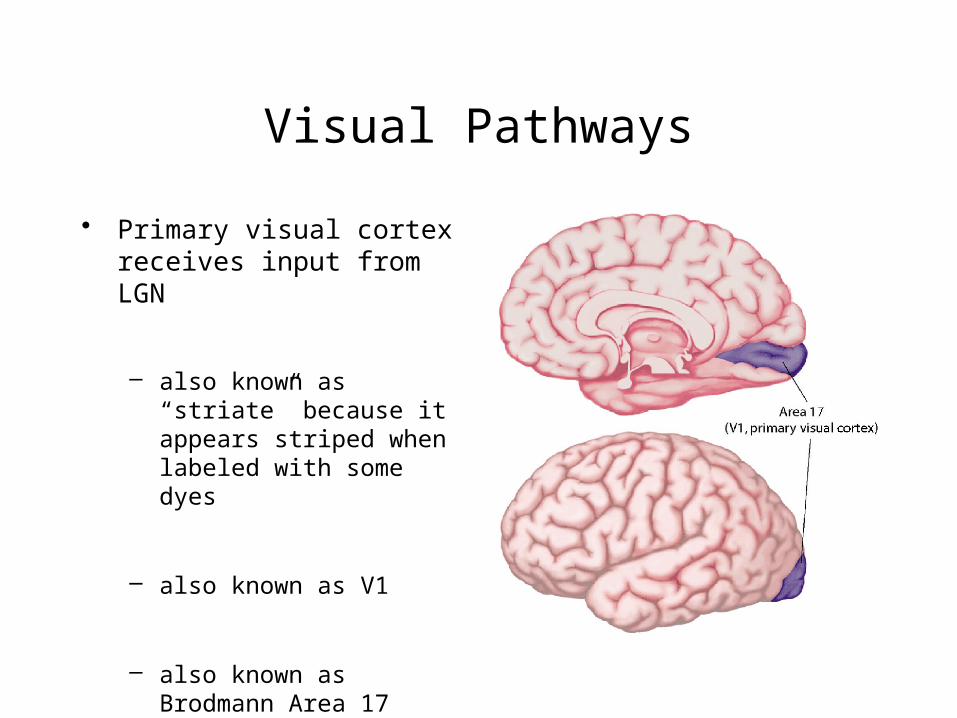
• Primary visual cortex receives input from LGN
– also known as “striate” because it appears striped when labeled with some dyes
– also known as V1
– also known as Brodmann Area 17
Visual Pathways
W. W. Norton
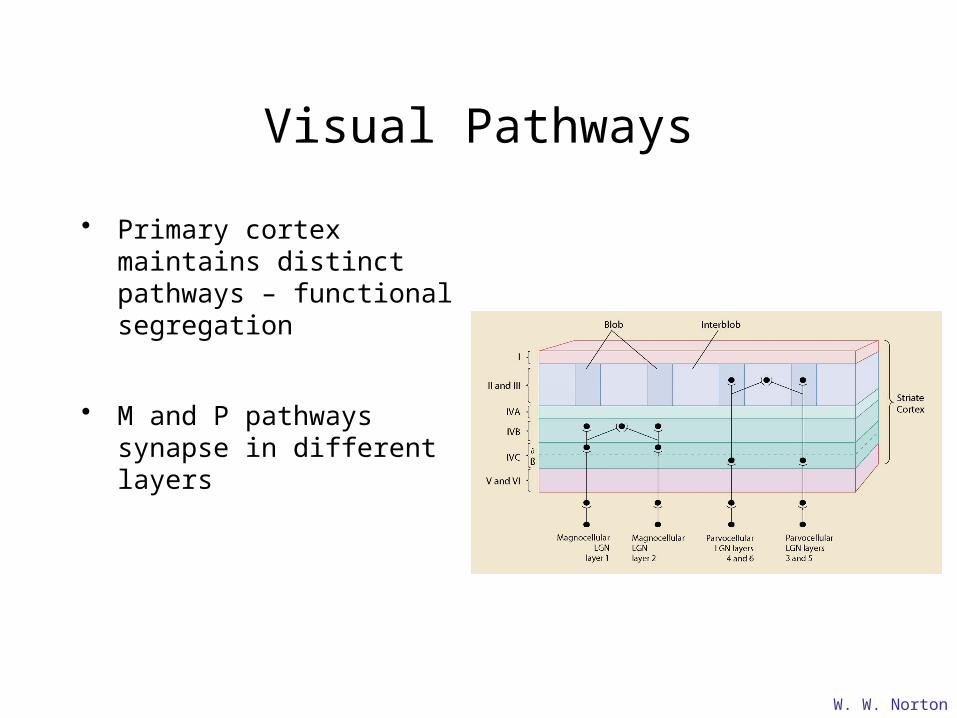
• Primary cortex maintains distinct pathways – functional segregation
• M and P pathways synapse in different layers

The Role of “Extrastriate” Areas
• Different visual cortex regions contain cells with different tuning properties
The Role of “Extrastriate” Areas
• Consider two plausible models:
1. System is hierarchical:
– each area performs some elaboration on the input it is given and then passes on that elaboration as input to the next “higher” area
2. System is analytic and parallel:
– different areas elaborate on different features of the input
The Role of “Extrastriate” Areas
• Functional imaging (PET) investigations of motion and colour selective visual cortical areas
• Zeki et al.
• Subtractive Logic– stimulus alternates between two scenes that differ only in
the feature of interest (i.e. colour, motion, etc.)

The Role of “Extrastriate” Areas
• Identifying colour sensitive regions
Subtract Voxel intensities during these scans…
…from voxel intensities during these scans
…etc.Time ->
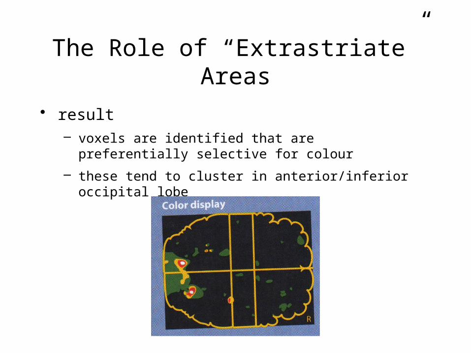
The Role of “Extrastriate” Areas
• result– voxels are identified that are preferentially selective for
colour
– these tend to cluster in anterior/inferior occipital lobe

The Role of “Extrastriate” Areas
• similar logic was used to find motion-selective areas
Subtract Voxel intensities during these scans…
…from voxel intensities during these scans
…etc.Time ->
MOVING STATIONARY MOVING STATIONARY

The Role of “Extrastriate” Areas
• result– voxels are identified that are preferentially selective for
motion
– these tend to cluster in superior/dorsal occipital lobe near TemporoParietal Junction
– Akin to Human V5

The Role of “Extrastriate” Areas
• Thus PET studies doubly-dissociate colour and motion sensitive regions


![Computed Tomography of Chiasmal Optic Neuritis - · PDF fileComputed Tomography of Chiasmal Optic Neuritis ... During the episode of acute visual loss, ... [1 , 13']; in a study of](https://static.fdocuments.us/doc/165x107/5aba40537f8b9ad1768b4f94/computed-tomography-of-chiasmal-optic-neuritis-tomography-of-chiasmal-optic-neuritis.jpg)


![An accidental diagnosis of optic nerve meningioma in a ... · An accidental diagnosis of optic nerve meningioma in a patient affected by Thyroid Eye Disease ... visual pathway [5]](https://static.fdocuments.us/doc/165x107/5c877bee09d3f2bc6b8bcda8/an-accidental-diagnosis-of-optic-nerve-meningioma-in-a-an-accidental-diagnosis.jpg)