The Lower Limb - fac.ksu.edu.sa · The bony pelvis • Protective osseofibrous ring for the pelvic...
62
The Lower Limb Anatomy RHS 241 Lecture 2 Dr. Einas Al-Eisa
Transcript of The Lower Limb - fac.ksu.edu.sa · The bony pelvis • Protective osseofibrous ring for the pelvic...

The Lower Limb
AnatomyRHS 241Lecture 2
Dr. Einas Al-Eisa

The bony pelvis
• Protective osseofibrous ring for the pelvic viscera
• Transfer of forces to: – acetabulum & head of femur (when standing)– ischial tuberosities (when sitting)
• Large surface area for the attachment of muscles

The bony pelvis
Orientation
• When in the anatomical position: the ASIS and pubic symphysis lie within the same vertical line

Articulations
• Anteriorly: Pubic symphysis
• Posteriorly: sacroiliac joints

What are the primary functions of the sacroiliac joints when in the
anatomical position?

Innominate bone
Ilium Ischium Pubis

Where (i.e., in which region approximately) does fusion of the
three components of an innominate bone occur to form a single bone as seen in adults?

Pelvis: lateral view

Ilium
• Iliac crest• Anterior superior iliac spine (ASIS)• Anterior inferior iliac spine• Posterior superior iliac spine (PSIS)• Auricular surface (articular surface of SIJ)• Supracristal line: joins the highest points
of the iliac crests (to locate the position the spinous process of L4)


Pubis
• Body • Pubic crest (superior margin of the body)
• Pubic tubercles (prominent lateral eminence of pubic crest)
• Superior & inferior rami (project laterally from the body)

Ischium
• Ischial tuberosity
• Greater sciatic notch (superior to the iliac spine)
• Lesser sciatic notch (inferior to the iliac spine)


Pelvis: medial view


Femur
• Long bone:Shaft (diaphysis)Enlarged proximal & distal ends (epiphysis)
• Angle of obliquity: in the anatomical position, the distal ends are closer to midline than proximal ends (more in females)

Long Bones

Femur- proximal end
• Head: articulates with acetabulum (hip J)
• Pit (fovea of head): attachment of a ligament
• Neck: directed laterally, downward, & posteriorly (common site of fractures)

Right femur

Femur- proximal end
• Greater trochanter: attachment of muscles• Lesser trochanter: attachment of muscles
• Intertrochanteric line: anterior at the junction of the neck & shaft
• Intertrochanteric crest: prominent; posterior at the junction of the neck & shaft

Femur- shaft
• Linea aspera: posterior (roughened by the attachment of thigh muscles)
• Popliteal surface: posterior
• Supraconylar lines: medially & laterally

Right femur

Femur- distal end
• Why is the distal end of femur enlarged?
• Medial & lateral condyles: articulate with the tibia (knee joint)
• Intercondylar notch (fossa): posterior
• Patellar surface: anterior (articulates with the patella to for the femoropatellar joint)

Hip joint
• Synovial• Ball & socket (multiaxial)
• Articular sufaces:Head of femurLunate surface of acetabulum (U-shaped)Acetabular labrum: complete ring of fibrocartilage


What function is served by the acetabular labrum?

Ligaments
• Capsular ligaments of the hip are strong and taut (particularly in the extended position)
• The joint is most vulnerable to dislocationwhen in flexion and external rotation (e.g., sitting in a car)

Ligaments
• Iliofemoral: anterior two bands of fibers (medial & lateral)known clinically as the ‘Y’ ligament of Bigelow ASIS to intertrochanteric line prevent overextension of hip

Ligaments
• Pubofemoral:anterior & inferiorextends laterally from the pubic bone to the intertrochanteric line prevent over-abduction

Hip Joint: Anterior View

Ligaments
• Ischiofemoral:posteriorfrom the ischium, arches forward over the neck of the femur attaches anteriorly to the intertrochanteric line prevent hyperextension of hip
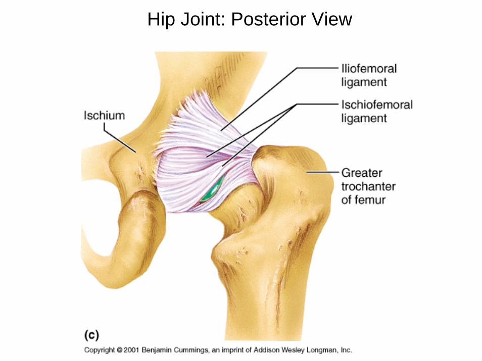
Hip Joint: Posterior View

Ligaments
• Transverse ligament of the acetabulum:inferior bridges the ends (horns) of the lunate articularsurface

Ligaments
• Ligament of the head of femur:Carries a blood vessel to the femoral head Attached to the pit (fovea) of the femur


Gluteal region
• Sciatic nerve enters the gluteal region near the midpoint of a line joining the PSIS & ischial tuberosity
• Common site for intramuscular injections: upper lateral quadrant (to avoid damage to the sciatic nerve)


Sacral Plexus

Routes for neurovascular structures from the pelvis to gluteal region & thigh
= osseofibrous spaces or holes through which nerves and blood vessels enter and leave the thigh from the pelvis

Routes for neurovascular structures
• Inguinal route: formed by the inguinal ligament and the innominate bone (anterior)
• Obturator route: the opening or canal within the obturator membrane
• Gluteal route: osseofibrous ring formed by the greater sciatic notch and accessory ligaments of the SIJc


Muscles of the gluteal region
• Gluteus maximus: inferior gluteal nerve extension & lateral rotation
• Gluteus medius: superior gluteal nerve abduction
• Gluteus minimus: superior gluteal nerve

Gluteal Region & Posterior thigh

Muscles of the gluteal region
• Gluteus medius & minimus: chief abductors (on a free limb)
• During walking, they contract on the weight-bearing side to prevent the pelvis on the non-weight-bearing side from dropping
• Trendlenburg gait?

When standing on booth feet and shifting your weight from one limb
to the other, on which side are the gluteus medius & minimus
contracting?

Short lateral rotators of the thigh
• Piriformis (anterior surface of sacrum to greater trochanter)
• Obturator internus (to greater trochanter)• Obturator externus (to greater trochanter)
• The gemelli muscles (superior & inferior)• Quadratus femoris (ischial tuberosity to
intertrochanteric crest)

Gluteal Region

Gluteal Region: Deep

Short lateral rotators of the thigh
• Common function: assist lateral rotation of the femur
• Oriented like the fascicles of the gluteus maximus
• Most of those muscles lie posterior to the neck of the femur

What is the strongest lateral rotator of the thigh?

Structure of spinal nerves


Spinal nerves

Sacral Plexus
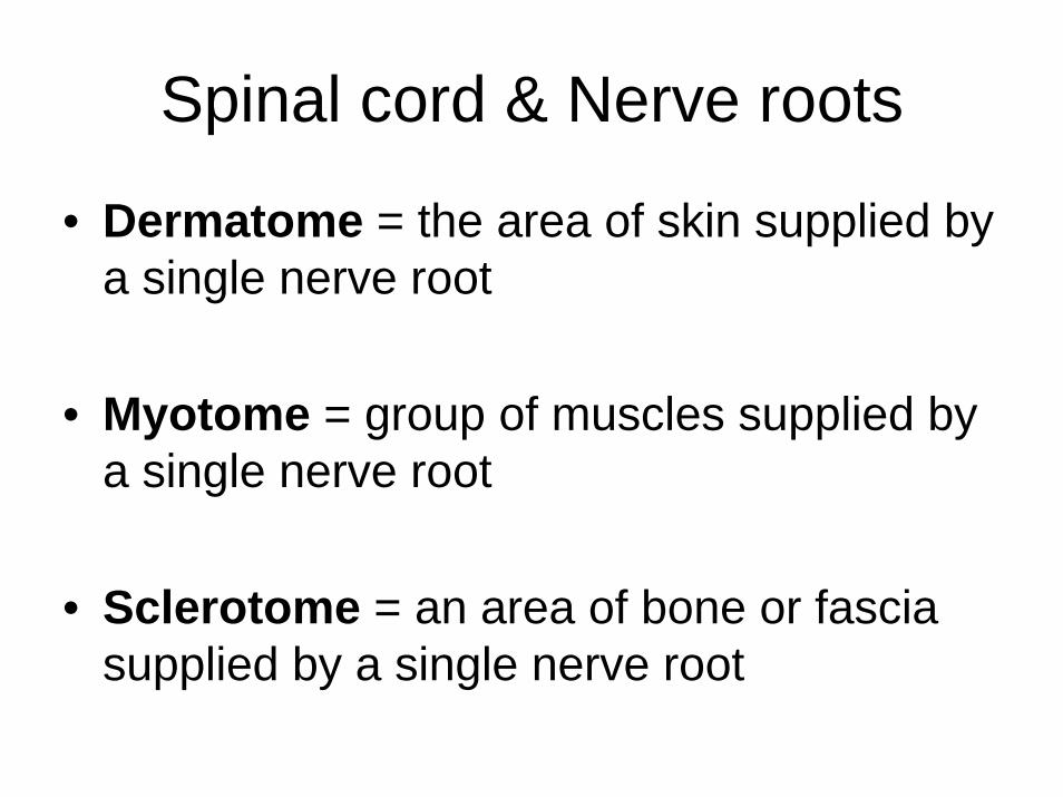
Spinal cord & Nerve roots
• Dermatome = the area of skin supplied by a single nerve root
• Myotome = group of muscles supplied by a single nerve root
• Sclerotome = an area of bone or fascia supplied by a single nerve root

Neurological testing
• Dermatome: may exhibit sensory changes for light touch and pin prick
• Myotome: assessed by performing isometric resisted tests held for 3-5 seconds (L1-L2: hip flexion, L3: knee extension, L4: ankle dorsiflexion & inversion, L5: extension of big toe, S1-S2: plantar flexion & knee flexion, S3-S4: muscles of the pelvic floor & bladder)

Neurological testing
• Reflexes:
• The qudriceps reflex L3
• The achilles tendon reflex S1

Referred Pain
• Pain felt in a part of the body that is usually far from the tissue that have caused it.
• May be due to misinterpretation by the brain as to the source of the painful stimulus.
• Indicates that one of the structures innervated by a nerve root is causing signs & symptoms in other tissues supplied by that same nerve root.

Radiating (radicular) Pain
= Pain felt in a dermatome, myotome, or sclerotome because of direct involvement of a spinal nerve root.