The effects of Bordetella pertussis on dendritic cell ...
67
San Jose State University SJSU ScholarWorks Master's eses Master's eses and Graduate Research Summer 2010 e effects of Bordetella pertussis on dendritic cell imprinting of CD4+ T cells Sana Waheed San Jose State University Follow this and additional works at: hps://scholarworks.sjsu.edu/etd_theses is esis is brought to you for free and open access by the Master's eses and Graduate Research at SJSU ScholarWorks. It has been accepted for inclusion in Master's eses by an authorized administrator of SJSU ScholarWorks. For more information, please contact [email protected]. Recommended Citation Waheed, Sana, "e effects of Bordetella pertussis on dendritic cell imprinting of CD4+ T cells" (2010). Master's eses. 3837. DOI: hps://doi.org/10.31979/etd.5ehv-ca54 hps://scholarworks.sjsu.edu/etd_theses/3837
Transcript of The effects of Bordetella pertussis on dendritic cell ...
San Jose State UniversitySJSU ScholarWorks
Master's Theses Master's Theses and Graduate Research
Summer 2010
The effects of Bordetella pertussis on dendritic cellimprinting of CD4+ T cellsSana WaheedSan Jose State University
Follow this and additional works at: https://scholarworks.sjsu.edu/etd_theses
This Thesis is brought to you for free and open access by the Master's Theses and Graduate Research at SJSU ScholarWorks. It has been accepted forinclusion in Master's Theses by an authorized administrator of SJSU ScholarWorks. For more information, please contact [email protected].
Recommended CitationWaheed, Sana, "The effects of Bordetella pertussis on dendritic cell imprinting of CD4+ T cells" (2010). Master's Theses. 3837.DOI: https://doi.org/10.31979/etd.5ehv-ca54https://scholarworks.sjsu.edu/etd_theses/3837
THE EFFECTS OF BORDETELLA PERTUSSIS ON DENDRITIC CELL IMPRINTING
OF CD4+ T CELLS
A Thesis
Presented to
The Faculty of the Department of Biological Sciences
San José State University
In Partial Fulfillment
of the Requirements for the Degree
Master of Science
by
Sana Waheed
August 2010

© 2010
Sana Waheed
ALL RIGHTS RESERVED

The Designated Thesis Committee Approves the Thesis Titled
THE EFFECTS OF BORDETELLA PERTUSSIS ON DENDRITIC CELL IMPRINTING
OF CD4+ T CELLS
by
Sana Waheed
APPROVED FOR THE DEPARTMENT OF BIOLOGICAL SCIENCES
SAN JOSÉ STATE UNIVERSITY
August 2010 Dr. Tzvia Abramson Department of Biological Sciences Dr. John Boothby Department of Biological Sciences Dr. Kenneth Youngman Department of Biological Sciences

ABSTRACT
THE EFFECTS OF BORDETELLA PERTUSSIS ON DENDRITIC CELL IMPRINTING OF CD4+ T CELLS
by Sana Waheed
Bordetella pertussis is an aerobic gram-negative bacterial pathogen that causes
the human respiratory disease whooping cough. Despite widespread vaccination,
whooping cough is reemerging due to decreased vaccine efficacy. One of the hallmarks
of infection is lymphocytosis, which is induced by the pertussis toxin. Lymphocytes such
as CD4+ T cells navigate to infected tissues through surface-trafficking molecules, which
are imprinted during their interaction with tissue-associated dendritic cells. We
hypothesized that the pertussis toxin affects the imprinting process resulting in altered
expression of trafficking molecules on CD4+ T cells. We tested this hypothesis using a
mouse model of infection. Imprinting levels on CD4+ T cells were compared to
Bordetella parapertussis, a related strain that lacks pertussis toxin. Our results indicated
that 5 days post-infection, the percentage of lung dendritic cells increased and adopted a
mature phenotype (displaying an increased capability to migrate and present antigen to T
cells) in response to B. pertussis infection, and there was an overall downregulation of
trafficking molecules on CD4+ T cells. However, 25 days post-infection with B.
pertussis, dendritic cells continued to express elevated levels of MHC class II, and the
expression of trafficking markers on CD4+ T cells also increased compared to uninfected
controls. These results enable identification of molecules that are specific for
lymphocyte trafficking to the respiratory airways and contribute to knowledge useful in
the development of better vaccines.

v
ACKNOWLEDGEMENTS
I would like to begin by thanking Dr. Tzvia Abramson for accepting me as her
master’s student and getting me interested in research. I never thought I would enjoy
working in a lab, but thanks to her guidance and encouragement, I discovered how much
I enjoyed research and what I was capable of accomplishing both in and out of the lab. I
also want to say thank you to Dr. John Boothby and Dr. Ken Youngman for their advice
and for reviewing and commenting on the manuscript. Additionally, thank you to the
staff at San José State University, including Tim Andriese, Art Valencia, Marco Parent,
and especially the veterinary staff, Larry Young and Nelia Medeiros, for their help and
support.
This research project was the combined efforts of several people and I could not
have done it without the constant help of Brian Kwong and Nicole Tarlton throughout the
course of this project. Also thank you to Caroline Green for all her help and advice as
well as all the students who helped with the experiments. I also want to say thank you to
Linh Nguyen, Luis A. Zuniga, Jr., and Cecilia Oderup, who are our collaborators from
Dr. Eugene Butcher’s lab at Stanford University, for their advice and training. Also,
thank you to Lusijah Rott for taking the time to train us to use the LSRII.
Finally, thank you to my brother Omar Waheed for proofreading my thesis, and
last but not least, a big thank you to my parents, Khalid and Shamsunnisa Waheed, for
always supporting me and encouraging me throughout my life and educational career. I
could not have made it this far without you.

vi
TABLE OF CONTENTS
LIST OF FIGURES…………………………………………………………………vii
LIST OF TABLES………………………………………………………………….viii
PREFACE……………………………………………………….…………………..1
CHAPTER I
INTRODUCTION…………………………………………………………. 2
CHAPTER II
THE EFFECTS OF BORDETELLA PERTUSSIS ON DENDRITIC
CELL IMPRINTING OF CD4+ T CELLS…….………………………..….5
CHAPTER III
CONCLUSION……………………………………………………………..42
REFERENCES……………………………………………………………………...45
APPENDICES
A. TIME COURSE EXPERIMENT TO DETERMINE PEAK
INFECTION..……….……..………………….….………….………….51
B. SUPPLEMENTARY DATA.…………………………..……………….52
vii
LIST OF FIGURES
FIGURE 1. Experimental design.…………………………………………………18
FIGURE 2. DC maturation 5 days post-infection with B. pertussis and B.
parapertussis………………………………………………………………………..20
FIGURE 3. Imprinting of trafficking molecules, 5 days post-infection, occurs
when DCs interact with CD4+ T cells………………………………………………25
FIGURE 4. DC maturation 25 days post-infection with B. pertussis and
B. parapertussis……………………………………………………………………..28
FIGURE 5. The levels of trafficking molecules increase 25 days post-infection
in B. pertussis infection..….…………………………………………………….…..30
FIGURE 6. A time course of infection shows the peak infection days for
B. pertussis and B. parapertussis…..………………….……………………………51
FIGURE 7. DCs mature in response to B. pertussis infection even with smaller
doses of inoculum ………………………………………………………………….52
FIGURE 8. DCs mature in response to Bordetella and downregulation of homing
molecules on CD4+ T cells was observed in B. pertussis co-cultures……….……. 54
FIGURE 9. 25 days post-infection, the pattern of trafficking markers changes
compared to peak infection…………………………………………………………58

viii
LIST OF TABLES
TABLE I. DC phenotypic markers.…………………………………………….......19
TABLE II. Trafficking markers and their functions…..…….…………………..….24

1
PREFACE
This thesis is comprised of three chapters and the appendices. Chapter I provides
an overview of the research project and includes information regarding each part of the
project. The second chapter is presented in journal format according to the guidelines set
by the Journal of Immunology. The final chapter includes the conclusions from the
research as well as a discussion of future directions. The appendices provide additional,
supplementary data associated with this study.

2
CHAPTER I
INTRODUCTION

3
Overview of the Research Project
Immune cells such as lymphocytes circulate between the blood, lymph nodes, and
organs due to chemotactic interactions between secreted and surface molecules in tissues
and surface molecules on immune cells. This process is termed lymphocyte trafficking
and is responsible for the recruitment of T lymphocytes to sites of infection (1).
Lymphocyte trafficking (homing) to infected organs and tissues is crucial for the
elimination of airborne pathogens and for restoration of a homeostatic environment such
that unnecessary inflammation and tissue damage is prevented. T lymphocytes are able
to home to specific organs due to a special combinatorial pattern of surface trafficking
molecules that are “imprinted” on their surface during interaction with dendritic cells.
The trafficking molecules responsible for exclusive homing to the airways remain
unknown. We report here our study of CD4+ T cell trafficking using two respiratory
pathogens, B. pertussis, which produces pertussis toxin and, B. parapertussis, which does
not. Pertussis toxin is responsible for lymphocytosis (extremely elevated levels of
lymphocytes in the bloodstream) and is known to inhibit G proteins, which are
responsible for cell signaling leading to chemotaxis or cell migration. Therefore we
designed this study to observe the effects of pertussis toxin on dendritic cell imprinting of
trafficking molecules on CD4+ T cells that could lead to preventing their exit from the
bloodstream and entry into the lungs.
This project had two main objectives. The first objective was to analyze the
presence of homing molecules on CD4+ T cells in three different compartments (lymph
nodes, blood, and lungs), which was conducted primarily by Brian Kwong. Imprinting of
4
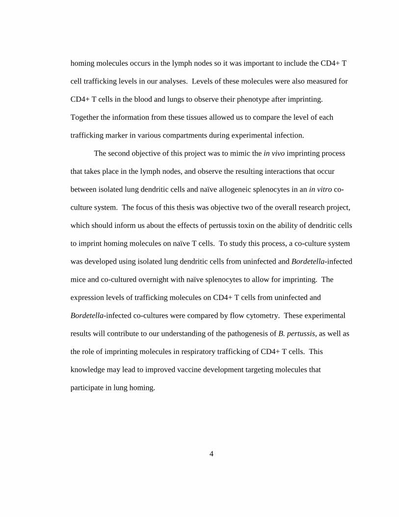
homing molecules occurs in the lymph nodes so it was important to include the CD4+ T
cell trafficking levels in our analyses. Levels of these molecules were also measured for
CD4+ T cells in the blood and lungs to observe their phenotype after imprinting.
Together the information from these tissues allowed us to compare the level of each
trafficking marker in various compartments during experimental infection.
The second objective of this project was to mimic the in vivo imprinting process
that takes place in the lymph nodes, and observe the resulting interactions that occur
between isolated lung dendritic cells and naïve allogeneic splenocytes in an in vitro co-
culture system. The focus of this thesis was objective two of the overall research project,
which should inform us about the effects of pertussis toxin on the ability of dendritic cells
to imprint homing molecules on naïve T cells. To study this process, a co-culture system
was developed using isolated lung dendritic cells from uninfected and Bordetella-infected
mice and co-cultured overnight with naïve splenocytes to allow for imprinting. The
expression levels of trafficking molecules on CD4+ T cells from uninfected and
Bordetella-infected co-cultures were compared by flow cytometry. These experimental
results will contribute to our understanding of the pathogenesis of B. pertussis, as well as
the role of imprinting molecules in respiratory trafficking of CD4+ T cells. This
knowledge may lead to improved vaccine development targeting molecules that
participate in lung homing.

5
CHAPTER II
THE EFFECTS OF BORDETELLA PERTUSSIS ON DENDRITIC CELL IMPRINTING
OF CD4+ T CELLS

6
THE EFFECTS OF BORDETELLA PERTUSSIS ON DENDRITIC CELL IMPRINTING
OF CD4+ T CELLS 1
Sana Waheed,* Tzvia Abramson,2*
*Department of Biological Sciences, San José State University,
San José, CA 95192
1 This work was supported by funds from Dr. Tzvia Abramson and California State University Minigrant (2008). 2 Address correspondence and reprint requests to Tzvia Abramson, Department of Biological Sciences, One Washington Square, San José State University, San José, CA 95192 Abbreviations used in this paper: DCs, dendritic cells; B. pertussis, Bordetella pertussis; B. parapertussis, Bordetella parapertussis; PTx, Pertussis Toxin; MHC, major histocompatibility complex; CD, cluster of differentiation; PBS, phosphate buffered saline; FCS, fetal calf serum; FSC, Forward Scatter; SSC, Side Scatter; PSGL-1, P-selectin glycoprotein ligand-1; VLA-1, very late antigen-1; LFA-1, lymphocyte function-associated antigen-1; VLA-4, very late antigen-4; CCR5, chemokine receptor type 5; CCR6, chemokine receptor type 6; CXCR3, CXC chemokine receptor 3; VCAM-1, vascular cell adhesion molecule-1; CLA, cutaneous lymphocyte antigen.

7
Abstract
Bordetella pertussis is an aerobic gram-negative bacterial pathogen that causes
the human respiratory disease whooping cough. Despite widespread vaccination,
whooping cough is reemerging due to decreased vaccine efficacy. One of the hallmarks
of infection is lymphocytosis, which is induced by the pertussis toxin. Lymphocytes such
as CD4+ T cells navigate to infected tissues through surface-trafficking molecules, which
are imprinted during their interaction with tissue-associated dendritic cells. We
hypothesized that the pertussis toxin affects the imprinting process resulting in altered
expression of trafficking molecules on CD4+ T cells. We tested this hypothesis using a
mouse model of infection. Imprinting levels on CD4+ T cells were compared to
Bordetella parapertussis, a related strain that lacks pertussis toxin. Our results indicated
that 5 days post-infection, the percentage of lung dendritic cells increased and adopted a
mature phenotype (displaying an increased capability to migrate and present antigen to T
cells) in response to B. pertussis infection, and there was an overall downregulation of
trafficking molecules on CD4+ T cells. However, 25 days post-infection with B.
pertussis, dendritic cells continued to express elevated levels of MHC class II, and the
expression of trafficking markers on CD4+ T cells also increased compared to uninfected
controls. These results enable identification of molecules that are specific for
lymphocyte trafficking to the respiratory airways and contribute to knowledge useful in
the development of better vaccines.

8
Introduction
Bordetella pertussis is an aerobic gram-negative bacterium that causes the
respiratory disease whooping cough in humans. Considerable mortality was attributed to
B. pertussis in the early twentieth century, which declined once a vaccine was introduced.
However, whooping cough is reemerging in both adults and children due to drop in
vaccine efficacy (2-3). Vaccinated adults can be carriers of pertussis and manifest a
prolonged, but relatively mild respiratory disease, which may be transmitted to
unvaccinated infants (4). In infants the manifestations can include vomiting, pneumonia,
hypoxia, seizures or apnea. Throughout the world there are 300,000 pertussis-related
deaths each year, mostly in children (5). Although these cases are mainly seen in
developing areas of the world, there have been incidents of pertussis outbreaks in the
United States and, according to the Centers for Disease Control and Prevention, the U.S.
had over 10,000 reported cases of pertussis in 2007 (6). The onset of the disease is
characterized by a persistent, forceful whoop-like cough that can last for several weeks to
months, even after the clearance of the bacteria. The hallmark of the disease is
leukocytosis with extreme lymphocytosis, which is attributed to one of the bacterial
virulence factors, pertussis toxin (PTx) (7-10).
Bordetella pertussis is an extracellular pathogen that colonizes the upper
respiratory system and bronchial epithelial cells. The respiratory system is constantly
exposed to airborne antigens. The mucosal immunity maintains a steady state in which
harmless antigens do not trigger unnecessary immune responses (11-12). Tissue resident
DCs sample the airways and are key players in supporting immunological tolerance in

9
response to non-pathogenic antigens (11). Bordetella pertussis overcomes this
suppressive mechanism and disrupts these homeostatic conditions in the airways. Several
bacterial factors contribute to the pathogenesis of B. pertussis in the respiratory tract.
PTx is a secreted AB subunit exotoxin that is associated with numerous pathogenic
functions and immunomodulation. It is responsible for inhibiting chemotaxis in T cells,
macrophages, neutrophils, and reduced B cell survival (13). The A subunit is the
enzymatically active component that is responsible for ADP-ribosylation and inhibition
of the alpha subunit of G proteins (Gα) on target host cells (14). G proteins are coupled
to chemokine receptors and mediate cell signaling. PTx prevents activation of Gα and
interferes with signaling that leads to chemotaxis and cell migration (8, 15).
Administration of soluble PTx was reported to delay the initial recruitment of immune
cells such as neutrophils and macrophages, which are responsible for engulfing and
killing bacteria (16-20). Impaired neutrophil and macrophage function can lead to
colonization of B. pertussis in the respiratory tract
During respiratory infection, DCs sample and pinocytose (internalize) antigen
from the tracheal lumen and tissue-penetrating antigens. Immediately after antigen
internalization, DCs develop a mature phenotype that is comprised of upregulation of
surface maturation markers such as MHC class II, CD40, and CD86, which facilitate DC
entry into the lymphatics and migration to the draining lymph nodes where antigen
presentation to T cells occurs (21-23). The interaction of mature DCs with naïve T cells
in the lymph nodes results in T cell activation, signaling, and upregulation of trafficking
receptors, a mechanism called imprinting. Mature DCs derived from a specific tissue

10
will then imprint T cells with trafficking receptors that direct them to the area of infection
where the antigen was originally encountered by the DC (24).
Lymphocyte trafficking is a multi-step process that allows activated T cells to exit
the lymph nodes, enter the bloodstream, extravasate, and home to infected tissue (1, 24-
26). The first step of this process is a rolling/tethering action mediated by selectins that
loosely bind the T cell to inflamed endothelium and slows its transit in the bloodstream.
This is followed by attachment and activation of the T cell by chemokine receptors. The
third step is mediated by integrins, which completely halt the T cell in the bloodstream
and mediate extravasation.
The concept of trafficking receptor imprinting on lymphocytes is well
documented in the gut and skin where the trafficking molecules and environmental
factors influencing this interaction have been identified (26). For example, DCs from gut
origin imprint T cells in the presence of retinoic acid (a dietary environmental factor)
with high levels of CCR9 and α4β7, which are the surface molecules that direct T cells to
the small intestine (1, 25, 27-30). Trafficking of T cells to the skin is mediated by DCs
and activated Vitamin D3 derivatives that imprint CCR10, CLA, and CCR4, which leads
T cells to the skin (1, 31-33). As of now, the imprinting factors that direct T cell
trafficking exclusively to the respiratory system have not been described (34).
In this thesis, we studied the interactions between DCs and T cells during a
respiratory infection with B. pertussis. We hypothesized that B. pertussis (via PTx) alters
DC imprinting of trafficking molecules on T cells. This may lead to a high degree of
lymphocytosis in the circulation with reduced levels of trafficking molecules that can

11
orient lymphocyte homing to the lungs. We tested our hypothesis using B. pertussis and
Bordetella parapertussis experimental infections in mice. Bordetella parapertussis lacks
PTx and was used as a control. This comparison elucidated the effects of PTx on G
protein-coupled receptor signaling, as well as DC and T cell interactions leading to T cell
homing (35-36). Since defective homing could lead to delayed clearance during B.
pertussis infection, knowledge in this area should contribute to understanding the
pathogenesis of this bacterium and aid in vaccine development based on lung associated
trafficking molecules.

12
Materials and Methods
All studies using animals have been approved by the Institutional Animal Care and Use
Committee (IACUC) (Protocol #921) at San José State University, San José, CA.
Bacteria
Two Bordetella strains were used, B. pertussis 338, which is a nalidixic acid-resistant
derivative of the Tohama strain, and B. parapertussis, which was obtained from ATCC
(#9305). Both strains were grown on Bordet-Gengou blood plates, and single colonies
were used to inoculate Stainer-Scholte broth containing Heptakis (Sigma-Aldrich, St.
Louis, MO) prior to experimental infection.
Experimental infection
Six-week-old female Balb/c mice were inoculated intranasally with 20 µl of 5x10⁶ colony
forming units of B. pertussis or B. parapertussis. Uninfected control mice received 20 µl
of PBS (37-38). Two time-points of experimental infection were established, 5 days
post-infection (peak infection) and 25 days post-infection, which were determined by
performing a time-course experiment where bacterial load in the lungs was measured for
ten days and again on day 25 (see Appendix A). To confirm infection and determine the
bacterial load, lungs from experimentally infected mice were harvested, homogenized,
and plated on Bordet-Gengou plates prior to each experiment.
13
Preparation of single cell suspensions from lungs and spleen
Mice were exsanguinated followed by perfusion of lungs with 30-50 ml of PBS. The
lungs and trachea were removed, minced and digested in RPMI-1640, 10% FCS, 75U/ml
DNase I (Sigma-Aldrich) and 250 U/ml Type I Collagenase (Worthington Biochemical
Corporation, Lakewood, NJ) in a 37ºC orbital shaker for 45 to 60 minutes. The digested
lungs were passed through a 40-micron nylon mesh strainer and cells were washed with
RPMI-1640 to remove residual digest medium. Digested lung tissue was resuspended in
40% Percoll (GE Lifescience, Piscataway, NJ), slowly underlaid with 70% Percoll and
centrifuged for 25 minutes to create a density gradient. Cells at the 40-70% interface
(mononuclear cells) were collected and used for enrichment of DCs. The spleens from
uninfected allogeneic mice were harvested and pressed through a nylon mesh to obtain a
single cell suspension (39). Splenocytes were incubated with 3 ml red blood cell lysis
buffer (Sigma-Aldrich) for 5 minutes and washed with RPMI-1640 with 10% FCS. Cell
pellets from the lungs were resuspended in buffer (PBS, 0.5% FCS, 2 mM EDTA)
suitable for MACS columns.
Enrichment of lung DCs
All cell enrichments were performed using magnetic microbeads (Miltenyi Biotech,
Auburn, CA). For DC enrichment from lungs, CD11c microbeads (N418) were used and
lung mononuclear cells were enriched for CD11c+ cells using positive selection with a
MiniMACS separator. Briefly, lung mononuclear cells were incubated with 100 µl of
CD11c microbeads for 15 minutes at 4°C. Cells were washed with MACS buffer (PBS,

14
0.5% FCS, 2 mM EDTA) and resuspended in 500 µl of buffer. One MS column was used
to enrich for CD11c+ cells.
T cell activation and co-cultures
For co-culturing experiments, 24-well plates were coated with the purified monoclonal
antibodies CD3 (1 µg/ml) and CD28 (1 µg/ml) in PBS, and incubated at 4ºC, as adapted
from Sigmundsdottir (33) and Campanelli (39). Enriched lung DCs were cultured with
splenocytes from uninfected allogeneic mice at a 1:25 ratio in RPMI-1640 supplemented
with 10% FCS, penicillin, streptomycin, IL-2 (12.5 ng/ml) and IL-12 (2.5 ng/ml) (R&D
Systems, Minneapolis, MN). Cells were co-cultured overnight at 37ºC with 5% CO2 to
allow for imprinting, followed by flow cytometry analysis. T cell activation was
determined using intracellular antibody staining with Ki-67 (B56) and interferon γ
(XMG1.2), which served as markers of T cell proliferation in an allogeneic system.
Briefly, cells were stained for extracellular markers and resuspended in 250 µl
Fixation/Permeabilization (BD Biosciences, Franklin Lakes, NJ) solution and incubated
at 4°C for 20 minutes. Cells were washed twice with 1 ml of 1× Perm/Wash Buffer (BD
Biosciences) and then resuspended in 100 µl of Perm/Wash Buffer containing the
antibody or negative control. Cells were incubated at room temperature in the dark for 30
minutes before being washed twice with 1 ml/wash of 1× Perm/Wash Buffer and
resuspended in staining buffer (0.1% Sodium Azide, 1% FCS, PBS).

15
Flow cytometry
Analysis of surface and intracellular markers was done by flow cytometry. Lung derived
DC phenotyping was measured by the expression of myeloid associated markers [CD11c
(N418), CD11b (M1/70)] and maturation markers [MHC class II (M5/114.15.2), CD40
(3/23), CD86 (GL-1)]. Additional trafficking markers were evaluated on DCs as detailed
for T cells. Expression of trafficking molecules on CD4+ T cells included CD4 (RM4-5,
BD Biosciences and Invitrogen, Carlsbad, California), CD38 (90) and the following
trafficking molecules: CD11a (M17/4), CD49f (GoH3), CD162 (2PH1, BD Biosciences),
CXCR3 (220803, R&D Systems), CD49a (HMalpha1, ABDSerotec, Raleigh, NC),
CD49d (9C10 MFR4.B), and CD103 (2E7). All antibodies were purchased from
BioLegend (San Diego, CA) unless otherwise indicated. The levels of these markers
were compared for B. pertussis and B. parapertussis infections and normalized to
uninfected controls as explained in the figure legends. Samples were acquired using the
FACS Calibur (BD Biosciences) at San José State University and the LSRII (BD
Biosciences) at Stanford University, and were analyzed using Flowjo.

16
Results
Experimental design
Balb/c mice were inoculated intranasally with PBS (uninfected controls), B.
pertussis or B. parapertussis for a period of 5 and 25 days. At each time-point lungs
were harvested, mononuclear cells were obtained and enriched for DCs (CD11c+ cells)
using Miltenyi Microbeads (Fig. 1). DCs were enumerated and phenotyped for each
treatment using multicolor flow cytometry (Fig. 2, 4). Imprinting of trafficking
molecules was observed in co-cultures of lung DCs with naïve allogeneic splenocytes
from Swiss Webster mice (Fig. 3, 5).
Peak infection: DCs matured in response to Bordetella infection
Five days post-infection we found 23% of CD11c+ cells in uninfected control
lungs, whereas B. parapertussis had 33.1% and B. pertussis had 70.6% (Fig. 2A). The
percent DCs was estimated by the overall analysis of CD11c+ cells isolated from the
lungs. DC phenotype was characterized using CD11c+ and a second marker (Table I)
using flow cytometry (Fig. 2B). Levels of maturation markers were assessed with MHC
class II, CD40, and CD86 (Fig C-D). These molecules bind to the T cell receptor,
CD40L, and CD28 respectively on CD4+ T cells, forming an immunological synapse that
results in T cell activation and stimulation (11). Since B. pertussis is an extracellular
bacterial pathogen, we analyzed MHC class II, which is the molecule responsible for
presenting antigen to CD4+ T cells. Lung DCs from uninfected mice revealed low levels
of MHC class II, CD40, and CD86 (7.72%, 10.64%, 6.11% respectively), whereas B.

17
parapertussis-infected DCs showed increased expression (20.64%, 13.67%, 14.04%).
Comparatively, lung DCs from B. pertussis infection displayed the highest levels of
maturation (64.92%, 36.64%, 60.26% respectively).
The phenotypic marker CD11b is present on myeloid DCs of lung origin (40).
Relatively low expression of CD11b (10.21%) was observed in lung DCs from
uninfected control mice while this percentage increased in B. parapertussis infection
(20.5%), but even more so in B. pertussis infection (61.04%). Additionally, CCR5 was
analyzed to observe DC migratory abilities to inflamed tissues. There was low
expression of CCR5 in uninfected DCs (7.76%) compared to B. parapertussis (13.95%,)
and B. pertussis (55.84%). Together, this data indicate that DCs mature during B.
pertussis infection and do not appear to be inhibited by PTx.

18
FIGURE 1. Experimental design. Balb/c mice were infected and lungs were harvested 5 and 25 days post-infection. Lungs were processed and mononuclear cells were obtained to further enrich for CD11c+ cells. DCs were enumerated, phenotyped and analyzed for imprinting abilities in co-cultures with allogeneic splenocytes from Swiss Webster mice.
Uninfected (PBS) B. pertussis B. parapertussis
Time-points of 5 or 25 days post-infection
Infect Balb/c mice on Day 0
Harvest lungs and digest (separately)
Obtain mononuclear cells with Percoll gradient
Enrich for DCs using CD11c microbeads
DC phenotyping using multicolor flow cytometry
Imprinting of trafficking molecules from co-culture of
lung DCs with allogeneic splenocytes from Swiss
Websters
Enumerate DCs with multicolor flow cytometry

19
TABLE I. DC phenotypic markers. The following markers were used to phenotype DCs and determine maturation levels for uninfected, B. pertussis and B. parapertussis infected lungs.
Marker Function CD11c Expressed primarily on DCs CD40 Co-stimulatory molecule CD86 Co-stimulatory molecule MHC class II Extracellular antigen presentation molecule CCR5 Allows DC migration to inflamed tissue (24) CD11b Integrin expressed on DCs and monocytes

20
FIGURE 2. DC maturation 5 days post-infection with B. pertussis and B. parapertussis. Mononuclear cells were isolated from lungs and enriched for DCs. Percent of CD11c+ cells was estimated using quadrants that were placed according to positive controls (A). Mononuclear cells were gated on monocytes, and doublets (cell clumping) were removed (B). CD11c+ cells (DCs) were analyzed for a second phenotypic marker as detailed in C. (See Table I for a description of the markers). For DC maturation, we measured levels of CD40, CD86, MHC class II; myeloid associated marker CD11b, as well as ability to migrate to lymph nodes with CCR5 (C-D). Data are representative of at least 4 independent experiments.
Percent CD11c+ Cells per Treatment
0
10
20
30
40
50
60
70
80
Uninfected B. parapertussis B. pertussis
Treatment
% C
D11
c+ c
ells
Uninfected B. parapertussis B. pertussis
2A
2B
FSC
SSC
CD11c
DC Phenotypic Marker
FSC-A
FSC-W

21
2C Uninfected B. parapertussis B. pertussis
CD11c
CD40
CD86
CCR5
CD11b
MHC II

22
Infection with Bordetella alters trafficking receptor levels on naïve CD4+ T cells
In order to verify the imprinting ability of lung DCs derived from experimentally
infected mice, we co-cultured lung DCs with naïve splenocytes from allogeneic mice.
Establishing an allogeneic system ensures that imprinting occurs on proliferating T cells,
where DCs interact through MHC class II with the T cell receptor and trigger non-
specific T cell proliferation. This leads to transcription of certain patterns of molecules
that guide the T cell to the site of infection (33). These homing molecules are a
combination of chemokine receptors and integrins that interact with inflamed
endothelium and allow transmigration of the T cell from the bloodstream into the tissue.
2D DC Phenotype 5 Days Post-Infection
0
10
20
30
40
50
60
70
MHC II CD40 CD86 CD11b CCR5
% E
xpre
ssio
n o
n C
D11
c+ C
ells
Uninfected B. parapertussis B. pertussis

23
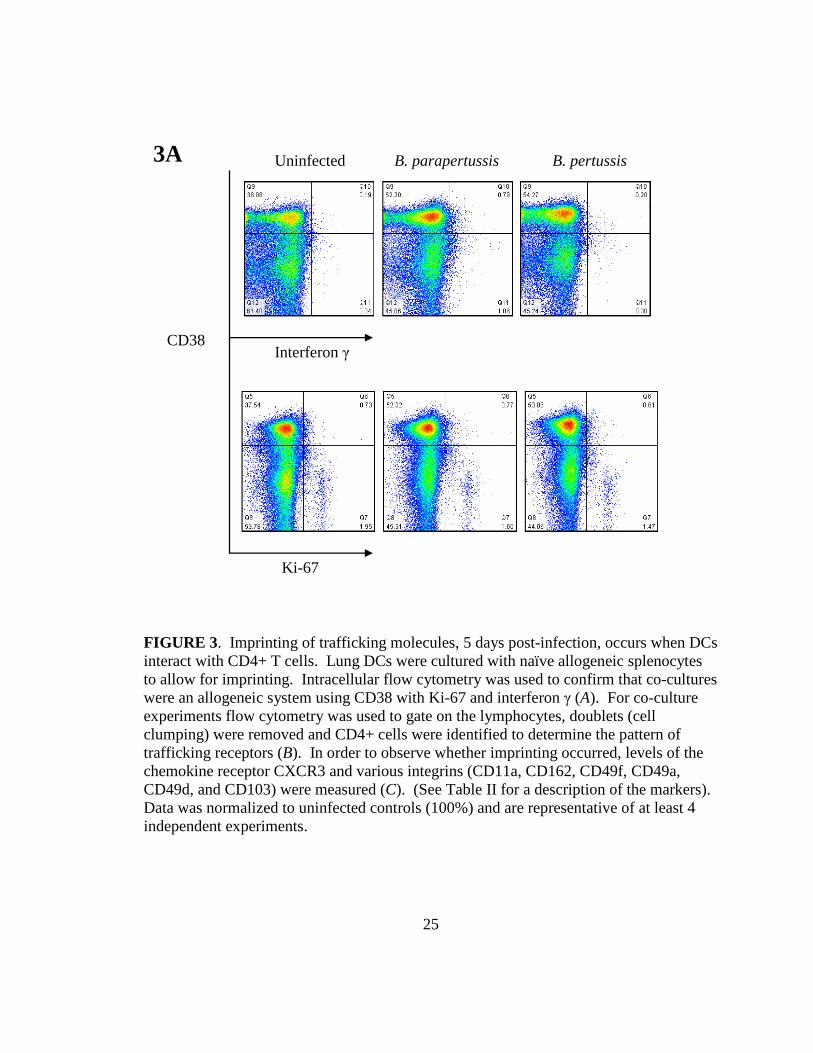
The proliferative nature of the allogeneic system was confirmed by intracellular
staining for interferon γ (secreted during T cell activation) and Ki-67 (a nuclear factor of
proliferation) in activated CD38+ cells (Fig. 3A). A proliferating population was
observed in each treatment; 0.5-1.8% of cells were positive for interferon γ and roughly
2% were positive for Ki-67 indicating that we had a proliferating T cell system. DC
ability to imprint trafficking markers on allogeneic T cells was then analyzed. Since little
is known about specific lung homing markers, we analyzed a panel of trafficking
molecules to determine whether the levels of imprinted homing molecules were altered in
infected co-cultures compared to uninfected controls (Table 2). Our results indicate an
overall downregulation of homing molecules on CD4+ T cells during experimental B.
pertussis infection compared to uninfected controls at 5 days post-infection. These
results were normalized to uninfected control values set at 100%.
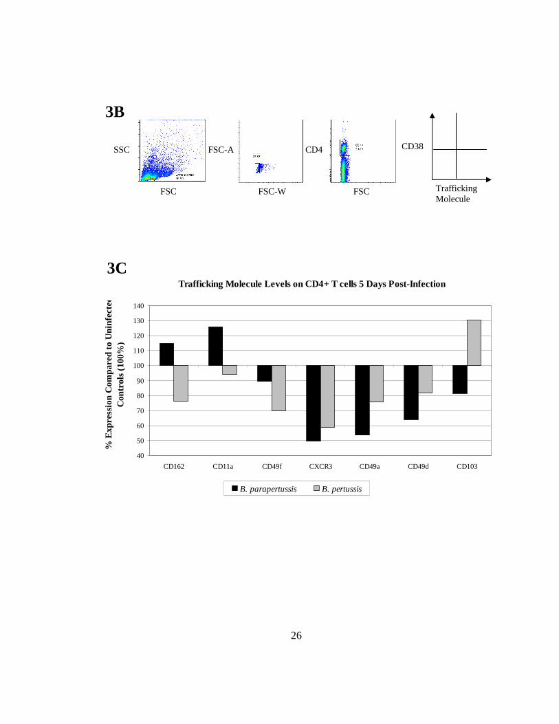
Recent studies indicate that CD11a, CD162, and CD103 are important markers
for trafficking and cell retention in the lungs (28, 41-45). On day 5 of infection, CD4+ T
cells from B. parapertussis co-cultures showed upregulation of CD11a (125.9%) and
CD162 (114.9%) but downregulation of CD103 (81.5%) compared to uninfected controls
(100%) (Fig. 3C). In contrast B. pertussis co-cultures showed downregulation of CD11a
(94.4%) and CD162 (76.1%) with upregulation of CD103 (130.3%). Additionally, the
integrins CD49a and CD49d can dimerize with CD29 and have been reported to migrate
to the respiratory system (21, 46-47). However, downregulation was observed in both B.
parapertussis and B. pertussis infections for CD49a (53.6%, 75.6% respectively) and
CD49d (63.9%, 81.9% respectively) compared to uninfected controls (100%). Another

24
adhesion molecule, CD49f, may play a role in lung homing and was downregulated more
in B. pertussis co-cultures (69.9%) than in B. parapertussis (89.6%) (27). There is also
evidence in the literature that CXCR3 is involved in T cell homing to the lungs (11, 48-
50). Five days post-infection with Bordetella, CXCR3 was downregulated in both B.
parapertussis (49.6%) and B. pertussis (58.7%,) compared to uninfected controls (100%).
TABLE II. Trafficking markers and their functions. A variety of markers that are implicated in the homing process were analyzed, such as molecules involved in tethering or rolling and other integrins that mediate attachment to the endothelium and lead to extravasation.
Marker Function Receptor Family
CD162 (PSGL-1) Adhesion (43) Selectin CXCR3 Chemotaxis of T cells (49) Chemokine receptor CD11a (LFA-1) Cell adhesion, costimulation (45) Integrin CD49a (α1) Involved in T cell adhesion (46) Integrin CD49d (α4) Cell migration, adhesion, homing (47) Integrin CD49f (α6) Cell adhesion, migration (27) Integrin CD103 (αε) Tissue retention of lymphocytes (42) Integrin CD38 Activation marker ---
25
FIGURE 3. Imprinting of trafficking molecules, 5 days post-infection, occurs when DCs interact with CD4+ T cells. Lung DCs were cultured with naïve allogeneic splenocytes to allow for imprinting. Intracellular flow cytometry was used to confirm that co-cultures were an allogeneic system using CD38 with Ki-67 and interferon γ (A). For co-culture experiments flow cytometry was used to gate on the lymphocytes, doublets (cell clumping) were removed and CD4+ cells were identified to determine the pattern of trafficking receptors (B). In order to observe whether imprinting occurred, levels of the chemokine receptor CXCR3 and various integrins (CD11a, CD162, CD49f, CD49a, CD49d, and CD103) were measured (C). (See Table II for a description of the markers). Data was normalized to uninfected controls (100%) and are representative of at least 4 independent experiments.
3A Uninfected B. parapertussis B. pertussis
Interferon γ CD38
Ki-67
26
Trafficking Molecule Levels on CD4+ T cells 5 Days Post-Infection
40
50
60
70
80
90
100
110
120
130
140
CD162 CD11a CD49f CXCR3 CD49a CD49d CD103
% E
xpre
ssio
n C
ompa
red
to U
ninf
ecte
d C
ontr
ols
(100
%)
B. parapertussis B. pertussis
3C
3B
CD38
Trafficking Molecule
FSC
SSC FSC-A
FSC-W
CD4
FSC

27
Twenty-five days post-infection: DC maturation levels decrease except for some markers
in B. pertussis infection
In our previous experiment for 5 days post-infection, we observed an increase in
both percent CD11c+ cells and DC maturation levels during experimental Bordetella
infection. In order to establish the phenotypic state of DCs 25 days post-infection, lungs
were harvested and lung mononuclear DCs were isolated. In the lungs, the percent gated
CD11c+ cells decreased for B. pertussis (20.7%) and B. parapertussis (23.1%) infections
to levels similar to uninfected controls (20.4%) (Fig. 4A). DC phenotypic levels were
then analyzed where lung DCs from B. parapertussis infection had reduced expression of
the maturation marker CD86 (4.14%) compared to B. pertussis (8.04%) (Fig. 4B). In
contrast, MHC class II remained upregulated in B. pertussis (18.3%) whereas the levels
for uninfected controls and B. parapertussis decreased to 6.74% and 7.57% respectively.
The lung-associated marker CD11b was 5.24% for lung DCs from uninfected
controls, whereas DCs from B. pertussis infection had elevated levels (13.1%) while DCs
from B. parapertussis were downregulated (6.56%). The DC migratory marker CCR5
was upregulated on DCs from B. pertussis (7.24%) compared to B. parapertussis (3.59%)
and uninfected controls (3.75%). Together, this indicates that the effect of Bordetella on
DC phenotype diminished 25 days post-infection with the exception of B. pertussis
infection where MHC class II, CD86, CD11b, and CCR5 remained elevated.

28
FIGURE 4. DC maturation 25 days post-infection with B. pertussis and B. parapertussis. After 25 days of infection lung mononuclear cells were obtained, enriched for DCs and gated similarly to that shown in Fig. 2B. We assessed the percent CD11c+ cells in each treatment (as determined using quadrants from flow cytometry dot plots) (A) and evaluated DC maturation with MHC class II, CD40, CD86; myeloid and lung associated markers CD11b, CD11c, and migratory capability with CCR5 (B). Data are representative of at least 3 independent experiments.
Percent CD11c+ Cells per Treatment
0
5
10
15
20
25
Uninfected B. parapertussis B. pertussis
Treatment
% C
D11
c+ C
ells
Uninfected
4A
DC Phenotype 25 Days Post-Infection
0
5
10
15
20
25
MHC II CD40 CD86 CD11b CCR5
Per
cent
Exp
ress
ion
on C
D11
c+ c
ells
Uninfected B. parapertussis B. pertussis
4B

29
Trafficking receptor levels decreased in B. parapertussis co-cultures but increased in B.
pertussis co-cultures 25 days after infection
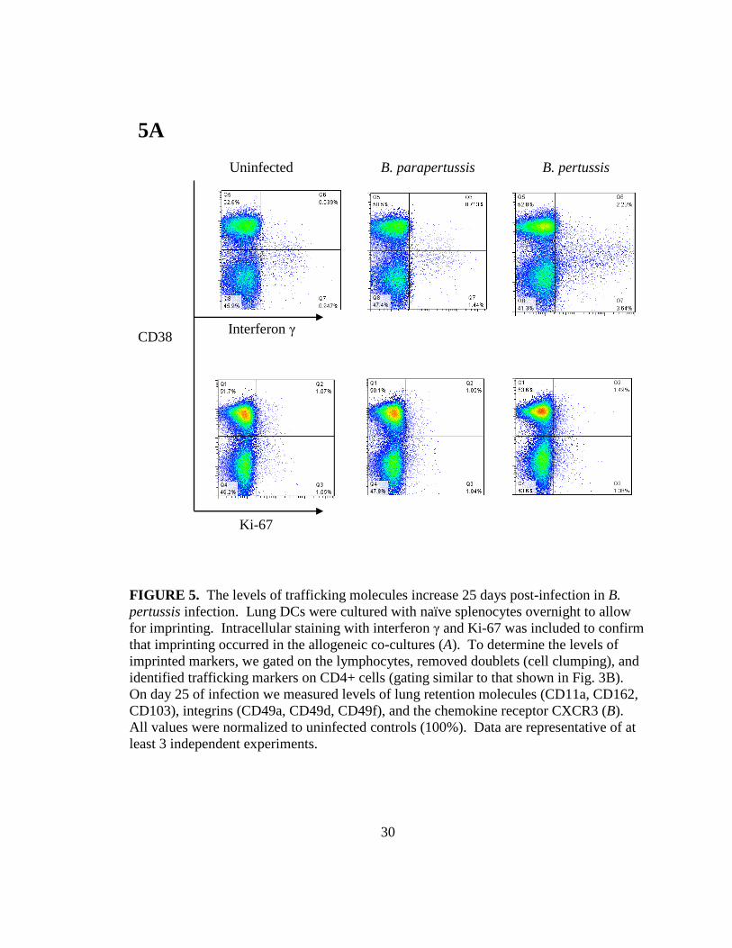
Intracellular flow cytometry results of interferon γ and Ki-67 confirmed the
proliferative ability of each co-culture, as illustrated by the interferon γ positive and Ki-
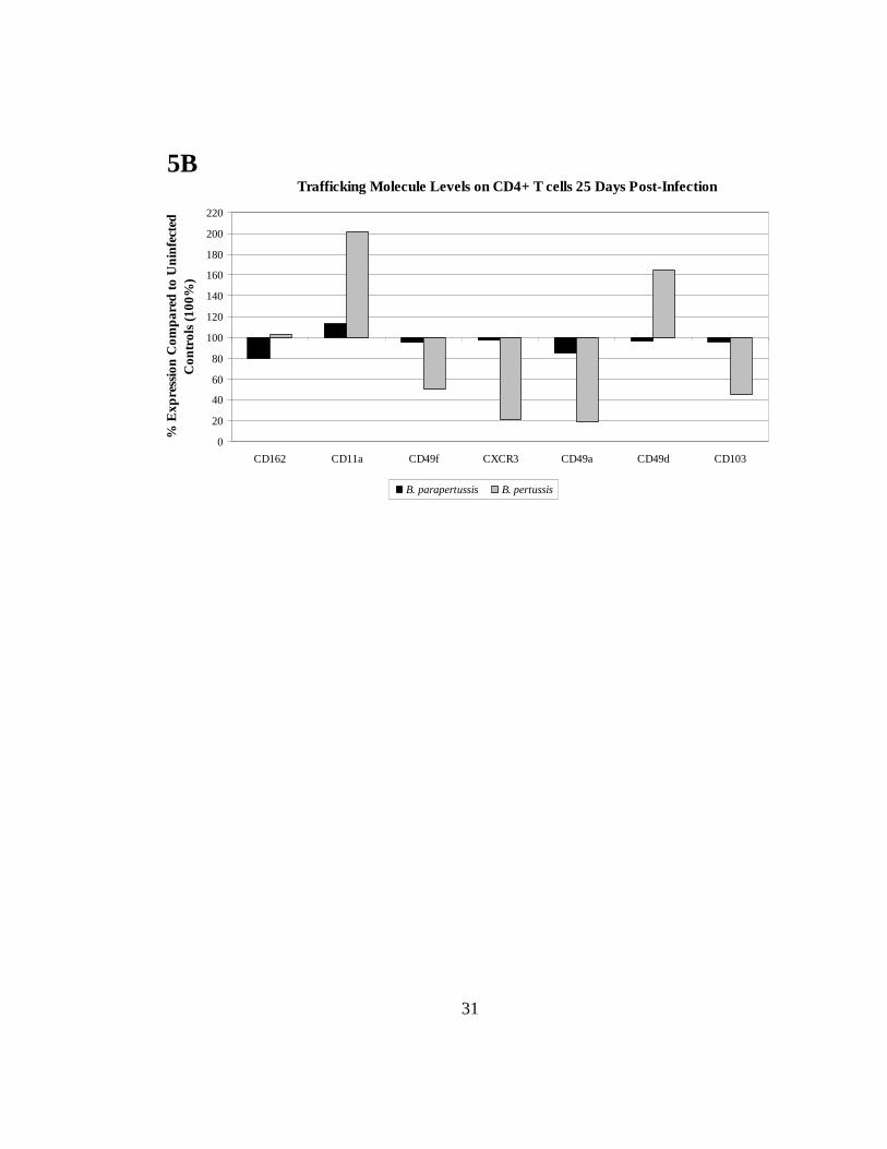
67 positive populations (Fig. 5A). The level of trafficking receptors on CD4+ T cells was
measured to determine whether DCs continued to actively imprint 25 days post-infection.
In B. pertussis-infected co-cultures, the adhesion and lung retention marker CD11a was
upregulated to 201.1% whereas it was downregulated in B. parapertussis-infected co-
cultures (113.5%) when compared to uninfected controls (100%) (Fig. 5B).
The integrins CD49d and CD49f are associated with migration and had reduced
expression in B. parapertussis co-cultures (96.7% and 95.1% respectively) when
compared to B. pertussis co-cultures, which displayed elevated levels of CD49d (164.6%)
but maintained downregulation of CD49f (50.5%). For B. pertussis co-cultures, CXCR3
remained downregulated (21.4%) compared to uninfected controls (100%). This suggests
that 25 days post-infection, B. pertussis-infected DCs can imprint some trafficking
molecules on CD4+ T cells, while this is not the case for B. parapertussis-infected co-
cultures, where these molecules are expressed at low levels.
30
FIGURE 5. The levels of trafficking molecules increase 25 days post-infection in B. pertussis infection. Lung DCs were cultured with naïve splenocytes overnight to allow for imprinting. Intracellular staining with interferon γ and Ki-67 was included to confirm that imprinting occurred in the allogeneic co-cultures (A). To determine the levels of imprinted markers, we gated on the lymphocytes, removed doublets (cell clumping), and identified trafficking markers on CD4+ cells (gating similar to that shown in Fig. 3B). On day 25 of infection we measured levels of lung retention molecules (CD11a, CD162, CD103), integrins (CD49a, CD49d, CD49f), and the chemokine receptor CXCR3 (B). All values were normalized to uninfected controls (100%). Data are representative of at least 3 independent experiments.
5A
Interferon γ
Ki-67
Uninfected B. parapertussis B. pertussis
CD38
31
Trafficking Molecule Levels on CD4+ T cells 25 Days Post-Infection
0
20
40
60
80
100
120
140
160
180
200
220
CD162 CD11a CD49f CXCR3 CD49a CD49d CD103
% E
xpre
ssio
n C
omp
ared
to
Un
infe
cted
C
ontr
ols
(100
%)
B. parapertussis B. pertussis
5B

32
Discussion
Unlike other areas of the body such as the skin and gut where specific homing
molecules that govern lymphocyte trafficking have been established (26), a unique
trafficking molecule pattern to the respiratory airways remains to be defined. We were
interested in elucidating the role PTx has on DC imprinting of trafficking molecules on
CD4+ T cells that guides them to the respiratory system. PTx inhibits G protein
activation and hinders chemotaxis of cells (8, 13, 15). Therefore, we established an
experimental respiratory infection model using two bacteria from the Bordetella species:
B. pertussis (produces PTx) and B. parapertussis (lacks PTx).
DC origin and their level of maturation are critical components for imprinting.
Therefore, we utilized flow cytometry to determine DC phenotype for uninfected controls
and Bordetella-infected DCs. At 5 days post-infection, CD11c+ cells increased 1.4 times
in B. parapertussis infection, whereas there was a 3-fold increase in B. pertussis infection
compared to uninfected controls. This increase in CD11c+ cells may either be due to
recruitment of DCs in the lungs during a state of infection or inability to leave the lungs
and migrate to the lymph nodes, a phenomenon that could be attributed to PTx inhibition
of chemokine receptors such as CCR7. However, by 25 days post-infection, the percent
CD11c+ cells in the lungs decreased for both infections, possibly due to a halt in
recruitment of these cells to the lungs.
In contrast to those who report partial DC maturation during B. pertussis infection
(51), our results indicated that 5 days post-infection, lung DCs from B. pertussis infection
had upregulated MHC class II, CD40 and CD86 and had generally higher levels of

33
maturation compared to that in B. parapertussis infection or uninfected controls. This
discrepancy between our results and those of others may be attributed to the timing of
post-infection sampling or differences in bacterial strains. The phenotype of DCs
changed 25 days post-infection for B. parapertussis so that MHC class II, CD86, CD11b,
and CCR5 were downregulated compared to B. pertussis. However MHC class II
remained upregulated in B. pertussis, which may be due to continued antigen presentation
to CD4+ T cells in the regional lymph nodes even at late stages of infection. Together
this information indicated that 5 days post-infection, B. pertussis does not prevent DCs
from acquiring a mature phenotype that is required for imprinting CD4+ T cells.
However, by 25 days after infection, B. parapertussis-infected lungs appeared to revert to
steady-state conditions, which may not be the case for B. pertussis infection. An
important chemokine receptor, CCR7, is required for entry into lymph nodes (24). In
order to further characterize the migratory ability of B. pertussis-infected DCs, CCR7
assessment will be included in future experiments.
Once the DC phenotype was established, isolated lung DCs were co-cultured
with naïve splenocytes harvested from allogeneic mice, and imprinting of trafficking
molecules on CD4+ T cells was analyzed. CD11a and CD162 have been reported to be
important for adhesion and retention of cells in the lungs (43, 45). Interestingly, we
found that on day 5 post-infection, both CD11a and CD162 were downregulated in B.
pertussis infection but upregulated in B. parapertussis, which suggests that PTx has a
role in suppressing these molecules during peak infection. However, on day 25 post-

34
infection, CD11a levels were elevated, suggesting that this marker is upregulated only at
later stages of infection, perhaps after the effects of PTx have diminished.
Bordetella pertussis also showed elevated levels of CD103 on day 5 post-
infection, while it was downregulated in B. parapertussis co-cultures. CD103 has been
shown to be involved in tissue retention of lymphocytes (42), and perhaps CD4+ T cells
are retained in the tissue instead of migrating during B. pertussis infection. There is also
evidence that CXCR3 is important for CD4+ T cell recruitment to the airways (11) and
both Bordetella infections showed decreased levels of CXCR3 on day 5 of infection,
while B. pertussis infection continued to show downregulation on day 25. This indicates
that perhaps both species prevent initial T cell recruitment to the lung through this
chemokine receptor.
The results from this research project suggest that B. pertussis downregulates
certain trafficking molecules during peak infection; however, at later stages of infection
these molecules are upregulated. Since B. pertussis is a prolonged respiratory disease, it
is possible that imprinting of trafficking molecules on CD4+ T cells persists during later
stages of infection, perhaps due to the decreased effects of PTx. Our experiments
indicate that the lung environment for B. parapertussis returned to a state of homeostasis
25 days after infection, whereas this is not the case for B. pertussis infection.
Further studies can also include additional trafficking molecules such as CLA,
CCR9 and α4β7 due to their presence on T cells that traffic exclusively to the skin and
gut respectively. Therefore, low levels of these molecules may be observed on T cells
that home to the respiratory system (30, 48, 52). Additional experiments may include in

35
vitro infection of uninfected lung DCs pulsed with PTx, to assess the antigen specific
response and imprinting of trafficking molecules that are PTx-dependent. Antigen pulsed
DCs could be co-cultured with naïve splenocytes to allow for imprinting, and results can
be compared to in vivo experimental Bordetella infection. In order to further confirm the
role of PTx, an experimental infection with B. parapertussis complemented with soluble
PTx could be included and compared to B. pertussis (18). This information will increase
our understanding of the pathogenesis of B. pertussis, and the role of PTx in preventing
lymphocyte recruitment to the respiratory system through trafficking molecule
modulation. This line of research may identify the molecules responsible for the
exclusive recruitment of CD4+ T cells to the airways, leading to a more efficacious
vaccine design that targets trafficking molecules important in B. pertussis infection.

36
References
1. Butcher EC, Picker LJ. 1996. Lymphocyte homing and homeostasis. Science 272: 60-6.
2. Carbonetti NH. 2007. Immunomodulation in the pathogenesis of Bordetella pertussis infection and disease. Curr Opin Pharmacol 7: 272-8.
3. McIntyre P, Wood N. 2009. Pertussis in early infancy: disease burden and preventive strategies. Curr Opin Infect Dis 22: 215-23.
4. Mattoo S, Cherry JD. 2005. Molecular pathogenesis, epidemiology, and clinical manifestations of respiratory infections due to Bordetella pertussis and other Bordetella subspecies. Clin Microbiol Rev 18: 326-82.
5. Crowcroft NS, Pebody RG. 2006. Recent developments in pertussis. Lancet 367: 1926-36.
6. Pertussis. Department of Health and Human Services. 2009. Centers for Disease Control and Prevention. 19 Feb 2010. <http://www.cdc.gov/ncidod/dbmd/Diseaseinfo/pertussis_t.htm>.
7. Bernales R, Eastman J, Kaplan J. 1976. Quantitation of circulating T and B lymphocytes in children with whooping cough. Pediatr Res 10: 965-7.
8. Mooi FR. 1988. Virulence factors of Bordetella pertussis. Antonie Van Leeuwenhoek 54: 465-74.
9. Munoz JJ, Arai H, Bergman RK, Sadowski PL. 1981. Biological activities of crystalline pertussigen from Bordetella pertussis. Infect Immun 33: 820-6.
10. Pittman M. 1979. Pertussis toxin: the cause of the harmful effects and prolonged immunity of whooping cough. A hypothesis. Rev Infect Dis 1: 401-12.

37
11. Kohlmeier JE, Woodland DL. 2009. Immunity to respiratory viruses. Annu Rev Immunol 27: 61-82.
12. Weiner HL. 2004. Tolerogenic dendritic cells and T regulatory cells in the mucosa. Sociedade Brasileira de Imunologia. 24 Mar 2009 <http://www.sbi.org.br/sbinarede/SBInarede25/howard.htm>.
13. Mills KH. 2001. Immunity to Bordetella pertussis. Microbes Infect 3: 655-77.
14. Smith AM, Guzman CA, Walker MJ. 2001. The virulence factors of Bordetella pertussis: a matter of control. FEMS Microbiol Rev 25: 309-33.
15. Aepfelbacher M, Aktories K, Just I. 2000. Bacterial protein toxins. Berlin ; New York: Springer. xxxii, p. 167-185.
16. Andreasen C, Carbonetti NH. 2008. Pertussis toxin inhibits early chemokine production to delay neutrophil recruitment in response to Bordetella pertussis respiratory tract infection in mice. Infect Immun 76: 5139-48.
17. Carbonetti NH, Artamonova GV, Andreasen C, Bushar N. 2005. Pertussis toxin and adenylate cyclase toxin provide a one-two punch for establishment of Bordetella pertussis infection of the respiratory tract. Infect Immun 73: 2698-703.
18. Carbonetti NH, Artamonova GV, Mays RM, Worthington ZE. 2003. Pertussis toxin plays an early role in respiratory tract colonization by Bordetella pertussis. Infect Immun 71: 6358-66.
19. Kirimanjeswara GS, Agosto LM, Kennett MJ, Bjornstad ON, Harvill ET. 2005. Pertussis toxin inhibits neutrophil recruitment to delay antibody-mediated clearance of Bordetella pertussis. J Clin Invest 115: 3594-601.
20. Meade BD, Kind PD, Ewell JB, McGrath PP, Manclark CR. 1984. In vitro inhibition of murine macrophage migration by Bordetella pertussis lymphocytosis-promoting factor. Infect Immun 45: 718-25.

38
21. Holt PG, Strickland DH, Wikstrom ME, Jahnsen FL. 2008. Regulation of immunological homeostasis in the respiratory tract. Nat Rev Immunol 8: 142-52.
22. Jahnsen FL, Strickland DH, Thomas JA, Tobagus IT, Napoli S, Zosky GR, Turner DJ, Sly PD, Stumbles PA, Holt PG. 2006. Accelerated antigen sampling and transport by airway mucosal dendritic cells following inhalation of a bacterial stimulus. J Immunol 177: 5861-7.
23. Kim CH, Campbell DJ, Butcher EC. 2001. Nonpolarized memory T cells. Trends Immunol 22: 527-30.
24. Sallusto F, Mackay CR, Lanzavecchia A. 2000. The role of chemokine receptors in primary, effector, and memory immune responses. Annu Rev Immunol 18: 593-620.
25. Mora JR. 2008. Homing imprinting and immunomodulation in the gut: role of dendritic cells and retinoids. Inflamm Bowel Dis 14: 275-89.
26. Sigmundsdottir H, Butcher EC. 2008. Environmental cues, dendritic cells and the programming of tissue-selective lymphocyte trafficking. Nat Immunol 9: 981-7.
27. Abitorabi MA, Mackay CR, Jerome EH, Osorio O, Butcher EC, Erle DJ. 1996. Differential expression of homing molecules on recirculating lymphocytes from sheep gut, peripheral, and lung lymph. J Immunol 156: 3111-7.
28. Butcher EC, Williams M, Youngman K, Rott L, Briskin M. 1999. Lymphocyte trafficking and regional immunity. Adv Immunol 72: 209-53.
29. Iwata M, Hirakiyama A, Eshima Y, Kagechika H, Kato C, Song SY. 2004. Retinoic acid imprints gut-homing specificity on T cells. Immunity 21: 527-38.
30. Kunkel EJ, Campbell JJ, Haraldsen G, Pan J, Boisvert J, Roberts AI, Ebert EC, Vierra MA, Goodman SB, Genovese MC, Wardlaw AJ, Greenberg HB, Parker CM, Butcher EC, Andrew DP, Agace WW. 2000. Lymphocyte CC chemokine receptor 9 and epithelial thymus-expressed chemokine (TECK) expression distinguish the small

39
intestinal immune compartment: Epithelial expression of tissue-specific chemokines as an organizing principle in regional immunity. J Exp Med 192: 761-8.
31. Campbell JJ, Haraldsen G, Pan J, Rottman J, Qin S, Ponath P, Andrew DP, Warnke R, Ruffing N, Kassam N, Wu L, Butcher EC. 1999. The chemokine receptor CCR4 in vascular recognition by cutaneous but not intestinal memory T cells. Nature 400: 776-80.
32. Reiss Y, Proudfoot AE, Power CA, Campbell JJ, Butcher EC. 2001. CC chemokine receptor (CCR)4 and the CCR10 ligand cutaneous T cell-attracting chemokine (CTACK) in lymphocyte trafficking to inflamed skin. J Exp Med 194: 1541-7.
33. Sigmundsdottir H, Pan J, Debes GF, Alt C, Habtezion A, Soler D, Butcher EC. 2007. DCs metabolize sunlight-induced vitamin D3 to 'program' T cell attraction to the epidermal chemokine CCL27. Nat Immunol 8: 285-93.
34. de Bree GJ, van Leeuwen EM, Out TA, Jansen HM, Jonkers RE, van Lier RA. 2005. Selective accumulation of differentiated CD8+ T cells specific for respiratory viruses in the human lung. J Exp Med 202: 1433-42.
35. Arico B, Gross R, Smida J, Rappuoli R. 1987. Evolutionary relationships in the genus Bordetella. Mol Microbiol 1: 301-8.
36. Arico B, Rappuoli R. 1987. Bordetella parapertussis and Bordetella bronchiseptica contain transcriptionally silent pertussis toxin genes. J Bacteriol 169: 2847-53.
37. Wolfe DN, Kirimanjeswara GS, Harvill ET. 2005. Clearance of Bordetella parapertussis from the lower respiratory tract requires humoral and cellular immunity. Infect Immun 73: 6508-13.
38. Zachariadis O, Cassidy JP, Brady J, Mahon BP. 2006. Gammadelta T cells regulate the early inflammatory response to Bordetella pertussis infection in the murine respiratory tract. Infect Immun 74: 1837-45.

40
39. Campanelli AP, Roselino AM, Cavassani KA, Pereira MS, Mortara RA, Brodskyn CI, Goncalves HS, Belkaid Y, Barral-Netto M, Barral A, Silva JS. 2006. CD4+CD25+ T cells in skin lesions of patients with cutaneous leishmaniasis exhibit phenotypic and functional characteristics of natural regulatory T cells. J Infect Dis 193: 1313-22.
40. Gonzalez-Juarrero M, Shim TS, Kipnis A, Junqueira-Kipnis AP, Orme IM. 2003. Dynamics of macrophage cell populations during murine pulmonary tuberculosis. J Immunol 171: 3128-35.
41. Dunne PJ, Moran B, Cummins RC, Mills KH. 2009. CD11c+CD8alpha+ dendritic cells promote protective immunity to respiratory infection with Bordetella pertussis. J Immunol 183: 400-10.
42. Suffia I, Reckling SK, Salay G, Belkaid Y. 2005. A role for CD103 in the retention of CD4+CD25+ Treg and control of Leishmania major infection. J Immunol 174: 5444-55.
43. Clark JG, Mandac-Dy JB, Dixon AE, Madtes DK, Burkhart KM, Harlan JM, Bullard DC. 2004. Trafficking of Th1 cells to lung: a role for selectins and a P-selectin glycoprotein-1-independent ligand. Am J Respir Cell Mol Biol 30: 220-7.
44. Ghosh S, Chackerian AA, Parker CM, Ballantyne CM, Behar SM. 2006. The LFA-1 adhesion molecule is required for protective immunity during pulmonary Mycobacterium tuberculosis infection. J Immunol 176: 4914-22.
45. Thatte J, Dabak V, Williams MB, Braciale TJ, Ley K. 2003. LFA-1 is required for retention of effector CD8 T cells in mouse lungs. Blood 101: 4916-22.
46. Ray SJ, Franki SN, Pierce RH, Dimitrova S, Koteliansky V, Sprague AG, Doherty PC, de Fougerolles AR, Topham DJ. 2004. The collagen binding alpha1beta1 integrin VLA-1 regulates CD8 T cell-mediated immune protection against heterologous influenza infection. Immunity 20: 167-79.

41
47. Xu B, Wagner N, Pham LN, Magno V, Shan Z, Butcher EC, Michie SA. 2003. Lymphocyte homing to bronchus-associated lymphoid tissue (BALT) is mediated by L-selectin/PNAd, alpha4beta1 integrin/VCAM-1, and LFA-1 adhesion pathways. J Exp Med 197: 1255-67.
48. Campbell JJ, Brightling CE, Symon FA, Qin S, Murphy KE, Hodge M, Andrew DP, Wu L, Butcher EC, Wardlaw AJ. 2001. Expression of chemokine receptors by lung T cells from normal and asthmatic subjects. J Immunol 166: 2842-8.
49. Kohlmeier JE, Cookenham T, Miller SC, Roberts AD, Christensen JP, Thomsen AR, Woodland DL. 2009. CXCR3 directs antigen-specific effector CD4+ T cell migration to the lung during parainfluenza virus infection. J Immunol 183: 4378-84.
50. Kohlmeier JE, Miller SC, Smith J, Lu B, Gerard C, Cookenham T, Roberts AD, Woodland DL. 2008. The chemokine receptor CCR5 plays a key role in the early memory CD8+ T cell response to respiratory virus infections. Immunity 29: 101-13.
51. Ross PJ, Lavelle EC, Mills KH, Boyd AP. 2004. Adenylate cyclase toxin from Bordetella pertussis synergizes with lipopolysaccharide to promote innate interleukin-10 production and enhances the induction of Th2 and regulatory T cells. Infect Immun 72: 1568-79.
52. Picker LJ, Martin RJ, Trumble A, Newman LS, Collins PA, Bergstresser PR, Leung DY. 1994. Differential expression of lymphocyte homing receptors by human memory/effector T cells in pulmonary versus cutaneous immune effector sites. Eur J Immunol 24: 1269-77.

42
CHAPTER III
CONCLUSION
43
Conclusions
These experiments were designed to study the interactions between DCs and
CD4+ T cells that lead to the imprinting of trafficking markers, which are especially
important for respiratory illnesses such as B. pertussis. The discovery of lung specific
trafficking markers will aid in the development of improved vaccines that target the
upregulation of such molecules and will enhance the clearance of respiratory pathogens.
Our results from this research project indicated that DCs are activated and adopt a mature
phenotype from B. pertussis infection on day 5, and that perhaps PTx interferes with the
imprinting process, as observed by the downregulation of CD11a and CD162 on CD4+ T
cells, which are important molecules for T cell retention in the lungs. However, 25 days
post-infection with B. pertussis there is continued elevated expression of MHC class II,
CCR5, and CD11b in addition to upregulation of CD11a and CD49d, which could be due
to the fading effects of PTx. This suggests that B. pertussis may initially alter the DC
imprinting process to avoid T cell recruitment to the lungs.
Future directions
In order to further investigate the effects B. pertussis has on the respiratory
system, we plan to perform immunohistochemical analysis of lung sections from
uninfected and Bordetella-infected mice. These lung sections will illustrate the degree of
inflammation and tissue damage inflicted by the bacteria compared to uninfected lungs.
We will also include fluorescence tagged antibodies specific to T cells to determine
whether these cells are recruited to the lungs. In order to broaden our analysis of

44
lymphocyte trafficking we will also incorporate CD8+ T cells to observe whether PTx
differentially imprints homing molecules on this cell type during infection.

45
REFERENCES
1. Butcher EC, Picker LJ. 1996. Lymphocyte homing and homeostasis. Science 272: 60-6.
2. Carbonetti NH. 2007. Immunomodulation in the pathogenesis of Bordetella pertussis infection and disease. Curr Opin Pharmacol 7: 272-8.
3. McIntyre P, Wood N. 2009. Pertussis in early infancy: disease burden and preventive strategies. Curr Opin Infect Dis 22: 215-23.
4. Mattoo S, Cherry JD. 2005. Molecular pathogenesis, epidemiology, and clinical manifestations of respiratory infections due to Bordetella pertussis and other Bordetella subspecies. Clin Microbiol Rev 18: 326-82.
5. Crowcroft NS, Pebody RG. 2006. Recent developments in pertussis. Lancet 367: 1926-36.
6. Pertussis. Department of Health and Human Services. 2009. Centers for Disease Control and Prevention. 19 Feb 2010. <http://www.cdc.gov/ncidod/dbmd/Diseaseinfo/pertussis_t.htm>.
7. Bernales R, Eastman J, Kaplan J. 1976. Quantitation of circulating T and B lymphocytes in children with whooping cough. Pediatr Res 10: 965-7.
8. Mooi FR. 1988. Virulence factors of Bordetella pertussis. Antonie Van Leeuwenhoek 54: 465-74.
9. Munoz JJ, Arai H, Bergman RK, Sadowski PL. 1981. Biological activities of crystalline pertussigen from Bordetella pertussis. Infect Immun 33: 820-6.
10. Pittman M. 1979. Pertussis toxin: the cause of the harmful effects and prolonged immunity of whooping cough. A hypothesis. Rev Infect Dis 1: 401-12.
46
11. Kohlmeier JE, Woodland DL. 2009. Immunity to respiratory viruses. Annu Rev Immunol 27: 61-82.
12. Weiner HL. 2004. Tolerogenic dendritic cells and T regulatory cells in the mucosa. Sociedade Brasileira de Imunologia. 24 Mar 2009 <http://www.sbi.org.br/sbinarede/SBInarede25/howard.htm>.
13. Mills KH. 2001. Immunity to Bordetella pertussis. Microbes Infect 3: 655-77.
14. Smith AM, Guzman CA, Walker MJ. 2001. The virulence factors of Bordetella pertussis: a matter of control. FEMS Microbiol Rev 25: 309-33.
15. Aepfelbacher M, Aktories K, Just I. 2000. Bacterial protein toxins. Berlin ; New York: Springer. xxxii, p. 167-185.
16. Andreasen C, Carbonetti NH. 2008. Pertussis toxin inhibits early chemokine production to delay neutrophil recruitment in response to Bordetella pertussis respiratory tract infection in mice. Infect Immun 76: 5139-48.
17. Carbonetti NH, Artamonova GV, Andreasen C, Bushar N. 2005. Pertussis toxin and adenylate cyclase toxin provide a one-two punch for establishment of Bordetella pertussis infection of the respiratory tract. Infect Immun 73: 2698-703.
18. Carbonetti NH, Artamonova GV, Mays RM, Worthington ZE. 2003. Pertussis toxin plays an early role in respiratory tract colonization by Bordetella pertussis. Infect Immun 71: 6358-66.
19. Kirimanjeswara GS, Agosto LM, Kennett MJ, Bjornstad ON, Harvill ET. 2005. Pertussis toxin inhibits neutrophil recruitment to delay antibody-mediated clearance of Bordetella pertussis. J Clin Invest 115: 3594-601.
20. Meade BD, Kind PD, Ewell JB, McGrath PP, Manclark CR. 1984. In vitro inhibition of murine macrophage migration by Bordetella pertussis lymphocytosis-promoting factor. Infect Immun 45: 718-25.
47
21. Holt PG, Strickland DH, Wikstrom ME, Jahnsen FL. 2008. Regulation of immunological homeostasis in the respiratory tract. Nat Rev Immunol 8: 142-52.
22. Jahnsen FL, Strickland DH, Thomas JA, Tobagus IT, Napoli S, Zosky GR, Turner DJ, Sly PD, Stumbles PA, Holt PG. 2006. Accelerated antigen sampling and transport by airway mucosal dendritic cells following inhalation of a bacterial stimulus. J Immunol 177: 5861-7.
23. Kim CH, Campbell DJ, Butcher EC. 2001. Nonpolarized memory T cells. Trends Immunol 22: 527-30.
24. Sallusto F, Mackay CR, Lanzavecchia A. 2000. The role of chemokine receptors in primary, effector, and memory immune responses. Annu Rev Immunol 18: 593-620.
25. Mora JR. 2008. Homing imprinting and immunomodulation in the gut: role of dendritic cells and retinoids. Inflamm Bowel Dis 14: 275-89.
26. Sigmundsdottir H, Butcher EC. 2008. Environmental cues, dendritic cells and the programming of tissue-selective lymphocyte trafficking. Nat Immunol 9: 981-7.
27. Abitorabi MA, Mackay CR, Jerome EH, Osorio O, Butcher EC, Erle DJ. 1996. Differential expression of homing molecules on recirculating lymphocytes from sheep gut, peripheral, and lung lymph. J Immunol 156: 3111-7.
28. Butcher EC, Williams M, Youngman K, Rott L, Briskin M. 1999. Lymphocyte trafficking and regional immunity. Adv Immunol 72: 209-53.
29. Iwata M, Hirakiyama A, Eshima Y, Kagechika H, Kato C, Song SY. 2004. Retinoic acid imprints gut-homing specificity on T cells. Immunity 21: 527-38.
30. Kunkel EJ, Campbell JJ, Haraldsen G, Pan J, Boisvert J, Roberts AI, Ebert EC, Vierra MA, Goodman SB, Genovese MC, Wardlaw AJ, Greenberg HB, Parker CM, Butcher EC, Andrew DP, Agace WW. 2000. Lymphocyte CC chemokine receptor 9 and epithelial thymus-expressed chemokine (TECK) expression distinguish the small

48
intestinal immune compartment: Epithelial expression of tissue-specific chemokines as an organizing principle in regional immunity. J Exp Med 192: 761-8.
31. Campbell JJ, Haraldsen G, Pan J, Rottman J, Qin S, Ponath P, Andrew DP, Warnke R, Ruffing N, Kassam N, Wu L, Butcher EC. 1999. The chemokine receptor CCR4 in vascular recognition by cutaneous but not intestinal memory T cells. Nature 400: 776-80.
32. Reiss Y, Proudfoot AE, Power CA, Campbell JJ, Butcher EC. 2001. CC chemokine receptor (CCR)4 and the CCR10 ligand cutaneous T cell-attracting chemokine (CTACK) in lymphocyte trafficking to inflamed skin. J Exp Med 194: 1541-7.
33. Sigmundsdottir H, Pan J, Debes GF, Alt C, Habtezion A, Soler D, Butcher EC. 2007. DCs metabolize sunlight-induced vitamin D3 to 'program' T cell attraction to the epidermal chemokine CCL27. Nat Immunol 8: 285-93.
34. de Bree GJ, van Leeuwen EM, Out TA, Jansen HM, Jonkers RE, van Lier RA. 2005. Selective accumulation of differentiated CD8+ T cells specific for respiratory viruses in the human lung. J Exp Med 202: 1433-42.
35. Arico B, Gross R, Smida J, Rappuoli R. 1987. Evolutionary relationships in the genus Bordetella. Mol Microbiol 1: 301-8.
36. Arico B, Rappuoli R. 1987. Bordetella parapertussis and Bordetella bronchiseptica contain transcriptionally silent pertussis toxin genes. J Bacteriol 169: 2847-53.
37. Wolfe DN, Kirimanjeswara GS, Harvill ET. 2005. Clearance of Bordetella parapertussis from the lower respiratory tract requires humoral and cellular immunity. Infect Immun 73: 6508-13.
38. Zachariadis O, Cassidy JP, Brady J, Mahon BP. 2006. Gammadelta T cells regulate the early inflammatory response to Bordetella pertussis infection in the murine respiratory tract. Infect Immun 74: 1837-45.

49
39. Campanelli AP, Roselino AM, Cavassani KA, Pereira MS, Mortara RA, Brodskyn CI, Goncalves HS, Belkaid Y, Barral-Netto M, Barral A, Silva JS. 2006. CD4+CD25+ T cells in skin lesions of patients with cutaneous leishmaniasis exhibit phenotypic and functional characteristics of natural regulatory T cells. J Infect Dis 193: 1313-22.
40. Gonzalez-Juarrero M, Shim TS, Kipnis A, Junqueira-Kipnis AP, Orme IM. 2003. Dynamics of macrophage cell populations during murine pulmonary tuberculosis. J Immunol 171: 3128-35.
41. Dunne PJ, Moran B, Cummins RC, Mills KH. 2009. CD11c+CD8alpha+ dendritic cells promote protective immunity to respiratory infection with Bordetella pertussis. J Immunol 183: 400-10.
42. Suffia I, Reckling SK, Salay G, Belkaid Y. 2005. A role for CD103 in the retention of CD4+CD25+ Treg and control of Leishmania major infection. J Immunol 174: 5444-55.
43. Clark JG, Mandac-Dy JB, Dixon AE, Madtes DK, Burkhart KM, Harlan JM, Bullard DC. 2004. Trafficking of Th1 cells to lung: a role for selectins and a P-selectin glycoprotein-1-independent ligand. Am J Respir Cell Mol Biol 30: 220-7.
44. Ghosh S, Chackerian AA, Parker CM, Ballantyne CM, Behar SM. 2006. The LFA-1 adhesion molecule is required for protective immunity during pulmonary Mycobacterium tuberculosis infection. J Immunol 176: 4914-22.
45. Thatte J, Dabak V, Williams MB, Braciale TJ, Ley K. 2003. LFA-1 is required for retention of effector CD8 T cells in mouse lungs. Blood 101: 4916-22.
46. Ray SJ, Franki SN, Pierce RH, Dimitrova S, Koteliansky V, Sprague AG, Doherty PC, de Fougerolles AR, Topham DJ. 2004. The collagen binding alpha1beta1 integrin VLA-1 regulates CD8 T cell-mediated immune protection against heterologous influenza infection. Immunity 20: 167-79.

50
47. Xu B, Wagner N, Pham LN, Magno V, Shan Z, Butcher EC, Michie SA. 2003. Lymphocyte homing to bronchus-associated lymphoid tissue (BALT) is mediated by L-selectin/PNAd, alpha4beta1 integrin/VCAM-1, and LFA-1 adhesion pathways. J Exp Med 197: 1255-67.
48. Campbell JJ, Brightling CE, Symon FA, Qin S, Murphy KE, Hodge M, Andrew DP, Wu L, Butcher EC, Wardlaw AJ. 2001. Expression of chemokine receptors by lung T cells from normal and asthmatic subjects. J Immunol 166: 2842-8.
49. Kohlmeier JE, Cookenham T, Miller SC, Roberts AD, Christensen JP, Thomsen AR, Woodland DL. 2009. CXCR3 directs antigen-specific effector CD4+ T cell migration to the lung during parainfluenza virus infection. J Immunol 183: 4378-84.
50. Kohlmeier JE, Miller SC, Smith J, Lu B, Gerard C, Cookenham T, Roberts AD, Woodland DL. 2008. The chemokine receptor CCR5 plays a key role in the early memory CD8+ T cell response to respiratory virus infections. Immunity 29: 101-13.
51. Ross PJ, Lavelle EC, Mills KH, Boyd AP. 2004. Adenylate cyclase toxin from Bordetella pertussis synergizes with lipopolysaccharide to promote innate interleukin-10 production and enhances the induction of Th2 and regulatory T cells. Infect Immun 72: 1568-79.
52. Picker LJ, Martin RJ, Trumble A, Newman LS, Collins PA, Bergstresser PR, Leung DY. 1994. Differential expression of lymphocyte homing receptors by human memory/effector T cells in pulmonary versus cutaneous immune effector sites. Eur J Immunol 24: 1269-77.

51
APPENDIX A: TIME COURSE EXPERIMENT TO DETERMINE PEAK
INFECTION
Time Course Of Infection
2 3 4 5 6 7 8 9 10102
103
104
105
106
107
108
B. pertussis
B. parapertussis
Days Post Infection
Col
ony
For
min
g U
nits
(CFU
) per
Lun
g
FIGURE 6. A time course of infection shows the peak infection days for B. pertussis and B. parapertussis. In order to determine the optimal day for peak infection, we administered 5x10⁶ CFU per 20 µl by intranasal administration and performed a time course experiment with two Balb/c mice per time-point. Lungs were harvested at each time-point, homogenized and plated on Bordet-Gengou blood plates. By day 25 post-infection, zero colonies of B. pertussis and B. parapertussis were recovered from lungs (data not shown). Experiment and analysis was done by Nicole Tarlton.

52
APPENDIX B: SUPPLEMENTARY DATA
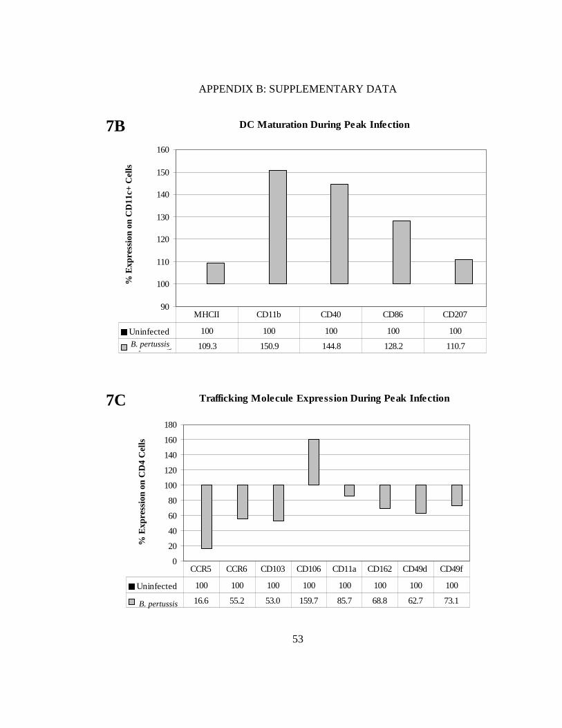
FIGURE 7. DCs mature in response to B. pertussis infection even with smaller doses of inoculum. BALB/c mice were infected intranasally with 5x10⁵ CFU in 20 µl and percent DCs were measured in each treatment at a peak infection time of 10 days (A). Maturation markers (MHC class II, CD40, CD86) and lung-associated markers (CD11b, CD207) were also measured (B). Bordetella pertussis-infected DCs were co-cultured with naïve splenocytes and levels of CCR5, CCR6, CD103, CD106, CD11a, CD162, CD49d, and CD49f were assessed (C). All values for the co-cultures were normalized to uninfected controls (100%).
Percent CD11c+ Cells per Treatment
0
0.5
1
1.5
2
2.5
3
3.5
Uninfected B. pertussis
Treatment
% C
D11
c+ C
ells
7A
53
APPENDIX B: SUPPLEMENTARY DATA
Trafficking Molecule Expression During Peak Infection
0
20
40
60
80
100
120
140
160
180
% E
xpre
ssio
n on
CD
4 C
ells
Uninfected 100 100 100 100 100 100 100 100
B. pertussis 16.6 55.2 53.0 159.7 85.7 68.8 62.7 73.1
CCR5 CCR6 CD103 CD106 CD11a CD162 CD49d CD49f
7C
B. pertussis
DC Maturation During Peak Infection
90
100
110
120
130
140
150
160
% E
xpre
ssio
n on
CD
11c+
Cel
ls
Uninfected 100 100 100 100 100
B. pertussis 109.3 150.9 144.8 128.2 110.7
MHCII CD11b CD40 CD86 CD207
B. pertussis
7B

54
APPENDIX B: SUPPLEMENTARY DATA
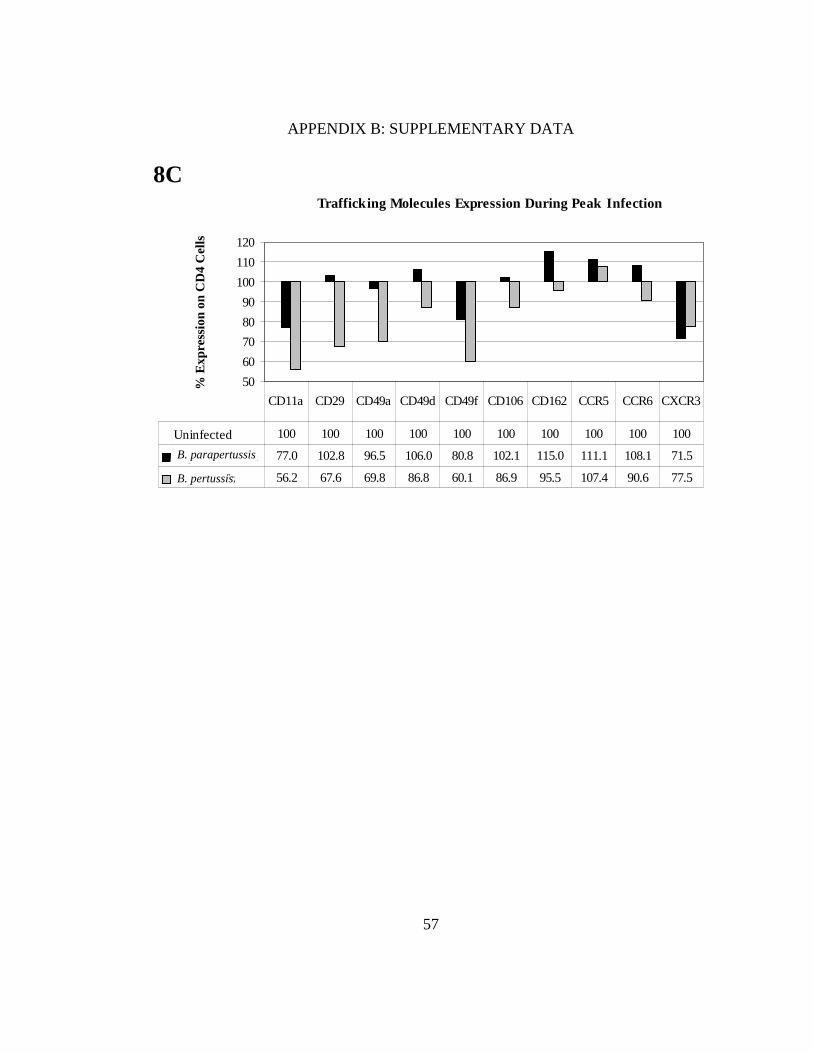
FIGURE 8. DCs mature in response to Bordetella and downregulation of homing molecules on CD4+ T cells was observed in B. pertussis co-cultures. We assessed the percentage of CD11c+ cells in each treatment using quadrants that were placed according to positive controls (A). DC maturation markers observed included MHC class II, CD40, CD80, CD86; myeloid and lung associated markers, CD11b, CD207 and migratory makers CD103, CCR5, CCR6, CCR7 (B). After co-culturing, levels of the chemokine receptors CCR5, CCR6, CXCR3, and adhesion markers CD11a, CD29, CD49a, CD49d, CD106, and CD162 were measured (C). All values were normalized to uninfected controls (100%).
Percent CD11c+ Cells per Treatment
0
10
20
30
40
50
Uninfected B. parapertussis B. pertussis
Treatment
% C
D11
c+ C
ells
8A

55
APPENDIX B: SUPPLEMENTARY DATA
CD207
CD40
CD86
Uninfected B. parapertussis B. pertussis
CD11c
DC Phenotypic Marker
CCR5
8B

56
APPENDIX B: SUPPLEMENTARY DATA
CD103
MHCII
CD11b
CD80
Uninfected B. parapertussis B. pertussis
CCR7
CCR6
57
APPENDIX B: SUPPLEMENTARY DATA
Trafficking Molecules Expression During Peak Infection
50
60
70
80
90
100
110
120
% E
xpre
ssio
n on
CD
4 C
ells
Uninfected 100 100 100 100 100 100 100 100 100 100
B. parapertussis 77.0 102.8 96.5 106.0 80.8 102.1 115.0 111.1 108.1 71.5
B. pertussis 56.2 67.6 69.8 86.8 60.1 86.9 95.5 107.4 90.6 77.5
CD11a CD29 CD49a CD49d CD49f CD106 CD162 CCR5 CCR6 CXCR3
8C
B. parapertussis
B. pertussis
58
APPENDIX B: SUPPLEMENTARY DATA
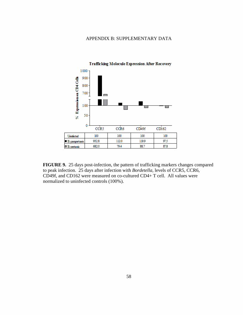
FIGURE 9. 25 days post-infection, the pattern of trafficking markers changes compared to peak infection. 25 days after infection with Bordetella, levels of CCR5, CCR6, CD49f, and CD162 were measured on co-cultured CD4+ T cell. All values were normalized to uninfected controls (100%).