The Corynebacterium diphtheriaeshaft pilin SpaA is …nsmn1.uh.edu/yeo/doc/BCHS6229/Presentation...
13
The Corynebacterium diphtheriae shaft pilin SpaA is built of tandem Ig-like modules with stabilizing isopeptide and disulfide bonds Hae Joo Kang a , Neil G. Paterson a , Andrew H. Gaspar b , Hung Ton-That b,c,1 , and Edward N. Baker a,1 a Maurice Wilkins Centre for Molecular Biodiscovery and School of Biological Sciences, University of Auckland, Auckland 1020, New Zealand; b Department of Molecular, Microbial, and Structural Biology, University of Connecticut Health Center, Farmington, CT 06030; and c Department of Microbiology and Molecular Genetics, University of Texas Health Science Center, Houston, TX 77030 Edited by David S. Eisenberg, University of California, Los Angeles, CA, and approved August 12, 2009 (received for review June 17, 2009) Cell-surface pili are important virulence factors that enable bacterial pathogens to adhere to specific host tissues and modulate host immune response. Relatively little is known about the structure of Gram-positive bacterial pili, which are built by the sortase-catalyzed covalent crosslinking of individual pilin proteins. Here we report the 1.6-Å resolution crystal structure of the shaft pilin component SpaA from Corynebacterium diphtheriae, revealing both common and unique features. The SpaA pilin comprises 3 tandem Ig-like domains, with characteristic folds related to those typically found in non-pilus adhesins. Whereas both the middle and the C-terminal domains contain an intramolecular Lys–Asn isopeptide bond, previously de- tected in the shaft pilins of Streptococcus pyogenes and Bacillus cereus, the middle Ig-like domain also harbors a calcium ion, and the C-terminal domain contains a disulfide bond. By mass spectrometry, we show that the SpaA monomers are cross-linked in the assembled pili by a Lys–Thr isopeptide bond, as predicted by previous genetic studies. Together, our results reveal that despite profound dissimi- larities in primary sequences, the shaft pilins of Gram-positive patho- gens have strikingly similar tertiary structures, suggesting a modular backbone construction, including stabilizing intermolecular and in- tramolecular isopeptide bonds. crystal structure polymerization bacterial pilus mass spectrometry pilin motif P ili are long, thin protein assemblies that extend from the cell surface of many bacteria and play pivotal roles in coloniza- tion and pathogenesis. Like Gram-negative bacteria, many Gram-positive pathogens express pili on their surface (1–3). These structures have aroused great interest because of their direct roles in infection and pathogenesis and their importance as vaccine candidates (2, 3). They also use covalent isopeptide (amide) bonds, both intermolecular and intramolecular, to give strength and stability, and thus present a new paradigm among protein polymers (4–6). Unlike Gram-negative pili, whose subunits associate via non- covalent interactions, these Gram-positive pili are formed by covalent polymerization of pilin subunits, orchestrated by transpeptidase enzymes called sortases (6, 7). The general principles of assembly were first established through studies on the SpaA pili expressed by Corynebacterium diphtheriae. These pili, encoded by the gene cluster spaA-spaB-srtA-spaC, comprise a polymeric shaft formed by SpaA, SpaC located at the tip, and SpaB found at the base and occasionally along the shaft (6, 8–10). In our current model, successive major pilin SpaA subunits are joined by the action of the pilus-specific sortase SrtA. Cleavage between Thr-494 and Gly-495 of the LPXTG motif near the SpaA C terminus is followed by presumed amide bond formation between the new C terminus and Lys-190 from a conserved YPKN pilin motif in the next subunit (6). Finally, the entire assembly is covalently attached to the cell wall peptidogly- can by a housekeeping sortase (9, 11). Presumably, the tip pilin SpaC is linked to the SpaA shaft via the same reaction as recently identified for the tip pilin BcpB of Bacillus cereus (12). It remains unclear how the minor pilin SpaB is incorporated into the pilus structure, although recent evidence indicates that SpaB forms the basal subunit for tethering the pilus to the cell wall and that it, too, is attached to the polymer via a specific Lys residue (9). A major step forward in understanding Gram-positive pilus structure and assembly came with the structural analysis of the major pilin Spy0128 from Streptococcus pyogenes (5). Spy0128 does not have a recognizable pilin motif, but the crystal structure revealed columns of molecules resembling a putative polymer assembly and identified a candidate lysine; this was then confirmed by mass spectral analysis of native S. pyogenes pili. The structure also revealed unexpected internal crosslinks in the form of self-generated isopeptide bonds, 1 in each domain of the 2-domain structure, joining Lys and Asn side chains. These are strategically located to give strength and stability to the pilus assembly. The major pilins of different Gram-positive bacteria show wide variations in size and sequence, making it difficult to predict whether the structural principles seen for S. pyogenes apply also to other Gram-positive pili. Here we present the high-resolution crystal structure of SpaA, the archetypal major pilin from C. diphtheriae. This reveals a modular structure comprising 3 tandem Ig-like domains, 2 of which contain internal Lys–Asn isopeptide bonds like those in Spy0128. We also confirm, by mass spectrometry, the identity of the lysine used in polymerization and note a pilus-like assembly of SpaA molecules in the crystal. These results point to a likely common architecture for the backbones of many Gram-positive pili and consolidate a new paradigm for the structure, stability, and assembly of these remarkable covalent polymers. Results Structure Determination. A construct comprising residues 53– 486 of C. diphtheriae SpaA was expressed in Escherichia coli, puri- fied, and crystallized. This lacks residues 1–52 encompassing the signal peptide and ends 4 residues before the sortase-recognition LPXTG motif. The crystal structure, with 1 SpaA molecule per asymmetric unit, was solved by single wavelength anomalous dispersion methods and refined at 1.6-Å resolution (R 19.3%, R free 22.0%) [supporting information (SI) Table S1]. Only the Author contributions: H.J.K., N.G.P., A.H.G., H.T.-T., and E.N.B. designed research; H.J.K., N.G.P., and A.H.G. performed research; H.J.K. contributed new reagents/analytic tools; H.J.K., N.G.P., and E.N.B. analyzed data; and H.J.K., H.T.-T., and E.N.B. wrote the paper. The authors declare no conflict of interest. This article is a PNAS Direct Submission. Data deposition: Atomic coordinates and structure factors have been deposited in the Protein Data Bank, www.pdb.org (PDB ID code 3HR6). 1 To whom correspondence may be addressed. E-mail: [email protected] or [email protected]. This article contains supporting information online at www.pnas.org/cgi/content/full/ 0906826106/DCSupplemental. www.pnas.orgcgidoi10.1073pnas.0906826106 PNAS October 6, 2009 vol. 106 no. 40 16967–16971 BIOCHEMISTRY
Transcript of The Corynebacterium diphtheriaeshaft pilin SpaA is …nsmn1.uh.edu/yeo/doc/BCHS6229/Presentation...

The Corynebacterium diphtheriae shaft pilin SpaAis built of tandem Ig-like modules with stabilizingisopeptide and disulfide bondsHae Joo Kanga, Neil G. Patersona, Andrew H. Gasparb, Hung Ton-Thatb,c,1, and Edward N. Bakera,1
aMaurice Wilkins Centre for Molecular Biodiscovery and School of Biological Sciences, University of Auckland, Auckland 1020, New Zealand; bDepartmentof Molecular, Microbial, and Structural Biology, University of Connecticut Health Center, Farmington, CT 06030; and cDepartment of Microbiologyand Molecular Genetics, University of Texas Health Science Center, Houston, TX 77030
Edited by David S. Eisenberg, University of California, Los Angeles, CA, and approved August 12, 2009 (received for review June 17, 2009)
Cell-surface pili are important virulence factors that enable bacterialpathogens to adhere to specific host tissues and modulate hostimmune response. Relatively little is known about the structure ofGram-positive bacterial pili, which are built by the sortase-catalyzedcovalent crosslinking of individual pilin proteins. Here we report the1.6-Å resolution crystal structure of the shaft pilin component SpaAfrom Corynebacterium diphtheriae, revealing both common andunique features. The SpaA pilin comprises 3 tandem Ig-like domains,with characteristic folds related to those typically found in non-pilusadhesins. Whereas both the middle and the C-terminal domainscontain an intramolecular Lys–Asn isopeptide bond, previously de-tected in the shaft pilins of Streptococcus pyogenes and Bacilluscereus, the middle Ig-like domain also harbors a calcium ion, and theC-terminal domain contains a disulfide bond. By mass spectrometry,we show that the SpaA monomers are cross-linked in the assembledpili by a Lys–Thr isopeptide bond, as predicted by previous geneticstudies. Together, our results reveal that despite profound dissimi-larities in primary sequences, the shaft pilins of Gram-positive patho-gens have strikingly similar tertiary structures, suggesting a modularbackbone construction, including stabilizing intermolecular and in-tramolecular isopeptide bonds.
crystal structure � polymerization � bacterial pilus � mass spectrometry �pilin motif
P ili are long, thin protein assemblies that extend from the cellsurface of many bacteria and play pivotal roles in coloniza-
tion and pathogenesis. Like Gram-negative bacteria, manyGram-positive pathogens express pili on their surface (1–3).These structures have aroused great interest because of theirdirect roles in infection and pathogenesis and their importanceas vaccine candidates (2, 3). They also use covalent isopeptide(amide) bonds, both intermolecular and intramolecular, to givestrength and stability, and thus present a new paradigm amongprotein polymers (4–6).
Unlike Gram-negative pili, whose subunits associate via non-covalent interactions, these Gram-positive pili are formed bycovalent polymerization of pilin subunits, orchestrated bytranspeptidase enzymes called sortases (6, 7). The generalprinciples of assembly were first established through studies onthe SpaA pili expressed by Corynebacterium diphtheriae. Thesepili, encoded by the gene cluster spaA-spaB-srtA-spaC, comprisea polymeric shaft formed by SpaA, SpaC located at the tip, andSpaB found at the base and occasionally along the shaft (6,8–10). In our current model, successive major pilin SpaAsubunits are joined by the action of the pilus-specific sortaseSrtA. Cleavage between Thr-494 and Gly-495 of the LPXTGmotif near the SpaA C terminus is followed by presumed amidebond formation between the new C terminus and Lys-190 froma conserved YPKN pilin motif in the next subunit (6). Finally, theentire assembly is covalently attached to the cell wall peptidogly-can by a housekeeping sortase (9, 11). Presumably, the tip pilinSpaC is linked to the SpaA shaft via the same reaction as recently
identified for the tip pilin BcpB of Bacillus cereus (12). It remainsunclear how the minor pilin SpaB is incorporated into the pilusstructure, although recent evidence indicates that SpaB formsthe basal subunit for tethering the pilus to the cell wall and thatit, too, is attached to the polymer via a specific Lys residue (9).
A major step forward in understanding Gram-positive pilusstructure and assembly came with the structural analysis of themajor pilin Spy0128 from Streptococcus pyogenes (5). Spy0128does not have a recognizable pilin motif, but the crystalstructure revealed columns of molecules resembling a putativepolymer assembly and identified a candidate lysine; this wasthen confirmed by mass spectral analysis of native S. pyogenespili. The structure also revealed unexpected internal crosslinksin the form of self-generated isopeptide bonds, 1 in eachdomain of the 2-domain structure, joining Lys and Asn sidechains. These are strategically located to give strength andstability to the pilus assembly.
The major pilins of different Gram-positive bacteria showwide variations in size and sequence, making it difficult topredict whether the structural principles seen for S. pyogenesapply also to other Gram-positive pili. Here we present thehigh-resolution crystal structure of SpaA, the archetypal majorpilin from C. diphtheriae. This reveals a modular structurecomprising 3 tandem Ig-like domains, 2 of which contain internalLys–Asn isopeptide bonds like those in Spy0128. We alsoconfirm, by mass spectrometry, the identity of the lysine used inpolymerization and note a pilus-like assembly of SpaA moleculesin the crystal. These results point to a likely common architecturefor the backbones of many Gram-positive pili and consolidate anew paradigm for the structure, stability, and assembly of theseremarkable covalent polymers.
ResultsStructure Determination. A construct comprising residues 53–486of C. diphtheriae SpaA was expressed in Escherichia coli, puri-fied, and crystallized. This lacks residues 1–52 encompassing thesignal peptide and ends 4 residues before the sortase-recognitionLPXTG motif. The crystal structure, with 1 SpaA molecule perasymmetric unit, was solved by single wavelength anomalousdispersion methods and refined at 1.6-Å resolution (R � 19.3%,Rfree � 22.0%) [supporting information (SI) Table S1]. Only the
Author contributions: H.J.K., N.G.P., A.H.G., H.T.-T., and E.N.B. designed research; H.J.K.,N.G.P., and A.H.G. performed research; H.J.K. contributed new reagents/analytic tools;H.J.K., N.G.P., and E.N.B. analyzed data; and H.J.K., H.T.-T., and E.N.B. wrote the paper.
The authors declare no conflict of interest.
This article is a PNAS Direct Submission.
Data deposition: Atomic coordinates and structure factors have been deposited in theProtein Data Bank, www.pdb.org (PDB ID code 3HR6).
1To whom correspondence may be addressed. E-mail: [email protected] [email protected].
This article contains supporting information online at www.pnas.org/cgi/content/full/0906826106/DCSupplemental.
www.pnas.org�cgi�doi�10.1073�pnas.0906826106 PNAS � October 6, 2009 � vol. 106 � no. 40 � 16967–16971
BIO
CHEM
ISTR
Y

N-terminal Glu-53, an external loop 69–79, and the C-terminalresidues 485–486 could not be modeled for lack of interpretableelectron density.
Modular Structure of SpaA. SpaA is folded into 3 tandem Ig-typedomains, giving an elongated molecule of �105 Å in length.Whereas the middle domain (M-domain; residues 193–351) andthe C-terminal domain (C-domain; residues 352–484) are ar-ranged linearly and share an extended strand P (Fig. 1), theN-terminal domain (N-domain; residues 54–192) sits on theM-domain at an angle of �20° to the long axis of the molecule.The SpaA molecules pack in columns through the crystal, in amanner resembling a pilus assembly; the N-domain of eachmolecule abuts against the C-domain of the next (Fig. 1 A).
The N- and C-domains both have the inverse IgG fold firstdescribed for the CnaB domains of the collagen binding proteinCna from Staphylococcus aureus (13). This comprises a �-sand-wich of 7 strands. When superimposed, the 2 CnaB-type domainsof SpaA show an rmsd in C� positions of 2.2 Å over 88 equivalentresidues and share 14% sequence identity. A distinctive featureof the N-domain is the presence of 2 helices between strands Band C that partially cover one side of the core, whereas theC-domain uniquely contains an elongated �-ribbon, formed bystrands S and T, running toward the M-domain. In contrast, theM-domain of SpaA has the CnaA fold, first seen in the N2domain of S. aureus CnaA (14). This comprises 9 �-strands thatform a partially open �-barrel.
The closest structural homologues of the N- and C-domains ofSpaA are the 2 CnaB-type domains of the Streptococcus agalac-tiae minor pilin GBS52 (15). In particular, the SpaA C-domainhas significant sequence identity (25%) and structural similarity(rmsd 1.7 Å over 91 equivalent C� atoms) with the N2 domainof GBS52. The 2 domains of the S. pyogenes major pilin Spy0128also share the same CnaB-type fold, albeit with some elabora-tions (5). Structural superpositions of the Spy0128 domains onto the SpaA N- and C-domains give rmsds in C� positionsranging from 2.5 to 3.1 Å and sequence identities ranging from3% to 17%. The M-domain shows strong structural homologywith the N2 domain of CnaA, despite minimal sequence identity(�8%); the rmsd is 3.4 Å over 123 equivalent C� atoms. Othersimilar CnaA-type domains include the N3 domain of S. aureusclumping factor A and the N2 domain of the Enterococcusfaecalis collagen-binding protein Ace (16, 17).
Internal Isopeptide Bonds and Other Stabilizing Features. The M- andC-domains of SpaA both contain stabilizing internal isopeptidebonds, formed by intramolecular reaction between the Lys�-amino group and the carboxyamide group of Asn. These wereclearly apparent in the initial experimentally phased electrondensity map, in which continuous density linked the side chainsof Lys-199 and Asn-321 in the M-domain and Lys-363 andAsn-482 in the C-domain (Fig. 2). The existence of theseLys–Asn isopeptide bonds was confirmed by electrospray ion-ization–time-of-f light mass spectrometry. The protein Mr wasmeasured as 46,795.4 Da, �34 Da less than that calculated fromthe amino acid sequence, 46,829.6 Da, consistent with loss of 2units of NH3 through formation of 2 isopeptide bonds. Confir-mation was obtained by digestion of the recombinant protein andanalysis by liquid chromatography–tandem mass spectrometry
Fig. 1. Crystal structure of SpaA. (A) End-to-end stacking of successivemolecules, in which the C-domain of one molecule, SpaAn, packs against theN-domain of the next, SpaAn � 1. For each molecule, the N-domain is in gold,M-domain green, and C-domain blue. The M-domain Ca2� ion is shown as agray sphere. Residues that form isopeptide bonds in the M- and C-domains areshown in red stick mode, and the disulfide bond in the C-domain is in yellow.The pilin motif lysine, Lys-190, in each molecule is labeled K. A black bar showsthe distance between the C terminus of one molecule and Lys-190 on the next.(B) Schematic representation of the fold of each domain colored as in A.Helices are labeled 1 to 3, and �-strands A to W. The isopeptide bond crosslinksare shown with red bars and the disulfide bond with a yellow bar. Anarrowhead points to Lys-190. Strands and loops connected by inter- or in-tramolecular isopeptide bonds are highlighted on a beige background. (C)Close view of the packing of adjacent molecules. Lys-190 projects upward,between the disordered AB loop (gray line) and the N-domain, toward the Cterminus of the next molecule (C). A broken line shows where the 10 missingC-terminal residues would bridge the �19-Å gap to Lys-190 on the nextmolecule. The conserved Trp-181 of the pilin motif is in the interface betweenmolecules.
Fig. 2. Internal isopeptide bonds in SpaA. Residues involved in bond forma-tion are in stick mode, colored by atom type, with surrounding hydrophobicresidues also shown. Hydrogen bonds are shown with broken lines, distancesin Å. The electron density is from an ( Fobs - Fcal ) � Phical map, contoured at 3�.(A) The M-domain isopeptide bond formed between Lys-199 and Asn-321,with catalytic Asp-241. (B) The C-domain isopeptide bond between Lys-363and Asn-482, with catalytic Glu-446.
16968 � www.pnas.org�cgi�doi�10.1073�pnas.0906826106 Kang et al.

(LC-MS/MS). Parent ions with mass-to-charge ratio (m/z)996.83� and 1067.53� contained the M-domain isopeptide bondbetween Lys-199 and Asn-321, and a parent ion with m/z 751.73�
contained the C-domain linkage between Lys-363 and Asn-482(Tables S2 and S3).
Similar Lys-Asn isopeptide bonds were first observed in thestructure of Spy0128, where an associated Glu residue was shownto be essential for the intramolecular reaction to occur (5). InSpaA, the same role is played by Asp-241 for the M-domainisopeptide bond and Glu-446 for the C-domain bond (Fig. 2).
Two isomeric forms of isopeptide bond are found in SpaA. TheM-domain bond (Lys-199–Asn-321) has a cis configuration, as forboth isopeptide bonds of Spy0128, allowing its NH and O moietiesto form a bidentate hydrogen-bonded interaction with the Asp-241carboxyl group (Fig. 2). In contrast, the C-domain bond (Lys-363–Asn-482) has a trans configuration and only a single hydrogen bondwith the carboxyl group of Glu-446. The hydrogen bonding patternsimply that both carboxyl groups are protonated. Both isopeptidebonds are located in the interior of their respective domains,surrounded by hydrophobic residues. Both also stack against aro-matic residues, Tyr-219 in the M-domain and Phe-365 in theC-domain (Fig. 2). Other surrounding hydrophobic residues includePhe-306, Val-352, Val-221, and Leu-243 in the M-domain andPhe-378, Ile-361, Ile-480, and Ala-376 in the C-domain. Thishydrophobic environment favors a nonprotonated Lys amino groupand protonated Glu/Asp carboxyl group, thus facilitating the in-tramolecular reaction (5, 18).
The SpaA structure has 2 other notable stabilizing features. Inthe M-domain a metal binding site is formed by the loop joiningstrands H and I, with the metal ion coordinated by 8 oxygenatoms, from Asp-204, Asp-205, Gln-208, Gly-210, Glu-215, and2 water molecules (Fig. 1). The coordination environment andaverage metal-ligand bond length (2.48 Å) are indicative of aCa2� ion, presumably cell derived. This is a unique feature, notseen before in any other CnaB- or CnaA-like domain, and itspersistence despite the use of 1 mM EDTA in buffers implies ahigh affinity. In the C-domain, a disulfide bond joins Cys-383 onstrand Q to Cys-443 on strand U (Fig. 1). The electron densityshows that this bond is incompletely formed, with approximately40% of molecules having both Cys reduced. This may result fromthe DTT needed for tag cleavage, and we anticipate that thedisulfide would be fully formed in vivo, in the oxidizing extra-cellular environment.
Interestingly, whereas SpaA has no internal isopeptide bondin its N-domain, such a bond is present in the N-domain of the3-domain major pilin BcpA from B. cereus, joining Lys-37 andAsn-163 (4). These residues are conserved in other pilins butreplaced by Ala-61 and His-191, respectively, in SpaA (Fig. S1).In the SpaA structure, the Ala and His side chains are closeenough such that if replaced by Lys and Asn, as in other pilins,an isopeptide bond could be formed. A conserved Glu that couldcatalyze Lys–Asn bond formation is present in the other pilinsbut in SpaA is replaced by Gln-153, positioned close to Ala-61and His-191 (Fig. S2).
Sequence Elements Implicated in Pilus Assembly. Sequence compar-isons and mutagenesis have identified 2 conserved sequencemotifs that contain residues essential for pilus assembly. Thepilin motif (consensus WxxxVxVYPK) contains the lysine that isjoined to the C terminus of the next molecule during polymer-ization; Lys-190 in SpaA. We sought to confirm the role ofLys-190 in SpaA polymer formation, using mass spectrometry.Purified SpaA pilus polymers were separated by SDS-PAGE anddigested in-gel with trypsin and AspN endopeptidase. Thedigestion products were analyzed by LC-MS/MS and the inter-subunit amide bond identified from a peptide peak with m/z585.73�. The Mr of this peptide corresponded exactly with thatexpected, allowing for the loss of 18 Da due to water elimination
during amide bond formation. The fragment ion spectrauniquely identified the peptides surrounding the pilin motifLys-190 and the sortase-cleaved C-terminal Thr-494, respec-tively (Fig. 3 and Table S4).
In the SpaA structure, the pilin motif is located on �G, the laststrand of the N-domain. Lys-190 is close to the point where �Gcrosses to the M-domain, becoming �H (Fig. 1). Nine residuesbefore Lys-190 is the conserved Trp-181, the first residue of thepilin motif, and 9 residues after is Lys-199, which forms theM-domain isopeptide bond. The side chain of Lys-190 projectsinto a cleft between the main body of the N-domain and a mobileloop, residues 63–83 (Fig. 1C). Head-to-tail packing of mole-cules in the crystal places the �-amino group of Lys-190 �19 Åfrom the C terminus of the next molecule, ample distance toaccommodate the 10 missing residues between the last modeledresidue, Lys-484, and the true sortase cleavage site, Thr-494. Italso places Trp-181 in the interface between the 2 molecules(Fig. 1C); the conservation of this residue supports the idea thatthe crystal packing models the true biologic assembly. The factthat the M-domain isopeptide bond closely follows Lys-190, onthe same extended �-strand, suggests why polymer formation isabrogated by deletion of the equivalent isopeptide bond in B.cereus BcpA (4); local structural destabilization could preventproper presentation of the essential lysine to the sortase.
The second sequence motif implicated in assembly is theE-box motif (consensus YxLxETxAPxGY). This contains aconserved glutamate, Glu-446 in SpaA, which is essential for theincorporation of the minor pilins SpaB and SpaC (7). Intrigu-ingly, Glu-446 proves to be the catalytic Glu that mediatesformation of the Lys-363–Asn-462 intramolecular bond. BecauseGlu-446 is internal, we infer that it is the stability imparted bythe intramolecular crosslink that is essential for minor pilinincorporation.
DiscussionThe discovery of thin, hair-like pili on the surface of C. diph-theriae in 2003, and their characterization as covalent polymers,was a milestone in understanding colonization and infection byGram-positive bacteria (6). Similar pilus assemblies are foundfor such important human pathogens as Group A and B strep-
Fig. 3. Intersubunit isopeptide bonds in SpaA. Fragmentation spectra of theparent ion at m/z 585.73� containing the intersubunit bond between SpaALys-190 and Thr-494 are shown. Ion types are indicated, and internal ions areshown in italics. The structure of crosslinked peptide is shown above thespectra. Daughter ions produced during MS/MS of these peptides are sum-marized in Table S4.
Kang et al. PNAS � October 6, 2009 � vol. 106 � no. 40 � 16969
BIO
CHEM
ISTR
Y

tococci, Streptococcus pneumoniae, and B. cereus (19–21), but thepilin subunits involved show large variations in size and sequencethat mask any possible structural homology.
The present structure for the C. diphtheriae major pilin SpaAresolves this question, revealing a modular assembly that utilizesIg-like domains similar to those used in the S. pyogenes majorpilin Spy0128, despite very low sequence identity. These domainscorrespond to 2 types of Ig-like domain, the CnaB and CnaAfolds that are widely used in the cell-surface adhesins known asMSCRAMMS (microbial surface components recognizing ad-hesive matrix molecules) (22). Prototype CnaB and CnaA do-mains are present in the multidomain S. aureus Cna protein,which has a structural B-region with repeating doublets of CnaBdomains, preceded by a collagen-binding A-region with 2 CnaAdomains (22, 23).
Spy0128, the major pilin of S. pyogenes, is one of the smallestshaft pilins (32.5 kDa) and comprises 2 tandem CnaB domains(5). SpaA, in contrast, is significantly larger (47 kDa) and has asingle CnaA-type domain, the M-domain, inserted between 2CnaB-type domains. This mosaic architecture suggests an evo-lutionary process in which copies or pieces of older genes areassembled to form new genes. It seems likely that all of the majorpilins of sortase-assembled Gram-positive pili conform to thesame structural principles. In many cases, for example the majorpilins SpaD and SpaH that form the 2 other types of pilusproduced by C. diphtheriae, sufficient sequence identity exists toinfer similar structures; these share �24% identity with SpaA,including the intramolecular isopeptide bond-forming residues,the pilin motif, and the Cys residues. In others, such as Spy0128,there is much less sequence similarity.
Sequence comparisons with the major pilins from otherGram-positive bacteria, such as S. pneumoniae, S. agalactiae, andB. cereus, reveal N- and C-terminal regions that are similar to theN- and C-domains of SpaA, including the N-domain pilin motifand the C-domain isopeptide bond-forming residues. The middleregions are more variable; some may form CnaA-type domainslike the SpaA M-domain, with substantial insertions/deletions,whereas others may have CnaB-type middle domains or adoptentirely different folds.
There is growing evidence that this modular architectureextends also to the minor pilin subunits. GBS52, a minor pilinfrom S. agalactiae that seems to correspond functionally to SpaB,comprises 2 CnaB-type domains (15), and its C-terminal N2domain closely resembles the SpaA C-domain, including theinternal isopeptide bond. Sequence comparisons show that theS. pyogenes minor pilin Cpa also has a C-terminal domainhomologous with the C-domain of the S. pyogenes major pilinSpy0128, again including an internal isopeptide bond (5). Thisleads to the concept that incorporation of the minor pilins isfacilitated by their structural resemblance to the pilins thatcomprise the polymeric shaft.
The 2 internal isopeptide bonds in SpaA confirm that suchcrosslinks are a common feature of Gram-positive pili. Firstdiscovered in the crystal structure of Spy0128 and confirmed bymass spectral analysis of native GAS pili (5), they have also beenfound in the B. cereus major pilin BcpA, which has 3 such bonds(4). The combined sequence and structural data from these 3characterized major pilins now enable internal isopeptide bondsto be inferred from the sequences of other major pilins. Re-evaluation of the crystal structures of GBS52 and the A- andB-domains of S. aureus Cna further showed that similar isopep-tide bonds are also present in minor pilins and adhesins (5).
The SpaA structure further defines the determinants ofintramolecular isopeptide bond formation. Mutagenesis hasshown that a catalytic carboxyl group is essential for bondformation (4, 5, 24); this can be Asp or Glu, as shown by the useof Asp-241 in the M-domain and Glu-446 in the C-domain. Theisopeptide moiety can have either cis or trans configuration, with
corresponding bidentate or monodentate interaction with theessential carboxyl side chain. The main requirement is proximityof the Lys–Asn pair and a hydrophobic environment in whichboth the Lys and Asp/Glu are uncharged. The locations of theseisopeptide bonds seem to be characteristic of the folds of thedomains in which they occur, and hence probably reflect theirevolutionary history. In the CnaB-like C-domain of SpaA, theisopeptide bond joins the first and last �-strands, as in bothCnaB-type domains of Spy0128. In contrast, in the CnaA-typeM-domain, the isopeptide bond bridges the first and second-laststrands, linking 2 opposing �-sheets.
Given the widespread occurrence of internal isopeptidecrosslinks in the cell-surface proteins of Gram-positive bacteria,what is their structural and functional importance? We haveshown that the intramolecular isopeptide bonds in Spy0128strongly enhance thermodynamic stability and resistance toproteolysis (24). SpaA additionally contains a disulfide bond,conserved in the other C. diphtheriae major pilins, which mightbe expected to further enhance stability. The major pilins of S.pyogenes, B. cereus, and S. pneumoniae lack Cys residues, how-ever, and given that many Gram-positive bacteria lack thedisulfide formation machinery of Gram-negative bacteria (25,26), we speculate that isopeptide bonds have evolved as analternative means of stabilization. As amide bonds they wouldalso be less prone to chemical disruption than disulfide bonds, aproperty that may be important for such thin, exposed assem-blies, which do not seem to form higher-order bundles.
We further hypothesize that their strategic location gives me-chanical (force-bearing) stability. Both in these pili, typified bySpy0128 and SpaA, and in multidomain adhesins such as Cna, analmost linear chain of covalent connectivity can be traced along thelong axis (5, 27). In SpaA this begins with Lys-190, the site ofattachment to the preceding subunit, and extends through the M-and C-domains to the next intermolecular linkage, possibly explain-ing why the N-domain does not require an isopeptide bond. Theattachment of adhesins to host cells subjects them to significanttensile stress, and their structures are thought to have evolved bothto withstand stress and to use it to optimize binding (28).
There is also growing evidence that the structural stabilizationconferred by the internal isopeptide bonds can be critical formolecular recognition. Defective sortase recognition or failureto present the Lys residue of the pilin motif appropriately wouldexplain the loss of pilus assembly when intramolecular isopeptidebonds in SpaA or BcpA are deleted by mutation of the catalyticAsp/Glu. The isopeptide bonds in CnaA and GBS52 maysimilarly influence the binding of partner molecules, becausetheir isopeptide bond-containing domains are important forspecific binding of collagen (CnaA) and capable of binding tohuman pulmonary epithelial cells (GBS52) (15, 29).
Finally, an intriguing feature of the crystal structures of bothSpaA and Spy0128 is the way the pilin molecules pack end-to-endin columns in the crystal, resembling assembled pili. The inter-molecular contacts do not seem to be particularly extensive(buried surface 850 Å2 for Spy0128 and 814 Å2 for SpaA), yet ineach case the molecules pack such that the critical lysine residue(Lys-161 in Spy0128, Lys-190 in SpaA) is brought close to the Cterminus of the next molecule. Importantly, in SpaA the con-served Trp-181 of the pilin motif forms part of this interface,consistent with a role in oligomer formation. The roles of theconserved Tyr-188 and Pro-189 of the pilin motif are less clear,but patches of electron density around them may represent partsof the unmodeled AB loop. These residues may interact tran-siently with this loop, which could in turn mediate the pilin–pilinand/or pilin–sortase interactions.
Materials and MethodsCloning and Protein Purification. DNA encoding amino acids 53–486 of SpaAfrom C. diphtheriae was amplified by PCR from genomic DNA, cloned, over-
16970 � www.pnas.org�cgi�doi�10.1073�pnas.0906826106 Kang et al.

expressed in E. coli as an N-terminally His-tagged protein, and purified bynickel-affinity chromatography (30). After His-tag removal and final size-exclusion chromatography, the protein was concentrated to 100 mg/mL in 10mM Tris-HCl (pH 8.0) and 50 mM NaCl. Selenomethionine (SeMet)-substitutedSpaA was produced using the methionine biosynthesis inhibition method (31)and similarly purified, but with 5 mM DTT and 1 mM EDTA in the final gelfiltration buffer.
Crystallization and Structure Determination. Crystals were grown in sittingdrops comprising 100 nL protein (100 mg/mL) and 100 nL precipitant. The bestnative SpaA crystals were obtained with 20% PEG 3350, 0.1 M NaI, and 0.1 MNaF as precipitant and SeMet-SpaA crystals with 20% PEG 3350 and 0.2 M Naformate. Crystals were flash-cooled without further cryoprotection. X-raydiffraction data were collected on beamline PX1 at the Australian Synchro-tron, to 1.6 Å and 1.8 Å resolutions, respectively, for native and SeMet-SpaAcrystals. Data were processed and scaled with MOSFLM and SCALA (32). All 4Se atoms were located by SHELX (33) with refinement and phase determina-tion in autoSHARP (34). Density modification and model building with PHENIX(35, 36) placed 408 of 436 residues, and model building was completed usingCOOT (37). The model was refined using REFMAC (38). Data collection, phas-ing, and refinement statistics are in Table S1. Structural superpositions weredone with SSM (39).
Isolation of SpaA Pili. Engineered SpaA pili were produced by transformationof the plasmid pAG153, encoding SpaA and SrtA, into a C. diphtheriae strain
that lacks spaA and srtA, followed by expression and purification of thepolymers (6). These procedures are described more fully in SI Materials andMethods. The use of a His-tagged SpaA construct with a SrtA construct lacking13 C-terminal residues means that the engineered SpaA pili are secreted intothe culture medium and can be purified by nickel-affinity chromatography aspreviously described (6).
Proteolytic Digestion and Mass Spectral Analyses. Purified SpaA pili andrecombinant SpaA protein were digested and analyzed according to previousprotocols (5). Briefly, SDS-PAGE gel bands containing recombinant SpaA orSpaA pili were cut out and incubated with trypsin (Promega) followed by AspNendopeptidase (Roche). Peptides in the m/z range 300–1,600 were analyzedusing a Q-STAR XL Hybrid MS/MS system (Applied Biosystems). Searchesagainst SpaA sequence using Mascot search engine version 2.0.05 (MatrixScience) identified linear peptides, and the unmatched peptides were thensearched manually to identify those containing noncontiguous peptidescrosslinked by isopeptide bonds, either intramolecular or intermolecular. Fulldetails are in SI Materials and Methods.
ACKNOWLEDGMENTS. We thank Tom Caradoc-Davies for help with datacollection, Martin Middleditch for help with mass spectrometry, and Asis Dasfor critical insights. This work was supported by the Health Research Counciland the Marsden Fund of New Zealand (E.N.B.) and National Institutes ofHealth Grant AI061381 (to H.T.-T). Data collection was undertaken on the PX1beamline at the Australian Synchrotron, Victoria, Australia, with support fromthe New Zealand Synchrotron Group Ltd.
1. Ton-That H, Schneewind O (2004) Assembly of pili in Gram-positive bacteria. TrendsMicrobiol 12:228–234.
2. Proft T, Baker EN (2009) Pili in Gram-negative and Gram-positive bacteria—structure,assembly and their role in disease. Cell Mol Life Sci 66:613–635.
3. Telford JL, Barocchi MA, Margarit I, Rappuoli R, Grandi G (2006) Pili in gram-positivepathogens. Nat Rev Microbiol 4:509–519.
4. Budzik JM, et al. (2008) Amide bonds assemble pili on the surface of bacilli. Proc NatlAcad Sci USA 105:10215–10220.
5. Kang HJ, Coulibaly F, Clow F, Proft T, Baker EN (2007) Stabilizing isopeptide bondsrevealed in Gram-positive bacterial pilus structure. Science 318:1625–1628.
6. Ton-That H, Schneewind O (2003) Assembly of pili on the surface of Corynebacteriumdiphtheriae. Mol Microbiol 50:1429–1438.
7. Ton-That H, Marraffini LA, Schneewind O (2004) Sortases and pilin elements involvedin pilus assembly of Corynebacterium diphtheriae. Mol Microbiol 53:251–261.
8. Gaspar AH, Ton-That H (2006) Assembly of distinct pilus structures on the surface ofCorynebacterium diphtheriae. J Bacteriol 188:1526–1533.
9. Mandlik A, Das A, Ton-That H (2008) The molecular switch that activates the cell wallanchoring step of pilus assembly in Gram-positive bacteria. Proc Natl Acad Sci USA105:14147–14152.
10. Swierczynski A, Ton-That H (2006) Type III pilus of corynebacteria: Pilus length isdetermined by the level of its major pilin subunit. J Bacteriol 188:6318–6325.
11. Swaminathan A, et al. (2007) Housekeeping sortase facilitates the cell wall anchoringof pilus polymers in Corynebacterium diphtheriae. Mol Microbiol 66:961–974.
12. Budzik JM, Oh SY, Schneewind O (2009) Sortase D forms the covalent bond that linksBcpB to the tip of Bacillus cereus pili. J Biol Chem 284:12989–12997.
13. Deivanayagam CC, et al. (2000) Novel fold and assembly of the repetitive B region ofthe Staphylococcus aureus collagen-binding surface protein. Structure 8:67–78.
14. Symersky J, et al. (1997) Structure of the collagen-binding domain from a Staphylo-coccus aureus adhesin. Nat Struct Biol 4:833–838.
15. Krishnan V, et al. (2007) An IgG-like domain in the minor pilin GBS52 of Streptococcusagalactiae mediates lung epithelial cell adhesion. Structure 15:893–903.
16. Deivanayagam CC, et al. (2002) A novel variant of the immunoglobulin fold in surfaceadhesins of Staphylococcus aureus: Crystal structure of the fibrinogen-bindingMSCRAMM, clumping factor A. EMBO J 21:6660–6672.
17. Liu Q, et al. (2007) The Enterococcus faecalis MSCRAMM ACE binds its ligand by thecollagen hug model. J Biol Chem 282:19629–19637.
18. Wikoff WR, et al. (2000) Topologically linked protein rings in the bacteriophage HK97capsid. Science 289:2129–2133.
19. Barocchi MA, et al. (2006) A pneumococcal pilus influences virulence and host inflam-matory responses. Proc Natl Acad Sci USA 103:2857–2862.
20. Budzik JM, Schneewind O (2006) Pili prove pertinent to enterococcal endocarditis.J Clin Invest 116:2582–2584.
21. Lauer P, et al. (2005) Genome analysis reveals pili in Group B Streptococcus. Science309:105.
22. Patti JM, Allen BL, McGavin MJ, Hook M (1994) MSCRAMM-mediated adherence ofmicroorganisms to host tissues. Annu Rev Microbiol 48:585–617.
23. Rich RL, et al. (1998) Domain structure of the Staphylococcus aureus collagen adhesin.Biochemistry 37:15423–15433.
24. Kang HJ, Baker EN (2009) Intramolecular isopeptide bonds give thermodynamic andproteolytic stability to the major pilin protein of Streptococcus pyogenes. J Biol Chem,in press.
25. Dutton RJ, et al. (2008) Bacterial species exhibit diversity in their mechanisms andcapacity for protein disulfide bond formation. Proc Natl Acad Sci USA 105:11933–11938.
26. Heras B, et al. (2009) DSB proteins and bacterial pathogenicity. Nat Rev Microbiol7:215–225.
27. Yeates TO, Clubb RT (2007) How some pili pull. Science 318:1558–1559.28. Sokurenko EV, Vogel V, Thomas WE (2008) Catch-bond mechanism of force-enhanced
adhesion: counterintuitive, elusive, but…widespread? Cell Host Microbe 4:314–323.29. Zong Y, et al. (2005) A ‘Collagen Hug’ model for Staphylococcus aureus CNA binding
to collagen. EMBO J 24:4224–4236.30. Kang HJ, Paterson NG, Baker EN (2009) Expression, purification, crystallization and
preliminary crystallographic analysis of SpaA, a major pilin from Corynebacteriumdiphtheriae. Acta Crystallogr Sect F Struct Biol Cryst Commun, in press.
31. Van Duyne GD, Standaert RF, Karplus PA, Schreiber SL, Clardy J (1993) Atomic structuresof the human immunophilin FKBP-12 complexes with FK506 and rapamycin. J Mol Biol229:105–124.
32. Collaborative Computational Project, Number 4 (1994) The CCP4 suite: Programs forprotein crystallography. Acta Crystallogr D Biol Crystallogr 50:760–763.
33. Schneider TR, Sheldrick GM (2002) Substructure solution with SHELXD. Acta CrystallogrD Biol Crystallogr 58:1772–1779.
34. Vonrhein C, Blanc E, Roversi P, Bricogne G (2007) Automated structure solution withautoSHARP. Methods Mol Biol 364:215–230.
35. Adams PD, et al. (2004) Recent developments in the PHENIX software for automatedcrystallographic structure determination. J Synchrotron Radiat 11:53–55.
36. Terwilliger TC (2003) Automated main-chain model building by template matchingand iterative fragment extension. Acta Crystallogr D Biol Crystallogr 59:38–44.
37. Emsley P, Cowtan K (2004) Coot: Model-building tools for molecular graphics. ActaCrystallogr D Biol Crystallogr 60:2126–2132.
38. Murshudov GN, Vagin AA, Dodson EJ (1997) Refinement of macromolecular structuresby the maximum-likelihood method. Acta Crystallogr D Biol Crystallogr 53:240–255.
39. Krissinel E, Henrick K (2004) Secondary-structure matching (SSM), a new tool for fastprotein structure alignment in three dimensions. Acta Crystallogr D Biol Crystallogr60:2256–2268.
Kang et al. PNAS � October 6, 2009 � vol. 106 � no. 40 � 16971
BIO
CHEM
ISTR
Y

BIOCHEMISTRYCorrection for ‘‘The Corynebacterium diphtheriae shaft pilinSpaA is built of tandem Ig-like modules with stabilizing isopep-tide and disulfide bonds,’’ by Hae Joo Kang, Neil G. Paterson,Andrew H. Gaspar, Hung Ton-That, and Edward N. Baker,which appeared in issue 40, October 6, 2009, of Proc Natl AcadSci USA (106:16967–16971; first published September 21, 2009;10.1073/pnas.0906826106).
The authors note that, due to a printer’s error, on page 16967,the keyword ‘‘mas spectromrtry’’ should instead have appearedas ‘‘mass spectrometry.’’ The online version has been corrected.
www.pnas.org/cgi/doi/10.1073/pnas.0911293106
CELL BIOLOGYCorrection for ‘‘CBP and p300 are cytoplasmic E4 polyubiquitinligases for p53,’’ by Dingding Shi, Marius S. Pop, RomanKulikov, Ian M. Love, Andrew Kung, and Steven R. Grossman,which appeared in issue 38, September 22, 2009, of Proc NatlAcad Sci USA (106:16275–16280; first published September 4,2009; 10.1073/pnas.0904305106).
The authors note that the author name Andrew Kung shouldinstead have appeared as Andrew L. Kung. The online versionhas been corrected. The corrected author line and authorcontributions footnote appear below.
Dingding Shi, Marius S. Pop, Roman Kulikov, Ian M. Love,Andrew L. Kung, and Steven R. Grossman
Author contributions: D.S., M.S.P., R.K., and S.R.G. designed research; D.S., M.S.P., R.K., andI.M.L. performed research; A.L.K. contributed new reagents/analytic tools; D.S., M.S.P., R.K.,and S.R.G. analyzed data; and S.R.G. wrote the paper.
www.pnas.org/cgi/doi/10.1073/pnas.0910983106
ECOLOGYCorrection for ‘‘Adaptive shell color plasticity during the earlyontogeny of an intertidal keystone snail,’’ by Patricio H. Man-rı́quez, Nelson A. Lagos, Marı́a Elisa Jara, and Juan CarlosCastilla, which appeared in issue 38, September 22, 2009, of ProcNatl Acad Sci USA (106:16298–16303; first published September2, 2009; 10.1073/pnas.0908655106).
The authors note that on page 16298, right column, first fullparagraph, the first sentence appeared incorrectly in part. ‘‘Inthe rocky intertidal habitats dominated by mussel beds (darkcolored) and barnacle stands (light colored), we found that morethan 95% of early postmetamorphic stages of C. concholepas (�2to 20 mm periostomal length, PL) showed a striking color-matching with the most abundant prey (Fig. 2 A and B)’’ shouldinstead have appeared as ‘‘In the rocky intertidal habitatsdominated by mussel beds (dark colored) and barnacle stands(light colored), we found that more than 95% of early postmeta-morphic stages of C. concholepas (�2 to 20 mm peristomallength, PL) showed a striking color-matching with the mostabundant prey (Fig. 2 A and B).’’ Additionally, the authors notethat on page 16301, right column, first full paragraph, the secondsentence did not appear in full due to a printer’s error. Thesentence should instead have appeared as ‘‘This modulation ofthe shell coloration is restricted to snails of small size (ca. lessthan 3 cm), coincidentally the size at which C. concholepasappear to escape predation by crabs (author’s personal obser-vations).’’ These errors do not affect the conclusions of thearticle.
www.pnas.org/cgi/doi/10.1073/pnas.0910777106
PNAS � October 27, 2009 � vol. 106 � no. 43 � 18427
CORR
ECTI
ON
S

Supporting InformationKang et al. 10.1073/pnas.0906826106SI Materials and MethodsConstruction of Plasmid pAG153 Encoding SpaA and SrtA. PrimersSpaA-498–5 (aaaagatcttgagttcgattggctttttttc) and SpaA-KpnI-3(aaaggta ccgttctttttcttgttgtcgatc) were used to PCR-amplify thepromoter and coding sequence of spaA (devoid of the sequenceencoding the last 39 C-terminal residues) from plasmid pHTT11DNA while appending BglII and KpnI sites for cloning purposes(1). Primers Kpn-MH6–5 (aaaggtaccatgcatcaccatcaccatcacgccggatttgaactgccac) and SrtA�13-Bg-3 (cgcagatctttaggtgatgatgatg-gcgatg) were used to amplify a DNA fragment containing thecoding sequence for 6 histidine residues and the last 39 residuesof spaA fused to the coding sequence of srtA lacking the codingsequences for the last 13 residues, while appending KpnI andBglII sites. Both DNA fragments were digested with BglII andKpnI restriction enzymes and ligated with the cleaved BglII siteof the E. coli/Corynebacterium shuttle vector pCGL0243 (1) togenerate pAG153. This recombinant plasmid was transformedinto a C. diphtheriae strain that lacks spaA and srtA.
Isolation of SpaA Pili. Overnight cultures of C. diphtheriae har-boring pAG153 were diluted 1:10 in a defined medium supple-mented with 50 �g/mL kanamycin. The supernatant of theovernight culture was collected by centrifugation and saturatedwith (NH4)2SO4 to 30% and then 75% for 1 h at 4 °C. Precip-itated proteins were solubilized in 15 mL of EQ buffer (150 mMNaCl and 50 mM Tris-HCl, pH 7.5) and dialyzed twice in EQbuffer for 24 h at 4 °C. Dialyzed material was centrifuged to
remove insoluble proteins, and soluble His6-tagged SpaA pilusproteins were purified using nickel-affinity chromatography aspreviously described (1).
Proteolytic Digestion and Mass Spectral Analyses. Purified SpaA piliand recombinant SpaA protein were digested and analyzed in asimilar way to that previously described (2). SDS-PAGE gelbands containing �10 �g of SpaA or SpaA pili were diced andwashed with 50% acetonitrile and 25 mM NH4HCO3 to removethe gel stain. The gel pieces were incubated with 100% aceto-nitrile and dried under vacuum before adding proteases dis-solved in 25 mM NH4HCO3 and 10% acetonitrile. The gel pieceswere incubated with trypsin (Promega) for 4 h followed by AspNendopeptidase (Roche) for 16 h at 37 °C. For SpaA pili, 1.5 �gof trypsin and 2 �g of AspN were used. For recombinant SpaA,500 ng of trypsin and 200 ng of AspN were used. The digestedsamples were analyzed using a Q-STAR XL Hybrid MS/MSsystem (Applied Biosystems) and on a 0.3 � 150-mm C18 columnwith 0.1% aqueous formic acid (A) and 0.1% formic acid inacetonitrile (B), in which a linear gradient of 10–35% B wasapplied to separate the peptides. Data within the m/z range300–1,600 were acquired, and 3 product ion scans were per-formed on the 3 most abundant multiply charged precursors.Initial searches against SpaA sequence using Mascot searchengine version 2.0.05 (Matrix Science) identified linear peptidesonly. Unmatched peptides were manually searched to identifypeptides crosslinked by the isopeptide bonds, either intramolec-ular or intermolecular.
1. Ton-That H, Schneewind O (2003) Assembly of pili on the surface of Corynebacteriumdiphtheriae. Mol Microbiol 50:1429–1438.
2. Kang HJ, Coulibaly F, Clow F, Proft T, Baker EN (2007) Stabilizing isopeptide bondsrevealed in Gram-positive bacterial pilus structure. Science 318:1625–1628.
Kang et al. www.pnas.org/cgi/content/short/0906826106 1 of 7
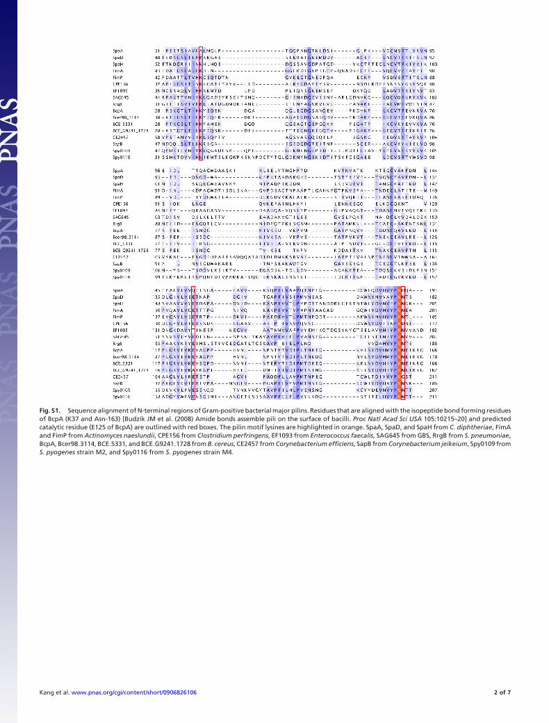
Fig. S1. Sequence alignment of N-terminal regions of Gram-positive bacterial major pilins. Residues that are aligned with the isopeptide bond forming residuesof BcpA (K37 and Asn-163) [Budzik JM et al. (2008) Amide bonds assemble pili on the surface of bacilli. Proc Natl Acad Sci USA 105:10215–20] and predictedcatalytic residue (E125 of BcpA) are outlined with red boxes. The pilin motif lysines are highlighted in orange. SpaA, SpaD, and SpaH from C. diphtheriae, FimAand FimP from Actinomyces naeslundii, CPE156 from Clostridium perfringens, EF1093 from Enterococcus faecalis, SAG645 from GBS, RrgB from S. pneumoniae,BcpA, Bcer98�3114, BCE�5331, and BCE�G9241�1728 from B. cereus, CE2457 from Corynebacterium efficiens, SapB from Corynebacterium jeikeium, Spy0109 fromS. pyogenes strain M2, and Spy0116 from S. pyogenes strain M4.
Kang et al. www.pnas.org/cgi/content/short/0906826106 2 of 7

Fig. S2. Ribbon representation of SpaA N-domain. Residues of Ala-61, Gln-153, His-191, and Lys-190 of SpaA are shown in stick mode.
Kang et al. www.pnas.org/cgi/content/short/0906826106 3 of 7
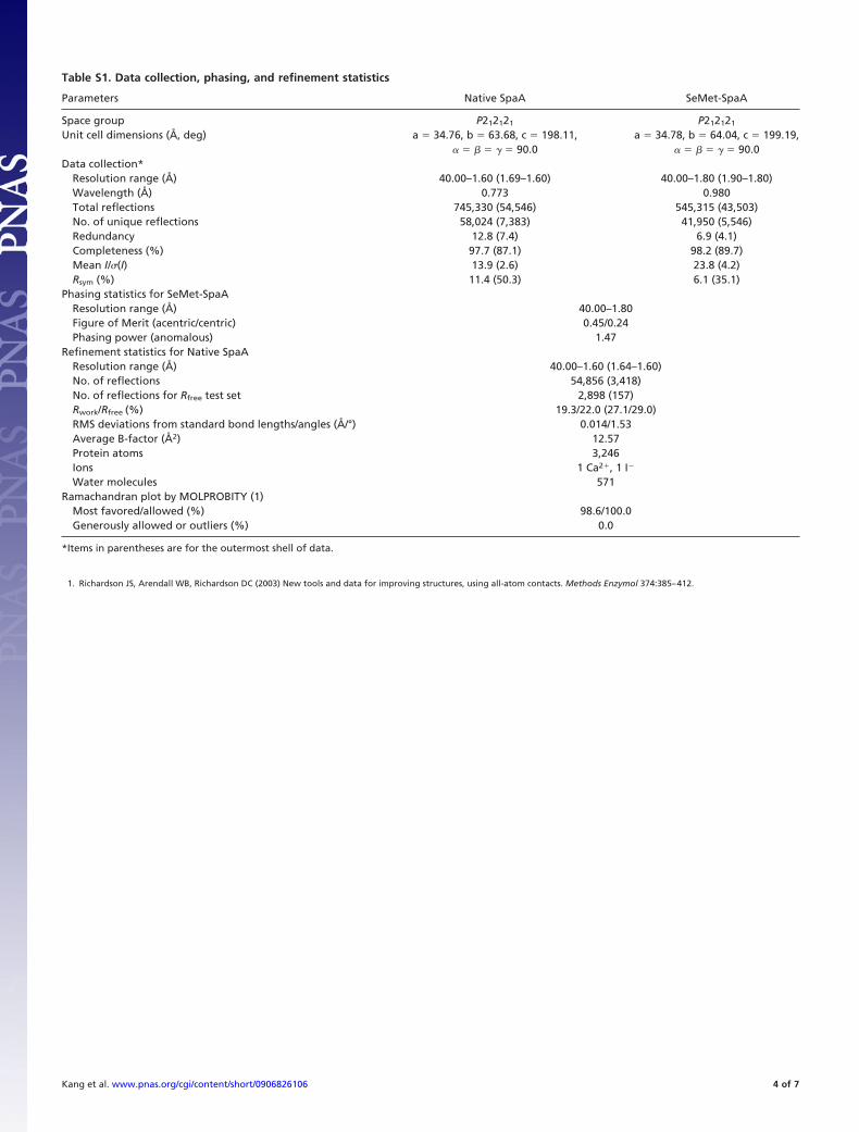
Table S1. Data collection, phasing, and refinement statistics
Parameters Native SpaA SeMet-SpaA
Space group P212121 P212121
Unit cell dimensions (Å, deg) a � 34.76, b � 63.68, c � 198.11,� � � � � � 90.0
a � 34.78, b � 64.04, c � 199.19,� � � � � � 90.0
Data collection*Resolution range (Å) 40.00–1.60 (1.69–1.60) 40.00–1.80 (1.90–1.80)Wavelength (Å) 0.773 0.980Total reflections 745,330 (54,546) 545,315 (43,503)No. of unique reflections 58,024 (7,383) 41,950 (5,546)Redundancy 12.8 (7.4) 6.9 (4.1)Completeness (%) 97.7 (87.1) 98.2 (89.7)Mean I/�(I) 13.9 (2.6) 23.8 (4.2)Rsym (%) 11.4 (50.3) 6.1 (35.1)
Phasing statistics for SeMet-SpaAResolution range (Å) 40.00–1.80Figure of Merit (acentric/centric) 0.45/0.24Phasing power (anomalous) 1.47
Refinement statistics for Native SpaAResolution range (Å) 40.00–1.60 (1.64–1.60)No. of reflections 54,856 (3,418)No. of reflections for Rfree test set 2,898 (157)Rwork/Rfree (%) 19.3/22.0 (27.1/29.0)RMS deviations from standard bond lengths/angles (Å/°) 0.014/1.53Average B-factor (Å2) 12.57Protein atoms 3,246Ions 1 Ca2�, 1 I�
Water molecules 571Ramachandran plot by MOLPROBITY (1)
Most favored/allowed (%) 98.6/100.0Generously allowed or outliers (%) 0.0
*Items in parentheses are for the outermost shell of data.
1. Richardson JS, Arendall WB, Richardson DC (2003) New tools and data for improving structures, using all-atom contacts. Methods Enzymol 374:385–412.
Kang et al. www.pnas.org/cgi/content/short/0906826106 4 of 7
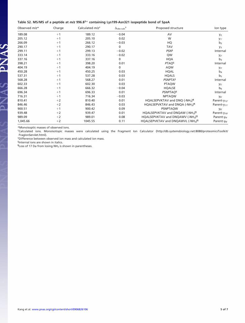
Table S2. MS/MS of a peptide at m/z 996.83� containing Lys199-Asn321 isopeptide bond of SpaA
Observed m/z* Charge Calculated m/z† �obs-calc‡ Proposed structure Ion type
189.08 �1 189.12 �0.04 AV y2
205.12 �1 205.10 0.02 W y1’
266.09 �1 266.12 �0.03 HQ b2
290.17 �1 290.17 0 TAV y3
299.11 �1 299.13 �0.02 PSN§ Internal333.14 �1 333.16 �0.02 QW y2’
337.16 �1 337.16 0 HQA b3
398.21 �1 398.20 0.01 PTAQ§ Internal404.19 �1 404.19 0 AQW y3’
450.28 �1 450.25 0.03 HQAL b4
537.31 �1 537.28 0.03 HQALS b5
568.28 �1 568.27 0.01 PSNPTA§ Internal602.33 �1 602.30 0.03 PTAQW y5’
666.28 �1 666.32 �0.04 HQALSE b6
696.34 �1 696.33 0.01 PSNPTAQ§ Internal716.31 �1 716.34 �0.03 NPTAQW y6’
810.41 �2 810.40 0.01 HQALSEPVKTAV and DNQ (-NH3)¶ Parent-y12’
846.46 �2 846.43 0.03 HQALSEPVKTAV and DNQA (-NH3)¶ Parent-y11’
900.51 �1 900.42 0.09 PSNPTAQW y8’
939.48 �2 939.47 0.01 HQALSEPVKTAV and DNQAW (-NH3)¶ Parent-y10’
989.09 �2 989.01 0.08 HQALSEPVKTAV and DNQAWV (-NH3)¶ Parent-y9’
1,045.66 �2 1045.55 0.11 HQALSEPVKTAV and DNQAWVL (-NH3)¶ Parent-y8’
*Monoisoptic masses of observed ions.†Calculated ions. Monoisotopic masses were calculated using the Fragment Ion Calculator (http://db.systemsbiology.net:8080/proteomicsToolkit/FragIonServlet.html).
‡Difference between observed ion mass and calculated ion mass.§Internal ions are shown in italics.¶Loss of 17 Da from losing NH3 is shown in parentheses.
Kang et al. www.pnas.org/cgi/content/short/0906826106 5 of 7
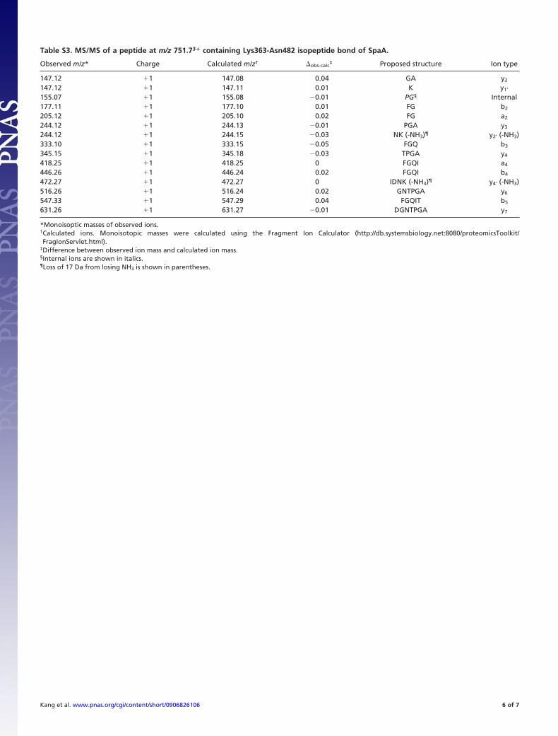
Table S3. MS/MS of a peptide at m/z 751.73� containing Lys363-Asn482 isopeptide bond of SpaA.
Observed m/z* Charge Calculated m/z† �obs-calc‡ Proposed structure Ion type
147.12 �1 147.08 0.04 GA y2
147.12 �1 147.11 0.01 K y1’
155.07 �1 155.08 �0.01 PG§ Internal177.11 �1 177.10 0.01 FG b2
205.12 �1 205.10 0.02 FG a2
244.12 �1 244.13 �0.01 PGA y3
244.12 �1 244.15 �0.03 NK (-NH3)¶ y2’ (-NH3)333.10 �1 333.15 �0.05 FGQ b3
345.15 �1 345.18 �0.03 TPGA y4
418.25 �1 418.25 0 FGQI a4
446.26 �1 446.24 0.02 FGQI b4
472.27 �1 472.27 0 IDNK (-NH3)¶ y4’ (-NH3)516.26 �1 516.24 0.02 GNTPGA y6
547.33 �1 547.29 0.04 FGQIT b5
631.26 �1 631.27 �0.01 DGNTPGA y7
*Monoisoptic masses of observed ions.†Calculated ions. Monoisotopic masses were calculated using the Fragment Ion Calculator (http://db.systemsbiology.net:8080/proteomicsToolkit/FragIonServlet.html).
‡Difference between observed ion mass and calculated ion mass.§Internal ions are shown in italics.¶Loss of 17 Da from losing NH3 is shown in parentheses.
Kang et al. www.pnas.org/cgi/content/short/0906826106 6 of 7

Table S4. Daughter ions produced during MS/MS of a peptide (m/z 585.73�) that was generated from trypsin and AspN digestion ofSpaA pili (this peptide contains the intersubunit isopeptide bond between SpaA Lys190 and Thr494)
Observed m/z* Charge Calculated m/z† �obs-calc‡ Proposed structure Ion type
106.05 �1 106.05 0 S y1
187.15 �1 187.11 0.04 DV a2
211.14 �1 211.16 �0.02 PL§ Internal215.11 �1 215.10 0.01 DV b2
215.11 �1 215.14 �0.03 EL a2’
219.13 �1 219.13 0 LS y2
243.13 �1 243.13 0 EL b2’
312.22 �1 312.20 0.02 PLT §–H2O Internal340.22 �1 340.19 0.03 ELP b3’
352.15 �1 352.16 �0.01 DVH b3
418.22 �1 418.23 �0.01 QALS y4
451.21 �1 451.23 �0.02 DVHV b4
472.57 �3 472.58 �0.01 DVHVYPKHQ and PLT (- H2O)¶ Parent-y3-b2’
496.26 �3 496.27 �0.01 DVHVYPKHQA and PLT (- H2O)¶ Parent-y2-b2’
533.97 �3 534.96 0.01 DVHVYPKHQAL and PLT (- H2O)¶ Parent-y1-b2’
555.32 �1 555.29 0.03 HQALS y5
568.99 �3 568.97 0.02 DVHVYPKHQALS and PLT (- H2O)¶ Parent-b2’
614.33 �1 614.29 0.04 DVHVY b5
*Monoisoptic masses of observed ions.†Calculated ions. Monoisotopic masses were calculated using the Fragment Ion Calculator (http://db.systemsbiology.net:8080/proteomicsToolkit/FragIonServlet.html).
‡Difference between observed ion mass and calculated ion mass.§Internal ions are shown in italics.¶Loss of 18 Da from losing H2O is shown in parentheses
Kang et al. www.pnas.org/cgi/content/short/0906826106 7 of 7