THE CELL !!JAY AMBE!! - DR. NAITIK TRIVEDI
35
DR. NAITIK D TRIVEDI & DR. UPAMA N. TRIVEDI THE CELL https://www.drnaitiktrivedi.com/ 1 !!JAY AMBE!! 2. THE CELL PREPARED BY DR. NAITIK D. TRIVEDI, M. PHARM, PH. D LECTURER AT GOVERNMENT AIDED, A. R. COLLEGE OF PHARMACY & G. H. PATEL INSTITUTE OF PHARMACY, VALLABH VIDYANAGAR, ANAND. Mobile: +91 - 9924567864 E-mail: [email protected] & DR. UPAMA N. TRIVEDI, M. PHARM, PH. D ASSOCIATE PROFESSOR & HoD (Pharm.D), INDUBHAI PATEL COLLEGE OF PHARMACY AND RESEARCH CENTRE, DHARMAJ. E-mail: [email protected]
Transcript of THE CELL !!JAY AMBE!! - DR. NAITIK TRIVEDI

DR. NAITIK D TRIVEDI
&
DR. UPAMA N. TRIVEDI
THE CELL
https://www.drnaitiktrivedi.com/ 1
!!JAY AMBE!!
2. THE CELL
PREPARED BY
DR. NAITIK D. TRIVEDI,
M. PHARM, PH. D
LECTURER AT GOVERNMENT AIDED,
A. R. COLLEGE OF PHARMACY & G. H. PATEL INSTITUTE OF
PHARMACY, VALLABH VIDYANAGAR, ANAND.
Mobile: +91 - 9924567864
E-mail: [email protected]
&
DR. UPAMA N. TRIVEDI,
M. PHARM, PH. D
ASSOCIATE PROFESSOR & HoD (Pharm.D),
INDUBHAI PATEL COLLEGE OF PHARMACY AND RESEARCH
CENTRE, DHARMAJ.
E-mail: [email protected]

DR. NAITIK D TRIVEDI
&
DR. UPAMA N. TRIVEDI
THE CELL
https://www.drnaitiktrivedi.com/ 2
! !JAY AMBE! !
STRUCTURE AND FUNCTION OF CELL
INTRODUCTIONS:
CELL: It is living structural and functional units of body enclosed by membrane.
CYTOLOGY: It is the branch of science concern with the study of cells.

DR. NAITIK D TRIVEDI
&
DR. UPAMA N. TRIVEDI
THE CELL
https://www.drnaitiktrivedi.com/ 3
PARTS OF THE CELLS:
It is mainly divided in to three main parts:
1) Plasma membrane:
It is the outer surface of cells. It’s separates cells from internal environments to
external environments.
It is a selective barrier that regulates the flow of materials into and out of a cell. This
selectivity helps to maintain the normal cellular activities.
2) Cytoplasm:
It consist all the cellular contains between plasma membrane and nucleus.
It consist two components:
a) Cytosol: The fluid portion of cytoplasm contains water, dissolved solutes and
suspended particles.
b) Organelles: This is surrounded by cytosol. Each type of organelles has characteristics
shapes and specific functions. Eg: Ribosomes, Endoplasmic Reticulum, Golgi
complex, Lysosomes, Peroxisomes and Mitochondria.
3) Nucleus:
It is large organelles. It is a house for most of DNA.
Within the nucleus, each chromosomes a single molecules of DNA associated
with several proteins, contains thousand of hereditary units called genes that
control cellular structures and functions.
►THE PLASMA MEMBRANE◄
It is a thin barrier that separates the internal components of a cell from extracellular
materials. It is also known as cell membrane.
It is well describe by fluid mosaic model. According to this model, the molecular
arrangement of the plasma membrane resembles an ever-moving sea of fluid lipids
that contains a mosaic of many proteins.

DR. NAITIK D TRIVEDI
&
DR. UPAMA N. TRIVEDI
THE CELL
https://www.drnaitiktrivedi.com/ 4
Some proteins floats freely like ice bridges in the lipid sea, whereas others are anchored at
specific location like boat at a dock.
MEMBRANE CHEMISTRY AND ANATOMY:
It consist 50:50 mix by weight of protein and lipids that are held together noncovalent
interactions.
In plasma membrane protein are large molecules than the lipid. So one protein
molecules surrounded by around 50 lipids molecules.
A) Membrane lipids:
The plasma membrane is made up by lipid bilayer.
It consist three types of lipids,
a) Phospholipids: 75% of membrane lipids are phospholipids. It contains phosphate
groups.
b) Cholesterol: 20% of membrane lipids are cholesterol. Which is a steroid attached
with –OH group.
c) Glycolipids: 5% of membrane lipids are glycolipids. Attached with carbohydrate
groups.
The lipid bilayer is amphipathic because it consist both polar and non polar parts.
In phospholipids, the polar part is the phosphate containing head which is hydrophilic
(water loving). The non polar part contains two long fatty acid tails which are
hydrophobic (water hating) hydrocarbon chains.
Cholesterol molecules are weakly amphipathic.
In glycolipids carbohydrate groups act head as polar group while their fatty tail act as
non polar group.
B) Membrane proteins:
Plasma membrane consist two types of proteins
a) Integrated proteins:
It extends across the phospholipids bilayer among the fatty acid tail.
Most of integral proteins are glycoprotein, it is attached with sugar groups.
The portion of the attaché sugar group faces the extracellular fluids.
b) Peripheral proteins:
They do not extend across the phospholipids bilayer.
They are loosely attached to the inner and outer surface of the membrane and
are easily separated from it.

DR. NAITIK D TRIVEDI
&
DR. UPAMA N. TRIVEDI
THE CELL
https://www.drnaitiktrivedi.com/ 5
Role and functions of the Proteins:
1) Act as channels:
It act as channels that means some proteins have a pore though which certain
substance can flow into or out of the cells.
2) Act as transporter:
It acts as transporter that means it works as carrier for moving the substance from
one side to other side.
3) Works as receptors:
It works as receptors that means it identify and attach to a specific molecules such
as a hormone, a neurotransmitter etc.
A molecule that specifically binds to receptors by forces other than covalent bonds
is called as legend of those receptors.
4) Works as enzymes:
These are mainly take part in catalyzing reaction inside or out side of the cells.
5) Act as a cytoskeleton anchor:
In side the cells they all provides the structural stability and maintain the shape of
cells.
They also participate in the movements of the cell.
6) Work as a cell identity marker:
It works as a cell identity marker that means distinguishes your cells from anyone
else’s.
Most of glycoproteins and glycolipids work as a cell identity marker.
MEMBRANE PHYSIOLOGY:
1) Communication:
It plays a main role in cellular communication.
This includes interactions with other body cells, foreign cells and ligands such as
hormones, neurotransmitters, enzymes, nutrients and antibodies in the extracellular
fluid.
2) Electrochemical gradients:
The membrane maintains the electrical and chemical difference (gradient) between
the inside and out side of the membrane is known as electrochemical gradient.
In the extracellular fluids, the main cation (positively charged ion) is Na+ and main
anion (Negative Charged ion) is Cl-.

DR. NAITIK D TRIVEDI
&
DR. UPAMA N. TRIVEDI
THE CELL
https://www.drnaitiktrivedi.com/ 6
In the cytosol the main cation is K+ and anions are organic phosphate (PO4-) and
amino acids in proteins. .
The electrochemical gradient arises because inside surface of the membrane is more
negatively charged than the outer surface. As a result there is a voltage is form is
known as membrane potential across the membrane.
The voltage across the plasma membrane of cells through out your body is between -
20 and -200 milllivolts (mV).
The negative sign in front of the number means inside is negatively relative to the
outside.
3) Selective permeability:
It regulates the entry and exit of the materials.
It permits passage of certain substance and restricts the others.
Eg.: water is passage more easily than other substances.
The selectivity is depends on several factors such as;
a) Lipid solubility:
Substances that dissolve in lipids (Nonpolar, Hydrophobic molecules) pass easily
through out the phospholipids bilayers.
b) Size:
Large molecules like as proteins cannot pass through plasma membrane.
Small uncharged polar molecules can pass through the phospholipids bilayer.
c) Charges:
It is impermeable to all charged molecules and ions but some charged molecules
can pass through the pores of the membrane.
Presence of channels and transporters:
Polar and charged substance cannot pass by the phospholipids bilayer but they
can pass by the help of several proteins either they form the water filled pores or
act as transporters.
Transporters pick up the molecules from one side of the membrane and leave it
to other side.

DR. NAITIK D TRIVEDI
&
DR. UPAMA N. TRIVEDI
THE CELL
https://www.drnaitiktrivedi.com/ 7
►MOVEMENTS OF MATERIALS ACROSS THE PLASMA MEMBRANE: ◄
Movement of the material across the plasma membrane is describe by the two
processes;
A) Passive process
B) Active process
A) Passive process:
“When the movement is Depend on concentration gradient means process is held from higher
concentration to lower concentration is known as passive transport.”
It is also known as nonionic diffusion as well as downhill process.
Passive transfer is energy independent.
Passive transport is best express by Fick’s first law of diffusion, which state that the
drug molecules transport from a region of higher concentration to lower concentration
until equilibrium attained and that the rare is directly proportional to the concentration
of gradient across the membrane.
dQ = DAKm/w (CGIT – C)
dt h
Where,
dQ/dt = rate of drug transport (amount/time)
D = Diffusion coefficient of the drug through the membrane. (area/time)
A = Surface area and h = thickness of the membrane.
Km/w = partition coefficient of the drug between the lipoidal membrane and the aqueous GIT
fluid.
(CGIT – C) = Different in concentration between GI fluid and plasma.
Above equation shows that;
The drug or transportation is down hill process.
The rate of transfer is proportional to concentration gradient between GI fluid and
plasma compartment.
Greater the area and lesser the thickness of the membrane faster the transfer.
The another passive transport processes are;
i) Diffusion:
All the substance has their own kinetics energy. So movement of the molecules or
ions due to their kinetics energy is known as diffusion.

DR. NAITIK D TRIVEDI
&
DR. UPAMA N. TRIVEDI
THE CELL
https://www.drnaitiktrivedi.com/ 8
When the two such areas are connected and one side area have more particles than the
other sides it will create the concentration gradient.
So the substances move by their kinetics energy from higher concentration to lower
concentration until the equilibrium rich. It is known as net diffusion.
Eg.: if we have a two compartment vessels fill up by water then add crystal of dye in
one compartment so the dye diffusion is held from dye added water to water because
of concentration different.
Lipid soluble molecules such as oxygen, nitrogen, steroids, fat soluble vitamins (A, D,
E & K), glycerol etc are cross the plasma membrane by simple diffusion.
Diffusion is important in the movement of oxygen and carbon dioxide between blood
and body cell and between blood and air within the lungs during breathing.
Small molecules that are not lipid soluble may diffuse into or out of cells through
water filled pores of integral proteins.
Eg.: Sodium ions (Na+), Potassium ions (K+), Calcium ions (Ca+), Chloride ions (Cl-),
Bicarbonate ions (HCO3-) and urea.
ii) Osmosis:
Another passive process is osmosis.
In this process water is move by osmosis across a membrane from an area of higher
water concentration (lower solute concentration in water) to an area of lower water
concentration (higher solute concentration in water).
For the description of this process take a sac made of cellophane (selective permeable
membrane) contain a sugar solution and it immersed in a beaker of pure water. It is
only permeable for water not for sugar because sugar molecules are large in size.
The water concentration on the two sides are different means lower water
concentration inside the sac because the additional of sugar.
iii) Pore transport:
It is also known as connective transport, bulk flow transport or filtration.
The process is important for the absorption of lower weight and lower size molecules.
Urea, water and sugars are transfer by this mechanism.
B) Active process:
“When the movement is against the concentration gradient energy is a required mean the
transport of molecules is occurring by the help of ATP is known as active process”.
The drug is transported from a region of lower to higher concentration i.e against the
concentration gradient.

DR. NAITIK D TRIVEDI
&
DR. UPAMA N. TRIVEDI
THE CELL
https://www.drnaitiktrivedi.com/ 9
It is known as uphill transport.
Energy is required for this transfer.
Substances that transport actively are sodium, potassium, calcium, glucose, certain
amino acids and vitamins like niacin, pyridoxine and ascorbic acid.
It includes the different processes like;
i) Primary active transport:
Movement of ions or molecules across a selectively permeable membrane from a
region of lower to higher concentration by pump protein that use energy from the
splitting of ATP.
Eg.: Sodium ions (Na+), Potassium ions (K+), Calcium ions (Ca+), Chloride ions (Cl-) and
other ions.
ii) Secondary active transport:
When the simultaneously movements of two substance is held in which one substance
is Na+ or Transport by using energy is known as secondary active transport.
Eg.: Glucose into cells lining of the small intestine and the kidney tubules.
In this transport if the both substances move in a same direction is known as
symporters and if the both substances movement are in opposite direction is known as
antiporters.
Eg.: Sodium ions (Na+), Potassium ions (K+), Calcium ions (Ca+) etc
iii) Endocytosis:
It is a minor transport mechanism which involves engulfing extracellular materials
within a cell.
Vitamins like A, D, E, K and drugs like insulin refer this phenomenon.
Endocytosis includes two types of processes;
a) Phagocytosis:
It is known as cell eating.
Absorption of solid particles.
b) Pinocytosis:
It is known as cell drinking.
Absorption of fluid solute.

DR. NAITIK D TRIVEDI
&
DR. UPAMA N. TRIVEDI
THE CELL
https://www.drnaitiktrivedi.com/ 10
►CYTOPLASM◄
It consist all the cellular contains between plasma membrane and nucleus.
It consist two components:
1. Cytosol:
It is the unsaturated soluble portion of the cells.
Chemically it is 75-90 % water plus solid components (protein, carbohydrate, lipids
and inorganic substance).
Inorganic substance and smaller organic substance such as simple sugar and amino
acid are soluble in water and are present as solute. While larger particle such as
protein and polysaccharide glycogen found as colloidal particle in surrounding
medium and they are not dissolved.
The cytosol receives raw material from the external environment and gain usable
energy from them by decomposition reaction.
2. Organelles
These are specialized structures that have characteristics appearance and specific role
in growth, maintenance, repair and control.
The number and types of organelles vary in different kinds of cells depending on their
function. Different types of organelles are:
A) Mitochondria:
Mitochondria are the largest components of the cytoplasm.
They are the power house of the cell and each cell may contain from 50 to 2500
mitochondria depending upon the respiratory activity of the cells.

DR. NAITIK D TRIVEDI
&
DR. UPAMA N. TRIVEDI
THE CELL
https://www.drnaitiktrivedi.com/ 11
Eg: The cell of skeletal muscle, kidney and liver contain large number of mitochondria
while heart muscles contain less.
They are vary in shape and size (0.5 to 3μ long and 0.1 to 0.6μ wide).
They have two membranes, the outer is smooth but the inner is arranged series of
folds form ridges known as cristae.
Mitochondria consist central cavity enclosed by inner membrane is known as matrix.
Folds increase the inner surface area which useful for the cellular respiration.
The matrix and cristae contains the catalytic enzyme which produce the ATP.
Mitochondria swell in hypotonic solution and shrink hypertonic solution.
The mitochondria contain large number of enzyme system known as “cylophorase”
which are involved in:
a) Oxidation of pyruvic acid in Kreb’s cycle via acetyl CoA
b) Electron transport and oxidative phosphorylation
c) Synthesis of fatty acids
Although each cell’s nucleolus contains genes from both your mother and father,
mitochondria genes usually are inherited only from mother because the head of the
sperm which is the part that penetrates and fertilizes an egg have lacks mitochondria.
B) Endoplasmic Reticulum:

DR. NAITIK D TRIVEDI
&
DR. UPAMA N. TRIVEDI
THE CELL
https://www.drnaitiktrivedi.com/ 12
This is the complicated and organized system of membranes in the cytoplasm of the
cell.
This membrane is constituted of protein lipid double layer and is very well developed
in tissue with active protein synthesis.
There are two types of endoplasmic reticules one is rough or granular endoplasmic
reticulum consist ribosomes on their surface and second is smooth endoplasmic
reticulum or agranular which has no ribosomes.
Rough endoplasmic reticulum is associated with the protein synthesis.
It serves as temporary storage area for newly synthesized molecules and may add
sugar groups to certain proteins. Eg.: Glycoproteins.
Smooth endoplasmic reticulum is the site for fatty acids, phospholipids and steroidal
synthesis.
Enzyme within the smooth ER can inactive or detoxified a verity of chemicals
including alcohols, pesticides, and carcinogens.
C) Ribosome:
Ribosomes are tiny granules that contain ribosomal RNA (rRNA) and many
ribosomal proteins.
The size of the ribosomes ranges from 15 to 20 millimicrone and the diameter being
150Ao.
The rRNA synthesized by DNA in nucleus.
Functionally the ribosomes are the sites of protein synthesis.
Some ribosomes are known as free ribosomes, float in cytosol. They are not attached
to other organelles. Free ribosomes are form singly or in clustered form.
Other ribosomes attached to a cellular structure called endoplasmic reticulum.
D) Golgi Complex:
The golgi complex or apparatus is an organelles located near the nucleus.
It consist flattened sac called cisterns which consists small Golgi vesicles.
The Golgi complex processes sorts, packages and deliver proteins and lipids to
plasma membrane and forms lysosomes and secretary vesicles.
All the proteins are export from the cells by similar rout i.e ribosomes (site of protein
synthesis) – rough endoplasmic reticulum cistern – transport vesicles – Golgi complex
– secretary vesicles – release to exterior of the cells by exocytosis.
A trip through a Golgi complex normally occurs as follows:

DR. NAITIK D TRIVEDI
&
DR. UPAMA N. TRIVEDI
THE CELL
https://www.drnaitiktrivedi.com/ 13
Within the cistern of rough ER, protein becomes surrounded by a piece of the ER
membrane which form a transport vesicle.
The transport vesicle leaves the ER and moves toward the Golgi complex.
Here, the vesicle fuses within the side of the Golgi stack closest to the ER which is
term as cis or entry cistern. As a result of this fusion the protein enters the Golgi
complex.
Then they move cis cistern to middle cistern and then next cistern.
As the proteins pass through the Golgi cistern, they are modified in various ways
depending on their function and destination. Finally they enter to trans or exit cistern.
The trans or exist cistern modified the protein in to vesicles.
Some vesicles become seretory vesicles and discharge their contain in to the
extracellular fluid by exocytosis process.
E) Lysosomes:
Lysosomes are membrane enclosed vesicles that form in the Golgi complex.
Inside the lysosomes, there are as many as 40 kinds of powerful digestive (hydrolytic)
enzymes capable of breaking down a wide variety of molecules.
Some disorders are caused by faulty lysosomes.
Eg.: Tay-Sachs disease is an inherited which is cause by absence of single lysosomes
enzyme. This enzyme is essential for the break down of membrane glycolipids which
is essential for to prevent the nerve cells function. In absence of this enzyme nerve
cells get damage and produce blindness in child, demented, die usually before the age
of 5.
Lysosomes work best at acidic pH. The lysosomes membrane have active transport
pump that drive hydrogen ion (H+) into the lysosomes. So the interior of a lysosomes
has a pH 5, which is 100 times more acidic than the cytosolic pH of 7.
Lysosomes digest the bacteria and other substance that enter the cell by phagocytosis
or pinocytosis.
Lysosomal enzymes may also destroy their own host cells called autolysis.
Lysosomes digest the old organelles and return the digested components to the cytosol
for reuse. So old organelles are replaced by new organelles this process is known as
autophagy.
Lysosomal enzymes may digest the cellular debris at the site of injury.
F) Peroxisomes:
It have the similar structure to lysosomes but smaller than the lysosomes.

DR. NAITIK D TRIVEDI
&
DR. UPAMA N. TRIVEDI
THE CELL
https://www.drnaitiktrivedi.com/ 14
It contains one or more enzymes that are used in oxygen to oxidize process. Such
reaction produces hydrogen peroxide.
G) The cytoskeleton:
Coordination of the cellular movements and cellular shape is maintained by the
cytoskeleton.
The cytoskeleton is responsible for the movement of whole cells, such as phagocytes
and for movement of organelles and some chemicals within the cells.
There are three main types of the proteins comprise the cytoskeletons:
a) Microfilaments:
It has rod like structures of varying length that are formed from the protein actin.
In muscles tissue actin filament (thin filament) and myosin filaments (thick
filaments) are important for the muscles contraction.
In nonmuscle cells, actin microfilaments provide support and shape.
b) Microtubules:
It is larger than microfilaments.
They are relatively straight and cylindrical in structure that consist protein is
known as tubulin.
Microtubules also work as conveyer belt for the movement of various substances.
c) Intermediate filaments:
It is strong and tough.
It holds the organelles in their position.
H) Centrosome and Centriols:
Near the nucleus is a dense area of cytoplasmic material with radiant microtubules
called as centrosome.
Centrosomes serve as center for organizing microtubules in non-dividing cells.
It also forms the mitotic spindle during cell division.
Within the Centrosomes, a pair of cylindrical structures known as Centriols.
Each Centriols is composed of nine cluster of three microtubules arranged in a
circular pattern.

DR. NAITIK D TRIVEDI
&
DR. UPAMA N. TRIVEDI
THE CELL
https://www.drnaitiktrivedi.com/ 15
►NUCLEUS◄
Nucleus is a spherical or oval in shape and is the heaviest or largest structure in the
cell. Within the nucleus are most of the hereditary unit of the cell known as genes,
which control cellular structure and direct many cellular activities.
The nuclear genes are arranged side bi side and form a specific structure known as
chromosomes.
Human somatic cells have 46 chromosomes, 23 inherited from each parents.
A double membrane called the nuclear envelope separates the nucleus from the
cytoplasm. Both layers of the nuclear layer envelop are phospholipids bilayer similar
to plasma membrane.
The two membranes are separated from each other by the perinuclear cisterns and join
at interval to form pores which allows the passage of materials from the cytoplasm to
the nucleus and vice versa an these pores are 10 times more larger than the pores of
channels in the plasma membrane, even larger molecules such as RNA and various
protein can also pass.
Inside the nucleus one or more spherical bodice are present known as nucleoli
(Singular is nucleolus). They contain bunch of protein, DNA and RNA that are not
enclosed by membrane.
Nucleolus or Nucleoli contains ribosomes as well as ribosomal RNA and it play a key
role in protein synthesis.
A chromosome is a very long DNA molecule that is coiled and packed in amazing
compact structure together with several proteins.
In chromosomes two identical pair consist nucleoprotein strands that are joined at
centromere and separated during cell division is known as chromatid. It forms a tough
thread like structure in not dividing 46 chromosomes.

DR. NAITIK D TRIVEDI
&
DR. UPAMA N. TRIVEDI
THE CELL
https://www.drnaitiktrivedi.com/ 16
Chromatin has a “beads on a string (thread)” structure. Each beads known as
nucleosomes consist eight proteins molecules called as histone which are wrapped by
double strand DNA twice around it.
►PROTEIN SYNTHESIS◄
Cells are basically protein factories that constantly synthesize large number of diverse
protein. The, protein determine the physical and chemical characteristics of cells and
therefore of organisms.
Some proteins are structural to form plasma membranes, microfilaments, microtubules,
Centriols, mitochondria and other parts of cells.
Other proteins serve as hormones, antibodies and contractile elements in muscle tissue also it
act as enzyme.
This process can be divided into two parts:
1. Transcription
Before the synthesis of a protein begins, the corresponding RNA molecule is
produced by RNA transcription.
Three forms of RNA are made from the DNA template,
a) messenger RNA (mRNA) which direct synthesis of a polypeptide chain,
b) transfer RNA (tRNA) which bind to amino acid during translation and
c) ribosomal RNA (rRNA) which comes together with ribosomal protein to make up
ribosomes.
In protein synthesis, one strand of the DNA double helix is used as a template by the RNA
polymerase to synthesize a messenger RNA (mRNA) this strand refer as sense strand and the

DR. NAITIK D TRIVEDI
&
DR. UPAMA N. TRIVEDI
THE CELL
https://www.drnaitiktrivedi.com/ 17
other strand that not transcribed known as antisense strand, during the transcription the
changes in to the nitrogen base are as under;
DNA RNA
A U
T A
G C
C G
A U
T A
Template DNA base sequence Complementary RNA sequence
Within DNA are region known as intron that do not synthesis of part of protein and intron are
located between regions called exons that do code for proteins.
Initially mRNA transcript include both introns and exons then RNA region corresponding to
DNA introns are deleted (cut out) and the exon are spliced (rejoined) and finally mRNA
migrates from the nucleus to the cytoplasm, this process is known as mRNA splicing.
In the cytoplasm, the start the next step which is translation
2. Translation
It is the process where the nucleotide sequence in a molecule of mRNA specifies the
amino acid sequence for protein molecules.
In the mRNA molecules, each set of three consecutive nucleotide bases is called
codon and specifies one amino acid.
Most mRNA molecules contain 300-3000 nucleotides so it form 100 to 1000 amino
acid because three nucleotide code for one amino acids.
Translation process following steps;
Initiation:
a) In the cytoplasm, the small ribosomal subunit binds to one end of the mRNA
molecules and finds the start codon, a sequence where translation will begin. Then the
large ribosomal subunit joins in the process.
b) In the cytosol, tRNA binds to one kind of amino acid and brings it to the ribosomes.
One end of the tRNA carries amino acid and another part of each tRNA has a triplet
of nucleotides called as anticodon. This anticodon of the tRNA attach to
complementary codon on mRNA.
c) Eg.: if the mRNA codon is AUG then tRNA have the anticodon UAC would attach.
d) In the starting of this process tRNA brings methionine amino acid.

DR. NAITIK D TRIVEDI
&
DR. UPAMA N. TRIVEDI
THE CELL
https://www.drnaitiktrivedi.com/ 18
Elongation:
e) Once the first tRNA attach to mRNA, the ribosomes moves exactly three nucleotides
along the mRNA and the tRNA carry its amino acid on that particular nucleotides or
codon.
f) When the second tRNA brings the next amino acid first tRNA again goes back in to
the cytoplasm. The proper amino acids are brought into line, one by one peptide
bonds form between them and protein progressively lengthens.
g) Each time the ribosome moves one codon along mRNA and empty tRNA is eject. The
released tRNA can pick up another amino acid.
Termination:
h) When the specified protein is complete, synthesis is terminated by a special stop
codon.
i) Then assemble protein is then released from the ribosomes.
j) After protein synthesis small and large ribosomal subunits separate.
►CELL CYCLE◄
Cell Cycle
Positive control Negative Control
(Stimulate Cell Division) (Inhibit Cell Division)
In the normal cell cycle include,
G1 Phase Pre (Nucleic acid) Synthesis Interval
S Phase Synthesis of DNA Occur
G2 Phase Post Synthetic Interval
M Phase Mitosis occur produce two G1 Cells Which
either enter in to Next Cycle or Pass in to non
proliferative phase (G0-quiescent phase).
G1 G0
S (synthesis) phase, M (mitosis) phase, G1 (Check point 1 between M and S phase where
cell is preparing for S phase by synthesis messenger RNAs and proteins need for DNA
replication), G2 (check point 2 between S and M phase where double the number of
chromosomes), Go is the quiescent phase where the cell is not constantly divide, here the
Rb protein is hypophosporylated. If the DNA or cell is damaged the repairing of cell is

DR. NAITIK D TRIVEDI
&
DR. UPAMA N. TRIVEDI
THE CELL
https://www.drnaitiktrivedi.com/ 19
take place either in check point 1 or check point 2. If the repair is fails then cell goes in to
the apoptosis.
In normal human, cell proliferation and cell destruction process is controlled by positive
(Stimulate Cell Growth) and negative regulation (Inhibit Cell Growth).
In positive control, two families of proteins; cyclin and cyclin dependent kinases (cdks)
have a major role. Each cdk is inactive until it binds to a cyclin, the binding enabling the
cdk to phosphorylate the protein(s) necessary for a particular step in the cell cycle. After
the phosphorylation take place cyclin is degraded by ubiquitin/protease system. There are
eight groups of cyclins. Those important in the control of the cell cycle are cyclins A, B,
D and E. Each cyclin is associated with and activates the particular cdk(s). Cyclin A
activates cdks 1 and 2; cyclin B, cdk 1; cyclin D, cdks 4 and 6; cyclin E, cdk 2.
In negative regulation, the mediators either stop the cell cycle or produce cell death
(apoptosis). Different mediators are Rb protein that holds the cycle in Go phase while it is
hypophosporylated. Another two families inhibitors are, one is CIP family (cdk inhibitor
proteins, also termed KIP or kinase inhibitory proteins) – p21, p27, and p57. Other is Ink
family (inhibitor of kinase) – p16, p19 and p15. p 21 is the under control of the gene p51.
Genetically regulate positive and negative regulators in control of cell proliferation.
Significance of Cell cycles:
It replace dead cell and injured cell.
Add new cells in place of dead cells and injured cells.
It maintains the normal body structure of the cell and in regulation condition protects
us from the various diseases like skin cancer.
It pass the genetically information from parents to daughter cells.
It maintains the normal homeostasis mechanism of body.
When needed it produce the apoptosis so inhibit the unwanted cell growth.

DR. NAITIK D TRIVEDI
&
DR. UPAMA N. TRIVEDI
THE CELL
https://www.drnaitiktrivedi.com/ 20
It maintain the genetically information for individual means it differentiate two person
by genetically.
►NORMAL CELL DIVISION◄
“Cell division is the process by which cells produce themselves.” Two kind of cell division
are recognizing;
1) Somatic cell division:
In this process parent cell produces two identical daughter cells. This process consists
of
a) Nuclear division known as mitosis and
b) Cytoplasmic division known as cytokinesis.
In this process daughter cell have same number and kind of chromosomes as the
original parent cell.
2) Reproductive cell division:
In this process sperm and egg cells are produce. These are the cell which produce new
organism.
The process consists of a special nuclear division called as meiosis (reduction
division) followed by cytokinesis.
1) SOMATIC CELL DIVISION:
Human cells, except for egg and sperm, contain 23 pairs of chromosomes.
Two chromosomes that belong to a pair, one contributed by mother and one
contributed by father known as homologues chromosomes.
It is describe by following two steps,
a) Interphase: Cell is between divisions, chromosomes are not seen under light
microscope. It include G1 Phase, S Phase and G2 Phase
b) Cell division: parent cell produce two identical daughter cells, chromosomes
are visible under light microscope. It include Mitosis and Cytokinesis.

DR. NAITIK D TRIVEDI
&
DR. UPAMA N. TRIVEDI
THE CELL
https://www.drnaitiktrivedi.com/ 21
BRIEF INTRODUCTION OF CELL DIVISION

DR. NAITIK D TRIVEDI
&
DR. UPAMA N. TRIVEDI
THE CELL
https://www.drnaitiktrivedi.com/ 22
Nuclear Division Mitosis
The process called mitosis is the distribution of the two sets of chromosomes into two
separate and equal nuclei. It results in the exact duplication of genetic information.

DR. NAITIK D TRIVEDI
&
DR. UPAMA N. TRIVEDI
THE CELL
https://www.drnaitiktrivedi.com/ 23
Mitosis is divided in to four stages prophase, metaphase, anaphase and telophase.
1) Prophase:
It is the first stage of mitosis. Each prophase chromosomes contains a pair of identical
double strand chromatids.
Each chromatid pair is held together by a small spherical body known as centromere
that is required for the proper segregation of chromosomes.
Attached to the outside of each centromere is a protein complex known as the
kinetochore.
Later in prophase the nucleoli disappear and nuclear envelop breaks down and
dissolve in to cytosol.
In addition each centrosomes and its Centriols move to opposite pole (ends) of the
cell.
So the centrosomes start to form mitotic spindle, a football shaped assembly of
microtubules that are responsible for the movement of chromosomes.
As the mitotic spindle develop three types of microtubules form:
1) nonkinetochore microtubules grow from centrosomes, extend inward but do not
bind to kinetochores.
2) Kinetochore microtubules grow from centrosomes, extend inward and attached to
kinetochore
3) Aster microtubules go out of centromers.
The spindle is the attachment site for the chromosomes and it also distributes
chromosomes to opposite poles of the cells.
2) Metaphase:
In this phase, the centromeres of the chromatids pairs line up at the exact centre of the
mitotic spindle. This portion is known as equatorial plane region.
3) Anaphase:
It is characterized by the splitting and separation of centromers and the movment of
two sister chromatids of each pair toward opposite poles of the cells.
Separated chromatids known as daughter chromosomes.
The movement is due to shortening of kinetochore microtubules and elongation of the
nonkerinetochore microtubules. These processes increase the distance between
separated chromosomes. They also pulled by the microtubules so appears as a V
shapes from the centromeres.
DR. NAITIK D TRIVEDI
&
DR. UPAMA N. TRIVEDI
THE CELL
https://www.drnaitiktrivedi.com/ 24
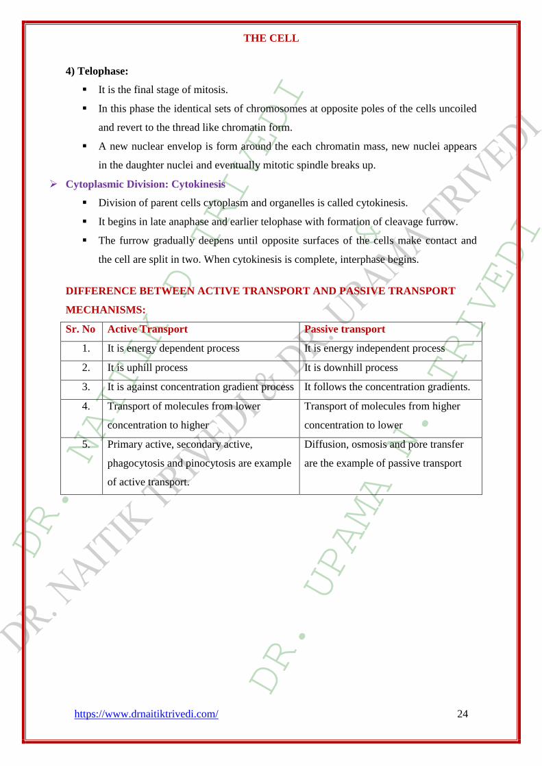
4) Telophase:
It is the final stage of mitosis.
In this phase the identical sets of chromosomes at opposite poles of the cells uncoiled
and revert to the thread like chromatin form.
A new nuclear envelop is form around the each chromatin mass, new nuclei appears
in the daughter nuclei and eventually mitotic spindle breaks up.
Cytoplasmic Division: Cytokinesis
Division of parent cells cytoplasm and organelles is called cytokinesis.
It begins in late anaphase and earlier telophase with formation of cleavage furrow.
The furrow gradually deepens until opposite surfaces of the cells make contact and
the cell are split in two. When cytokinesis is complete, interphase begins.
DIFFERENCE BETWEEN ACTIVE TRANSPORT AND PASSIVE TRANSPORT
MECHANISMS:
Sr. No Active Transport Passive transport
1. It is energy dependent process It is energy independent process
2. It is uphill process It is downhill process
3. It is against concentration gradient process It follows the concentration gradients.
4. Transport of molecules from lower
concentration to higher
Transport of molecules from higher
concentration to lower
5. Primary active, secondary active,
phagocytosis and pinocytosis are example
of active transport.
Diffusion, osmosis and pore transfer
are the example of passive transport

DR. NAITIK D TRIVEDI
&
DR. UPAMA N. TRIVEDI
THE CELL
https://www.drnaitiktrivedi.com/ 25
CELL JUNCTION
Unicellular organisms use to adhere to the environment, nutrition or pathogenesis.
Multicellular organisms require adhesion for cells to adhere to each other and the
extracellular matrix. Cell adhesion occurs through specific cellular specializations and
molecules and has both static and dynamic functions.
Cell junction molecules
The molecules responsible for creating cell junctions include various cell adhesion molecules.
There are four main types:
1. Selectins,
2. cadherins,
3. integrins, and
4. the immunoglobulin super family.
1. Selectins are cell adhesion molecules that play an important role in the initiation of
inflammatory processes. The functional capacity of selectin is limited to leukocyte
collaborations with vascular endothelium. There are three types of selectins found in
humans; L-selectin, P-selectin and E-selectin. L-selectin deals with lymphocytes,
monocytes and neutrophils, P-selectin deals with platelets and endothelium and E-selectin
deals only with endothelium. They have extracellular regions made up of an amino-
terminal lectin domain, attached to a carbohydrate ligand, growth factor-like domain
(EGF) and short repeat units (numbered circles) that match the complimentary binding
protein domains.
2. Cadherins are calcium-dependent adhesion molecules. Cadherins are extremely important
in the process of morphogenesis – fetal development. Together with an alpha-beta catenin
complex, the cadherin can bind to the microfilaments of the cytoskeleton of the cell. This
allows for homophilic cell–cell adhesion. The β-catenin–α-catenin linked complex at the
adherens junctions allows for the formation of a dynamic link to the actin cytoskeleton.
3. Integrins act as adhesion receptors, transporting signals across the plasma membrane in
multiple directions. These molecules are an invaluable part of cellular communication, as
a single ligand can be used for many integrins. Unfortunately these molecules still have a
long way to go in the ways of research.
4. Immunoglobulin superfamily are a group of calcium independent proteins capable of
homophilic and heterophilic adhesion. Homophilic adhesion involves the
immunoglobulin-like domains on the cell surface binding to the immunoglobulin-like
domains on an opposing cell’s surface while heterophilic adhesion refers to the binding of
the immunoglobulin-like domains to integrins and carbohydrates instead.

DR. NAITIK D TRIVEDI
&
DR. UPAMA N. TRIVEDI
THE CELL
https://www.drnaitiktrivedi.com/ 26
Cell adhesion is a vital component of the body. Loss of this adhesion effects cell structure,
cellular functioning and communication with other cells and the extracellular matrix and can
lead to severe health issues and diseases.
TYPES OF CELL JUNCTIONS

DR. NAITIK D TRIVEDI
&
DR. UPAMA N. TRIVEDI
THE CELL
https://www.drnaitiktrivedi.com/ 27
1. Anchoring Junctions:
a. Adherens junctions (zonula adherens)
b. Desmosomes(macula adherens) and
c. Hemidesmosomes
2. Gap junctions (communicating junction)
3. Tight junctions (occluding junctions)
1. Anchoring junctions:
Cells within tissues and organs must be anchored to one another and attached to
components of the extracellular matrix. Cells have developed several types of
junctional complexes to serve these functions, and in each case, anchoring proteins
extend through the plasma membrane to link cytoskeletal proteins in one cell to
cytoskeletal proteins in neighboring cells as well as to proteins in the extracellular
matrix.
Three types of anchoring junctions are observed, and differ from one another in the
cytoskeletal protein anchor as well as the transmembrane linker protein that extends
through the membrane:
Junction Cytoskeletal anchor Transmembrane linker Ties cell to:
Adherens junctions Actin filaments Cadherin / Integrins Other cells / EC matrix
Desmosomes Intermediate filaments Cadherin Other cells
Hemidesmosomes Intermediate filaments Integrins EC matrix
Anchoring-type junctions not only hold cells together but provide tissues with
structural cohesion. These junctions are most abundant in tissues that are subject to
constant mechanical stress such as skin and heart.
a. Adherens junctions (Zonula Adherens)
Adherens junctions share the characteristic of anchoring cells through their
cytoplasmic actin filaments. Similarly to desmosomes and hemidesmosomes, their
transmembrane anchors are composed of cadherins in those that anchor to other cells
and integrins in those that anchor to extracellular matrix. There is
considerable morphologic diversity among adherens junctions. Those that tie cells to
one another are seen as isolated streaks or spots, or as bands that completely encircle
the cell. The band-type of adherens junctions is associated with bundles of actin
filaments that also encircle the cell just below the plasma membrane. Spot-like
DR. NAITIK D TRIVEDI
&
DR. UPAMA N. TRIVEDI
THE CELL
https://www.drnaitiktrivedi.com/ 28
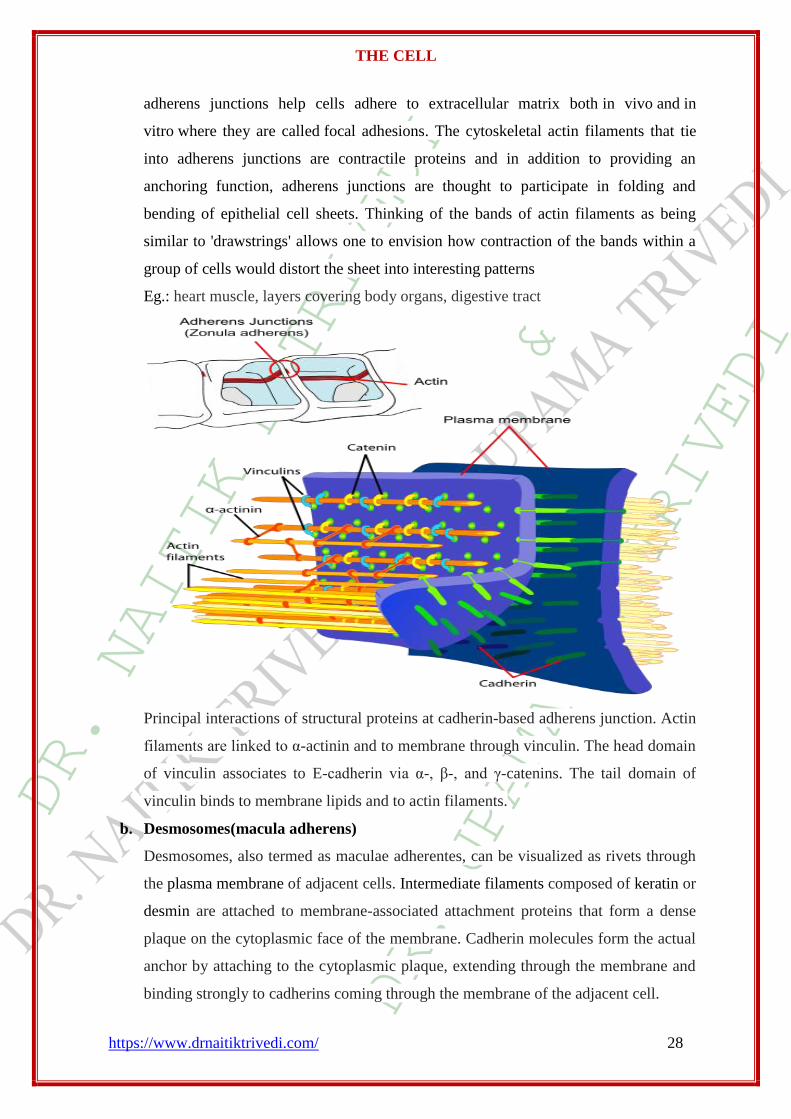
adherens junctions help cells adhere to extracellular matrix both in vivo and in
vitro where they are called focal adhesions. The cytoskeletal actin filaments that tie
into adherens junctions are contractile proteins and in addition to providing an
anchoring function, adherens junctions are thought to participate in folding and
bending of epithelial cell sheets. Thinking of the bands of actin filaments as being
similar to 'drawstrings' allows one to envision how contraction of the bands within a
group of cells would distort the sheet into interesting patterns
Eg.: heart muscle, layers covering body organs, digestive tract
Principal interactions of structural proteins at cadherin-based adherens junction. Actin
filaments are linked to α-actinin and to membrane through vinculin. The head domain
of vinculin associates to E-cadherin via α-, β-, and γ-catenins. The tail domain of
vinculin binds to membrane lipids and to actin filaments.
b. Desmosomes(macula adherens)
Desmosomes, also termed as maculae adherentes, can be visualized as rivets through
the plasma membrane of adjacent cells. Intermediate filaments composed of keratin or
desmin are attached to membrane-associated attachment proteins that form a dense
plaque on the cytoplasmic face of the membrane. Cadherin molecules form the actual
anchor by attaching to the cytoplasmic plaque, extending through the membrane and
binding strongly to cadherins coming through the membrane of the adjacent cell.

DR. NAITIK D TRIVEDI
&
DR. UPAMA N. TRIVEDI
THE CELL
https://www.drnaitiktrivedi.com/ 29
Eg.: Skin, lining of internal body cavity surfaces
It disappear when cells are transformed
c. Hemidesmosomes
Hemidesmosomes form rivet-like links between cytoskeleton and extracellular matrix
components such as the basal laminae that underlie epithelia. Like desmosomes, they
tie to intermediate filaments in the cytoplasm, but in contrast to desmosomes, their
transmembrane anchors are integrins rather than cadherins.
Present in tissues subject to shear or lateral stress
2. Gap junctions (communicating junction)
Communicating junctions, or gap junctions allow for direct chemical communication
between adjacent cellular cytoplasm through diffusion without contact with the
extracellular fluid. This is possible due to six connexin proteins interacting to form a

DR. NAITIK D TRIVEDI
&
DR. UPAMA N. TRIVEDI
THE CELL
https://www.drnaitiktrivedi.com/ 30
cylinder with a pore in the centre called a connexon. The connexon complexes
stretches across the cell membrane and when two adjacent cell connexons interact,
they form a complete gap junction channel. Connexon pores vary in size, polarity and
therefore can be specific depending on the connexin proteins that constitute each
individual connexon. Whilst variation in gap junction channels do occur, their
structure remains relatively standard, and this interaction ensures efficient
communication without the escape of molecules or ions to the extracellular fluid.
Gap junctions play vital roles in the human body, including their role in the uniform
contractile of the heart muscle. They are also relevant in signal transfers in the brain,
and their absence shows a decreased cell density in the brain. Retinal and skin
cells are also dependent on gap junctions in cell differentiation and proliferation.
Used for rapid communication in heart muscle, smooth muscle, embryo blastocyst
cells, electrical and chemical integration as a single functional unit. Also in embryonic
development
Direct communication between cells (open & close) of signaling molecules in ATP,
cyclic adenosine monophosphate (cAMP), inositol triphosphate (IP3), glucose,
glutathione, glutamate, sodium, potassium and calcium ions.
3. Tight junctions (occluding junctions)
Found in vertebrate epithelia, tight junctions act as barriers that regulate the
movement of water and solutes between epithelial layers. Tight junctions are
classified as a paracellular barrier which is defined as not having directional

DR. NAITIK D TRIVEDI
&
DR. UPAMA N. TRIVEDI
THE CELL
https://www.drnaitiktrivedi.com/ 31
discrimination; however, movement of the solute is largely dependent upon size and
charge. There is evidence to suggest that the structures in which solutes pass through
are somewhat like pores.
Physiological pH plays a part in the selectivity of solutes passing through tight
junctions with most tight junctions being slightly selective for cations. Tight junctions
present in different types of epithelia are selective for solutes of differing size, charge,
and polarity.
Proteins: There have been approximately 40 proteins identified to be involved in tight
junctions. These proteins can be classified into four major categories; scaffolding
proteins, signalling proteins, regulation proteins, and transmembrane proteins.
Scaffolding Proteins – organise the transmembrane proteins, couple
transmembrane proteins to other cytoplasmic proteins as well as to actin filaments.
Signaling Proteins – involved in junctions assembly, barrier regulation, and gene
transcription.
Regulation Proteins – regulate membrane vesicle targeting.
Transmembrane Proteins – including junctional adhesion molecule (JAM),
occludin, and claudin. It is believed that claudin is the protein molecule
responsible for the selective permeability between epithelial layers.
General principal of cell communication:
Communication between cells is mediated mainly by extracellular signal molecules. Some of
these operate over long distances, signaling to cells far away; others signal only to immediate
neighbors. Most cells in multicellular organisms both emit and receive signals. Reception of
the signals depends on receptor proteins, usually (but not always) at the cell surface, which
bind the signal molecule. The binding activates the receptor, which in turn activates one or
more intracellular signaling pathways. These relay chains of molecules—mainly intracellular
signaling proteins—process the signal inside the receiving cell and distribute it to the
appropriate intracellular targets. These targets are generally effector proteins, which are
altered when the signaling pathway is activated and implement the appropriate change of cell
behavior. Depending on the signal and the nature and state of the receiving cell, these
effectors can be gene regulatory proteins, ion channels, components of a metabolic pathway,
or parts of the cytoskeleton—among other things.

DR. NAITIK D TRIVEDI
&
DR. UPAMA N. TRIVEDI
THE CELL
https://www.drnaitiktrivedi.com/ 32
Intracellular signaling pathway activation by extracellular signal molecule
The signal molecule usually binds to a receptor protein that is embedded in the plasma
membrane of the target cell and activates one or more intracellular signaling pathways
mediated by a series of signaling proteins. Finally, one or more of the intracellular signaling
proteins alters the activity of effector proteins and thereby the behavior of the cell.
Signaling is endocrine, paracrine, synaptic, or direct cell contact
- signal transduction is mediated by receptor proteins
- Receptors bind primary signal (ligand)
- Some amplification event occurs
- Example: ligand gated ion channel opens: Influx of ions triggers change in activity
(vesicle fusion in nerve end, contraction in muscle)
- Example: ligand binds to 7-pass membrane receptor catalyzes GTP exchange to Ga-
subunit of trimeric G-protein active Ga-subunit-GTP is allosteric activator of effector
enzymes:

DR. NAITIK D TRIVEDI
&
DR. UPAMA N. TRIVEDI
THE CELL
https://www.drnaitiktrivedi.com/ 33
- ADENYLATE CYCLASE: makes cyclic AMP
- PHOSPHOLIPASE C: makes DAG and IP3
these second messengers activate target enzymes Trigger cascades
- Must shut off cascade: removal of ligand, hydrolysis of GTP, phosphodiesterase,
protein phosphatases, Ca++ ion pumps
The biding of extracellular signal molecules to either cell surface receptors or
intracellular receptors
A. Most signal molecules are hydrophilic and are therefore unable to cross the target
cell’s plasma membrane directly; instead, they bind to cell-surface receptors, which in
turn generate signals inside the target cell
B. Some small signal molecules, by contrast, diffuse across the plasma membrane and
bind to receptor proteins inside the target cell— either in the cytosol or in the nucleus
(as shown here). Many of these small signal molecules are hydrophobic and nearly
insoluble in aqueous solutions; they are therefore transported in the bloodstream and
other extracellular fluids bound to carrier proteins, from which they dissociate before
entering the target cell.

DR. NAITIK D TRIVEDI
&
DR. UPAMA N. TRIVEDI
THE CELL
https://www.drnaitiktrivedi.com/ 34
FORMS OF INTERCELLULAR SIGNALING
1. Direct cell contact: ex. delta/notch
Cells that maintain an intimate membrane-to-membrane interface can engage in
contact-dependent signaling.
2. Paracrine: local ex. nitric oxide, histamines, prostaglandins
Paracrine signals are released by cells into the extracellular fluid in their
neighborhood and act locally.
3. Synaptic: ex. Neurotransmitters
Neuronal signals are transmitted along axons to remote target cells.
4. Endocrine: long distance ex. estrogen, epinephrine
Hormones produced in endocrine glands are secreted into the bloodstream and are
often distributed widely throughout the body

DR. NAITIK D TRIVEDI
&
DR. UPAMA N. TRIVEDI
THE CELL
https://www.drnaitiktrivedi.com/ 35
IMPORTANT QUESTIONS:
1) Draw the neat labeled diagram of cell.
2) Write a short note on plasma membrane.
3) Write the functions of membrane proteins
4) How plasma membrane is selectively permeable?
5) Write fluid mosaic model of plasma membrane.
6) Enlist the various transport mechanism and explain active transport process.
7) Write difference between active and passive transport.
8) Write a short note on mitochondria.
9) Explain the detail mechanism of protein synthesis.
10) Write short note on cell cycle.
11) Explain the process of mitosis in cell division.
12) Write short note on molecules of cell junctions.
13) Classify cell junctions. Explain anchoring junctions.
14) Write short note on gap junction.
! ! JAY AMBE! !