Supporting Online Material for -...
12
www.sciencemag.org/cgi/content/full/312/5778/1389/DC1 Supporting Online Material for Onset and Progression in Inherited ALS Determined by Motor Neurons and Microglia Séverine Boillée, Koji Yamanaka,* Christian S. Lobsiger, Neal G. Copeland, Nancy A. Jenkins, George Kassiotis, George Kollias, Don W. Cleveland* *To whom correspondence should be addressed. E-mail: [email protected] (D.W.C.), [email protected] (K.Y.) Published 2 June 2006, Science 312, 1389 (2006) DOI: 10.1126/science.1123511 This PDF file includes: Materials and Methods SOM Text Figs. S1 to S3 References
Transcript of Supporting Online Material for -...
www.sciencemag.org/cgi/content/full/312/5778/1389/DC1
Supporting Online Material for
Onset and Progression in Inherited ALS Determined by Motor Neurons
and Microglia
Séverine Boillée, Koji Yamanaka,* Christian S. Lobsiger, Neal G. Copeland, Nancy A. Jenkins, George Kassiotis, George Kollias, Don W. Cleveland*
*To whom correspondence should be addressed. E-mail: [email protected] (D.W.C.),
[email protected] (K.Y.)
Published 2 June 2006, Science 312, 1389 (2006) DOI: 10.1126/science.1123511
This PDF file includes:
Materials and Methods SOM Text Figs. S1 to S3 References
Supporting Online Material for Boillee et al, “Onset and Progression in Inherited
ALS determined by Motor Neurons and Microglia”
Contents
Material and methods
Supporting text
Supplemental figures
Supporting references
Material and methods
Construction and mating of mice
For construction of the LoxSOD1G37R
transgene, a pair of 34 base loxP sequences
were cloned to each end of the human SOD1 gene carrying the G37R mutation (S1). The
final 12kb gene was excised (using Sal I and Not I), purified by agarose gel
electrophoresis, and microinjected into one cell stage hybrid (C57BL/6J x C3H/HeJ) F2
mouse embryos. Transgenic founders were identified by immunoblotting of tail protein
extracts with an SOD1 antibody that recognizes human and mouse SOD1 with equal
affinity (S2). Founders were crossed with C57BL/6 mice five times before mating to the
Islet-1-Cre or CD11b-Cre mice.
For generation of the CD11b-Cre mice, a 1.7 kb DNA fragment (position -1584 to
+85) of the human CD11b promoter (S3) was directly cloned by PCR from genomic
DNA derived from the human leukemic myelomonocytic cell line U937. A sense-strand
primer, 5’-CGGGGTACCGGTTCAAAGTGATTCTGC-3’, introduced a Kpn I
restriction site, while an antisense primer, 5’-
CCGCTCGAGTGGAAGGAGCCAGAACC-3’, created a newly formed Xho I restriction
site at position +85 of the promoter. The Kpn I-Xho I fragment was cloned into the
respective restriction sites of pBluescript (Stratagene) already containing a 1.1 kb Cre
recombinase gene with a nuclear localization site (isolated from the pMC-Cre plasmid
(kindly provided by Dr V. Episkopou, MRC Clinical Sciences Centre, Hammersmith
Hospital, London, UK)). Finally a polyadenylation signal from the human growth
hormone gene was added (kindly provided by Dr R. Palmiter, Howard Hughes Medical
Institute and Department of Biochemistry, University of Washington, Seattle, USA). The
final gene was isolated for microinjection into fertilized F1:CBAxC57BL/6 eggs as a Kpn
I-Not I fragment.
Heterozygous mice for the mutant human SOD1G37R
transgene (LoxSOD1G37R
)
were crossed either with heterozygous Islet1-Cre mice (S4) (in a C57BL/6 background -
the gift from Dr. Thomas Jessel) or heterozygous CD11b-Cre mice (also in a C57BL/6
background). Mice were genotyped by PCR for the presence of the mutant SOD1
transgene, as previously described (S5) and for the Cre transgene using the following
primers: sense, CCGGGCTGCCACGACCAA; antisense,
GGCGCGGCAACACCATTTTT. The CD11b-Cre transgene was identified using an
antisense primer, CAGGTATGCTCAGAAAACGCCT, and a sense primer
TGGGCCAACCCAAGAAACAAGT.
The feasibility of in vivo excision of the floxed transgene in LoxSOD1G37R
mice
was documented by mating to ZP3-Cre transgenic mice (S6). Resultant doubly transgenic
females were then mated with C57BL/6 males and gene excision rate was tested in the
progeny of these females.
For survival experiments, LoxSOD1G37R
/Cre+ mice were always compared with
their contemporaneously produced littermates without the Cre transgene. Time of disease
onset was retrospectively determined as the time when mice reached peak body weight.
We previously proposed that decline in peak body weight as the earliest observable
measure of disease onset, since there is high degree of correlation between the age at
which rotor rod performance declines and initial loss of body weight (S7, S8). Indeed, as
subsequently confirmed by others, peak body weight prior to weight loss is an objective,
easily measured parameter that defines earliest disease onset, initiating before any
observable motor performance decline such as grip strength, rotor-rod performance, and
cage activity (S9, S10). The time of early disease was defined at the time when
denervation-induced muscle atrophy had produced a 10% loss of maximal weight, and
endstage was determined by paralysis so severe that the animal could not right itself
within 20 seconds when placed on its side, an endpoint frequently used for SOD1 mutant
expressing mice (e.g., (S2, S11)) and one that was consistent with the requirements of the
Animal Care and Use Committee of the University of California. The early or later phase
of disease progression was defined by the duration between the onset and early disease or
between early disease and endstage, respectively.
Immunohistochemistry and antibodies
Mice were perfused transcardially with phosphate buffered saline (PBS) followed
by 4% paraformaldehyde in phosphate buffer. Before freezing, tissues were cryoprotected
for 48h in 30% sucrose in PBS. Thirty-micron (for spinal cords) and 20-micron (for L5
roots) cryosections were stained with the following antibodies: CD11b (1:100,
Chemicon), Iba1 (1:500, Wako chemicals), GFAP (1:4000, Dako). !-galactosidase
activity was measured by overnight incubation with X-Gal substrate (0.2 mg/ml) in PBS
containing 4 mM potassium ferrocyanide and 1mM magnesium chloride.
For motor root morphology, L5 roots transversely sectioned into 5mm blocks
were treated with 2% osmium tetroxide in 0.05M cacodylate buffer, washed, dehydrated,
and embedded with Epon (Electron Microscopy Sciences). One µm thick cross sections
were cut and stained with 1% toluidine blue, 1% sodium borate for 30 sec, rinsed and
dried.
Motor neuron numbers were determined from 30 µm serial sections across the
entire lumbar spinal cord and counted in every twelfth cresyl violet stained section
corresponding to a total of 23-26 sections per animal.
Immunofluorescence and quantification of fluorescence intensity of human
SOD1 in the motor roots
For immunofluorescence detection, lumbar spinal cord or L5 roots were fixed
with 4% paraformaldehyde in phosphate buffer, cryoprotected in 30% sucrose in PBS,
and frozen. Thirty-micron (for spinal cords) and 20-micron (for L5 roots) cryosections
were incubated with the following antibodies: human specific SOD1 (1:200, (S2, S12)),
Myelin Basic Protein (MBP, 1:100, Chemicon) or SMI-32 (1:2000). Bound antibodies
were detected with fluorescently tagged anti-rabbit, anti-rat, or anti-mouse antibodies,
respectively.

For quantification of fluorescence intensity, L5 root sections from 4 month old
presymptomatic mice of each genotype (Cre+ and Cre-), as well as non-transgenic
(C57BL/6) mice, were stained contemporaneously using identical conditions, and
fluorescence images were obtained with a Bio-Rad confocal microscope (MRC 1024)
under the identical image acquisition settings. The power of the laser and the time of the
exposure were identical for all samples. Multiple high magnification images covering an
entire root section (8-10 images) were collected. Fluorescence intensity was quantified by
Image-J software (a public domain Java image processing program inspired by NIH
Image) within all axons of an entire motor root. The averaged intensity of axons of the
non-transgenic mice was used as a background that was then subtracted from all
intensities measured from the LoxSOD1G37R
samples. The corrected intensities from both
genotypes were further normalized to the mean intensity of all axons from LoxSOD1G37R
/ Islet1-Cre– mice (mean=1.0). Histograms of normalized intensities of individual axons
were plotted in Figure 1C, D and Supplemental Figure 2A.
Lumbar spinal cord sections were stained in an analogous manner and the
fluorescence images were collected contemporaneously using identical conditions.
Cell preparation and culture
Microglial cells were harvested as previously described (S13, S14). Cortexes from
1-day-old mice were dissociated and plated in DMEM (Gibco) containing 10% heat
inactivated fetal bovine serum (Gibco). After 2 weeks, non adherent microglial cells were
isolated from primary glial cell cultures. Immunostaining with CD11b antibodies and
toluidine blue counterstaining demonstrated a purity of 98%. The cells were pelleted and
stored at -80C. Other remaining cells, mainly astrocytes, were trypsinized, pelleted,
stored at -80C and used as enriched astrocytes for DNA or protein extraction.
Quantification of SOD1 transgene content by real-time PCR
To measure SOD1 mutant transgene content, a real time PCR assay was
developed from the protocol from Howland et al (S15), modified as follows. For genomic
DNA extraction from peritoneal macrophages and microglial cells, the QIAamp DNA
micro kit (Qiagen) was used following the manufacturer’s instructions. For genomic
DNA extraction from tails or astrocytes, tissues or cell pellets were incubated overnight
at 55C in digestion buffer containing 50mM Tris pH8, 50 mM EDTA, 0.5 % SDS and 0.5
mg/ml proteinase K. Debris was pelleted by centrifugation for 10 min at 13,200 rpm. For
tail extracts, 5 !l of the crude extract was diluted with 95 !l of water and heat inactivated
at 95C for 15 min. Genomic DNA was extracted from astrocyte lysates using
phenol/chloroform and ethanol precipitation.
DNA (33 ng) was amplified with iQ Supermix (Bio-Rad) and 100 nM of each
primer and probe (IDT) in a Bio-Rad iCycler real time PCR machine using the following
protocol: 1 cycle 50C, 2 min; 1 cycle 95C, 10 min; 40 cycles 95C, 15 sec, 60C 1 min.
Specific primers and probe for the human SOD1 gene were: hSOD1-forward,
CAATGTGACTGCTGACAAAG; hSOD1-reverse, GTGCGGCCAATGATGCAAT;
and hSOD1 probe, fam-CCGATGTGTCTATTGAAGATTCTG-BHQ. Primers and probe
for the normalizer apolipoprotein B (apoB) were: apoB-forward,
CACGTGGGCTCCAGCATT; apoB-reverse, TCACCAGTCATTTCTGCCTTTG; and
apoB probe, Texas Red-CCAATGGTCGGGCACTGCTCAA-BHQ2.

To establish the sensitivity and linearity of the real-time PCR, genomic DNA
extracted from the tail segments from LoxSOD1 mice was mixed in different proportions
with comparable DNA C57/Bl6 and the Q-PCR was run with the same amount of total
DNA.
Lasermicrodissection of lumbar motor neurons
Lumbar spinal cords were isolated, washed, immediately frozen in OCT
compound (Fisher) and stored at –80 C. Cryosections (20 !m) were mounted on RNAse-
free PEN-Slides (Leica) and immediately frozen at –80 C. Motor neurons were identified
by a rapid Nissl-staining protocol (cresyl-violet acetate; Sigma) to preserve the integrity
of the RNA. In brief, sections were fixed for 30 sec in 75% ethanol, Nissl-stained for 30
sec, followed by ethanol dehydration and a final step in xylene for 30 sec. All solutions
were RNAse-free. Sections were dessicated for 1 hour before microdissection and
dissected motor neurons were collected in lysis buffer to protect the RNA (Absolutely
RNA-Nanoprep Kit; Stratagene). From each spinal cord, 1500 lumbar (L4-L6) ventral
horn motor neurons were excised by laser microdissection (Leica DM-LMD
Lasermicrodissection System), recovered, total RNA extracted (Nanoprep Kit), DNAse-
treated and concentration of the final RNA was determined by absorbance.
Real-time RT-PCR
Total RNA from 1500 motor neurons per animal was reverse transcribed using
oligo-dT (Superscript-III; Invitrogen) and 1/10 of the resulting cDNA was used for
Taqman real-time PCR (parameters for the PCR and primer/probe-set for the human
SOD1 gene, as described above). The assay was run in triplicate for n=2 animals per
genotype (LoxSOD1G37R
/Isl1Cre+ and LoxSOD1
G37R /Isl1Cre
-, respectively). No signal
was detected in the negative control in which the reverse transcriptase was omitted.
To establish the sensitivity of the real-time RT-PCR, total RNA (25 ng) extracted
from spinal cord of LoxSOD1 mice and one from C57/Bl6 mice were mixed in different
proportions. Equal amounts of total RNA were reverse-transcribed and a tenth of the
resulting cDNA was used for the real-time PCR.
Protein content analyzed by immunoblotting
Proteins from spinal cord, peritoneal macrophages, primary microglia, and
astrocytes were extracted with 50 mM Tris pH 7.4, 150 mM NaCl, 1% Triton X100 and
protease inhibitors (1mM PMSF, 1 µg/ml pepstatin, 1 µg/ml leupeptin, 1 µg/ml
chymotrypsin). The extract was clarified by centrifugation for 10 min at 13,000 g and
supernatants were electrophoresed on SDS-polyacryamide gels, transferred to membranes
and developed with ECL (Amersham Biosciences) or SuperSignal West Femto
Maximum Sensitivity Substrate (Pierce) using an anti-peptide antibody (to human SOD1
residues 125-137) that recognizes human and mouse SOD1 with equal affinity (S2, S16).
Supporting Text
LoxSOD1G37R
transgenic mice develop motor neuron disease from an SOD1
transgene that can be removed by Cre-mediated recombination
The LoxSOD1G37R
line with the highest level of mutant SOD1 expression in
spinal cords was identified (Fig. S1B). This line developed fatal progressive motor
neuron disease, including progressive weight loss from denervation induced muscle
atrophy and paralysis that was essentially indistinguishable from previously described
SOD1G37R
lines (S1). This LoxSOD1G37R
line reached end stage disease between 8.5 and
11 months accompanied by death of 55% of spinal motor neurons (Fig. S1C-F). To verify
that the SOD1G37R
gene could be efficiently excised by Cre expression, this line was
mated to ZP3-Cre mice, which express the Cre-recombinase in oocytes (S6) (Fig. S1G).
No progeny from ZP3-Cre/LoxSOD1G37R
females expressed any human SOD1 protein,
demonstrating efficient in vivo gene removal in the presence of Cre (Fig. S1H).
Cre-mediated gene inactivation of mutant SOD1 within motor neurons of
LoxSOD1G37R
/Islet-1 Cre+ mice.
Cre recombinase in Islet1-Cre (Isl1-Cre) mice is expressed in the nervous system
exclusively in progenitors of motor and dorsal root ganglion neurons, and was sufficient
to substantially reduce mutant SOD1 accumulation in most motor axons of L5 motor
roots of 4 month old Isl1-Cre+/LoxSOD1
G37R animals, with a mean reduction of mutant
SOD1 by "50% (Fig. 1A-E, S2A, B). In lumbar spinal cord, this was further confirmed
as the "30% decrease of human SOD1G37R
mRNA by quantitative RT-PCR of pooled
laser dissected motor neurons from L4-L6 lower spinal cord (Fig. S2C, D) as well as
immunofluorescence staining of human SOD1 in lumbar spinal motor neurons (Fig. S2E,
F).
CD11b-Cre transgenic mice activate Cre exclusively in
macrophage/microglial lineages
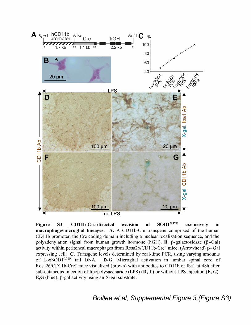
Specificity of Cre expression of CD11b-Cre mice was verified by mating to the
Rosa26 mouse that ubiquitously expresses a !-galactosidase (!-gal) transgene that can be
translated into functional !-gal only if Cre-mediated recombination removes a premature
translation terminator (S17). Fifty-eight percent (± 4.9%) of peritoneal macrophages
(n=3, Fig. S3B) of Rosa26/CD11b-Cre+ mice expressed !-gal, while none did in animals
without the CD11b-Cre gene. Although both neurons and astrocytes from Rosa26 mice
showed high levels of !-gal activity after germ line Cre expression (Fig. 2B), none could
be detected in either of these cell types in mice with the CD11b-encoded Cre (Fig. 2A).
In the lumbar spinal cords of Rosa26/CD11b-Cre+ mice !-gal was expressed only in a
few, small cells (Fig. 2A), identified as microglial cells by the presence of the microglia
specific proteins CD11b or Iba1 (S18) (Fig. S3E, G). Subcutaneous injection into
Rosa26/CD11b-Cre+ mice of lipopolysaccharide (LPS), known to induce microglial
activation and increase CD11b expression (S19, S20) (Fig. S3D, E), produced an increase
in !-gal expressing cells (Fig. S3E). Thus, CD11b-directed Cre expression within the
spinal cord is microglial cell specific and is further induced by activation.


Supplementary Figure Legends
Figure S1. LoxSOD1G37R
transgenic mice develop motor neuron disease from an
SOD1 transgene that can be removed by Cre-mediated recombination.
A. Schematic drawing of the LoxSOD1G37R
transgene. The human SOD1 gene with
mutation G37R was flanked by loxP sequences. B. Spinal cord extract from a
LoxSOD1G37R
mouse immunoblotted with an antibody that recognizes human and mouse
SOD1 with equal affinity (S2). B6, extract from a non-transgenic mouse. C. Onset, early
disease, and survival times of LoxSOD1G37R
mice. D, E. Ventral horn region of the
lumbar spinal cord from 8.5 month old (D) normal and (E) LoxSOD1G37R
mice (stained
with cresyl-violet). F. Numbers of ventral horn motor neurons from age-matched normal
and endstage LoxSOD1G37R
mice. G. Mating scheme for LoxSOD1G37R
and ZP3-Cre
mice. H. Spinal cord extracts from F2 mice in (G) immunoblotted for human and mouse
SOD1. Mutant SOD1 was absent from all F2 mice. Bar in D, E: 50µm.
Figure S2. Quantification of Cre-mediated gene inactivation of mutant SOD1
within motor neurons. A. Histograms of relative intensities of mutant SOD1
fluorescence in individual L5 motor axons measured from three LoxSOD1G37R
(Cre– #1-
3, blue) or LoxSOD1G37R
/Isl1Cre+ (Cre
+ #4-6, red) mice. B. Total accumulated mutant
SOD1 measured as relative total fluorescence intensity in motor axons from entire L5
roots (n=3 for each genotype). Bars indicate standard deviation. C. SOD1G37R
mRNA
levels determined by real-time PCR on reverse-transcribed spinal cord RNA using
various amounts of LoxSOD1G37R
RNA. D. LoxSOD1G37R
mRNA contents (n=2 animals
for each genotype) in laser micro-dissected lumbar motor neurons from
LoxSOD1G37R
/Isl1-Cre+ (Cre
+) and LoxSOD1
G37R (Cre
–) mice using RT followed by real-
time PCR. E, F. Representative immunofluorescence images of anterior horn regions of
lumbar spinal cord from 4 month old presymptomatic LoxSOD1G37R
/Isl1Cre+ (F: Isl1-
Cre+) and LoxSOD1
G37R littermates (E: Isl1-Cre
–), as indicated. Spinal cord sections
were stained with antibodies to human SOD1 (SOD1; green, left) and neurofilaments
(SMI32; red, right) and appropriate fluorescent secondary antibodies. Arrowheads
indicate motor neurons. Bars: 40!m.
Figure S3: CD11b-Cre-directed excision of SOD1G37R
exclusively in
macrophage/microglial lineages. A. A CD11b-Cre transgene comprised of human
CD11b promoter, the Cre coding domain including a nuclear localization sequence, and
the polyadenylation signal from human growth hormone (hGH). B. !-galactosidase
(!#Gal) activity within peritoneal macrophages from Rosa26/CD11b-Cre+ mice.
(Arrowhead) !#Gal expressing cell. C. Transgene levels determined by real-time PCR,
using varying amounts of LoxSOD1G37R
tail DNA. D-G. Microglial activation in lumbar
spinal cord of Rosa26/CD11b-Cre+ mice visualized (brown) with antibodies to CD11b or
Iba1 at 48h after sub-cutaneous injection of lipopolysaccharide (LPS) (D, E) or without
LPS injection (F, G). E, G (blue); !-gal activity using an X-gal substrate.
References for supporting online materials
S1. P. C. Wong et al., Neuron 14, 1105 (1995).
S2. A. M. Clement et al., Science 302, 113 (2003).
S3. D. D. Hickstein, D. M. Baker, K. A. Gollahon, A. L. Back, Proc Natl Acad Sci U
S A 89, 2105 (1992).
S4. S. Srinivas et al., BMC Dev Biol 1, 27 (2001).
S5. T. L. Williamson, D. W. Cleveland, Nat Neurosci 2, 50 (1999).
S6. M. Lewandoski, K. M. Wassarman, G. R. Martin, Curr Biol 7, 148 (1997).
S7. C. S. Lobsiger, M. L. Garcia, C. M. Ward, D. W. Cleveland, Proc Natl Acad Sci
U S A 102, 10351 (Jul 19, 2005).
S8. J. Liu, L. A. Shinobu, C. M. Ward, D. Young, D. W. Cleveland, J Neurochem 93,
875 (2005).
S9. B. Schutz et al., J Neurosci 25, 7805 (Aug 24, 2005).
S10. A. Matsumoto et al., J Neurosci Res 83, 119 (Jan, 2006).
S11. B. K. Kaspar, J. Llado, N. Sherkat, J. D. Rothstein, F. H. Gage, Science 301, 839
(2003).
S12. L. I. Bruijn et al., Neuron 18, 327 (1997).
S13. C. Thery, B. Chamak, M. Mallat, Eur J Neurosci 3, 1155 (1991).
S14. V. Zujovic, J. Benavides, X. Vige, C. Carter, V. Taupin, Glia 29, 305 (2000).
S15. D. S. Howland et al., Proc Natl Acad Sci U S A 99, 1604 (2002).
S16. C. A. Pardo et al., Proc Natl Acad Sci U S A 92, 954 (1995).
S17. P. Soriano, Nat Genet 21, 70 (1999).
S18. Y. Imai, I. Ibata, D. Ito, K. Ohsawa, S. Kohsaka, Biochem Biophys Res Commun
224, 855 (1996).
S19. M. B. Graeber, W. J. Streit, G. W. Kreutzberg, J Neurosci Res 21, 18 (1988).
S20. C. U. Kloss, M. Bohatschek, G. W. Kreutzberg, G. Raivich, Exp Neurol 168, 32
(2001).

Supporting Online Material
www.sciencemag.org
Material and Methods
Supporting Text
Figs. S1, S2, S3
Supporting References