Structural Biology of Proteins & Nucleic...
15
1 Structural Biology of Proteins & Nucleic Acids Dr. Stephen R. Holbrook Staff Scientist Departments of Structural Biology Computational & Theoretical Biology Physical Biosciences Division Lawrence Berkeley National Laboratory Why Structural Biology? Understand the Functions of Biological Macromolecules Determine Evolutionary Relationships Protein (and RNA) Engineering Ligand (Drug) Design
Transcript of Structural Biology of Proteins & Nucleic...

1
Structural Biology of Proteins & Nucleic Acids
Dr. Stephen R. Holbrook
Staff Scientist
Departments of Structural Biology
Computational & Theoretical Biology
Physical Biosciences Division
Lawrence Berkeley National Laboratory
Why Structural Biology?
Understand the Functions ofBiological Macromolecules
Determine Evolutionary Relationships
Protein (and RNA) Engineering
Ligand (Drug) Design

2
Topics to be Covered
Methods
Databases
Classification
Visualization
Modeling
Molecular Interactions/ Docking
Drug Design
Summary
Methods for Determinationof Three-Dimensional Structure
Experimental
X-ray crystallography Multi-dimensional NMR
Computational
• Molecular Modeling
• Fold Prediction/Threading• Ab initio

3
X-Ray Crystallographyof Biological
Macromoleculeshttp://www-structure.llnl.gov/Xray/101index.html
Advantages: Most powerful
Any size (ribosome, viruses)High resolution/overdeterminationWaters, Metals, LigandsDynamics (Thermal parameters)
Disadvantages
• Need large quantities/highly purified• Must crystallize• Can be time consuming
NMR StructureDeterminationof Biological
Macromoleculeshttp://www.cryst.bbk.ac.uk/PPS2/projects/schirra/html/home.htm
Advantages
Data collected in solution Can easily change conditions Do not need to crystallize Dynamics
Disadvantages
• Need milligram quantities (isotopes)• Size limited to < 20-30 kDa• Local information, not global• Lower “resolution”

4
http://www.rcsb.org/pdb/index.html
Databases: RCSB Protein Data Bank
Myosin is composed ofseveral protein chains:two large "heavy"chains and four small"light" chains. Thestructures available inthe PDB, such as the oneshown above, containonly part of the myosinmolecule. In theillustration above, fromPDB entry 1b7t, atomsin the heavy chain arecolored red on the left-hand side, and atoms inthe light chains arecolored orange andyellow. The wholemolecule is much larger,with a long tail that hasbeen clipped off to allowthe molecule to bestudied. Fortunately, thecrystal structuresinclude most of the"motor" domain, thepart of the molecule thatperforms the powerstroke, so we can look atthis process in detail.
Anatomy of a Molecular Muscle
5
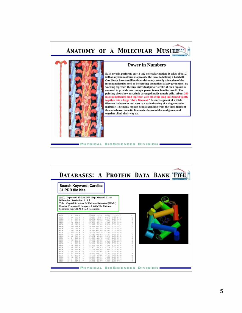
Power in Numbers
Each myosin performs only a tiny molecular motion. It takes about 2trillion myosin molecules to provide the force to hold up a baseball.Our biceps have a million times this many, so only a fraction of themyosin molecules need to be exerting themselves at any given time. Byworking together, the tiny individual power stroke of each myosin issummed to provide macroscopic power in our familiar world. Thepainting shows how myosin is arranged inside muscle cells. About 300myosin molecules bind together, with all of the long tails bound tightlytogether into a large "thick filament." A short segment of a thickfilament is shown in red, next to a scale drawing of a single myosinmolecule. The many myosin heads extending from the thick filamentthen reach over to actin filaments, shown in blue and green, andtogether climb their way up.
Anatomy of a Molecular Muscle
Databases: A Protein Data Bank FileSearch Keyword: Cardiac31 PDB file hits
1DTL Deposited: 12-Jan-2000 Exp. Method: X-rayDiffraction Resolution: 2.15 ÅTitle Crystal Structure Of Calcium-Saturated (3Ca2+)Cardiac Troponin C Complexed With The CalciumSensitizer Bepridil At 2.15 A Resolution
ATOM 1 N TYR A 5 15.850 -8.428 2.760 1.00 41.77 NATOM 2 CA TYR A 5 15.686 -9.919 2.617 1.00 42.13 CATOM 3 C TYR A 5 15.079 -10.614 3.847 1.00 41.62 CATOM 4 O TYR A 5 15.122 -10.097 4.971 1.00 41.33 OATOM 5 CB TYR A 5 17.022 -10.583 2.258 1.00 54.00 CATOM 6 CG TYR A 5 17.061 -11.068 0.834 1.00 56.45 CATOM 7 CD1 TYR A 5 16.046 -11.885 0.323 1.00 57.61 CATOM 8 CD2 TYR A 5 18.107 -10.720 0.000 1.00 57.88 CATOM 9 CE1 TYR A 5 16.081 -12.338 -0.982 1.00 59.55 CATOM 10 CE2 TYR A 5 18.154 -11.166 -1.305 1.00 60.05 CATOM 11 CZ TYR A 5 17.140 -11.979 -1.797 1.00 60.72 CATOM 12 OH TYR A 5 17.191 -12.449 -3.094 1.00 62.68 OATOM 13 N LYS A 6 14.517 -11.792 3.617 1.00 39.40 NATOM 14 CA LYS A 6 13.862 -12.533 4.676 1.00 38.68 CATOM 15 C LYS A 6 14.698 -12.685 5.921 1.00 37.49 CATOM 16 O LYS A 6 14.195 -12.489 7.038 1.00 36.25 OATOM 17 CB LYS A 6 13.429 -13.905 4.166 1.00 82.36 CATOM 18 CG LYS A 6 12.820 -14.819 5.228 1.00 82.36 CATOM 19 CD LYS A 6 13.729 -16.007 5.495 1.00 82.36 CATOM 20 CE LYS A 6 13.136 -16.935 6.543 1.00 82.36 CATOM 21 NZ LYS A 6 14.017 -18.119 6.781 1.00 82.36 NATOM 22 N ALA A 7 15.961 -13.041 5.729 1.00 41.54 NATOM 23 CA ALA A 7 16.867 -13.217 6.851 1.00 40.69 CATOM 24 C ALA A 7 17.090 -11.907 7.598 1.00 39.56 CATOM 25 O ALA A 7 16.991 -11.877 8.838 1.00 37.44 O
6
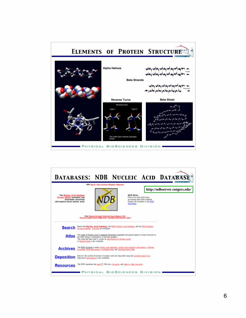
Elements of Protein Structure
Alpha Helices
Reverse Turns
Beta Strands
Beta Sheet
http://ndbserver.rutgers.edu/
Databases: NDB Nucleic Acid Database

7
tRNAPhe
6TNAHammerhead
Ribozyme1MME
HDVRibozyme
1DRZ P4-P6 Domain of
Group I Intron1GID
RNAs, Like Proteins, are Structurally Diverse
http://scop.berkeley.edu/
Classes: SCOP Structural Classes

8
SCOP Structural Hierarchy
Follow thestructuralhierarchy from thegeneral to thespecific
This is the“Fold”Level
SCOP Structural Hierarchy

9
SCOP Structural HierarchySuperfamilies are Specific
The TIM Barrel is a commonProtein fold first observedIn triosephosphate isomerase
SCOP Example: The TIM Barrel

10
SCOP Statistics
1368859564Total
1027252Small Proteins
191711Membrane & CellSurface Proteins
322525Multi-domain
Proteins
345237168Alpha and BetaProteins (a+b)
32315393Alpha and BetaProteins (a/b)
25115887All Beta Proteins
296197128All Alpha Proteins
Number ofsuperfamilies
Number offamilies
Number offoldsClass
http://www.expasy.ch/sprot/sprot-top.html
Tools and Databases

11
http://www.expasy.ch/sitemap.html
A Map to Tools and Databases
Swiss PDBViewer for Your PC (MAC)

12
Swiss PDBViewer Gallery
Building a Protein Model

13
Building a Protein Model
How do you construct a model of a new proteinfrom its amino acid sequence?
1. Identify the closest related protein ofknown structure
2. From the alignment - replace the aminoacid sidechains
3. For deletions - remove the deletedstructure
4. For insertions - Add loops or otherinsertions using structures of similarsequence found in other proteins
5. Energy refine the modeled structure
Procedure
Protein InteractionsProtein-Protein Complex 1DS6Crystal Structure Of A Rac-Rhogdi Protein-DNA Complex 1DMU
Restriction Endonuclease BgliBound to its DNA RecognitionSequence Protein-ligand Complex 1EET
HIV-1 Reverse Transcriptase InComplex With The InhibitorMsc204
14
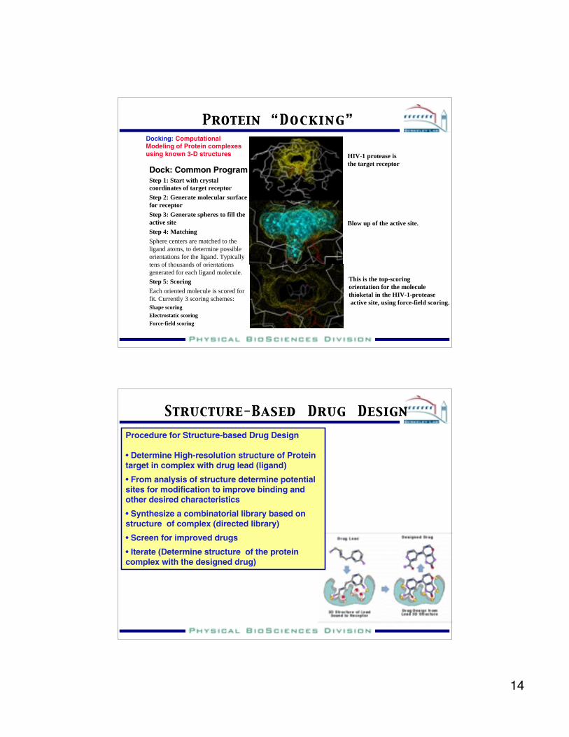
Protein “Docking”Docking: ComputationalModeling of Protein complexesusing known 3-D structures
Dock: Common ProgramStep 1: Start with crystalcoordinates of target receptor
Step 2: Generate molecular surfacefor receptor
Step 3: Generate spheres to fill theactive site
Step 4: Matching
Sphere centers are matched to theligand atoms, to determine possibleorientations for the ligand. Typicallytens of thousands of orientationsgenerated for each ligand molecule.
Step 5: Scoring
Each oriented molecule is scored forfit. Currently 3 scoring schemes:Shape scoringElectrostatic scoringForce-field scoring
Blow up of the active site.
HIV-1 protease isthe target receptor
This is the top-scoring orientation for the molecule thioketal in the HIV-1-protease active site, using force-field scoring.
Structure-Based Drug DesignProcedure for Structure-based Drug Design
• Determine High-resolution structure of Proteintarget in complex with drug lead (ligand)
• From analysis of structure determine potentialsites for modification to improve binding andother desired characteristics
• Synthesize a combinatorial library based onstructure of complex (directed library)
• Screen for improved drugs
• Iterate (Determine structure of the proteincomplex with the designed drug)

15
PDB file 1A30 displayed withSwiss PDB Viewer
HIV-1 protease with tripeptideinhibitor.
AIDS protease inhibitorsare examples of successfulapplications of Structure-based drug design
Structure-Based Drug Design
Summary
•Structural Biology is a broad discipline that attempts todescribe biological function in terms of 3-D MOLECULARSTRUCTURE
•High-throughput structure determination methods are beingdeveloped for crystallography and NMR - Structural genomics
•It is now possible to engineer proteins and nucleic acids fordesired or improved functions
•Structure-based drug design is now yielding medicallyimportant pharmaceuticals