Split Immunity: Immune Inhibition of Rat Gliomas by ... · Split Immunity: Immune Inhibition of Rat...
12
of February 27, 2013. This information is current as Unmodified Live Tumor Cells Gliomas by Subcutaneous Exposure to Split Immunity: Immune Inhibition of Rat Zvi Ram, Irun R. Cohen and Lea Eisenbach Machlenkin, Ofir Goldberger, Felix Mor, Shimon Slavin, Ilan Volovitz, Yotvat Marmor, Meir Azulay, Arthur http://www.jimmunol.org/content/187/10/5452 doi: 10.4049/jimmunol.1003946 October 2011; 2011; 187:5452-5462; Prepublished online 12 J Immunol Material Supplementary 6.DC1.html http://www.jimmunol.org/content/suppl/2011/10/12/jimmunol.100394 References http://www.jimmunol.org/content/187/10/5452.full#ref-list-1 , 13 of which you can access for free at: cites 56 articles This article Subscriptions http://jimmunol.org/subscriptions is online at: The Journal of Immunology Information about subscribing to Permissions http://www.aai.org/ji/copyright.html Submit copyright permission requests at: Email Alerts http://jimmunol.org/cgi/alerts/etoc Receive free email-alerts when new articles cite this article. Sign up at: Print ISSN: 0022-1767 Online ISSN: 1550-6606. Immunologists, Inc. All rights reserved. Copyright © 2011 by The American Association of 9650 Rockville Pike, Bethesda, MD 20814-3994. The American Association of Immunologists, Inc., is published twice each month by The Journal of Immunology at Weizmann Institute of Science on February 27, 2013 http://jimmunol.org/ Downloaded from
Transcript of Split Immunity: Immune Inhibition of Rat Gliomas by ... · Split Immunity: Immune Inhibition of Rat...

of February 27, 2013.This information is current as
Unmodified Live Tumor CellsGliomas by Subcutaneous Exposure to Split Immunity: Immune Inhibition of Rat
Zvi Ram, Irun R. Cohen and Lea EisenbachMachlenkin, Ofir Goldberger, Felix Mor, Shimon Slavin, Ilan Volovitz, Yotvat Marmor, Meir Azulay, Arthur
http://www.jimmunol.org/content/187/10/5452doi: 10.4049/jimmunol.1003946October 2011;
2011; 187:5452-5462; Prepublished online 12J Immunol
MaterialSupplementary
6.DC1.htmlhttp://www.jimmunol.org/content/suppl/2011/10/12/jimmunol.100394
Referenceshttp://www.jimmunol.org/content/187/10/5452.full#ref-list-1
, 13 of which you can access for free at: cites 56 articlesThis article
Subscriptionshttp://jimmunol.org/subscriptions
is online at: The Journal of ImmunologyInformation about subscribing to
Permissionshttp://www.aai.org/ji/copyright.htmlSubmit copyright permission requests at:
Email Alertshttp://jimmunol.org/cgi/alerts/etocReceive free email-alerts when new articles cite this article. Sign up at:
Print ISSN: 0022-1767 Online ISSN: 1550-6606. Immunologists, Inc. All rights reserved.Copyright © 2011 by The American Association of9650 Rockville Pike, Bethesda, MD 20814-3994.The American Association of Immunologists, Inc.,
is published twice each month byThe Journal of Immunology
at Weizm
ann Institute of Science on February 27, 2013http://jim
munol.org/
Dow
nloaded from
The Journal of Immunology
Split Immunity: Immune Inhibition of Rat Gliomas bySubcutaneous Exposure to Unmodified Live Tumor Cells
Ilan Volovitz,*,† Yotvat Marmor,* Meir Azulay,* Arthur Machlenkin,* Ofir Goldberger,*Felix Mor,* Shimon Slavin,‡ Zvi Ram,† Irun R. Cohen,* and Lea Eisenbach*
Gliomas that grow uninhibited in the brain almost never metastasize outside the CNS. The rare occurrences of extracranial me-tastasis are usually associated with a suppressed immune system. This observation raises the possibility that some gliomas might notgrow outside the CNS due to an inherent immune response, We report in this study that the highly malignant F98 Fischer ratundifferentiated glioma, which grows aggressively in the brain, spontaneously regresses when injected live s.c. We found that thisregression is immune-mediated and that it markedly enhances the survival or cures rats challenged with the same tumor intra-cranially either before or after the s.c. live-cell treatment. Adoptive transfer experiments showed the effect was immune-mediatedand that the CD8 T cell fraction, which exhibited direct tumor cytotoxicity, was more effective than the CD4 T cell fraction inmediating resistance to intracranial challenge of naive rats. Brain tumors from treated rats exhibited enhanced CD3+CD8+
CD42 and CD3+CD4+CD82 T cell infiltration and IFN-g secretion. The results in the F98 glioma were corroborated in the Lewisrat CNS-1 astrocytoma. In both tumor models, s.c. treatment with live cells was significantly better than immunization withirradiated cells. We propose in this study a location-based immunotherapeutic phenomenon we term “split immunity”: a tumorthat thrives in an immune-privileged site may be inhibited by injecting live, unmodified tumor cells into a site that is notprivileged, generating protective immunity that spreads back to the privileged site. Split immunity could explain several long-standing paradoxes regarding the lack of overt extracranial metastasis in patients with primary brain tumors. The Journal ofImmunology, 2011, 187: 5452–5462.
T he CNS is known to be an immune-privileged site; tumorallografts and xenografts, rejected at other sites in thebody, are accepted in the CNS in immunocompetent hosts
(1–3), and tumors in the brain are either ignored by the immunesystem or their growth is not controlled (1, 4).A peculiar phenomenon relating to brain tumors, especially to
the highly malignant glioblastoma multiforme (GBM), is the rarityof detectable systemic metastases. Approximately 10 cases ofspontaneous metastasis of unresected primary GBM were reportedin the medical literature in 50 y (5, 6); other cases of metastasisamounting to ,0.5% of patients (7) followed resection of theprimary CNS tumor and occurred mostly in immune-compromisedhosts (8, 9).In this paper, we studied the F98 rat brain tumor that is an
anaplastic glioma, syngeneic to the Fischer (F344) rat, and exhibitsan aggressive infiltrative behavior similar to that of human GBM.The tumor was induced chemically and therefore does not carry anyartificially introduced Ags. This tumor was shown to be refractory
to most therapeutic modalities and is uniformly lethal following aninoculum of as few as 1–10 tumor cells (10, 11).In the current study, nevertheless, we observed that the F98
tumor injected s.c. was rejected in 97% of the rats. This observationsuggested that the spontaneous peripheral rejection of an otherwiselethal brain tumor might be exploited against intracranial tumors.We report in this study that the antitumor immune response gen-erated by the injection of live F98 tumor cells outside the CNS isnot limited to the periphery only, but markedly inhibits the growthof tumors inside the CNS; complete tumor eradication was noted inabout half of the rats.Vaccination is classically defined as the exposure of an in-
dividual to an attenuated or inactivated pathogen (e.g., irradiated,fixed, genetically manipulated, etc.) that protects the host againsta virulent pathogen (12). In this article, we define a location-basedimmune phenomenon we term “split immunity,” a nonattenuatedlive tumor that when situated in one location is lethal to the host,but when situated in another location, the tumor generates im-munity that can spread back to the original site (e.g., the brain),thereby protecting the host.
Materials and MethodsAnimals
Female Fischer (F344) or Lewis rats were obtained from Harlan Israel. Therats were maintained in a specific pathogen-free environment in theWeizmann Institute of Science animal facilities. The rats were used at age6–10 wk unless specified otherwise. The animal experiments were super-vised by institutional animal welfare authorities.
Cells and culture
The F98 undifferentiated glioma is a chemically induced glioma syngeneicto the Fischer rat (13). The cells were obtained from the American TypeCulture Collection and maintained as suggested by the supplier.
The CNS-1 astrocytoma is a chemically induced glioma syngeneic to theLewis rat (14). The CNS-1 line was provided by its originator, William F.
*Department of Immunology, Weizmann Institute of Science, Rehovot 76100, Israel;†Cancer Immunotherapy Laboratory, Department of Neurosurgery, Tel Aviv Soura-sky Medical Center, Tel Aviv 64239, Israel; and ‡International Center for Cell Ther-apy and Cancer Immunotherapy, Weizmann Center, Tel-Aviv 64239, Israel
Received for publication December 2, 2010. Accepted for publication September 7,2011.
This work was partially supported by the Weizmann Institute of Science’s Yeda CEOfund.
Address correspondence and reprint requests to Dr. Ilan Volovitz, Cancer Immuno-therapy Laboratory, Department of Neurosurgery, Tel Aviv Sourasky Medical Center,6 Weizmann Street, Tel Aviv 64239, Israel. E-mail address: [email protected]
The online version of this article contains supplemental material.
Abbreviations used in this article: GBM, glioblastoma multiforme; IC, intracranially;IT, intratumoral; LU30, number of cells that could generate 30% lysis of the 5000target cells; MHC-II, MHC class II; TBI, total body irradiation.
Copyright! 2011 by TheAmericanAssociation of Immunologists, Inc. 0022-1767/11/$16.00
www.jimmunol.org/cgi/doi/10.4049/jimmunol.1003946
at Weizm
ann Institute of Science on February 27, 2013http://jim
munol.org/
Dow
nloaded from

Hickey (Dartmouth Medical Center, Lebanon, NH). The line was grown,as suggested by Prof. Hickey, in RPMI medium supplemented with 4 mML-glutamine, combined antibiotics, and 10% FCS. Care was taken to usesimilar cells of a low passage. All cultures were mycoplasma negative.
F98-luc is a subclone of F98 cells we transfected with the firefly lu-ciferase gene (pcDNA-III; Invitrogen, Carlsbad, CA) using the Lipofect-amine reagent (Invitrogen). The cells were grown in selection medium, theF98medium supplemented with 2mg/ml G-418 antibiotics (Sigma-Aldrich,St. Louis, MO). The F98-luc subclone was selected for its high photonefficiency and ability to express luciferase without a selection agent bothin vitro and in vivo for $3 mo.
Optical imaging of bioluminescent cells
In vivo bioluminescence imaging was performed using the In Vivo ImagingSystem IVIS-100 (Xenogen, Almeda, CA). After i.p. injection of the re-porter probe D-luciferin, 40 mg/kg body weight (Hopkinton, MA), ratswere anesthetized with 80–125 ml 1:2 ratio of xylazine 2% (Vitamed,Benyamina, Israel) and ketamine 100 mg/ml (Fort Dodge, Fort Dodge,IA). The rats were shaved and placed in a supine position in the IVIS(Xenogen) and repeatedly imaged for 2-min intervals until luminescencedeclined.
Bioluminescence was quantified in units of photon flux (P 3 s21) persimilar area measured using the Living Image 2.5 software overlay(Xenogen) and IGOR image analysis software (V 4.09A; WaveMetrics,Lake Oswego, OR). Only metabolically active cells contribute to thebioluminescence obtained by luciferase-transfected cells (15, 16). Bio-luminescence of a different luciferase-transfected F98 clone injected in-tracranially (IC) was shown to be highly correlative to the viable IC tumorsize measured by micro–positron-emission tomography (16) and serialhistological sections (15).
Separation of lymphocytes on MACS magnetic beads
The MACS technique (Miltenyi Biotec, Bergish Gladbach, Germany) wasused to separate CD4+ or CD8+ cell populations for various assays. CD4+-or CD8+-depleted or purified populations were typically .99% free fromunwarranted cells as ascertained by FACS.
In vitro cytotoxicity assays
In vitro cytotoxicity assays (5 and 16 h) were conducted as described before(17). Target cells were labeled overnight with [35S]methionine 5 mCi/ml(PerkinElmer, Waltham, MA). The supernatants were transferred toTopSeal-A, 96-well microplates (Packard Instruments, Downers Grove,IL), added to with 150 ml microscint 40 (Packard Instruments) scintillationfluid, and counted in a TopCount microplate scintillation and luminescenceb-counter (Packard Instruments). An LU30 is defined as the number ofcells that could generate 30% lysis of the 5000 target cells (18). Thenumber is calculated from the regression line of the four different E:Tratios (5:1–40:1). The graph depicts the number of LU30/107 cells.
Coculture proliferation assays
Inactivated tumor cells (irradiated 5000 cGy and treated with mitomycin Cat 80 mg/ml/107 cells) were used as stimulators in coculture proliferationassays. The cells were dispensed in stimulation medium (DMEM, 1%normal rat serum, 2 mM glutamine, combined antibiotics, 1 mM sodiumpyruvate, 5 3 1025 M 2-ME, and 1% nonessential amino acids) inquadruplicates in 96-well plates. Effector cells were either splenocytes(2.53 105 cells/well) or MACS-purified CD4 T cells (2.53 104 cells/well).Where indicated, 5000 cGy-irradiated naive rat thymocytes (2.5 3 105)served as APC.
After 2 d in culture, the wells were pulsed overnight with mediumcontaining methyl [3H]thymidine (5 mCi/ml; GE Healthcare). Cultureswere harvested into a 96 GF/C Unifilter (PerkinElmer) and Microscint-20scintillation fluid (PerkinElmer) added. The plates were read using a Top-count microplate scintillation and luminescence counter (Packard Instru-ments).
IFN-g secretion assays
Two days after coculture of immunocytes with tumor cells, as outlinedabove, assay supernatant from triplicate or quadruplicate microcultures wasassayed by ELISA using standard manufacturer protocols (BD-OptEIA,Franklin Lakes, NJ).
Disruption of IC and s.c. tumor to single cells
Brain and s.c. tumors were surgically removed from rats, cut into smallpieces, and resuspended in HBSS2+ at 200 mg dry tissue/ml. Trypsin 2.5%
(Life Technologies-Invitrogen, Carlsbad, CA) was added at a 1:1 ratio tothe tumor pieces and incubated at 37˚C for 30 min. Cells were then trit-urated using a Pasteur pipette, and the digestion was stopped by addingFCS at a 1:1 ratio. The cell slurry was then passed through a 40-mm cellstrainer (BD Biosciences, Franklin Lakes, NJ) to remove coarse debris.
FACS analysis and Abs
Brain tumor cells were stained and analyzed using a FACS intracellularcytokine secretion protocol similar to Lamoreaux et al. (19). Briefly, excisedand disrupted brain tumor cells were incubated for 5 h with PMA/ionomycin (Sigma-Aldrich) and then stained with ViViD, a violet LIVE/DEAD viability dye (Molecular Probes-Invitrogen). Cells were fixed,permeabilized, and washed using the Fix/Perm kit (BD Biosciences,Franklin Lakes, NJ) and then stained with the following anti rat AbsCD4-PE (W3/25), CD8a-PerCP (OX-8), CD3-Ax488 (1F4), IFN-g–Ax647(DB1) (all from BioLegend, CA). Single color indirect staining was doneusing anti-rat MHC class II (MHC-II)–RT1D monomorphic: (OX-17;Serotec, Oxford, U.K.); anti-rat TCR (R73; BioLegend) and a secondarydonkey anti-mouse Cy5 Ab (Jackson Immunoresearch Laboratories, WestGrove, PA). The cells were monitored by LSRII (BD Biosciences) andanalyzed using FlowJo flow cytometry analysis software (Tree Star).
Tumor cell purification using Percoll density gradientseparation
Density of F98 cells was determined to be 1.06 to 1.07 g/ml by running thetissue-cultured F98 cells in a stepwise Percoll gradient made by overlayingdifferent ratios of isotonic Percoll (9:1 Percoll [Pharmacia-Sigma] to PBS310) mixed with PBS2/2.
Trypsinized tumor cells (from IC or s.c. tumors) were run on a continuousPercoll gradient made by mixing 60% isotonic-Percoll mixed with 40%PBS2/2 run at 26,0003 g for 40 min on a fixed-angle rotor ultracentrifuge(Beckman Coulter, Brea, CA). The single cells were carefully loaded ontoone premade gradient, whereas another identical gradient was loaded withdensity beads (GE Healthcare, Buckinghamshire, U.K.). The two gradientswere then centrifuged at 2600 rpm, for 30 min, 20˚C, brake off. Cellsfound at 1.06 to 1.07 g/ml density were collected and counted for viablecells. The percent of the live cells found at the 1.06 to 1.07 density wascalculated as part of the total live cells found on the entire gradient.Percoll-purified live cells, 1.8 3 105 (30% of total gradient cells), andunsorted live trypsinized cells (6 3 105) consisting of similar amounts ofcells found at the 1.06 to 1.07 g/ml density were both injected s.c. to twogroups of rats to determine growth dynamics.
Inoculation with tumor cells s.c.
Rats were injected s.c. in the lower flank with 100 ml PBS containing live orirradiated F98 tumor cells. The average of two perpendicular diameters ofthe tumor was used to calculate the circular tumor area. Moribund ratsexhibiting signs of severe illness (immobility, difficulty to breathe, etc.) orrats with tumors.3.5 cm in mean diameter were euthanized by CO2. Timeto s.c. tumor rejection was calculated as the number of days between theinitial recording of an observable or palpable tumor to the day the tumorcould no longer be detected.
Inoculation with tumor cells IC
Rats were anesthetized with xylazine and ketamine, and their heads wereshaved and the skin opened to reveal the bregma. A hole was drilled witha microdrill at a point 2 mm anterior to the bregma and 4 mm to the right ofthe midline. The head was fixed in a stereotactic apparatus, and tumor cells(5 3 103, unless specified otherwise), suspended in 2 ml PBS, were slowlyinfused (2 ml/min) at a depth of 4 mm into the skull with a fine Hamiltonsyringe. Following the injection and needle removal, the opened skull wasswabbed with 70% alcohol, and the scalp was closed with 9-mm Autoclipswound clips (Clay Adams, Parsippany, NJ). All surgical procedures wereperformed inside a sterile hood.
Adoptive transfer of splenocytes and tumor challenge
Spleens were harvested from IC-rejector rats. RBCs were lysed using RBClysis buffer (Sigma-Aldrich), and the remaining splenocytes were restim-ulated with inactivated tumor cells at a tumor/splenocyte ratio of 1:50 for3 d in lymphocyte medium.
The rats were challenged either s.c. or IC with tumor cells; the next day(s.c.) or the previous day (IC), the rats were irradiated with total bodyirradiation (TBI) 500 cGy, and, 2 d following irradiation, groups wereinjected i.v. with 107 splenocytes unsorted, MACS depleted of either CD4or CD8 cells, or unsorted naive rat splenocytes. Naive rat splenocytes were
The Journal of Immunology 5453
at Weizm
ann Institute of Science on February 27, 2013http://jim
munol.org/
Dow
nloaded from
not restimulated prior to injection, as repeatedly, hardly any viable naivesplenocytes remained following the 3-d restimulation.
Statistical analysis
A Student’s unpaired two-tailed t test (for two groups) or ANOVA andTukey–Kramer tests (for three or more groups) were used for statisticalanalysis of quantitative normally distributed data in cytotoxicity, prolifer-ation, or cytokine secretion assays. Results in these assays are expressedas mean 6 SEM, unless specified otherwise. Nonparametric survival re-sults were analyzed using the log-rank test. Nonnormal distributed flowcytometric data were analyzed using Mann–Whitney U test.
ResultsGlioma cells grow progressively in the brain but are rejectedspontaneously in an s.c. site
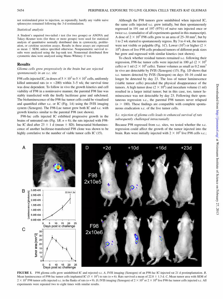
F98 cells injected IC, in doses of 53 102 to 53 105 cells, uniformlykilled untreated rats (n = !200) within 3–5 wk; the survival timewas dose dependent. To follow in vivo the growth kinetics and cellviability of F98 in a noninvasive manner, the parental F98 line wasstably transfected with the firefly luciferase gene and subcloned.The bioluminescence of the F98-luc tumor cells could be visualizedand quantified either s.c. or IC (Fig. 1A) using the IVIS imagingsystem (Xenogen). The F98-Luc tumor grew both IC and s.c. withgrowth kinetics similar to the parental F98 (not shown).F98-luc cells injected IC exhibited progressive growth in the
brains of untreated rats (Fig. 1B; n = 6); the rats injected with F98-luc IC died after 23 6 1 d (mean 6 SD). Intracranial biolumines-cence of another luciferase-transfected F98 clone was shown to behighly correlative to the number of viable tumor cells IC (15).
Although the F98 tumors grew uninhibited when injected IC,the same cells injected s.c. grew initially, but then spontaneouslyregressed in 191 out of 197 (97%) of naive rats injected once ortwice s.c. (cumulative of all experiments quoted in this manuscript).A dose of 2 3 105 F98 cells grew to an area of 25–30 mm2, but by1 to 2 wk started to spontaneously regress. By 3 to 4 wk, the tumorswere not visible or palpable (Fig. 1C). Lower (105) or higher (2 3106) doses of live F98 cells produced tumors of different peak sizesbut grew and regressed with similar kinetics (not shown).To check whether residual tumors remained s.c. following their
regression, F98-luc tumor cells were injected in 100 ml (2 3 105
cells) or 1 ml (2 3 106 cells). Tumor volumes as small as 0.2 mm3
in vivo are detectable by IVIS (Xenogen) (15). Fig. 1D shows thats.c. tumors detected by IVIS (Xenogen) on days 10–16 could nolonger be detected by day 23. The loss of tumor luminescence(viable tumor cells) preceded the physical disappearance of thetumors. A high tumor dose (2 3 106) and inoculum volume (1 ml)resulted in a larger initial tumor; but in this case, too, tumor lu-minescence was not detectable by day 23. Following their spon-taneous regression s.c., the parental F98 tumors never relapsed(n . 180). These findings are compatible with complete sponta-neous eradication s.c. of the live tumor cells.
S.c. rejection of glioma cells leads to enhanced survival of ratssubsequently challenged intracranially
Because F98 regressed from s.c. sites, we tested whether the s.c.regression could affect the growth of the tumor injected into thebrain. Rats were initially injected with 2 3 105 live F98 cells s.c.;
FIGURE 1. F98 glioma cells grow uninhibited IC and rejected s.c. A, IVIS imaging (Xenogen) of an F98-luc IC-injected rat 21 d postimplantation. B,Mean luminescence of F98-luc tumor cells implanted IC (5 3 103) to rats (n = 6). Rats survived a mean of 22.8 6 1.3 d. C, Mean tumor area with SEM of23 105 F98 tumor cells injected s.c. in the flanks of rats (n = 9). D, IVIS imaging (Xenogen) of 23 105 or 23 106 live F98-luc tumor cells injected s.c. Allexperiments were repeated two to eight times with similar results.
5454 PERIPHERAL EXPOSURE TO LIVE GLIOMA CELLS TREATS RAT GLIOMAS
at Weizm
ann Institute of Science on February 27, 2013http://jim
munol.org/
Dow
nloaded from
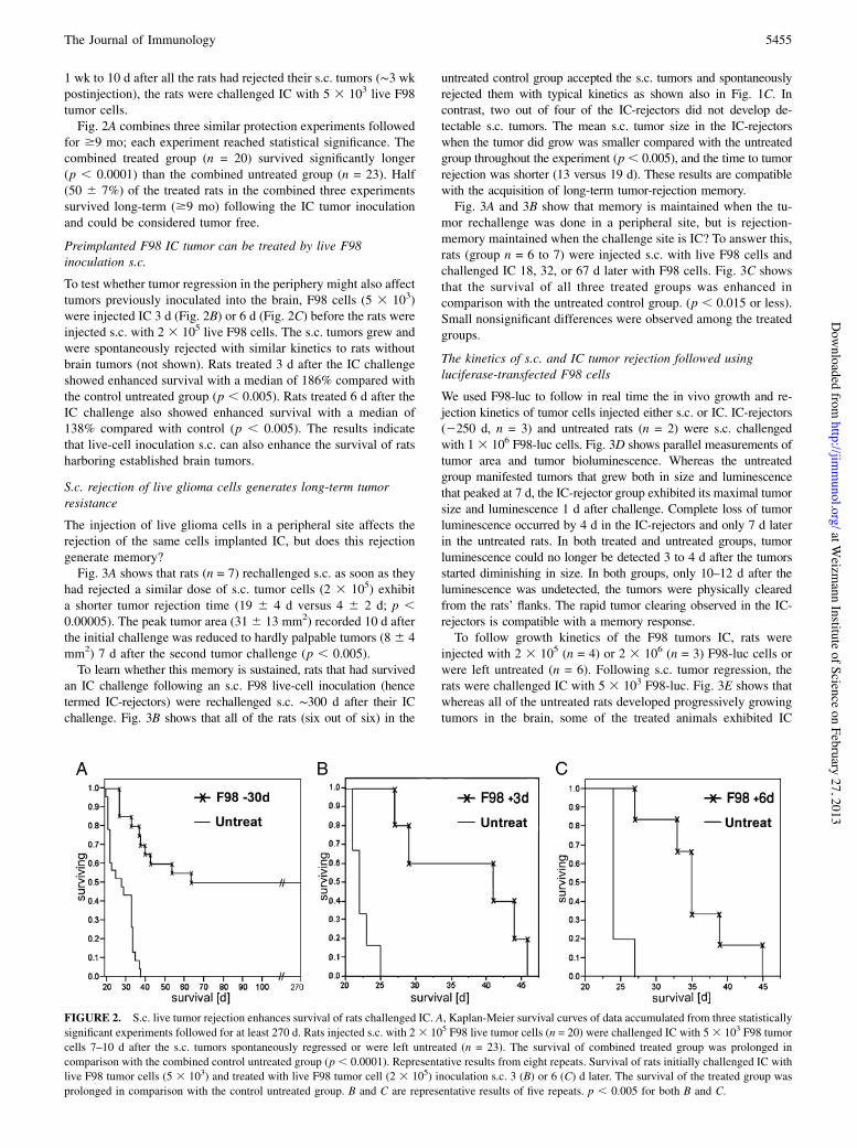
1 wk to 10 d after all the rats had rejected their s.c. tumors (!3 wkpostinjection), the rats were challenged IC with 5 3 103 live F98tumor cells.Fig. 2A combines three similar protection experiments followed
for $9 mo; each experiment reached statistical significance. Thecombined treated group (n = 20) survived significantly longer(p , 0.0001) than the combined untreated group (n = 23). Half(50 6 7%) of the treated rats in the combined three experimentssurvived long-term ($9 mo) following the IC tumor inoculationand could be considered tumor free.
Preimplanted F98 IC tumor can be treated by live F98inoculation s.c.
To test whether tumor regression in the periphery might also affecttumors previously inoculated into the brain, F98 cells (5 3 103)were injected IC 3 d (Fig. 2B) or 6 d (Fig. 2C) before the rats wereinjected s.c. with 2 3 105 live F98 cells. The s.c. tumors grew andwere spontaneously rejected with similar kinetics to rats withoutbrain tumors (not shown). Rats treated 3 d after the IC challengeshowed enhanced survival with a median of 186% compared withthe control untreated group (p , 0.005). Rats treated 6 d after theIC challenge also showed enhanced survival with a median of138% compared with control (p , 0.005). The results indicatethat live-cell inoculation s.c. can also enhance the survival of ratsharboring established brain tumors.
S.c. rejection of live glioma cells generates long-term tumorresistance
The injection of live glioma cells in a peripheral site affects therejection of the same cells implanted IC, but does this rejectiongenerate memory?Fig. 3A shows that rats (n = 7) rechallenged s.c. as soon as they
had rejected a similar dose of s.c. tumor cells (2 3 105) exhibita shorter tumor rejection time (19 6 4 d versus 4 6 2 d; p ,0.00005). The peak tumor area (316 13 mm2) recorded 10 d afterthe initial challenge was reduced to hardly palpable tumors (8 6 4mm2) 7 d after the second tumor challenge (p , 0.005).To learn whether this memory is sustained, rats that had survived
an IC challenge following an s.c. F98 live-cell inoculation (hencetermed IC-rejectors) were rechallenged s.c. !300 d after their ICchallenge. Fig. 3B shows that all of the rats (six out of six) in the
untreated control group accepted the s.c. tumors and spontaneouslyrejected them with typical kinetics as shown also in Fig. 1C. Incontrast, two out of four of the IC-rejectors did not develop de-tectable s.c. tumors. The mean s.c. tumor size in the IC-rejectorswhen the tumor did grow was smaller compared with the untreatedgroup throughout the experiment (p, 0.005), and the time to tumorrejection was shorter (13 versus 19 d). These results are compatiblewith the acquisition of long-term tumor-rejection memory.Fig. 3A and 3B show that memory is maintained when the tu-
mor rechallenge was done in a peripheral site, but is rejection-memory maintained when the challenge site is IC? To answer this,rats (group n = 6 to 7) were injected s.c. with live F98 cells andchallenged IC 18, 32, or 67 d later with F98 cells. Fig. 3C showsthat the survival of all three treated groups was enhanced incomparison with the untreated control group. (p , 0.015 or less).Small nonsignificant differences were observed among the treatedgroups.
The kinetics of s.c. and IC tumor rejection followed usingluciferase-transfected F98 cells
We used F98-luc to follow in real time the in vivo growth and re-jection kinetics of tumor cells injected either s.c. or IC. IC-rejectors(2250 d, n = 3) and untreated rats (n = 2) were s.c. challengedwith 1 3 106 F98-luc cells. Fig. 3D shows parallel measurements oftumor area and tumor bioluminescence. Whereas the untreatedgroup manifested tumors that grew both in size and luminescencethat peaked at 7 d, the IC-rejector group exhibited its maximal tumorsize and luminescence 1 d after challenge. Complete loss of tumorluminescence occurred by 4 d in the IC-rejectors and only 7 d laterin the untreated rats. In both treated and untreated groups, tumorluminescence could no longer be detected 3 to 4 d after the tumorsstarted diminishing in size. In both groups, only 10–12 d after theluminescence was undetected, the tumors were physically clearedfrom the rats’ flanks. The rapid tumor clearing observed in the IC-rejectors is compatible with a memory response.To follow growth kinetics of the F98 tumors IC, rats were
injected with 2 3 105 (n = 4) or 2 3 106 (n = 3) F98-luc cells orwere left untreated (n = 6). Following s.c. tumor regression, therats were challenged IC with 5 3 103 F98-luc. Fig. 3E shows thatwhereas all of the untreated rats developed progressively growingtumors in the brain, some of the treated animals exhibited IC
FIGURE 2. S.c. live tumor rejection enhances survival of rats challenged IC. A, Kaplan-Meier survival curves of data accumulated from three statisticallysignificant experiments followed for at least 270 d. Rats injected s.c. with 23 105 F98 live tumor cells (n = 20) were challenged IC with 53 103 F98 tumorcells 7–10 d after the s.c. tumors spontaneously regressed or were left untreated (n = 23). The survival of combined treated group was prolonged incomparison with the combined control untreated group (p, 0.0001). Representative results from eight repeats. Survival of rats initially challenged IC withlive F98 tumor cells (5 3 103) and treated with live F98 tumor cell (2 3 105) inoculation s.c. 3 (B) or 6 (C) d later. The survival of the treated group wasprolonged in comparison with the control untreated group. B and C are representative results of five repeats. p , 0.005 for both B and C.
The Journal of Immunology 5455
at Weizm
ann Institute of Science on February 27, 2013http://jim
munol.org/
Dow
nloaded from

tumor growth and then regression. In the group injected with 2 3105 live cells s.c., two out of four rats developed tumors that grewslower than those of the untreated rats. The other two out of fourrats in this group reached maximal tumor size by day 7 and thenexhibited gradual loss of tumor luminescence to no luminescenceby days 21–28. The rats treated with the higher dose (2 3 106) oflive cells s.c. had mean IC tumor luminescence by 7 d, similarto that of the untreated rats. The luminescence of their tumorsgradually decreased to no luminescence by 28 d. Significant dif-ferences in tumor luminescence to untreated rats were recordedfor the 2 3 105 treated rats on days 7–21 and for the 2 3 106
treated rats on days 15–21 (p, 0.025 or less). The results indicatethat although all live cell-treated animals supported initial ICgrowth of tumors, tumor growth IC was either delayed or thetumors were rejected.This dose-dependent response observed with F98-luc was noted
also using F98 parental cells; whereas treatment with 2 3 105 F98cells usually produced 50% long-term survivors, treatment with105 cells significantly enhanced survival, but produced consider-ably fewer (range 0–17%) long-term survivors (not shown).
Splenocytes obtained from IC-rejectors mediate tumor cellrejection
The observation of long term-tumor resistance suggests an immunemechanism. To test whether tumor rejection is immune mediated,rats were injected s.c. with live F98 tumor cells and 1 d laterunderwent lymphocyte ablation by TBI 500 cGy to facilitateadoptive transfer (20); 2 d later, the rats were injected i.v. with 107
splenocytes from untreated rats or IC-rejectors.Fig. 4A shows that the group receiving the IC-rejector spleno-
cytes had a significantly smaller mean s.c. tumor area than did thegroup that had received naive rat splenocytes; the rejecting ratsplenocytes restored in three out of four rats the capacity to rejectthe s.c. tumor. In the naive splenocyte-injected group, only one outof four rats ultimately rejected its s.c. tumor.These results indicate that the mild lymphoablation by TBI
compromised the rats’ ability to spontaneously reject a s.c. tumor.The replenishment of the irradiated recipients with IC-rejecting ratsplenocytes, but not with naive splenocytes, restored the rats’ability to reject the s.c. tumor. Similar results were observed usingF98-luc cells (not shown).
FIGURE 3. S.c. F98 rejection generates tumor-rejection memory. A, Rats rejecting a live s.c. tumor (23 105) challenge were rechallenged with live (23105) tumor cell s.c. in the opposite flank. Time to tumor rejection and peak tumor area were reduced in the second versus the first challenge (p , 0.005 orless). Repeated three times with similar results. B, A group of IC-rejectors 300 d post-IC challenge and an untreated group (naive) were challenged s.c. withlive F98 tumor cells (23 105). In the IC-rejector group, only two out of four rats developed tumors versus all six rats developing tumors in the naive group;these tumors were smaller than those recorded in the untreated group (p, 0.005 or less). Repeated twice with similar results. C, Groups of six to seven ratswere either untreated or injected s.c. with 2 3 105 live F98 tumor cells 18, 32, or 67 d prior to IC challenge with 5 3 103 F98 tumor cells. The survival oftreated groups was prolonged in comparison with the untreated control group (p , 0.015 or less). D, A group of three IC-rejectors 250 d following ICchallenge (Reject) and two control untreated rats were challenged s.c. with live F98-luc tumor cells (1 3 106). Tumor area and luminescence weremeasured. Mean tumor area is different between the groups at days 7 and 11 (p , 0.02 or less, t test); the mean tumor luminescence is different at day 7(p , 0.0001). E, Luminescence of the head area of individual rats challenged IC with 5 3 103 F98-luc tumor cells. Rats were either untreated (black line,n = 6) or injected with 2 3 105 (grey triangle, n = 4) or 2 3 106 (grey circle, n = 3) live F98-luc tumor cells s.c. Treated groups exhibit significantly lesstumor luminescence (p , 0.025 or less) on days 7–21 for the group treated with 2 3 105 cells (p , 0.025) and on days 15–21 for group treated with2 3 106 cells (p , 0.025). Repeated twice with similar results.
5456 PERIPHERAL EXPOSURE TO LIVE GLIOMA CELLS TREATS RAT GLIOMAS
at Weizm
ann Institute of Science on February 27, 2013http://jim
munol.org/
Dow
nloaded from

FIGURE 4. T cells from s.c. rejectors mediate tumor cell rejection. A, Rats were injected in the flank with live F98 tumor cells (2 3 105). The next day,the rats received TBI of 500 cGy. Two days later, the groups were injected with splenocytes obtained from an IC-rejector or naive rat. Significantdifferences in five out of seven time points (days 19–37) (p , 0.05 or less). The fraction of tumor-free animals by day 72 is displayed. B, One day afterreceiving TBI 500 cGy, rats were injected IC with live F98 tumor cells (5 3 103). The next day, groups were injected with total splenocytes or CD4/CD8MACS-depleted splenocytes taken from an IC-rejector. Survival of the rejecting-rat splenocytes and CD4-depleted rejecting-rat splenocyte-treated groupswas extended compared with the control group receiving naive-rat splenocytes (p , 0.02, p , 0.005, respectively). C, Tumor-restimulated splenocytesfrom an IC-rejector rat were sorted for CD4 cells (CD4 imm.), CD8 cells (CD8 imm.), or unsorted (Unsorted imm.). These and control naive splenocyteswere assayed for cytotoxicity. Depicted LU30/107 cells calculated from 40:1 up to 5:1 E:T ratio. D, Tumor-restimulated CD4+ splenocytes werecocultured either with the inactivated F98 or CNS-1 cells. Net cpm depicted was calculated by subtracting the thymidine incorporation cpm of the ir-radiated cells from the corresponding group. The proliferation to F98 cells in the CD4 rejecting + APC group was higher than the background pro-liferation to medium (p, 0.05). E, Tumor-restimulated IC-rejector splenocytes (imm.) or MACS-purified CD4/CD8 groups from it and naive splenocytesgroup were cocultured with irradiated tumor cells for cytokine secretion assay. IFN-g secretion to F98 in the Unsorted imm. and CD4 imm. groups washigher than naive (p , 0.05). Secretion of CD4 imm. to F98 was higher than to CNS-1 tumor cells (p , 0.05). F, Rats (five to nine per group) wereinjected s.c. with 63 105 viable single cells obtained from a harvested F98 brain tumor or 1.83 105 cells of the same preparation collected from the 1.06to 1.07 g/ml density of a continuous Percoll density gradient (both preparations contained equal amount of cells from the 1.06 to 1.07 g/ml, a densitypreviously determined to be the density of cultured F98 tumor cells), 2 3 105 of tissue-cultured F98 cells, or left untreated. Following s.c. rejection, allgroups were challenged IC with 5 3 103 F98 cells (tissue cultured). Survival of all treated groups is significantly enhanced versus the untreated group(p , 0.01 or less). G, Percent CTLs (CD3+CD8+CD42) and Th (CD3+CD4+CD82) of total live (ViViD2) cells found IT in brain tumors of live cell-treated (n = 5) and untreated rats (n = 4). Significantly more CTLs are found IT in brain tumors of treated rats than in untreated rats (p, 0.05). H, PercentIFN-g+ CTLs and IFN-g+ Th of total live cells found IT in brain tumors of treated and untreated rats. More IFN-g+ CTLs are found IT in brain tumors oftreated rats than in untreated rats (p = 0.05).
The Journal of Immunology 5457
at Weizm
ann Institute of Science on February 27, 2013http://jim
munol.org/
Dow
nloaded from
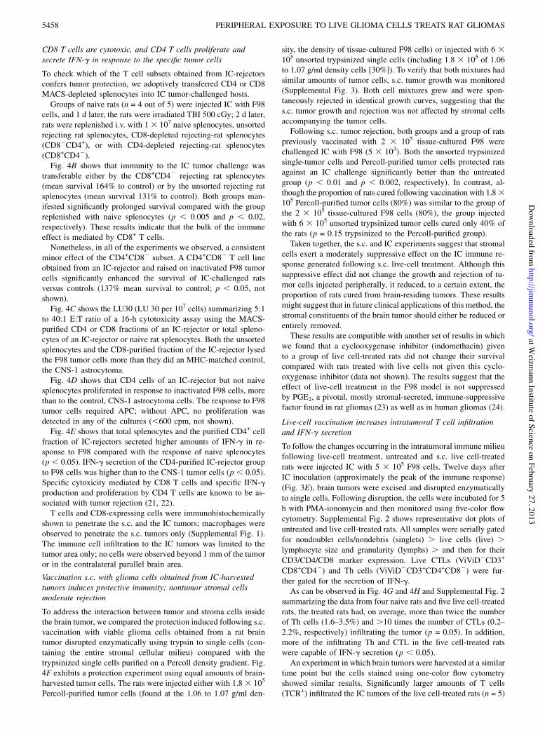
CD8 T cells are cytotoxic, and CD4 T cells proliferate andsecrete IFN-g in response to the specific tumor cells
To check which of the T cell subsets obtained from IC-rejectorsconfers tumor protection, we adoptively transferred CD4 or CD8MACS-depleted splenocytes into IC tumor-challenged hosts.Groups of naive rats (n = 4 out of 5) were injected IC with F98
cells, and 1 d later, the rats were irradiated TBI 500 cGy; 2 d later,rats were replenished i.v. with 13 107 naive splenocytes, unsortedrejecting rat splenocytes, CD8-depleted rejecting-rat splenocytes(CD82CD4+), or with CD4-depleted rejecting-rat splenocytes(CD8+CD42).Fig. 4B shows that immunity to the IC tumor challenge was
transferable either by the CD8+CD42 rejecting rat splenocytes(mean survival 164% to control) or by the unsorted rejecting ratsplenocytes (mean survival 131% to control). Both groups man-ifested significantly prolonged survival compared with the groupreplenished with naive splenocytes (p , 0.005 and p , 0.02,respectively). These results indicate that the bulk of the immuneeffect is mediated by CD8+ T cells.Nonetheless, in all of the experiments we observed, a consistent
minor effect of the CD4+CD82 subset. A CD4+CD82 T cell lineobtained from an IC-rejector and raised on inactivated F98 tumorcells significantly enhanced the survival of IC-challenged ratsversus controls (137% mean survival to control; p , 0.05, notshown).Fig. 4C shows the LU30 (LU 30 per 107 cells) summarizing 5:1
to 40:1 E:T ratio of a 16-h cytotoxicity assay using the MACS-purified CD4 or CD8 fractions of an IC-rejector or total spleno-cytes of an IC-rejector or naive rat splenocytes. Both the unsortedsplenocytes and the CD8-purified fraction of the IC-rejector lysedthe F98 tumor cells more than they did an MHC-matched control,the CNS-1 astrocytoma.Fig. 4D shows that CD4 cells of an IC-rejector but not naive
splenocytes proliferated in response to inactivated F98 cells, morethan to the control, CNS-1 astrocytoma cells. The response to F98tumor cells required APC; without APC, no proliferation wasdetected in any of the cultures (,600 cpm, not shown).Fig. 4E shows that total splenocytes and the purified CD4+ cell
fraction of IC-rejectors secreted higher amounts of IFN-g in re-sponse to F98 compared with the response of naive splenocytes(p , 0.05). IFN-g secretion of the CD4-purified IC-rejector groupto F98 cells was higher than to the CNS-1 tumor cells (p , 0.05).Specific cytoxicity mediated by CD8 T cells and specific IFN-gproduction and proliferation by CD4 T cells are known to be as-sociated with tumor rejection (21, 22).T cells and CD8-expressing cells were immunohistochemically
shown to penetrate the s.c. and the IC tumors; macrophages wereobserved to penetrate the s.c. tumors only (Supplemental Fig. 1).The immune cell infiltration to the IC tumors was limited to thetumor area only; no cells were observed beyond 1 mm of the tumoror in the contralateral parallel brain area.
Vaccination s.c. with glioma cells obtained from IC-harvestedtumors induces protective immunity; nontumor stromal cellsmoderate rejection
To address the interaction between tumor and stroma cells insidethe brain tumor, we compared the protection induced following s.c.vaccination with viable glioma cells obtained from a rat braintumor disrupted enzymatically using trypsin to single cells (con-taining the entire stromal cellular milieu) compared with thetrypsinized single cells purified on a Percoll density gradient. Fig.4F exhibits a protection experiment using equal amounts of brain-harvested tumor cells. The rats were injected either with 1.83 105
Percoll-purified tumor cells (found at the 1.06 to 1.07 g/ml den-
sity, the density of tissue-cultured F98 cells) or injected with 6 3105 unsorted trypsinized single cells (including 1.8 3 105 of 1.06to 1.07 g/ml density cells [30%]). To verify that both mixtures hadsimilar amounts of tumor cells, s.c. tumor growth was monitored(Supplemental Fig. 3). Both cell mixtures grew and were spon-taneously rejected in identical growth curves, suggesting that thes.c. tumor growth and rejection was not affected by stromal cellsaccompanying the tumor cells.Following s.c. tumor rejection, both groups and a group of rats
previously vaccinated with 2 3 105 tissue-cultured F98 werechallenged IC with F98 (5 3 103). Both the unsorted trypsinizedsingle-tumor cells and Percoll-purified tumor cells protected ratsagainst an IC challenge significantly better than the untreatedgroup (p , 0.01 and p , 0.002, respectively). In contrast, al-though the proportion of rats cured following vaccination with 1.83105 Percoll-purified tumor cells (80%) was similar to the group ofthe 2 3 105 tissue-cultured F98 cells (80%), the group injectedwith 6 3 105 unsorted trypsinized tumor cells cured only 40% ofthe rats (p = 0.15 trypsinized to the Percoll-purified group).Taken together, the s.c. and IC experiments suggest that stromal
cells exert a moderately suppressive effect on the IC immune re-sponse generated following s.c. live-cell treatment. Although thissuppressive effect did not change the growth and rejection of tu-mor cells injected peripherally, it reduced, to a certain extent, theproportion of rats cured from brain-residing tumors. These resultsmight suggest that in future clinical applications of this method, thestromal constituents of the brain tumor should either be reduced orentirely removed.These results are compatible with another set of results in which
we found that a cyclooxygenase inhibitor (indomethacin) givento a group of live cell-treated rats did not change their survivalcompared with rats treated with live cells not given this cyclo-oxygenase inhibitor (data not shown). The results suggest that theeffect of live-cell treatment in the F98 model is not suppressedby PGE2, a pivotal, mostly stromal-secreted, immune-suppressivefactor found in rat gliomas (23) as well as in human gliomas (24).
Live-cell vaccination increases intratumoral T cell infiltrationand IFN-g secretion
To follow the changes occurring in the intratumoral immune milieufollowing live-cell treatment, untreated and s.c. live cell-treatedrats were injected IC with 5 3 105 F98 cells. Twelve days afterIC inoculation (approximately the peak of the immune response)(Fig. 3E), brain tumors were excised and disrupted enzymaticallyto single cells. Following disruption, the cells were incubated for 5h with PMA-ionomycin and then monitored using five-color flowcytometry. Supplemental Fig. 2 shows representative dot plots ofuntreated and live cell-treated rats. All samples were serially gatedfor nondoublet cells/nondebris (singlets) . live cells (live) .lymphocyte size and granularity (lymphs) . and then for theirCD3/CD4/CD8 marker expression. Live CTLs (ViViD2CD3+
CD8+CD42) and Th cells (ViViD2CD3+CD4+CD82) were fur-ther gated for the secretion of IFN-g.As can be observed in Fig. 4G and 4H and Supplemental Fig. 2
summarizing the data from four naive rats and five live cell-treatedrats, the treated rats had, on average, more than twice the numberof Th cells (1.6–3.5%) and .10 times the number of CTLs (0.2–2.2%, respectively) infiltrating the tumor (p = 0.05). In addition,more of the infiltrating Th and CTL in the live cell-treated ratswere capable of IFN-g secretion (p , 0.05).An experiment in which brain tumors were harvested at a similar
time point but the cells stained using one-color flow cytometryshowed similar results. Significantly larger amounts of T cells(TCR+) infiltrated the IC tumors of the live cell-treated rats (n = 5)
5458 PERIPHERAL EXPOSURE TO LIVE GLIOMA CELLS TREATS RAT GLIOMAS
at Weizm
ann Institute of Science on February 27, 2013http://jim
munol.org/
Dow
nloaded from
compared with those of untreated rats (n = 5) (p = 0.05, notshown). Single cells obtained from s.c. tumors (n = 3) exhibitedsimilar T cell infiltration frequency as the IC tumors of live cell-treated rats (4 to 5% either, not shown).In this experiment, we also found increased numbers of MHC-
II–expressing cells within the tumors of live cell-treated rats (8versus 5%). MHC-II is expressed on activated microglia and astro-cytes (25) as well as on rat T cells following activation (26).Taken together, the results indicate that there are significantly
more T cells infiltrating brain tumors of live cell-treated rats,whereas there are almost no tumor-infiltrating CTLs in untreatedrats and large numbers of CTLs infiltrate the tumors of live cell-treated rats. The cells found inside the tumors of the treated ratswere also in a more activated state, as observed by the enhancedsecretion of IFN-g by T cells and by wider cellular expression ofMHC-II by tumor stromal cells. Tumor infiltration by T, NK, andNKT cells is a sign of improved prognosis in several human tu-mors (27).
S.c. inoculation with live tumor cells is more effective than s.c.immunization with irradiated tumor cells
Although we show the efficacy of live-cell treatment s.c. against ICtumors, one could argue that irradiated tumor cells are safer to useand might produce similar results when used for vaccination.To check the effects of vaccination s.c. with irradiated cells on
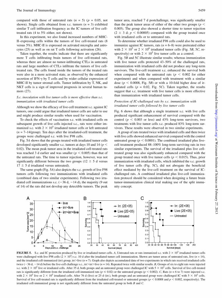
subsequent growth of live cells injected s.c., rats were either im-munized s.c. with 2 3 105 irradiated tumor cells or left untreated(n = 7–14/group). Ten days after the irradiated-cell treatment, thegroups were challenged s.c. with live F98 cells.Fig. 5A shows that the groups treated with irradiated tumor cells
developed significantly smaller s.c. tumors at days 10 and 14 (p ,0.02). The mean peak tumor area in the irradiated cell-treated ratswas reached 3 d earlier and was smaller (p , 0.005) than that ofthe untreated rats. The time to tumor rejection, however, was notsignificantly different between the two groups (12 6 5 d versus17 6 5 d irradiated versus untreated).The same graph (Fig. 5A) also depicts the s.c. growth of live F98
tumors cells following two immunizations with irradiated cells(combined data of two similar experiments). Following two irra-diated cell immunizations s.c. (236 d, 214 d), the majority (9 outof 14) of the rats did not develop any detectible tumors. The peak
tumor area, reached 7 d postchallenge, was significantly smallerthan the peak tumor areas of either of the other two groups (p ,0.005). The group also showed a shorter time to tumor rejection(2 6 3 d; p , 0.00005) compared with the group treated oncewith irradiated cells or to untreated rats.To determine whether irradiated F98 cells could also be used to
immunize against IC tumors, rats (n = 6–8) were pretreated eitherwith 2 3 105 or 2 3 106 irradiated tumor cells (Fig. 5B, 5C, re-spectively) or with 2 3 105 live tumor cells as a control.Fig. 5B and 5C illustrate similar results; whereas immunization
with live tumor cells protected 43–50% of the challenged rats,immunization with irradiated cells did not produce any long-termsurvivors. The live-cell treatment s.c. enhanced the survival bothwhen compared with the untreated rats (p , 0.002 for eitherexperiment) and when compared with treatment with a similardose (p , 0.0008, Fig. 5B) or with a 10-fold higher dose of ir-radiated cells (p = 0.02, Fig. 5C). Taken together, the resultssuggest that s.c. treatment with live tumor cells is more effectivethan immunization with irradiated tumor cells.
Protection of IC-challenged rats by s.c. immunization withirradiated tumor cells followed by live tumor cells
Fig. 6 shows that although a single treatment s.c. with live cellsproduced significant enhancement of survival compared with thecontrol (p , 0.003 or less) and 43% long-term survivors, twotreatments with live tumor cells s.c. produced 83% long-term sur-vivors. These results were observed in two similar experiments.A group of rats treated twice with irradiated cells and then twice
with live cells showed enhanced survival compared with the controluntreated group (p , 0.0001). The combined irradiated plus live-cell treatment produced 88–100% long-term surviving rats in twosimilar experiments. The survival of the irradiated plus live cell-treated group was also significantly enhanced compared with thegroup treated once with live tumor cells (p , 0.015). Thus, priorimmunization with irradiated cells, which inhibited the s.c. growthof live tumor cells (Fig. 5C), did not abrogate the beneficialeffect mediated by the live-cell treatment on the survival of IC-challenged rats. A combined irradiated plus live-cell immuniza-tion protocol should be considered when designing a future braintumor-immunization clinical trial making use of the split immu-nity concept.
FIGURE 5. S.c. and IC protection produced by live or irradiated tumor cells. A, Untreated rats or rats immunized s.c. with 2 3 105 irradiated tumor cellswere challenged with live F98 cells (2 3 105) s.c. 10 d after the irradiated tumor cell immunization. Shown are tumor areas of untreated rats, live (n = 14),and the irradiated cell-immunized (irr) group, irr.live (n = 7). Graph also depicts accumulated data of two experiments in which rats received irradiated cellstwice (236 d,214 d) before the live-cell challenge s.c., irr.irr.live (n = 14). Repeated twice with similar results. B, Groups of six to eight rats were injecteds.c. with 2 3 105 live or irradiated cells. After 35 d, both groups and an untreated group were challenged IC with 5 3 103 cells. Survival of live cell-treatedrats is significantly different from the irradiated cell-immunized rats (p # 0.02) or the untreated groups (p , 0.002). C, Rats (n = 6 to 7) were injected s.c.with 2 3 105 live or 2 3 105 irradiated cells. After 76 d (live) or 25 d (irr.), both groups and an untreated group were challenged IC with 5 3 103 cells.Survival of live cell-treated rats is significantly different from the irradiated cell-treated or untreated groups (p , 0.0008 and p , 0.002, respectively). Theirradiated cell-immunized group is not significantly different from the untreated group in both B and C.
The Journal of Immunology 5459
at Weizm
ann Institute of Science on February 27, 2013http://jim
munol.org/
Dow
nloaded from

DiscussionVaccination, as classically defined, is the immunization of an in-dividual with an attenuated/killed pathogen or to its antigenicdeterminants, leading to resistance to the live pathogen (12). Wedefine in this study a different, location-based immune phenom-enon we term “split immunity.” Split immunity refers to a situa-tion in which a nonattenuated tumor that is lethal in one location,when inoculated in another location, generates immunity that canspread back to the otherwise tolerant site, thus protecting the host.The differences observed in the efficacy of treatment with live
compared with irradiated (attenuated) cells might stem from bothquantitative and qualitative factors. Quantitatively, live tumor cellsmight present their Ags for a longer duration to the immune system(28); moreover, a growing tumor might present larger amounts oftumor Ags to the immune system.Ionizing irradiation has the capacity to induce immunogenic cell
death via membrane-expressed factors (e.g., CRT, HSP90) as wellas by secreted factors (e.g., HMGB1) (27). Tumor irradiation isa standard part of the treatment given to cancer patients, but al-though irradiation was shown to significantly enhance the survivalof high-grade glioma patients, it almost never provides cure (29).Qualitatively, the immune system responds differently to the
different protein repertoires of live cells versus those of dead ordying irradiated tumor cells. Major changes occur in the geneexpression profile of irradiated tumor cells (30–32) and in theirexpression of MHC molecules, costimulatory molecules, adhesionmolecules, death receptors, heat shock proteins, cytokines, andinflammatory mediators (33).Irradiated cells were extensively trialed for the immunotherapy
of most types of human cancers, including gliomas, usually incombination with an adjuvant; these treatments demonstrated poorresults (only 18 out of 531 patients exhibited a complete response in24 trials). This low response rate is similar to those observed in twotrials on primary brain tumors (2 out of 29 patients exhibited acomplete response) (34).Graf et al. (35) showed in four rat glioma models, including the
F98 model, that vaccination of rats with irradiated cells following
an IC challenge mobilizes myeloid suppressor cells into the tumorbed. The myeloid cells inhibited T cell functions, resulting inenhanced tumor growth (35). Chen et al. (36) showed that liveglioma cells that were attenuated by transfection with a membraneform of M-CSF induced a protective immune response in ratschallenged with the parental glioma cells IC. Vaccination withthe transfected cells, freeze-thawed, x-irradiated, or mitomycin Ctreated, did not produce any protective effect (36).The enhanced immunogenicity of live infectious pathogens com-
pared with killed pathogens has been repeatedly shown since theexperiments of Pasteur (37). In this study, too, the vaccination withunmodified live F98 cells exhibited the best overall survival ratesfor this tumor model (38). The exact nature of the differencesbetween the response induced following immunization with livecells versus irradiated cells in the context of split immunity is yetunknown and will be the subject of future research.We have confirmed the split immunity concept in another rat
glioma model, the CNS-1 astrocytoma, a carcinogen-induced tu-mor syngeneic to the Lewis rat (14). Contrary to the initial pub-lications claiming that CNS-1 is “…accepted without rejection innoncentral nervous system sites by Lewis rats,” we saw a smallbut reproducible fraction (!5%, n . 200, not shown) of rats inwhich CNS-1 s.c. tumors spontaneously regressed (14). Moreimportantly, treatment with live CNS-1 cells s.c. 3 d following anIC challenge with CNS-1 cells generated superior survival results(60% of rats surviving .6 mo) compared with immunization withirradiated CNS-1 cells (14% survival) or to nontreatment (0%survival) (Supplemental Fig. 4).How does the rejection of live glioma cells in the periphery
correspond with the well-documented immune suppression in-duced by glioma cells (39, 40)? Intratumoral and intrabrain im-mune suppressive milieu is mediated by a myriad of cells (e.g.,microglia/macrophages) and factors (e.g., TGF-b, PGE2, IL-10,gangliosides, MCP-1, Fas/Fas ligand, B7-H1, RCAS1, and CD70)(40). However, this immune suppression is not absolute. Animaltumor models have shown that peripheral vaccination with at-tenuated glioma cells could induce some effective antigliomaresponses (36, 41). In humans, Tang et al. (42) have shown thathigh-grade (but not low-grade) glioma patients produce a de novorobust CD8 T cell response to their cognate brain tumor cells.Moreover, they have shown that GBM patients exhibit normaladenovirus-specific memory T cell responses and functional den-dritic cells (42). Taken together with the data presented in thispaper, it is reasonable to conclude that the glioma-induced sup-pression is not absolute and does not preclude the ability to pro-duce robust peripherally generated immune responses against thesetumors.Two immune phenomena that share some similarity with split
immunity are concomitant and sinecomitant tumor immunity. Con-comitant immunity describes an immune response in a host witha primary progressive tumor that rejects the same tumor injectedin a different location (usually right and left flanks) (43). Sine-comitant tumor immunity describes a similar phenomenon but,in that case, the primary growing tumor is surgically removedbefore the introduction of the second tumor (44). Split immunitydefines a different phenomenon, a tumor that in one tissue kills thehost, and in another location it generates protective immunity.Although different proportions of the rats spontaneously rejecteds.c. live F98 (!97%) or CNS-1 (!5%) tumor cells, in both cases,peripheral exposure to the tumor cells induced protective immu-nity, whereas the IC inoculation always led to death. Moreover,differently from sine/concomitant immunity, the effective immu-nological information flow described by the split immunity con-cept is unidirectional: the inoculation of brain tumor cells IC
FIGURE 6. Protection from IC glioma by combination of irradiated (irr)and live tumor cell treatment. Groups of six to seven rats were immunizeds.c. with live F98 cells once (238 d) or live cells twice (238 d and 215 d)or with irradiated cells twice and then live cells twice (274 d, 252 d, 238d, and 215 d). The second injection of live cells was done after thecomplete rejection of live tumors s.c. All groups and a control untreatedgroup were challenged IC with 5 3 103 F98 cells (0 d). Kaplan-Meiersurvival curves are shown. Survival of live and the live . live cell-immunized rats are significantly different from the untreated group (p ,0.003 or less). Survival of irr . irr . live . live-treated group is signif-icantly different from the untreated group (p , 0.0001) and from the live-treated group (p , 0.015). Repeated twice with similar results.
5460 PERIPHERAL EXPOSURE TO LIVE GLIOMA CELLS TREATS RAT GLIOMAS
at Weizm
ann Institute of Science on February 27, 2013http://jim
munol.org/
Dow
nloaded from

before or after the s.c. challenge did not change the s.c. tumorgrowth/rejection dynamics, as would be expected if sine/concom-itant tumor immunity were the case.One may ask whether brain tumors generally tend to be rejected
when inoculated outside the brain. A recent review examines theeight most widely used brain tumor models in rat neuro-oncology:C6, 9L, T9, RG2, F98, BT4C, RT-2, and CNS-1 (45). Spontaneousrejection of unattenuated live cells was observed in the C6 (46),the 9L (47), and the T9 tumors (48). We add to this list the F98and, in some cases, the CNS-1 astrocytoma model (14).In 1979, Maat et al. (49) investigated the fate of 77 different
neurocarcinogen-induced CNS and peripheral nervous systemtumors inoculated s.c. as tumor pieces; these brain tumors, orig-inating from WAG/Rij or Sprague Dawley rats, were accepted s.c.by only 62% of syngeneic rat hosts. Most of the s.c. growingtumors were characterized histologically as non-CNS tumors (49).It is difficult to assess why certain brain tumors, such as the F98,are rejected spontaneously s.c. at different rates in the hands ofdifferent researchers (47). It is, however, apparent that whereastumor passage efficiency (i.e., the percent of animals acceptingwithout rejection a syngeneic tumor piece obtained from anotheranimal) in many animal brain tumor models is not 100%, passageefficiency of unmodified non-CNS animal tumors is in most, ifnot in all cases, 100%. Hewitt et al. (50) reported that of 20,000maintenance transplants of 27 different murine tumors passageds.c., none failed, and none regressed.Several explanations were given to the apparent lack of brain
tumor metastasis into peripheral sites (7–9). These explanationscannot account for the paradoxical differences in percentages ofovert metastasis found in the original brain tumor patient: !0.5%(51) compared with 23–46% donor-transmitted brain tumorsgrowing inside organs grafted into organ recipients that werepharmacologically immune suppressed (52).We propose in this study that the introduction of unmodified
CNS tumor cells into areas outside the CNS might in itself be animmunogenic event. This notion is partially supported by theobservation that inoculation into peripheral sites of unmodifiedtissues or cells obtained from immune privileged organs, such asbrain or testis, could induce experimental autoimmune encepha-lomyelitis (53) or orchitis (54), respectively, without the use of anadjuvant.As pointed out above, it is plausible to assume that brain tumor
cells reach peripheral sites in most patients (52). Why then do thesetumor cells not induce natural split immunity that spontaneouslyinhibits the IC brain tumors?We suggest that the induction of effective split immunity requires
peripheral exposure to a larger number of tumor cells than occursduring spontaneous metastasis from a brain tumor. We observedthat low or high doses of live cells inoculated s.c. (105, 23 105, or2 3 106) generate different survival rates (0–17, !50, and !100%,respectively). The s.c. localization of the live-cell treatment mayalso contribute to immunogenicity (55); we observed that i.p. in-jection of brain tumor cells did not induce effective split immunity(not shown).The phenomenon of split immunity might explain another para-
doxical observation: that the duration from diagnosis to death inglioblastoma patients having extracranial metastasis was 18.2 moon average (51), significantly longer (!65%) than the averageexpected life span of 11 mo for glioblastoma patients withoutextracranial metastases (56).High-grade brain tumors are almost uniformly lethal (56), and
no standard treatments appear to extend patient survival by morethan several weeks (57). Our data suggest that the split immunityapproach could be useful to treat tumors developing in immune-
privileged sites. This approach is planned to be translated frombench to bedside in the near future.
AcknowledgmentsWe thank Prof. W.F. Hickey (Dartmouth Medical Center) for sending theCNS-1 astrocytoma. All work was performed at the Weizmann Instituteof Science.
DisclosuresA patent on the split immunity mode of treatment was filed by theWeizmannInstitute of Science and the Tel-Aviv Sourasky Medical Center.
References1. Das, S., J. J. Raizer, and K. Muro. 2008. Immunotherapeutic treatment strategies
for primary brain tumors. Curr. Treat. Options Oncol. 9: 32–40.2. Murphy, J. B., and E. Sturm. 1923. Conditions determining the transplantability
of tissues in the brain. J. Exp. Med. 38: 183–197.3. Sampson, J. H., G. E. Archer, D. M. Ashley, H. E. Fuchs, L. P. Hale, G. Dranoff,
and D. D. Bigner. 1996. Subcutaneous vaccination with irradiated, cytokine-producing tumor cells stimulates CD8+ cell-mediated immunity against tumorslocated in the “immunologically privileged” central nervous system. Proc. Natl.Acad. Sci. USA 93: 10399–10404.
4. Gordon, L. B., S. C. Nolan, H. F. Cserr, P. M. Knopf, and C. J. Harling-Berg.1997. Growth of P511 mastocytoma cells in BALB/c mouse brain elicits CTLresponse without tumor elimination: a new tumor model for regional centralnervous system immunity. J. Immunol. 159: 2399–2408.
5. Chung, Y. H., S. L. Wong, and H. Y. Huang. 1999. Endobronchial metastasis ofglioblastoma multiforme diagnosed by fiberoptic bronchoscopic biopsy. J. For-mos. Med. Assoc. 98: 133–135.
6. Huang, P., A. Allam, A. Taghian, J. Freeman, M. Duffy, and H. D. Suit. 1995.Growth and metastatic behavior of five human glioblastomas compared with nineother histological types of human tumor xenografts in SCID mice. J. Neurosurg.83: 308–315.
7. Schuster, H., K. Jellinger, A. Gund, and H. Regele. 1976. Extracranial metastasesof anaplastic cerebral gliomas. Acta Neurochir. (Wien) 35: 247–259.
8. Bouillot-Eimer, S., H. Loiseau, and A. Vital. 2005. Subcutaneous tumoralseeding from a glioblastoma following stereotactic biopsy: case report and re-view of the literature. Clin. Neuropathol. 24: 247–251.
9. Mourad, P. D., L. Farrell, L. D. Stamps, M. R. Chicoine, and D. L. Silbergeld.2005. Why are systemic glioblastoma metastases rare? Systemic and cerebralgrowth of mouse glioblastoma. Surg. Neurol. 63: 511–519, discussion 519.
10. Clendenon, N. R., R. F. Barth, W. A. Gordon, J. H. Goodman, F. Alam,A. E. Staubus, C. P. Boesel, A. J. Yates, M. L. Moeschberger, R. G. Fairchild,et al. 1990. Boron neutron capture therapy of a rat glioma. Neurosurgery 26: 47–55.
11. Barth, R. F., and B. Kaur. 2009. Rat brain tumor models in experimental neuro-oncology: the C6, 9L, T9, RG2, F98, BT4C, RT-2 and CNS-1 gliomas. J.Neurooncol. 94: 299–312.
12. Paul, W. 1998. Fundumental Immunology. Lippincott Williams & Wilkins,Philadelphia, PA.
13. Ko, L., A. Koestner, and W. Wechsler. 1980. Morphological characterization ofnitrosourea-induced glioma cell lines and clones. Acta Neuropathol. 51: 23–31.
14. Kruse, C. A., M. C. Molleston, E. P. Parks, P. M. Schiltz, B. K. Kleinschmidt-DeMasters, and W. F. Hickey. 1994. A rat glioma model, CNS-1, with invasivecharacteristics similar to those of human gliomas: a comparison to 9L glio-sarcoma. J. Neurooncol. 22: 191–200.
15. Bryant, M. J., T. L. Chuah, J. Luff, M. F. Lavin, and D. G. Walker. 2008. A novelrat model for glioblastoma multiforme using a bioluminescent F98 cell line. J.Clin. Neurosci. 15: 545–551.
16. Cao, F., S. Lin, X. Xie, P. Ray, M. Patel, X. Zhang, M. Drukker, S. J. Dylla,A. J. Connolly, X. Chen, et al. 2006. In vivo visualization of embryonic stem cellsurvival, proliferation, and migration after cardiac delivery. Circulation 113:1005–1014.
17. Mandelboim, O., G. Berke, M. Fridkin, M. Feldman, M. Eisenstein, andL. Eisenbach. 1994. CTL induction by a tumour-associated antigen octapeptidederived from a murine lung carcinoma. Nature 369: 67–71.
18. Shimabukuro, T., and K. Naito. 2008. Tumor-infiltrating lymphocytes derivedfrom human renal cell carcinoma: clonal analysis of its characteristics. Int. J.Urol. 15: 241–244.
19. Lamoreaux, L., M. Roederer, and R. Koup. 2006. Intracellular cytokine opti-mization and standard operating procedure. Nat. Protoc. 1: 1507–1516.
20. Klebanoff, C. A., H. T. Khong, P. A. Antony, D. C. Palmer, and N. P. Restifo.2005. Sinks, suppressors and antigen presenters: how lymphodepletion enhancesT cell-mediated tumor immunotherapy. Trends Immunol. 26: 111–117.
21. Qin, Z., and T. Blankenstein. 2000. CD4+ T cell—mediated tumor rejectioninvolves inhibition of angiogenesis that is dependent on IFN gamma receptorexpression by nonhematopoietic cells. Immunity 12: 677–686.
22. Qin, Z., J. Schwartzkopff, F. Pradera, T. Kammertoens, B. Seliger, H. Pircher,and T. Blankenstein. 2003. A critical requirement of interferon gamma-mediatedangiostasis for tumor rejection by CD8+ T cells. Cancer Res. 63: 4095–4100.
23. Badie, B., J. M. Schartner, A. R. Hagar, S. Prabakaran, T. R. Peebles, B. Bartley,S. Lapsiwala, D. K. Resnick, and J. Vorpahl. 2003. Microglia cyclooxygenase-2
The Journal of Immunology 5461
at Weizm
ann Institute of Science on February 27, 2013http://jim
munol.org/
Dow
nloaded from

activity in experimental gliomas: possible role in cerebral edema formation.Clin. Cancer Res. 9: 872–877.
24. Mitsuhashi, M., J. Liu, S. Cao, X. Shi, and X. Ma. 2004. Regulation ofinterleukin-12 gene expression and its anti-tumor activities by prostaglandin E2derived from mammary carcinomas. J. Leukoc. Biol. 76: 322–332.
25. Neumann, H., J. Boucraut, C. Hahnel, T. Misgeld, and H. Wekerle. 1996.Neuronal control of MHC class II inducibility in rat astrocytes and microglia.Eur. J. Neurosci. 8: 2582–2590.
26. Reizis, B., C. Schramm, I. R. Cohen, and F. Mor. 1994. Expression of majorhistocompatibility complex class II molecules in rat T cells. Eur. J. Immunol. 24:2796–2802.
27. Zitvogel, L., L. Apetoh, F. Ghiringhelli, F. Andre, A. Tesniere, and G. Kroemer.2008. The anticancer immune response: indispensable for therapeutic success? J.Clin. Invest. 118: 1991–2001.
28. Song, S. U., Y. J. Hong, I. S. Oh, Y. Yi, K. B. Choi, J. W. Lee, K. W. Park,J. U. Han, J. K. Suh, and K. H. Lee. 2004. Regeneration of hyaline articularcartilage with irradiated transforming growth factor beta1-producing fibroblasts.Tissue Eng. 10: 665–672.
29. Miller, P. J., R. S. Hassanein, P. G. Giri, B. F. Kimler, P. O’Boynick, andR. G. Evans. 1990. Univariate and multivariate statistical analysis of high-gradegliomas: the relationship of radiation dose and other prognostic factors. Int. J.Radiat. Oncol. Biol. Phys. 19: 275–280.
30. Seidita, G., D. Polizzi, G. Costanzo, S. Costa, and A. Di Leonardo. 2000. Dif-ferential gene expression in p53-mediated G(1) arrest of human fibroblasts aftergamma-irradiation or N-phosphoacetyl-L-aspartate treatment. Carcinogenesis21: 2203–2210.
31. Magner, W. J., and T. B. Tomasi. 2005. Apoptotic and necrotic cells induced bydifferent agents vary in their expression of MHC and costimulatory genes. Mol.Immunol. 42: 1033–1042.
32. Klein, B., D. Loven, H. Lurie, E. Rakowsky, A. Nyska, I. Levin, and T. Klein.1994. The effect of irradiation on expression of HLA class I antigens in humanbrain tumors in culture. J. Neurosurg. 80: 1074–1077.
33. Friedman, E. J. 2002. Immune modulation by ionizing radiation and its impli-cations for cancer immunotherapy. Curr. Pharm. Des. 8: 1765–1780.
34. Neller, M. A., J. A. Lopez, and C. W. Schmidt. 2008. Antigens for cancer im-munotherapy. Semin. Immunol. 20: 286–295.
35. Graf, M. R., J. T. Sauer, and R. E. Merchant. 2005. Tumor infiltration by myeloidsuppressor cells in response to T cell activation in rat gliomas. J. Neurooncol. 73:29–36.
36. Chen, Y., T. Douglass, E. W. Jeffes, Q. Xu, C. C. Williams, N. Arpajirakul,C. Delgado, M. Kleinman, R. Sanchez, Q. Dan, et al. 2002. Living T9 gliomacells expressing membrane macrophage colony-stimulating factor produce im-mediate tumor destruction by polymorphonuclear leukocytes and macrophagesvia a “paraptosis”-induced pathway that promotes systemic immunity againstintracranial T9 gliomas. Blood 100: 1373–1380.
37. Germanier, R. 1984. Oral vaccination against enteric bacterial infections: anoverview. Infection 12: 138–142.
38. Biston, M. C., A. Joubert, J. F. Adam, H. Elleaume, S. Bohic, A. M. Charvet,F. Esteve, N. Foray, and J. Balosso. 2004. Cure of Fisher rats bearing radio-resistant F98 glioma treated with cis-platinum and irradiated with mono-chromatic synchrotron X-rays. Cancer Res. 64: 2317–2323.
39. Okada, H., G. Kohanbash, X. Zhu, E. R. Kastenhuber, A. Hoji, R. Ueda, andM. Fujita. 2009. Immunotherapeutic approaches for glioma. Crit. Rev. Immunol.29: 1–42.
40. Hussain, S. F., D. Yang, D. Suki, E. Grimm, and A. B. Heimberger. 2006. Innateimmune functions of microglia isolated from human glioma patients. J. Transl.Med. 4: 15.
41. Holladay, F. P., R. Choudhuri, T. Heitz, and G. W. Wood. 1994. Generation ofcytotoxic immune responses during the progression of a rat glioma. J. Neuro-surg. 80: 90–96.
42. Tang, J., P. Flomenberg, L. Harshyne, L. Kenyon, and D. W. Andrews. 2005.Glioblastoma patients exhibit circulating tumor-specific CD8+ T cells. Clin.Cancer Res. 11: 5292–5299.
43. Turk, M. J., J. A. Guevara-Patino, G. A. Rizzuto, M. E. Engelhorn, S. Sakaguchi,and A. N. Houghton. 2004. Concomitant tumor immunity to a poorly immu-nogenic melanoma is prevented by regulatory T cells. J. Exp. Med. 200: 771–782.
44. Kashida, T., N. Narasaki, K. Tsujihara, K. Naito, and S. Takeyama. 1998.Augmentation of sinecomitant immunity in mice by gamma-(9H-purine-6-yl)thiomethyl L-glutamate (6-MPG), a water-soluble derivative of 6-mercaptopu-rine. Biol. Pharm. Bull. 21: 16–21.
45. Barth, R. F., and B. Kaur. 2009. Rat brain tumor models in experimental neuro-oncology: the C6, 9L, T9, RG2, F98, BT4C, RT-2 and CNS-1 gliomas. J.Neurooncol. 94: 299–312.
46. Su, M. Y., J. A. Taylor, L. P. Villarreal, and O. Nalcioglu. 2000. Prediction ofgene therapy-induced tumor size changes by the vascularity changes measuredusing dynamic contrast-enhanced MRI. Magn. Reson. Imaging 18: 311–317.
47. Paul, D. B., R. F. Barth, W. Yang, G. H. Shen, J. Kim, and P. L. Triozzi. 2000.B7.1 expression by the weakly immunogenic F98 rat glioma does not enhanceimmunogenicity. Gene Ther. 7: 993–999.
48. Kida, Y., H. Cravioto, G. M. Hochwald, U. Hochgeschwender, and J. Ransohoff.1983. Immunity to transplantable nitrosourea-induced neurogenic tumors. II.Immunoprophylaxis of tumors of the brain. J. Neuropathol. Exp. Neurol. 42:122–135.
49. Maat, B., M. J. van Zwieten, D. W. van Bekkum, and C. Vecht. 1979. Trans-plantability and metastatic potential of chemically induced rat brain tumours.Biomedicine 31: 228–230.
50. Hewitt, H. B., E. R. Blake, and A. S. Walder. 1976. A critique of the evidence foractive host defence against cancer, based on personal studies of 27 murinetumours of spontaneous origin. Br. J. Cancer 33: 241–259.
51. Schweitzer, T., G. H. Vince, C. Herbold, K. Roosen, and J. C. Tonn. 2001.Extraneural metastases of primary brain tumors. J. Neurooncol. 53: 107–114.
52. Buell, J. F., T. M. Beebe, J. Trofe, T. G. Gross, R. R. Alloway, M. J. Hanaway,and E. S. Woodle. 2004. Donor transmitted malignancies. Ann. Transplant. 9:53–56.
53. Stosic-Grujicic, S., Z. Ramic, V. Bumbasirevic, L. Harhaji, and M. Mostarica-Stojkovic. 2004. Induction of experimental autoimmune encephalomyelitis inDark Agouti rats without adjuvant. Clin. Exp. Immunol. 136: 49–55.
54. Itoh, M., H. Terayama, M. Naito, Y. Ogawa, and S. Tainosho. 2005. Tissuemicrocircumstances for leukocytic infiltration into the testis and epididymis inmice. J. Reprod. Immunol. 67: 57–67.
55. Zinkernagel, R. M. 2000. Localization dose and time of antigens determineimmune reactivity. Semin. Immunol. 12: 163–171, discussion 257–344.
56. Ho, I. A., P. Y. Lam, and K. M. Hui. 2004. Identification and characterization ofnovel human glioma-specific peptides to potentiate tumor-specific gene delivery.Hum. Gene Ther. 15: 719–732.
57. Cohen, M. H., J. R. Johnson, and R. Pazdur. 2005. Food and Drug Adminis-tration Drug approval summary: temozolomide plus radiation therapy for thetreatment of newly diagnosed glioblastoma multiforme. Clin. Cancer Res. 11:6767–6771.
5462 PERIPHERAL EXPOSURE TO LIVE GLIOMA CELLS TREATS RAT GLIOMAS
at Weizm
ann Institute of Science on February 27, 2013http://jim
munol.org/
Dow
nloaded from