
Inhibition ofNonspecific Tumoricidal Activity Macrophages ... · INFECTION AND IMMUNITY, July 1981,...
9
Vol. 33, No. 1 INFECTION AND IMMUNITY, July 1981, p. 156-164 0019-9567/81/070156-09$02.00/0 Inhibition of Nonspecific Tumoricidal Activity by Activated Macrophages with Antiserum Against a Soluble Cytotoxic Factor DANIELA N. MANNEL, WERNER FALK,* AND MONTE S. MELTZER Laboratory of Immunobiology, National Cancer Institute, National Institutes of Health, Bethesda, Maryland 20205 Received 12 January 1981/Accepted 7 April 1981 Mouse peritoneal macrophages activated for tumor cytotoxicity by any of several in vivo or in vitro treatments released a soluble cytotoxin into culture fluids only after exposure to small amounts of bacterial lipopolysaccharides. This cytotoxic factor was physicochemically similar to the cytotoxic factor (tumor necrosis factor) in sera of BCG-infected mice injected with lipopolysaccharide. A rabbit antiserum against partially purified serum cytotoxic factor also inhibited the activity of macrophage-derived cytotoxic factor. Of special interest was the observation that rabbit anti-cytotoxic factor inhibited the cytotoxic activity of macrophages both in the presence and in the absence of exogenously added lipopolysaccharide. Inhibition was not complete but was consistent in all experi- ments. Thus, cytotoxic factor was implicated as a possible effector molecule in the nonspecific tumoricidal activity of activated macrophages. The tumor necrotic effects of bacterial lipo- polysaccharides (LPS) have been known for sev- eral decades (2, 23). Recent findings by Carswell et al. showed that this tumor necrosis activity is mediated by a factor found in sera of LPS- treated tumor-bearing animals (1). This factor was also found in sera of BCG-infected mice hours after LPS injection (BCG-LPS serum) (1). That the serum factor also has cytotoxic activity against tumor cells in vitro (but not against normal embryo cells) provides a relatively sim- ple and quantitative assay (1, 5, 8, 12, 20). We have previously shown that a cytotoxic factor (CF) physicochemically similar to that found in BCG-LPS sera can be detected in cul- tures of LPS-treated macrophages from BCG- infected mice (9, 10). This observation suggested that CF may contribute to the cytotoxic activity of activated macrophages (9). We have explored this possibility in this report by using the immunoglobulin G (IgG) fraction from a rabbit antiserum raised against serum- derived CF to define a possible role for this molecule in nonspecific macrophage-mediated tumor cytotoxicity. MATERIALS AND METHODS Mice. Male and female C3H/HeN and C57BL/6N mice, 6 to 12 weeks of age, were obtained from the Division of Research Services, National Institutes of Health. LPS. LPS from Escherichia coli K235 was prepared by the phenol-water extraction method of McIntire et al. (13). Lymphokine supernatant. C3H/HeN mice were immunized intradermally with 5 x 10⁶ viable Myco- bacterium bovis, strain BCG (Phipps substrain, TMC 1029, Trudeau Mycobacterial Collection, Saranac Lake, N.Y.) (22). Animals were sacrificed 3 to 6 weeks after BCG immunization. Spleens were aseptically re- moved and passed through no. 50 mesh stainless-steel sieves into tissue culture medium (RPMI 1640 with antibiotics; GIBCO Laboratories, Grand Island, N.Y.). Single-cell suspensions, obtained by serial aspirations through 19- and 23-gauge needles, were treated with tris(hydroxymethyl)aminomethane-buffered NH4Cl solution to lyse erythrocytes. Spleen cells were centri- fuged at 250 x g for 10 min at 4°C and suspended to a concentration of 5 x 10⁶ viable cells/ml in medium with 5% heat-inactivated fetal calf serum (GIBCO). Twenty-milliliter portions of the spleen cell suspension with 50 to 100 ltg of purified protein derivative (Con- nought Medical Research Laboratories, Toronto, Can- ada) per ml were incubated in upright 75-cm2 plastic tissue culture flasks (no. 3023; Falcon Plastics, Oxnard, Calif.) for 48 to 60 h at 37°C. Supernatant fluids were pooled, centrifuged at 450 x g for 15 min at 40C, and stored at -20°C until used. Preparation of cytotoxic serum. Mice were in- jected intravenously with 2 x 10⁶ viable BCG and injected intravenously 14 days later with 10,ug of LPS. Two hours after LPS injection, animals were exsan- guinated, and the serum (BCG-LPS serum) was pre- pared (8). Control serum was obtained from LPS- injected normal mice in a similar manner. All sera were stored at -20°C until used. Antisera. Female rabbits (New Zealand white) 156 at REGENSBURG on July 15, 2009 iai.asm.org Downloaded from
Transcript of Inhibition ofNonspecific Tumoricidal Activity Macrophages ... · INFECTION AND IMMUNITY, July 1981,...

Vol. 33, No. 1INFECTION AND IMMUNITY, July 1981, p. 156-1640019-9567/81/070156-09$02.00/0
Inhibition of Nonspecific Tumoricidal Activity by ActivatedMacrophages with Antiserum Against a Soluble Cytotoxic
FactorDANIELA N. MANNEL, WERNER FALK,* AND MONTE S. MELTZER
Laboratory ofImmunobiology, National Cancer Institute, National Institutes of Health, Bethesda,Maryland 20205
Received 12 January 1981/Accepted 7 April 1981
Mouse peritoneal macrophages activated for tumor cytotoxicity by any ofseveral in vivo or in vitro treatments released a soluble cytotoxin into culturefluids only after exposure to small amounts of bacterial lipopolysaccharides. Thiscytotoxic factor was physicochemically similar to the cytotoxic factor (tumornecrosis factor) in sera of BCG-infected mice injected with lipopolysaccharide. Arabbit antiserum against partially purified serum cytotoxic factor also inhibitedthe activity of macrophage-derived cytotoxic factor. Of special interest was theobservation that rabbit anti-cytotoxic factor inhibited the cytotoxic activity ofmacrophages both in the presence and in the absence of exogenously addedlipopolysaccharide. Inhibition was not complete but was consistent in all experi-ments. Thus, cytotoxic factor was implicated as a possible effector molecule inthe nonspecific tumoricidal activity of activated macrophages.
The tumor necrotic effects of bacterial lipo-polysaccharides (LPS) have been known for sev-eral decades (2, 23). Recent findings by Carswellet al. showed that this tumor necrosis activity ismediated by a factor found in sera of LPS-treated tumor-bearing animals (1). This factorwas also found in sera of BCG-infected micehours after LPS injection (BCG-LPS serum) (1).That the serum factor also has cytotoxic activityagainst tumor cells in vitro (but not againstnormal embryo cells) provides a relatively sim-ple and quantitative assay (1, 5, 8, 12, 20).We have previously shown that a cytotoxic
factor (CF) physicochemically similar to thatfound in BCG-LPS sera can be detected in cul-tures of LPS-treated macrophages from BCG-infected mice (9, 10). This observation suggestedthat CF may contribute to the cytotoxic activityof activated macrophages (9).We have explored this possibility in this report
by using the immunoglobulin G (IgG) fractionfrom a rabbit antiserum raised against serum-derived CF to define a possible role for thismolecule in nonspecific macrophage-mediatedtumor cytotoxicity.
MATERIALS AND METHODSMice. Male and female C3H/HeN and C57BL/6N
mice, 6 to 12 weeks of age, were obtained from theDivision of Research Services, National Institutes ofHealth.LPS. LPS from Escherichia coli K235 was prepared
by the phenol-water extraction method of McIntire etal. (13).Lymphokine supernatant. C3H/HeN mice were
immunized intradermally with 5 x 10⁶ viable Myco-bacterium bovis, strain BCG (Phipps substrain, TMC1029, Trudeau Mycobacterial Collection, SaranacLake, N.Y.) (22). Animals were sacrificed 3 to 6 weeksafter BCG immunization. Spleens were aseptically re-moved and passed through no. 50 mesh stainless-steelsieves into tissue culture medium (RPMI 1640 withantibiotics; GIBCO Laboratories, Grand Island, N.Y.).Single-cell suspensions, obtained by serial aspirationsthrough 19- and 23-gauge needles, were treated withtris(hydroxymethyl)aminomethane-buffered NH4Clsolution to lyse erythrocytes. Spleen cells were centri-fuged at 250 x g for 10 min at 4°C and suspended toa concentration of 5 x 10⁶ viable cells/ml in mediumwith 5% heat-inactivated fetal calf serum (GIBCO).Twenty-milliliter portions of the spleen cell suspensionwith 50 to 100ltg of purified protein derivative (Con-nought Medical Research Laboratories, Toronto, Can-ada) per ml were incubated in upright 75-cm2 plastictissue culture flasks (no. 3023; Falcon Plastics, Oxnard,Calif.) for 48 to 60 h at 37°C. Supernatant fluids werepooled, centrifuged at 450 x g for 15 min at 40C, andstored at -20°C until used.
Preparation of cytotoxic serum. Mice were in-jected intravenously with 2 x 10⁶ viable BCG andinjected intravenously 14 days later with 10,ug of LPS.Two hours after LPS injection, animals were exsan-guinated, and the serum (BCG-LPS serum) was pre-pared (8). Control serum was obtained from LPS-injected normal mice in a similar manner. All serawere stored at -20°C until used.
Antisera. Female rabbits (New Zealand white)156
at RE
GE
NS
BU
RG
on July 15, 2009 iai.asm
.orgD
ownloaded from

CYTOTOXIC FACTOR AND MACROPHAGE CYTOTOXICITY 157
were obtained from the Division of Research Services,National Institutes of Health. Animals were immu-nized with BCG-LPS serum-derived tumor CF par-
tially purified by Sephacryl S200 and diethylamino-ethyl-Sephacel (Pharmacia Fine Chemicals, Inc., Pis-cataway, N.J.) chromatography or with an equivalentfraction from control serum. Rabbits were injectedsubcutaneously in complete Freund adjuvant as de-scribed (8, 9). Each immunizing preparation containedabout 25 mg of protein per injection. The animals wereboosted with the same antigen preparations subcuta-neously in complete Freund adjuvant twice at 3 and 8weeks and again at periodic intervals without Freundadjuvant. Rabbits were bled from the ear artery, andserum was prepared. All antisera were heat inactivated(30 min at 56°C), and the proteins were precipitatedwith 50% saturated ammonium sulfate. Precipitateswere dissolved and extensively dialyzed against phos-phate-buffered saline (PBS). Heat-inactivated serum
was applied on protein A-Sepharose CL-4B (Phar-macia) at pH 7, and IgG was eluted with glycine bufferat pH 3. This IgG fraction was extensively dialyzedagainst Dulbecco minimal essential medium (DMEM).Normal rabbit IgG was obtained from Cappel Labo-ratories, Inc., Downingtown, Pa.
Peritoneal cells. Mice were treated intraperito-neally (i.p.) 1 day previously with 1 ml of PBS or 7days previously with: (i) 2 x 10⁶ viable BCG; (ii) 500ag of pyran copolymer (National Cancer Institute,Bethesda, Md.); (iii) 1.4 mg of killed Corynebacteriumparvum (Wellcome Research Laboratories, Becken-ham, England); (iv) 2% starch suspension (ConnoughtMedical Research Laboratories); or (v) 1:1,000 suspen-sion of latex beads (1-,um polystyrene beads; DowChemical Co., Midland, Mich.) in 0.5 ml of PBS each.Peritoneal exudate cells (PEC) were collected after i.p.injection of 8 to 10 ml of DMEM as described (21).This medium contained 2 g of NaHCO3 and 4.5 g ofglucose per liter, 10% fetal calf serum, and 50 ,ug ofgentamicin per ml. Peritoneal fluid was withdrawnthrough the anterior wall through a 19-gauge needle.Fluids from 3 to 10 mice were pooled, a sample was
taken for differential and total cell counts, and theremainder was centrifuged in polypropylene tubes (no.2074; Falcon Plastics) at 250 x g for 10 min at 4°C.Differential counts were made on Wright-stained cell
smears prepared by cytocentrifugation (Cytospin cen-
trifuge; Shandon Southern Instruments, Camberley,England). Washed PEC suspensions from each pool ofmice were adjusted to equal macrophage concentra-tions in medium used for cell collection. Macrophageswere exposed to dilutions of lymphokine supernatantsas adherent PEC for 6 h at 37°C in 5% C02 in moistair.CF assay. Tumor cells (mouse L929, American
Type Culture Collection, Rockville, Md., unless indi-cated otherwise) at 4 x 10³ cells per 6.4-mm culturewell (Costar 96, Cambridge, Mass.) were labeled in 0.1ml of Eagle minimal essential medium (EMEM) with25 mM N-2-hydroxyethylpiperazine-N'-2-ethanesul-fonic acid (HEPES) buffer, 5% heat-inactivated fetalcalf serum, and 0.5 MuCi of [methyl-3H]thymidine per
ml ([3H]TdR; specific activity, 6.7 Ci/mmol; New Eng-land Nuclear Corp., Boston, Mass.) for 18 to 24 h at37°C in 5% CO2 in moist air (7). Tumor cell monolayers
were washed twice after labeling and incubated withdilutions of cytotoxic samples in 0.2 ml of DMEM.After 48 h, labeled tumor cell monolayers lysed with0.5% sodium dodecyl sulfate (SDS) in water were usedto estimate total incorporated [3H]TdR. Cytotoxicitywas estimated by measuring release of [3H]TdR fromprelabeled tumor cells in duplicate cultures and ex-pressed as mean counts per minute + standard errorof the mean (SEM) or as percentage of SDS totalcounts.Macrophage cytotoxicity assay. A modified pro-
tocol of the cytotoxicity assay described by Meltzer etal. (15) was used. Tumor 1023 was induced in a C3H/HeNicr male mouse with pellets of 1% 3-methylchol-anthrene in paraffin. Target cell lines were establishedfrom enzyme-digested tumor fragments and weremaintained in culture with EMEM supplemented with10% fetal calf serum. [3H]TdR-prelabeled tumor cellswere obtained from washed and trypsin-digestedmonolayers (2 x 10⁶ to 3 x 10⁶ viable cells/75-cm2culture flask in 20 ml of EMEM with 25 mM HEPESbuffer, 5% fetal calf serum, and 1 M,Ci of [3H]TdR perml for 18 to 24 h at 370C in 5% C02 in moist air). Sixthousand tumor cells were added to 6.4-mm culturewells (Costar 96) with in vivo- or in vitro-activatedadherent PEC in a total volume of 0.2 ml of DMEM.After 48 h, labeled tumor cell monolayers lysed with0.5% SDS in water were used to estimate total incor-porated counts. Cytotoxicity was estimated by mea-surement of release of [3H]TdR from prelabeled tumorcells in duplicate cultures and expressed as meancounts per minute ± SEM or percentage of SDS totalcounts.
RESULTS
Correlation between macrophage tumor-icidal activity and release of CF. A solubleCF physicochemically similar to that found inBCG-LPS sera is detected in culture fluids ofLPS-treated macrophages from BCG-infectedanimals (9, 10). To determine whether CF playsa role in the nonspecific tumoricidal activity ofactivated macrophages in vitro, we attempted tocorrelate levels ofmacrophage tumoricidal activ-ity with release of CF into culture fluids (Table1). Macrophages from mice treated i.p. withviable BCG, killed C. parvum, or pyran copoly-mer were strongly cytotoxic to tumor cells invitro; cells from mice treated with PBS, starch,or latex beads had little or no cytotoxic activity.Levels of CF released by any of these cytotoxicor noncytotoxic macrophages, however, werenegligible. These observations confirm those inmany previous reports which failed to detectsoluble cytotoxins in cultures of activated mac-rophages (see reference 6 for review).The pattern of CF release by macrophage
cultures incubated with small amounts of LPS,however, was entirely different from that de-scribed above. Cultures of activated macro-phages from three different sources (BCG-, C.
VOL. 33, 1981
at RE
GE
NS
BU
RG
on July 15, 2009 iai.asm
.orgD
ownloaded from
158 MANNEL, FALK, AND MELTZER
TABLE 1. Correlation between macrophage tumoricidal activity and release of cytotoxic factora
Macrophages from mice treated Macrophage tumoricidal Release of CFbwith: activity -LPS +LPS
BCG 2,240 ± 130 (26) 470 ± 20 (11) 1,410 ± 60 (33)Pyran 2,380 ± 250 (27) 640 ± 10 (15) 1,600 ± 110 (38)C. parvum 2,020 ± 130 (23) 560 ± 20 (13) 1,640 ± 80 (39)PBS 770± 70 (9) 340 ± 30 (8) 480 ± 60 (11)Starch 620 ± 20 (7) 360 ± 80 (8) 360 ± 60 (8)Latex 790± 10 (9) 340 ± 40 (8) 400 ± 60 (9)No macrophages 680 ± 70 (8) 220 ± 70 (5)SDS total counts 8,740 ± 700 (100) 4,250 ± 170 (100)
a Adherent PEC from mice treated i.p. with 2 x 10⁶ viable BCG, 500 ,ug of pyran, 1.4 mg of C. parvum, 2%starch in water, or latex beads 7 days previously or with PBS 1 day previously were incubated with [3H]TdR-prelabeled tumor cells. Cytotoxicity (released [3H]TdR) was estimated at 48 h and expressed as mean countsper minute ± SEM for duplicate cultures and as percentage of SDS total counts (in parentheses).
b For release of CF, the cytotoxic activity of 2-h supernatants from 10' adherent PEC was tested on [3H]TdR-prelabeled L929 cells. Cytotoxicity (released [3H]TdR) was estimated at 48 h and expressed as mean counts perminute ± SEM for duplicate cultures and as percentage of SDS total counts (in parentheses).
parvum-, or pyran-treated mice) incubated with10 ng ofLPS per ml released significant levels ofCF. LPS alone (data not shown) or culture fluidsfrom LPS-treated inflammatory macrophageshad little or no cytotoxic activity (Table 1).Even though detectable levels of CF were
evident only in culture fluids of activated mac-rophages treated with an extraordinary stimulussuch as LPS, CF could still contribute to thecytotoxic effects of these cells in the absence ofLPS. For example, CF bound to macrophages orreleased only in the macrophage-tumor cell mi-croenvironment would not be detected in culturefluids. To examine this alternative, we at-tempted to inhibit CF activity with a partiallypurified and specific rabbit antiserum.Inhibition of the cytotoxic activity in
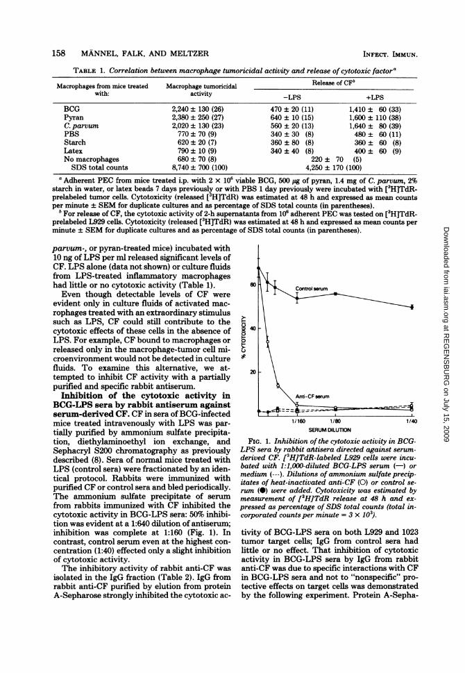
BCG-LPS sera by rabbit antiserum againstserum-derived CF. CF in sera of BCG-infectedmice treated intravenously with LPS was par-tially purified by ammonium sulfate precipita-tion, diethylaminoethyl ion exchange, andSephacryl S200 chromatography as previouslydescribed (8). Sera of normal mice treated withLPS (control sera) were fractionated by an iden-tical protocol. Rabbits were immunized withpurified CF or control sera and bled periodically.The ammonium sulfate precipitate of serumfrom rabbits immunized with CF inhibited thecytotoxic activity in BCG-LPS sera: 50% inhibi-tion was evident at a 1:640 dilution of antiserum;inhibition was complete at 1:160 (Fig. 1). Incontrast, control serum even at the highest con-centration (1:40) effected only a slight inhibitionof cytotoxic activity.The inhibitory activity of rabbit anti-CF was
isolated in the IgG fraction (Table 2). IgG fromrabbit anti-CF purified by elution from proteinA-Sepharose strongly inhibited the cytotoxic ac-
40
20-
Anti-CF serum
1/160 1/80 1/40SERUM DILUTION
FIG. 1. Inhibition of the cytotoxic activity in BCG-LPS sera by rabbit attisera directed against serum-derived CF. [3H]TdR-labeled L929 cells were incu-bated with 1:1,000-diluted BCG-LPS serum (-) ormedium (---). Dilutions of ammonium sulfate precip-itates of heat-inactivated anti-CF (0) or control se-rum (0) were added. Cytotoxicity was estimated bymeasurement of [3H]TdR release at 48 h and ex-pressed as percentage of SDS total counts (total in-corporated counts per minute = 3 x 10³).
tivity of BCG-LPS sera on both L929 and 1023tumor target cells; IgG from control sera hadlittle or no effect. That inhibition of cytotoxicactivity in BCG-LPS sera by IgG from rabbitanti-CF was due to specific interactions with CFin BCG-LPS sera and not to "nonspecific" pro-tective effects on target cells was demonstratedby the following experiment. Protein A-Sepha-
INFECT. IMMUN.
at RE
GE
NS
BU
RG
on July 15, 2009 iai.asm
.orgD
ownloaded from
CYTOTOXIC FACTOR AND MACROPHAGE CYTOTOXICITY 159
rose columns were exposed to equal concentra-tions of IgG from anti-CF and control sera.When BCG-LPS serum was passed over theanti-CF column, all cytotoxic activity was re-tained. In contrast, the titer of cytotoxic activityin BCG-LPS serum was unchanged after passageover the control serum column (Table 3).Effect of rabbit anti-CF on the tumorici-
dal activity ofactivated macrophages. Mac-rophages activated in vivo by the effects ofBCGinfection or activated in vitro by lymphokineswere cultured with tumor target cells in thepresence of IgG from sera of immunized (anti-CF), control (control sera), and untreated rab-bits (Fig. 2). Anti-CF exerted a significant inhib-itory effect on macrophage-mediated cytotoxic-ity with both BCG- and lymphokine-activatedcells. Inhibition was most evident at the lowereffector/target cell ratios. IgG from sera of un-treated rabbits had no effect on macrophagetumoricidal activity. In contrast, IgG from seraof control rabbits (animals immunized with serafrom normal mice injected with LPS) actuallyenhanced tumor cytotoxicity. Since both targetcells and mouse serum were ofC3H/HeN origin,enhancing activity in control serum could havebeen mediated by rabbit anti-C3H/HeN anti-bodies not present in sera of untreated rabbits.In fact, absorption of all rabbit sera with 1023target cells reduced this enhancement withoutaffecting the inhibitory effects of anti-CF (Fig.3).The inhibitory effects of anti-CF on tumor
cytotoxicity by activated macrophages was in-dependent of mouse strain and activation stim-ulus (Table 4). Effects of anti-CF were, however,dependent upon the effector/target cell ratioand the amount of anti-CF added during thecytotoxicity assay. Inhibition of macrophage tu-moricidal activity was not evident at effector/target cell ratios greater than or equal to 15:1 to20:1 or with less than 0.25 ,ug of anti-CF IgG perml (data not shown). This latter effect was notdue to loss by absorption or destruction of anti-body activity during the 48-h cytotoxicity assay.The titer of anti-CF in fluids of BCG-activatedmacrophage-tumor cell cultures (at 20:1 and 7:1effector/target cell ratios) remained unchangedthrough 48 h.Detectable levels of CF in fluids of activated
macrophage cultures are found only after expo-sure of cells to small amounts of LPS. CF isreleased within 2 h of LPS treatment. If LPS-treated macrophages are washed after this 2-hinterval, the cells remain cytotoxic but CF can-not be detected in culture fluids again through48 h (8). It is possible that anti-CF inhibits onlythe initial, LPS-dependent event. Later cyto-
VOL. 33, 1981
cocq0
-H -H -
-H -H CO0000 0
t-CO
10t- 10
o _- _9
eq eq eq
000
0-H
co
-8m-H
0eq8ct1
O
010
0C-CDa-
0-
U
._
00
0g
U
._
0
0
r.
0U
U40
01
0
A
0
C.
wU'4
i0
0
20
2
2204
0
2E
4
m
,20
.0
.0.0L.
0
144
.0
1..
Cc
C2
0
C..'0
C.)
..0
.5
.144
cc
OaH4
00 -
60,
0 0
CD
E.400
=. 0
o o
(1c.0.0
0~1" 0
-H
6,°0
*0 14s
cou2 =S
0'
E20 E.0.0 X
6ID
.=. z
1- M6-J
0_ _ _
r- -o --4
-H -H -H +1 +..
000001
+1 H+11+1 +1
CD CD CD CD CD
eq eq
+14 i+1 +1-.eq
aO C4
cq
o o o oC9CD c
-H8
t'
8
0.
0
0:=laC6.)
004=
sr _8 coO
.........._- _- _-- _- _- z
at RE
GE
NS
BU
RG
on July 15, 2009 iai.asm
.orgD
ownloaded from

160 MANNEL, FALK, AND MELTZER
TABLE 3. Absorption of cytotoxic activity from BCG-LPS sera by rabbit anti-CF bound to protein A-Sepharose
CytotoxicityEluate'
Ani-F gGProtein A coated with BfeEluateaAnti-CF IgG control IgG Buffer
1:4,000 590 ± 60 (13) 770 ± 190 (17) 790 ± 60 (18)1:2,000 590 ± 60 (13) 920 ± 160 (21) 910 ± 130 (20)1:1,000 650 ± 60 (14) 1,240 ± 210 (28) 1,210 ± 40 (27)1:500 610 ± 50 (14) 1,580 ± 50 (35) 1,680 ± 130 (37)Spontaneous release 600 ± 40 (13)SDS total counts 4,480 ± 240 (100)
a One milliliter samples of protein A-Sepharose CL-4B were each coated with buffer, 5 mg of anti-CF IgG, or5 mg of control IgG. Samples of 10 p1 of BCG-LPS serum were applied to the columns, and 5 ml of the eluatewas collected. [3H]TdR-labeled L929 celis were incubated with different dilutions of the eluates. Cytotoxicitywas estimated by [3H]TdR release at 48 h and expressed as mean counts per minute ± SEM for duplicatecultures and as percentage of SDS total counts (in parentheses). More than 50% of the cytotoxic activity fromBCG-LPS serum applied was recovered from the uncoated column (buffer).
Contro IgG
p Anti-~xCF Ig
PBS-Macrages+ LWr#vkines
BCG-Macophages
5:1 7:1EFFECTOR: TARGET
FIG. 2. Inhibition of macrophage tumctivity by IgG from rabbit anti-CF. Adhfrom mice treated i.p. with PBS 1 day preitreated in vitro with lymphokines (1:5 dilidium) for 6 h or adherent PEC from micewith 2 x 10⁶ viable BCG 7 days previincubated at different effector/target[3H]TdR-labeled tumor cells. The cells wein medium (O), rabbit IgG (U), anti-CFcontrol IgG (-). Final IgG concentrationlml. Cytotoxicity was estimated by [3H]Td)
toxic mechanisms may be unrelated to this earlyevent and thus may not be inhibited by anti-CF.
;4<To test this possibility, we examined effects ofanti-CF on tumor cytotoxicity by BCG-activatedmacrophages cultured with and without LPS for2 h. All cultures were then washed and incubatedwith anti-CF and target cells (Fig. 4). Anti-CFinhibited the cytotoxic activity of both LPS-treated and nontreated BCG-activated macro-
IG phages. The degrees of inhibition were compa-rable.
It is important to note that the measurementof macrophage-mediated tumor cytotoxicity byrelease of [3H]TdR used in these studies under-estimates the extent of macrophage-tumor cellinteraction. Macrophage-mediated cytostatic ef-fects are not measured by this assay. Moreover,
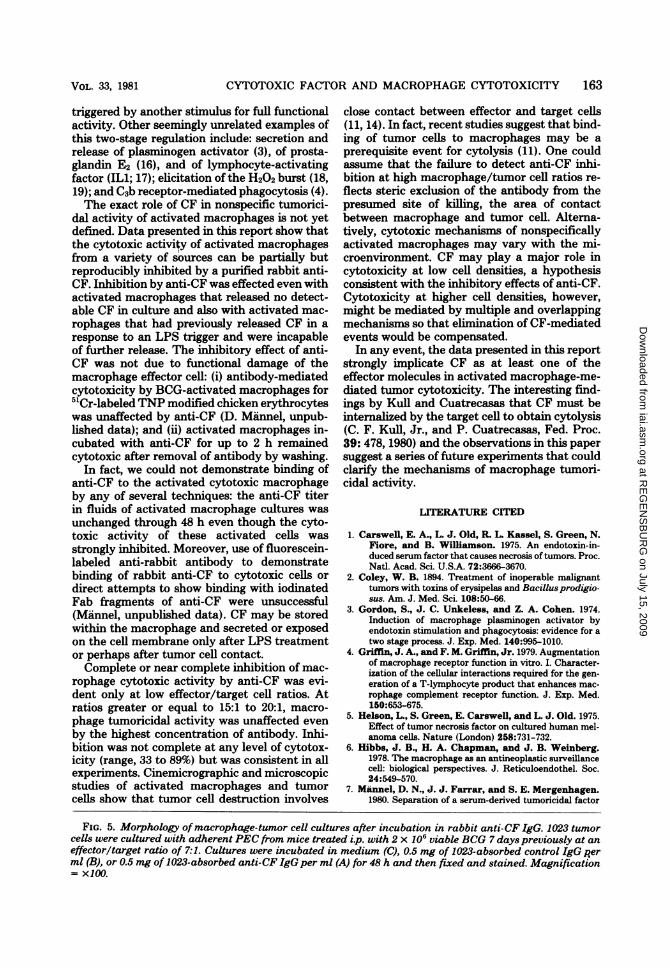
A4 dead tumor cells that have not yet autolyzedand released nuclear [3H]TdR by 48 h are like-wise not measured (15). Culture morphology(Fig. 5) demonstrates the striking effects of anti-CF on activated macrophage-tumor cell inter-actions. Cultures incubated in medium aloneshowed the strong cytopathic effects of activatedmacrophages on tumor cells (bottom). The celldensity did not increase over 48 h. Morphologyof cultures incubated with IgG from control or
-.'----- normal rabbit sera was comparable to that of9:1 cultures incubated in medium alone (middle). In
contrast, cell density of cultures in the presence)ricidal ac- of anti-CF was very high (top) and similar to theerent PEC cell density obtained when noncytotoxic residentviously and macrophages 'were cultured with tumor cells..uted in me- Since only tumor cells are able to grow underinfected lv.P these conditions, increased cell density can be
tzosy Werith attributed only to tumor cell growth.ere culturedIgG (0), orwas 0.5 mglR release at
48 h and expressed aspercentage ofSDS total counts(total incorporated counts per minute = 6 x 10³;spontaneous release = 7%).
40k
201-
0
aR40
20 -
INFECT. IMMUN.
at RE
GE
NS
BU
RG
on July 15, 2009 iai.asm
.orgD
ownloaded from

CYTOTOXIC FACTOR AND MACROPHAGE CYTOTOXICITY 161
DISCUSSION
Data presented in this report demonstrate fora variety of in vivo and in vitro treatments thatmacrophages activated for nonspecific tumor cy-totoxicity are also able to release soluble cyto-toxic factor after exposure to small amounts(nanograms) of LPS. Activation in vivo or invitro, however, was not sufficient for CF release.
60 r-4Contrc IgG
/ MediumAbsorbed
/ W control IgG,/ 1
Anti-CF IgGAbsbedanti-CF IgG
Over a broad range of experimental conditions,LPS was always necessary. Moreover, after theinitial LPS-induced release ofCF from activatedmacrophages, further release ofCF with or with-out LPS was not evident (8).These observations suggest that release of CF
from activated macrophages is yet another ex-ample ofmacrophage effector function regulatedby two stages: cells must be primed by onestimulus to enter into an inactive but receptivestage in which they can then respond or be
30 F
20I-
10
3:1 5:1 7:1 9:1EFFECTOR: TARGET
FIG. 3. Inhibition of macrophage tumoricidal ac-
tivity by anti-CF from rabbit sera absorbed with 1023fibrosarcoma cells. Two milliliters of IgG solutions(1 mg/ml) was absorbed three times with 25 x 10i1023 cells each for 30 min at 0°C. Adherent PECfrommice treated i.p. with 2 x 10⁶ viable BCG 7 dayspreviously were incubated at different effector/targetratios with [3H]TdR-labeled tumor cells. The cellswere cultured in medium (0), unabsorbed (---) or
absorbed (-) anti-CF IgG (0), or control IgG (0).Final IgG concentration was 0.5 mg/ml. Cytotoxicitywas estimated by [3H]TdR release at 48 h and ex-
pressed as percentage ofSDS total counts (total in-corporated counts per minute = 3.5 x 10³; sponta-neous release = 14%).
0' l
[=~----5:1 7:1
EFFECTOR: TARGET9:1
FIG. 4. Anti-CF inhibition of macrophage tumori-cidal activity after treatment with LPS. AdherentPEC of mice treated i.p. with 2 x 10⁶ viable BCG 7days previously were incubated in medium (-) or inmedium containing 10 ng ofLPSper ml (---) for 2 h.The cells were washed three times and cultured with[3H]TdR-labeled tumor cells in medium (P) or ab-sorbed anti-CFIgG (0). Final IgG concentration was0.5 mg/ml. Cytotoxicity was estimated by [3H]TdRrelease at 48 h and expressed as percentage of SDStotal counts (total incorporated counts per minute =
3 x 10³; spontaneous release = 5%).
TABLE 4. Inhibition ofmacrophage cytotoxicity by IgG against serum-derived CF'Cytotouicity
Mouse strain StimulusAnti-CF IgG Medium
C3H/HeN BCG 480 ± 50 (40) 1,060 ± 40 (89)Pyran 560 ± 30 (47) 1,030± 90 (86)C. parvum 770 ± 30 (64) 1,070 ± 80 (90)
C57BL/6N BCG 470 ± 40 (40) 780 ± 100 (65)Spontaneous release 170 ± 10 (14)SDS total counts 1,200 ± 90 (100)
'Adherent PEC from mice treated i.p. with 2 x 10⁶ viable BCG, 500 pg of pyran, or 1.4 mg of C. parvum 7days previously were incubated with [3H]TdR-labeled tumor cells at a 10:1 effector/target ratio. Cytotoxicitywas estimated by [3H]TdR release at 48 h and expressed as mean counts per minute ± SEM for duplicatecultures and as percentage of SDS total counts (in parentheses).
40 k0
0
20F-
VOL. 33, 1981
at RE
GE
NS
BU
RG
on July 15, 2009 iai.asm
.orgD
ownloaded from
CYTOTOXIC FACTOR AND MACROPHAGE CYTOTOXICITY 163
triggered by another stimulus for full functionalactivity. Other seemingly unrelated examples ofthis two-stage regulation include: secretion andrelease of plasminogen activator (3), of prosta-glandin E2 (16), and of lymphocyte-activatingfactor (IL1; 17); elicitation of the H202 burst (18,19); and C3b receptor-mediated phagocytosis (4).The exact role of CF in nonspecific tumorici-
dal activity of activated macrophages is not yetdefined. Data presented in this report show thatthe cytotoxic activity of activated macrophagesfrom a variety of sources can be partially butreproducibly inhibited by a purified rabbit anti-CF. Inhibition by anti-CF was effected even withactivated macrophages that released no detect-able CF in culture and also with activated mac-rophages that had previously released CF in aresponse to an LPS trigger and were incapableof further release. The inhibitory effect of anti-CF was not due to functional damage of themacrophage effector cell: (i) antibody-mediatedcytotoxicity by BCG-activated macrophages for51Cr-labeled TNP modified chicken erythrocyteswas unaffected by anti-CF (D. Mannel, unpub-lished data); and (ii) activated macrophages in-cubated with anti-CF for up to 2 h remainedcytotoxic after removal of antibody by washing.
In fact, we could not demonstrate binding ofanti-CF to the activated cytotoxic macrophageby any of several techniques: the anti-CF titerin fluids of activated macrophage cultures wasunchanged through 48 h even though the cyto-toxic activity of these activated cells wasstrongly inhibited. Moreover, use of fluorescein-labeled anti-rabbit antibody to demonstratebinding of rabbit anti-CF to cytotoxic cells ordirect attempts to show binding with iodinatedFab fragments of anti-CF were unsuccessful(Mannel, unpublished data). CF may be storedwithin the macrophage and secreted or exposedon the cell membrane only after LPS treatmentor perhaps after tumor cell contact.Complete or near complete inhibition of mac-
rophage cytotoxic activity by anti-CF was evi-dent only at low effector/target cell ratios. Atratios greater or equal to 15:1 to 20:1, macro-phage tumoricidal activity was unaffected evenby the highest concentration of antibody. Inhi-bition was not complete at any level of cytotox-icity (range, 33 to 89%) but was consistent in allexperiments. Cinemicrographic and microscopicstudies of activated macrophages and tumorcells show that tumor cell destruction involves
close contact between effector and target cells(11, 14). In fact, recent studies suggest that bind-ing of tumor cells to macrophages may be aprerequisite event for cytolysis (11). One couldassume that the failure to detect anti-CF inhi-bition at high macrophage/tumor cell ratios re-flects steric exclusion of the antibody from thepresumed site of killing, the area of contactbetween macrophage and tumor cell. Alterna-tively, cytotoxic mechanisms of nonspecificallyactivated macrophages may vary with the mi-croenvironment. CF may play a major role incytotoxicity at low cell densities, a hypothesisconsistent with the inhibitory effects of anti-CF.Cytotoxicity at higher cell densities, however,might be mediated by multiple and overlappingmechanisms so that elimination of CF-mediatedevents would be compensated.
In any event, the data presented in this reportstrongly implicate CF as at least one of theeffector molecules in activated macrophage-me-diated tumor cytotoxicity. The interesting find-ings by Kull and Cuatrecasas that CF must beinternalized by the target cell to obtain cytolysis(C. F. Kull, Jr., and P. Cuatrecasas, Fed. Proc.39: 478, 1980) and the observations in this papersuggest a series of future experiments that couldclarify the mechanisms of macrophage tumori-cidal activity.
LITERATURE CITED
1. Carswell, E. A., L J. Old, R. L. Kassel, S. Green, N.Fiore, and B. Williamson. 1975. An endotoxin-in-duced serum factor that causes necrosis of tumors. Proc.Natl. Acad. Sci. U.S.A. 72:3666-3670.
2. Coley, W. B. 1894. Treatment of inoperable malignanttumors with toxins of erysipelas and Bacillus prodigio-sus. Am. J. Med. Sci. 108:50-66.
3. Gordon, S., J. C. Unkeless, and Z. A. Cohen. 1974.Induction of macrophage plasminogen activator byendotoxin stimulation and phagocytosis: evidence for atwo stage process. J. Exp. Med. 140:995-1010.
4. Griffin, J. A., and F. M. Griffin, Jr. 1979. Augmentationof macrophage receptor function in vitro. I. Character-ization of the cellular interactions required for the gen-eration of a T-lymphocyte product that enhances mac-rophage complement receptor function. J. Exp. Med.150:653-675.
5. Helson, L., S. Green, E. Carswell, and L. J. Old. 1975.Effect of tumor necrosis factor on cultured human mel-anoma cells. Nature (London) 258:731-732.
6. Hibbs, J. B., H. A. Chapman, and J. B. Weinberg.1978. The macrophage as an antineoplastic surveillancecell: biological perspectives. J. Reticuloendothel. Soc.24:549-570.
7. Mannel, D. N., J. J. Farrar, and S. E. Mergenhagen.1980. Separation of a serum-derived tumoricidal factor
FIG. 5. Morphology ofmacrophage-tumor cell cultures after incubation in rabbit anti-CF IgG. 1023 tumorcells were cultured with adherent PEC from mice treated i.p. with 2 x 10' viable BCG 7 days previously at aneffector/target ratio of 7:1. Cultures were incubated in medium (C), 0.5 mg of 1023-absorbed control IgG perml (B), or 0.5 mg of 1023-absorbed anti-CF IgGper ml (A) for 48 h and then fixed and stained. Magnification= X100.
VOL. 33, 1981
at RE
GE
NS
BU
RG
on July 15, 2009 iai.asm
.orgD
ownloaded from

164 MANNEL, FALK, AND MELTZER
from a helper factor for plaque-forming cells. J. Immu-nol. 124:1106-1110.
8. Mannel, D. N., M. S. Meltzer, and S. E. Mergenhagen.1980. Generation and characterization of a lipopolysac-charide-induced and serum-derived cytotoxic factor fortumor cells. Infect. Immun. 28:204-211.
9. Mannel, D. N., R. N. Moore, and S. E. Mergenhagen.1980. Macrophages as a source of tumoricidal activity(tumor necrosis factor). Infect. Immun. 30:523-530.
10. Mannel, D. N., R. N. Moore, and S. E. Mergenhagen.1980. Endotoxin-induced tumor cytotoxic factor, p. 141-143. In D. Schlessinger (ed.), Microbiology-1980.American Society for Microbiology, Washington D.C.
11. Marino, P. A., and D. 0. Adams. 1980. Interaction ofBacillus Calmette-Guerin-activated macrophages andneoplastic cells in vitro: II. The relationship of selectivebinding to cytolysis. Cell. Immunol. 54:26-35.
12. Matthews, N., and J. F. Watkins. 1978. Tumour-necro-sis factor from the rabbit. I. Mode of activation, speci-ficity and physicochemical properties. Br. J. Cancer 38:302-309.
13. McIntire, F. C., H. W. Sievart, G. H. Barlow, R. A.Finley, and A. Y. Lee. 1967. Chemical, physical andbiological properties of lipopolysaccharide from Esche-richia coli K-235. Biochemistry 6:2363-2372.
14. Meltzer, M. S., R. W. Tucker, and A. C. Breuer. 1975.Interaction of BCG-activated macrophages with neo-plastic and nonneoplastic cell lines in vitro: cinemicro-graphic analysis. Cell. Immunol. 17:30-42.
15. Meltzer, M. S., R. W. Tucker, K. K. Sanford, and E.J. Leonard. 1975. Interaction of BCG-activated mac-rophages with neoplastic and nonneoplastic cell lines invitro: quantitation of the cytotoxic reaction by releaseof tritiated thymidine from prelabeled target cells. J.Natl. Cancer Inst. 54:1177-1184.
16. Meltzer, M. S., L. M. Wahl, E. J. Leonard, and C. A.
Nacy. 1980. Macrophage activation by lymphokines.Characterization of lymphokine signals for tumoricidaland microbicidal activities and for prostaglandin syn-thesis, p. 167-168. In A. L. de Weck (ed.), Proceedingsof the Second International Workshop. Academic Press,Inc., New York.
17. Mizel, S. B., and D. L. Rosenstreich. 1979. Regulationof lymphocyte-activation factor (LAF). Production andsecretion in P388D, cells: identification of high molec-ular weight precursor of LAF. J. Immunol. 122:2173-2179.
18. Nathan, C. F., L. H. Brukner, S. C. Silverstein, and Z.A. Cohn. 1979. Extracellular cytolysis by activatedmacrophages and granulocytes. I. Pharmacologic trig-gering of effector cells and the release of hydrogenperoxide. J. Exp. Med. 149:84-99.
19. Nathan, C. F., and R. K. Root. 1977. Hydrogen peroxiderelease from mouse peritoneal macrophages. Depend-ence on sequential activation and triggering. J. Exp.Med. 146:1648-1662.
20. Ostrove, J. M., and G. E. Gifford. 1979. Stimulation ofRNA synthesis in L929 cells by rabbit tumor necrosisfactor. Proc. Soc. Exp. Biol. Med. 160:354-358.
21. Ruco, L P., and M. S. Meltzer. 1978. Macrophage acti-vation for tumor cytotoxicity: development of macro-phage cytotoxic activity requires completion of a se-quence of short-lived intermediary reactions. J. Immu-nol. 121:2035-2042.
22. Ruco, L. P., and M. S. Meltzer. 1978. Macrophage acti-vation for tumor cytotoxicity: increased lymphokineresponsiveness of peritoneal macrophages, during acuteinflammation. J. Immunol. 120:1054-1062.
23. Shear, M. J. 1944. Chemical treatment of tumors. IX.Reactions of mice with primary subcutaneous tumorsto injection of a hemorrhage producing bacterial poly-saccharide. J. Natl. Cancer Inst. 4:461-476.
INFECT. IMMUN.
at RE
GE
NS
BU
RG
on July 15, 2009 iai.asm
.orgD
ownloaded from