MRI in treatment of adult gliomas - PBworks
9
Review The introduction of MRI into clinical practice has been among the most important of all advances in the care of patients with brain tumours. The crucial roles of neuro- imaging in neuro-oncology include refinement of preoperative differential diagnosis, precise anatomical localisation for operative planning (figure 1), detection of response to treatment and of tumour progression, and recognition of side-effects that are treatment related. However, there are many unsolved difficulties in the use of MRI in neuro-oncology. New techniques that allow analysis of the chemical composition of tumour tissue, capillary density, and the diffusion of water, have great potential but are not yet well validated. The specialty of molecular imaging is expanding rapidly, although only in the preclinical setting. Here we review the status of these techniques and other issues in neuro-oncology. Correlation between imaging appearance and histological features of gliomas Diffuse astrocytomas of adult cerebral hemispheres are unique among tumours in human beings for the scale to which their imaging features relate to clinical behaviour and histopathological characteristics. In low-grade, diffuse, fibrillary astrocytomas (WHO grade II; figure 2) there is mild expansion of the affected brain without evidence of abnormal gadolinium enhancement or substantial surrounding vasogenic oedema. These imaging findings indicate the histopathology of mildly increased cellularity without pronounced angiogenesis. In anaplastic astrocytomas (WHO grade III) there are typically nodular areas of gadolinium enhancement, showing the presence of newly formed blood vessels that have an abnormal blood–brain barrier. These tumours tend to produce moderate expansion of the involved brain regions, because of greater cellular proliferation, and might show evidence of substantial surrounding vasogenic oedema. In glioblastoma multiforme (WHO grade IV) there is pronounced mass effect and heterogeneous enhancement with centrally non- enhancing regions, which typically relate histologically to sections of necrosis. This tumour type also produces widespread, surrounding T2-weighted hyperintensity as a result of a combination of vasogenic oedema and infiltrating tumour (figure 2). An important point to recognise is not only that diffuse, infiltrating astrocytomas of the cerebral hemi- spheres disseminated throughout the brain at the time of diagnosis, but also that this feature is not shown well on imaging. Indeed, precisely because these tumours are infiltrating, intensification of local treatment, such as irradiation, does not substantially improve overall survival. 1 Lancet Oncol 2005; 6: 167–75 Division of Neuroradiology and Steven E and Catherine Pappas Center for Neuro-oncology (J W Henson MD, P Gaviani MD) and Division of Neuroradiology (R G Gonzalez MD), Massachusetts General Hospital, Boston, MA, USA Correspondence to: Dr John W Henson, Division of Neuroradiology and Pappas Center for Neuro-oncology, 55 Fruit Street, Yawkey 9 East, Boston, MA 02114, USA [email protected] http://oncology.thelancet.com Vol 6 March 2005 167 Diffuse astrocytomas of the adult cerebral hemispheres are unique among tumours in human beings in the extent to which their imaging features are related to histopathological characteristics and clinical behaviour. However, understanding is still restricted about the value of imaging features in the measurement of response and of progression in these tumours. The present approach used in clinical trials, which consists of an anatomical measurement of the enhancing tumour on MRI, has many problems, and might not be acceptable as a surrogate endpoint for survival in patients with glioblastoma who are enrolled in clinical trials. Dynamic imaging techniques, such as capillary permeability mapping, are being used in studies of new drugs that target specific molecular features of gliomas; however, the validity of these techniques has not been elucidated. Diffusion imaging can be valuable for fibre-tract mapping to assist surgical planning and might become useful in measuring early response to treatment in densely cellular tumours. Functional imaging techniques can be used to localise motor, sensory, and language-control areas before surgery. Intraoperative MRI has produced improvements in the extent of tumour resection, and molecular imaging is another technique on the horizon, which could come to have a role in clinical trials in the near future. Thus, as a rapidly expanding sphere of investigation, brain-tumour imaging is producing great excitement. The aim of these new techniques is to aid the identification of more effective treatments. MRI in treatment of adult gliomas John W Henson, Paola Gaviani, R Gilberto Gonzalez Figure 1: The anatomical association between a tumour (yellow) and the motor-control region for the hand (red) in a patient assessed with blood-oxygenation-level-dependent imaging Courtesy of Bradley R Buchbinder, Division of Neuroradiology, Boston, MA, USA
Transcript of MRI in treatment of adult gliomas - PBworks
Review
The introduction of MRI into clinical practice has beenamong the most important of all advances in the care ofpatients with brain tumours. The crucial roles of neuro-imaging in neuro-oncology include refinement ofpreoperative differential diagnosis, precise anatomicallocalisation for operative planning (figure 1), detectionof response to treatment and of tumour progression,and recognition of side-effects that are treatmentrelated. However, there are many unsolved difficultiesin the use of MRI in neuro-oncology. New techniquesthat allow analysis of the chemical composition oftumour tissue, capillary density, and the diffusion ofwater, have great potential but are not yet well validated.The specialty of molecular imaging is expandingrapidly, although only in the preclinical setting. Herewe review the status of these techniques and otherissues in neuro-oncology.
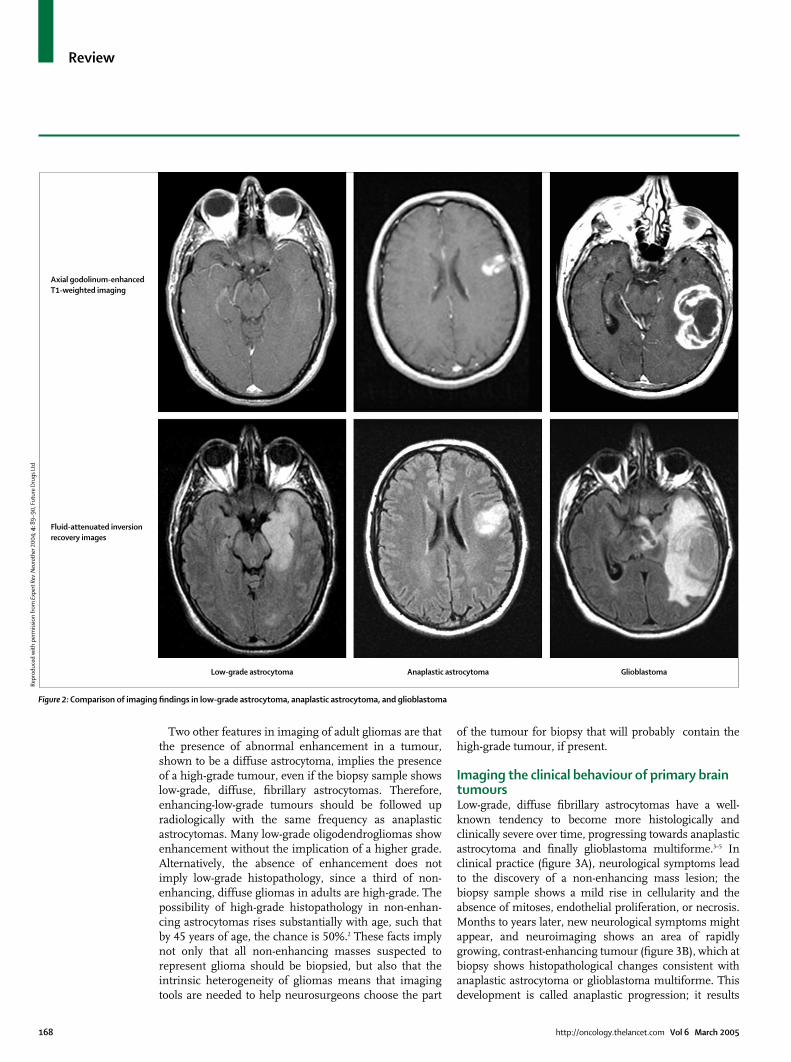
Correlation between imaging appearance andhistological features of gliomasDiffuse astrocytomas of adult cerebral hemispheres areunique among tumours in human beings for the scale towhich their imaging features relate to clinical behaviourand histopathological characteristics. In low-grade,diffuse, fibrillary astrocytomas (WHO grade II; figure 2)there is mild expansion of the affected brain withoutevidence of abnormal gadolinium enhancement orsubstantial surrounding vasogenic oedema. Theseimaging findings indicate the histopathology of mildlyincreased cellularity without pronounced angiogenesis.In anaplastic astrocytomas (WHO grade III) there aretypically nodular areas of gadolinium enhancement,showing the presence of newly formed blood vessels thathave an abnormal blood–brain barrier. These tumourstend to produce moderate expansion of the involvedbrain regions, because of greater cellular proliferation,and might show evidence of substantial surroundingvasogenic oedema. In glioblastoma multiforme (WHO
grade IV) there is pronounced mass effect andheterogeneous enhancement with centrally non-enhancing regions, which typically relate histologically tosections of necrosis. This tumour type also produceswidespread, surrounding T2-weighted hyperintensity asa result of a combination of vasogenic oedema andinfiltrating tumour (figure 2).
An important point to recognise is not only thatdiffuse, infiltrating astrocytomas of the cerebral hemi-spheres disseminated throughout the brain at the timeof diagnosis, but also that this feature is not shown wellon imaging. Indeed, precisely because these tumoursare infiltrating, intensification of local treatment,such as irradiation, does not substantially improveoverall survival.1
Lancet Oncol 2005; 6: 167–75
Division of Neuroradiology andSteven E and Catherine PappasCenter for Neuro-oncology(J W Henson MD, P Gaviani MD)and Division of Neuroradiology(R G Gonzalez MD),Massachusetts GeneralHospital, Boston, MA, USA
Correspondence to: Dr John W Henson, Division ofNeuroradiology and PappasCenter for Neuro-oncology,55 Fruit Street, Yawkey 9 East,Boston, MA 02114, [email protected]
http://oncology.thelancet.com Vol 6 March 2005 167
Diffuse astrocytomas of the adult cerebral hemispheres are unique among tumours in human beings in the extent towhich their imaging features are related to histopathological characteristics and clinical behaviour. However,understanding is still restricted about the value of imaging features in the measurement of response and ofprogression in these tumours. The present approach used in clinical trials, which consists of an anatomicalmeasurement of the enhancing tumour on MRI, has many problems, and might not be acceptable as a surrogateendpoint for survival in patients with glioblastoma who are enrolled in clinical trials. Dynamic imaging techniques,such as capillary permeability mapping, are being used in studies of new drugs that target specific molecularfeatures of gliomas; however, the validity of these techniques has not been elucidated. Diffusion imaging can bevaluable for fibre-tract mapping to assist surgical planning and might become useful in measuring early response totreatment in densely cellular tumours. Functional imaging techniques can be used to localise motor, sensory, andlanguage-control areas before surgery. Intraoperative MRI has produced improvements in the extent of tumourresection, and molecular imaging is another technique on the horizon, which could come to have a role in clinicaltrials in the near future. Thus, as a rapidly expanding sphere of investigation, brain-tumour imaging is producinggreat excitement. The aim of these new techniques is to aid the identification of more effective treatments.
MRI in treatment of adult gliomasJohn W Henson, Paola Gaviani, R Gilberto Gonzalez
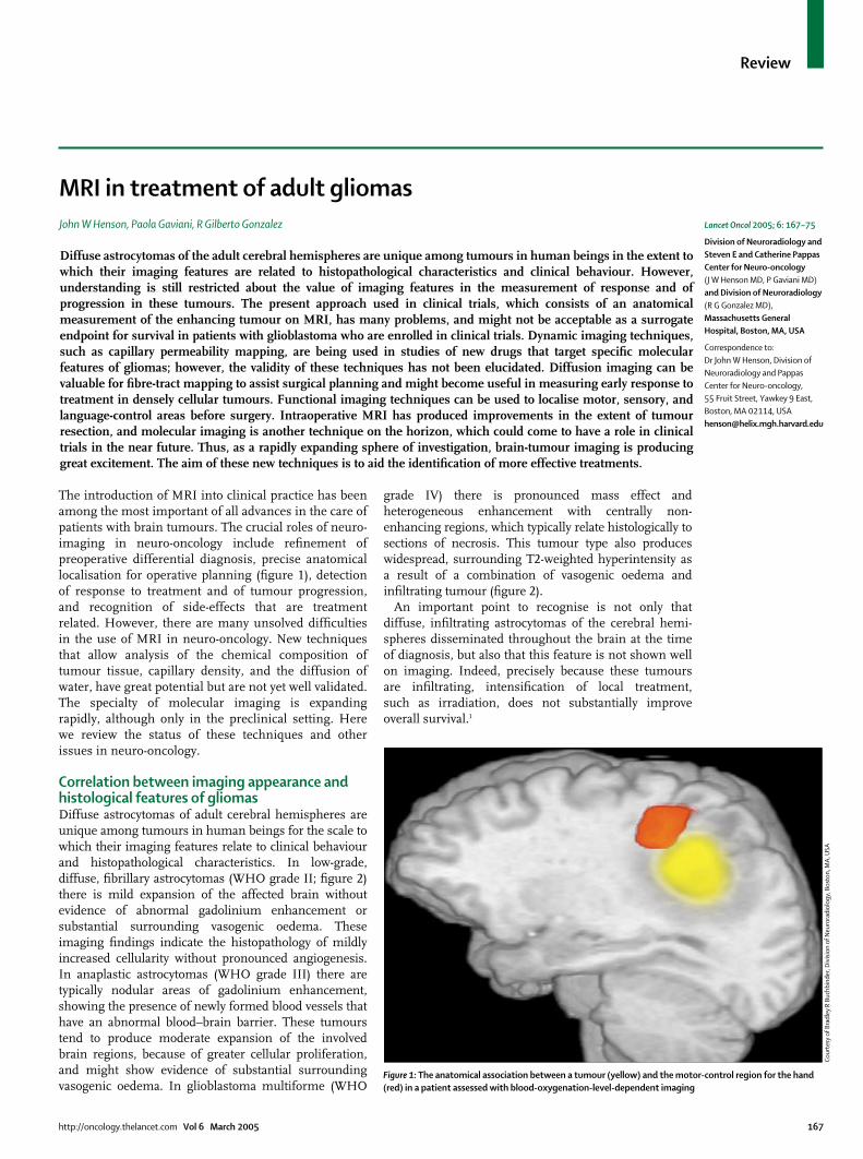
Figure 1: The anatomical association between a tumour (yellow) and the motor-control region for the hand(red) in a patient assessed with blood-oxygenation-level-dependent imaging
Cour
tesy
of B
radl
ey R
Buc
hbin
der,
Div
isio
n of
Neu
rora
diol
ogy,
Bos
ton,
MA
, USA
Review
Two other features in imaging of adult gliomas are thatthe presence of abnormal enhancement in a tumour,shown to be a diffuse astrocytoma, implies the presenceof a high-grade tumour, even if the biopsy sample showslow-grade, diffuse, fibrillary astrocytomas. Therefore,enhancing-low-grade tumours should be followed upradiologically with the same frequency as anaplasticastrocytomas. Many low-grade oligodendrogliomas showenhancement without the implication of a higher grade.Alternatively, the absence of enhancement does notimply low-grade histopathology, since a third of non-enhancing, diffuse gliomas in adults are high-grade. Thepossibility of high-grade histopathology in non-enhan-cing astrocytomas rises substantially with age, such thatby 45 years of age, the chance is 50%.2 These facts implynot only that all non-enhancing masses suspected torepresent glioma should be biopsied, but also that theintrinsic heterogeneity of gliomas means that imagingtools are needed to help neurosurgeons choose the part
of the tumour for biopsy that will probably contain thehigh-grade tumour, if present.
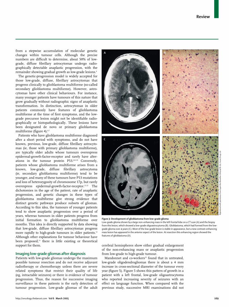
Imaging the clinical behaviour of primary braintumours Low-grade, diffuse fibrillary astrocytomas have a well-known tendency to become more histologically andclinically severe over time, progressing towards anaplasticastrocytoma and finally glioblastoma multiforme.3–5 Inclinical practice (figure 3A), neurological symptoms leadto the discovery of a non-enhancing mass lesion; thebiopsy sample shows a mild rise in cellularity and theabsence of mitoses, endothelial proliferation, or necrosis.Months to years later, new neurological symptoms mightappear, and neuroimaging shows an area of rapidlygrowing, contrast-enhancing tumour (figure 3B), which atbiopsy shows histopathological changes consistent withanaplastic astrocytoma or glioblastoma multiforme. Thisdevelopment is called anaplastic progression; it results
168 http://oncology.thelancet.com Vol 6 March 2005
Axial godolinum-enhancedT1-weighted imaging
Fluid-attenuated inversionrecovery images
Low-grade astrocytoma Anaplastic astrocytoma Glioblastoma
Figure 2: Comparison of imaging findings in low-grade astrocytoma, anaplastic astrocytoma, and glioblastoma
Repr
oduc
ed w
ith
perm
issi
on fr
om E
xper
t Rev
Neu
roth
er 2
004;
4:8
9–90
, Fut
ure
Dru
gs L
td
Review
from a stepwise accumulation of molecular geneticchanges within tumour cells. Although the precisenumbers are difficult to determine, about 50% of low-grade, diffuse fibrillary astrocytomas undergo radio-graphically detectable anaplastic progression, with theremainder showing gradual growth as low-grade lesions.4
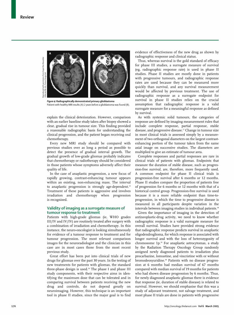
The genetic-progression model is widely accepted forthose low-grade, diffuse, fibrillary astrocytomas thatprogress clinically to glioblastoma multiforme (so-calledsecondary glioblastoma multiforme). However, astro-cytomas have other clinical behaviours. For instance,many younger patients have tumours of this nature thatgrow gradually without radiographic signs of anaplastictransformation. In distinction, astrocytomas in olderpatients commonly have features of glioblastomamultiforme at the time of first symptoms, and the low-grade precursor lesion might not be identifiable radio-graphically or histopathologically. These lesions havebeen designated de novo or primary glioblastomamultiforme (figure 4).6,7
Patients who have glioblastoma multiforme diagnosedafter a short period with symptoms, and do not haveknown, previous, low-grade, diffuse fibrillary astrocyto-mas (ie, those with primary glioblastoma multiforme),are typically older adults whose tumours overexpressepidermal-growth-factor-receptor and rarely have alter-ations in the tumour protein P53.6,8–10 Conversely,patients whose glioblastoma multiforme arises from aknown, low-grade, diffuse fibrillary astrocytoma(ie, secondary glioblastoma multiforme) tend to beyounger, and many of these tumours have P53 mutationsand loss of heterozygosity of chromosome 17p, but rarelyoverexpress epidermal-growth-factor-receptor.11,12 Thedichotomies in the age of the patient, rate of anaplasticprogression, and genetic changes in these types ofglioblastoma multiforme give strong evidence thatdistinct genetic pathways produce subsets of gliomas.According to this idea, the tumours of younger patientstend to show anaplastic progression over a period ofyears, whereas tumours in older patients progress frominitial formation to glioblastoma multiforme overmonths. This idea is directly supported by data showingthat low-grade, diffuse fibrillary astrocytomas progressmore rapidly to high-grade tumours in older patients.13
Although other explanations for tumour behaviour havebeen proposed,14 there is little existing or theoreticalsupport for them.
Imaging low-grade gliomas after diagnosisPatients with low-grade gliomas undergo the maximumpossible tumour resection and do not receive adjuvantradiotherapy or chemotherapy unless there are severerelated symptoms that restrict their quality of life(eg, intractable seizures) or there is evidence of tumourprogression. Thus, the reason for successive imagingsurveillance in these patients is the early detection oftumour progression. Low-grade gliomas of the adult
cerebral hemispheres show either gradual enlargementof the non-enhancing mass or anaplastic progressionfrom low-grade to high-grade tumour.
Mandonnet and co-workers15 found that in untreated,low-grade oligodendrogliomas there is about a 4 mmincrease in cross-sectional diameter of the tumour everyyear (figure 5). Figure 5 shows this pattern of growth in apatient with a left frontal, low-grade oligoastrocytomawho reported increasing severity of seizures with aneffect on language function. When compared with theprevious study, successive MRI examinations did not
http://oncology.thelancet.com Vol 6 March 2005 169
Figure 3: Development of glioblastoma from low-grade gliomaLow-grade glioma shown by a large non-enhancing mass in the left frontal lobe on a CT scan (A) and the biopsyfrom this lesion, which showed a low-grade oligoastrocytoma (B). Glioblastoma, which had formed from the low-grade glioma over 4 years (C). Most of the low-grade lesion is stable in appearance, but a new contrast-enhancingmass lesion has appeared in the anterior aspect of the lesion. At resection this enhancing region showed thefeatures of glioblastoma (D).
Repr
oduc
ed w
ith
perm
issi
on fr
om E
xper
t Rev
Neu
roth
er 2
004;
4:8
9–95
, Fut
ure
Dru
gs L
td
Review
explain the clinical deterioration. However, comparisonwith an earlier baseline study taken after biopsy showed aclear, gradual rise in tumour size. This finding provideda reasonable radiographic basis for understanding theclinical progression, and the patient began receiving oralchemotherapy.
Every new MRI study should be compared withprevious studies over as long a period as possible todetect the presence of gradual interval growth. Thegradual growth of low-grade gliomas probably indicatesthat chemotherapy or radiotherapy should be consideredin those patients whose symptoms adversely affect theirquality of life.
In the case of anaplastic progression, a new focus ofrapidly growing, contrast-enhancing tumour appearswithin an existing, non-enhancing mass. The intervalto anaplastic progression is strongly age-dependent.13
Treatment of these patients is aggressive and involvesirradiation and chemotherapy when progressionis recognised.
Validity of imaging as a surrogate measure oftumour response to treatmentPatients with high-grade gliomas (ie, WHO gradesIII/IV and IV/IV) are routinely treated after surgery witha combination of irradiation and chemotherapy. In thisinstance, the neuro-oncologist is looking simultaneouslyfor evidence of a tumour response to treatment and fortumour progression. The most relevant comparisonimages for the neuroradiologist and the clinician in thiscase are in most cases those from the most recentprevious study.
Great effort has been put into clinical trials of newdrugs for gliomas over the past 30 years. In the testing ofnew treatments for patients with gliomas, the standardthree-phase design is used.16 The phase I and phase IIIstudy components, with their respective aims in iden-tifying the maximum dose that can be tolerated and incomparing survival between patients receiving the newdrug and controls, do not depend greatly onneuroimaging. However, this technique is an importanttool in phase II studies, since the major goal is to find
evidence of effectiveness of the new drug as shown byradiographic response and clinical status.
Thus, whereas survival is the gold standard of efficacyfor phase III studies, a surrogate measure of survival(eg, radiographic response rate) is used in phase IIstudies. Phase II studies are mostly done in patientswith progressive tumours, and radiographic responserates are used because they can be measured morequickly than survival, and any survival measurementwould be affected by previous treatment. The use ofradiographic response as a surrogate endpoint forsurvival in phase II studies relies on the crucialassumption that radiographic response is a validsurrogate measure for a meaningful response as definedby survival.
As with systemic solid tumours, the categories ofresponse are defined by imaging measurement rules thatinclude complete response, partial response, stabledisease, and progressive disease.17 Change in tumour sizein most clinical trials is assessed simply by a measure-ment of two orthogonal diameters on the largest contrast-enhancing portion of the tumour taken from the sameaxial image on successive studies. The diameters aremultiplied to give an estimate of tumour area.
Complete responses and partial responses are rare inclinical trials of patients with gliomas. Endpoints thatmeasure the duration of stable disease, such as progres-sion-free survival, are, therefore, more frequently used.A common endpoint for phase II clinical trials isprogression-free survival after 6 months or 12 months.Phase II studies compare the proportion of patients freeof progression for 6 months or 12 months with that of ahistorical control group. Progression-free survival is usedbecause it is a more reliable endpoint than time-to-progression, in which the time to progressive disease ismeasured in all participants despite variation in theintervals between imaging studies in individual patients.18
Given the importance of imaging in the detection ofantineoplastic-drug activity, we need to know whetherradiographic response is a valid surrogate measure foroverall survival. Studies have provided strong evidencethat radiographic response predicts survival in anaplasticoligodendroglioma, for which response is associated withlonger survival and with the loss of heterozygosity ofchromosome 1p.19 For anaplastic astrocytomas, a studyby the Radiation Therapy Oncology Group randomlyassigned newly diagnosed patients to irradiation plusprocarbazine, lomustine, and vincristine with or withoutbromodeoxyuridine.18 Patients with no disease progres-sion at 6 months had median survival of 67 monthscompared with median survival of 19 months for patientswho had shown disease progression by 6 months. Thus,for newly diagnosed anaplastic gliomas there is evidencethat response (ie, duration of stable disease) is related tosurvival. However, we should emphasise that this was astudy of adjuvant treatment, not salvage treatment, andmost phase II trials are done in patients with progressive
170 http://oncology.thelancet.com Vol 6 March 2005
Figure 4: Radiographically demonstrated primary glioblastomaPatient with healthy MRI results (A) 2 years before a glioblastoma was found (B).
Repr
oduc
ed w
ith
perm
issi
on fr
om E
xper
t Rev
Neu
roth
er
2004
; 4:8
9–95
, Fut
ure
Dru
gs L
td

Review
disease. For the most common grade of glioma,glioblastoma multiforme, there is even less evidence thatradiographic response is valid as a surrogate measure ofoverall survival in the phase II setting.20 Thus, whetherduration of stable disease is a valid measure of a survival-associated response remains unclear.
Difficulties in measuring tumour size in clinicaltrialsTo complicate matters further, several features of gliomameasurement make response assessment difficult. Inthe standard approach used in clinical trials, twoorthogonal measurements are made of the gadolinium-enhancing portion of the tumour on the axial slicecontaining the largest lesion. That slice is measured onevery imaging study in the series. However, the lesionstend to be irregular in shape; so the standard cross-sectional measurements do not truly estimate the size ofthe lesion. Many tumours have large areas of cystic
necrosis, which although included in the measurementsare unlikely to respond to treatment. Diseaseprogression commonly occurs within a small region ofthe tumour, so successive linear measurements acrossthe same portion of the tumour from study to studymight not detect evidence of early progression.
Thus, the use of a method based on cross-sectionaldiameter to measure the proportion of abnormal enhan-cement within the tumour could be a suboptimumapproach for gauging response. A computer-aided volu-metric analysis is better than the cross-sectional-diameter method, especially with respect to early detec-tion of progression.21,22 Furthermore, the computer-aidedvolumetric approach shows substantially improvedreliability within and between observers than the cross-sectional-diameter method. In the volumetric approach,a technician uses a computer program to draw a regionof interest around a gadolinium-enhancing mass lesionon a single T1-weighted axial image. The computer then
http://oncology.thelancet.com Vol 6 March 2005 171
1997
9
8
7
6
5
4
3
2
10 50 100
Time (months)
Dia
met
er (c
m)
150 200
2002 2003
Figure 5: Patient with an oligoastrocytoma over 6 years of follow-up Comparison of axial fluid-attenuated inversion recovery images, taken several years apart, showed interval enlargement of the tumour, whereas a comparison of themost recent previous examination and the new examination (an interval of several months) did not clearly show enlargement. Each line in the graph representsincreasing diameter of a tumour over time.
Repr
oduc
ed w
ith
perm
issi
on fr
om E
xper
t Rev
Neu
roth
er 2
004;
4:8
9–95
, Fut
ure
Dru
gs L
td a
nd A
nn N
euro
l, Jo
hn W
iley
& S
ons
Inc.
Review
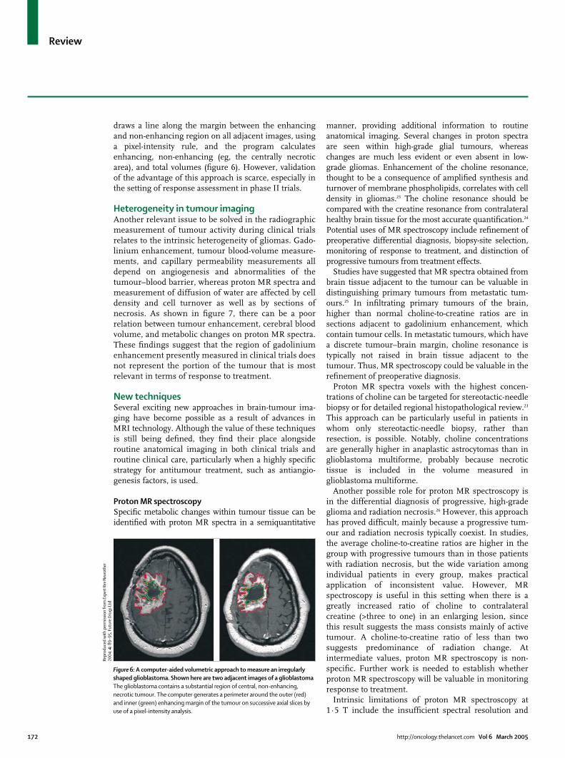
draws a line along the margin between the enhancingand non-enhancing region on all adjacent images, usinga pixel-intensity rule, and the program calculatesenhancing, non-enhancing (eg, the centrally necroticarea), and total volumes (figure 6). However, validationof the advantage of this approach is scarce, especially inthe setting of response assessment in phase II trials.
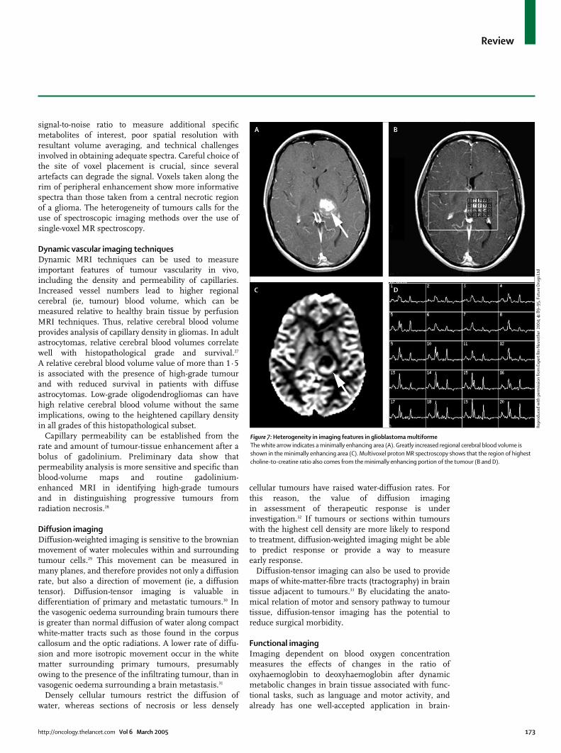
Heterogeneity in tumour imaging Another relevant issue to be solved in the radiographicmeasurement of tumour activity during clinical trialsrelates to the intrinsic heterogeneity of gliomas. Gado-linium enhancement, tumour blood-volume measure-ments, and capillary permeability measurements alldepend on angiogenesis and abnormalities of thetumour–blood barrier, whereas proton MR spectra andmeasurement of diffusion of water are affected by celldensity and cell turnover as well as by sections ofnecrosis. As shown in figure 7, there can be a poorrelation between tumour enhancement, cerebral bloodvolume, and metabolic changes on proton MR spectra.These findings suggest that the region of gadoliniumenhancement presently measured in clinical trials doesnot represent the portion of the tumour that is mostrelevant in terms of response to treatment.
New techniques Several exciting new approaches in brain-tumour ima-ging have become possible as a result of advances inMRI technology. Although the value of these techniquesis still being defined, they find their place alongsideroutine anatomical imaging in both clinical trials androutine clinical care, particularly when a highly specificstrategy for antitumour treatment, such as antiangio-genesis factors, is used.
Proton MR spectroscopy Specific metabolic changes within tumour tissue can beidentified with proton MR spectra in a semiquantitative
manner, providing additional information to routineanatomical imaging. Several changes in proton spectraare seen within high-grade glial tumours, whereaschanges are much less evident or even absent in low-grade gliomas. Enhancement of the choline resonance,thought to be a consequence of amplified synthesis andturnover of membrane phospholipids, correlates with celldensity in gliomas.23 The choline resonance should becompared with the creatine resonance from contralateralhealthy brain tissue for the most accurate quantification.24
Potential uses of MR spectroscopy include refinement ofpreoperative differential diagnosis, biopsy-site selection,monitoring of response to treatment, and distinction ofprogressive tumours from treatment effects.
Studies have suggested that MR spectra obtained frombrain tissue adjacent to the tumour can be valuable indistinguishing primary tumours from metastatic tum-ours.25 In infiltrating primary tumours of the brain,higher than normal choline-to-creatine ratios are insections adjacent to gadolinium enhancement, whichcontain tumour cells. In metastatic tumours, which havea discrete tumour–brain margin, choline resonance istypically not raised in brain tissue adjacent to thetumour. Thus, MR spectroscopy could be valuable in therefinement of preoperative diagnosis.
Proton MR spectra voxels with the highest concen-trations of choline can be targeted for stereotactic-needlebiopsy or for detailed regional histopathological review.23
This approach can be particularly useful in patients inwhom only stereotactic-needle biopsy, rather thanresection, is possible. Notably, choline concentrationsare generally higher in anaplastic astrocytomas than inglioblastoma multiforme, probably because necrotictissue is included in the volume measured inglioblastoma multiforme.
Another possible role for proton MR spectroscopy isin the differential diagnosis of progressive, high-gradeglioma and radiation necrosis.26 However, this approachhas proved difficult, mainly because a progressive tum-our and radiation necrosis typically coexist. In studies,the average choline-to-creatine ratios are higher in thegroup with progressive tumours than in those patientswith radiation necrosis, but the wide variation amongindividual patients in every group, makes practicalapplication of inconsistent value. However, MRspectroscopy is useful in this setting when there is agreatly increased ratio of choline to contralateralcreatine (>three to one) in an enlarging lesion, sincethis result suggests the mass consists mainly of activetumour. A choline-to-creatine ratio of less than twosuggests predominance of radiation change. Atintermediate values, proton MR spectroscopy is non-specific. Further work is needed to establish whetherproton MR spectroscopy will be valuable in monitoringresponse to treatment.
Intrinsic limitations of proton MR spectroscopy at1·5 T include the insufficient spectral resolution and
172 http://oncology.thelancet.com Vol 6 March 2005
Figure 6: A computer-aided volumetric approach to measure an irregularlyshaped glioblastoma. Shown here are two adjacent images of a glioblastomaThe glioblastoma contains a substantial region of central, non-enhancing,necrotic tumour. The computer generates a perimeter around the outer (red)and inner (green) enhancing margin of the tumour on successive axial slices byuse of a pixel-intensity analysis.
Repr
oduc
ed w
ith
perm
issi
on fr
om E
xper
t Rev
Neu
roth
er
2004
: 4:8
9–95
, Fut
ure
Dru
gs L
td
Review
signal-to-noise ratio to measure additional specificmetabolites of interest, poor spatial resolution withresultant volume averaging, and technical challengesinvolved in obtaining adequate spectra. Careful choice ofthe site of voxel placement is crucial, since severalartefacts can degrade the signal. Voxels taken along therim of peripheral enhancement show more informativespectra than those taken from a central necrotic regionof a glioma. The heterogeneity of tumours calls for theuse of spectroscopic imaging methods over the use ofsingle-voxel MR spectroscopy.
Dynamic vascular imaging techniques Dynamic MRI techniques can be used to measureimportant features of tumour vascularity in vivo,including the density and permeability of capillaries.Increased vessel numbers lead to higher regionalcerebral (ie, tumour) blood volume, which can bemeasured relative to healthy brain tissue by perfusionMRI techniques. Thus, relative cerebral blood volumeprovides analysis of capillary density in gliomas. In adultastrocytomas, relative cerebral blood volumes correlatewell with histopathological grade and survival.27
A relative cerebral blood volume value of more than 1·5is associated with the presence of high-grade tumourand with reduced survival in patients with diffuseastrocytomas. Low-grade oligodendrogliomas can havehigh relative cerebral blood volume without the sameimplications, owing to the heightened capillary densityin all grades of this histopathological subset.
Capillary permeability can be established from therate and amount of tumour-tissue enhancement after abolus of gadolinium. Preliminary data show thatpermeability analysis is more sensitive and specific thanblood-volume maps and routine gadolinium-enhanced MRI in identifying high-grade tumoursand in distinguishing progressive tumours fromradiation necrosis.28
Diffusion imagingDiffusion-weighted imaging is sensitive to the brownianmovement of water molecules within and surroundingtumour cells.29 This movement can be measured inmany planes, and therefore provides not only a diffusionrate, but also a direction of movement (ie, a diffusiontensor). Diffusion-tensor imaging is valuable indifferentiation of primary and metastatic tumours.30 Inthe vasogenic oedema surrounding brain tumours thereis greater than normal diffusion of water along compactwhite-matter tracts such as those found in the corpuscallosum and the optic radiations. A lower rate of diffu-sion and more isotropic movement occur in the whitematter surrounding primary tumours, presumablyowing to the presence of the infiltrating tumour, than invasogenic oedema surrounding a brain metastasis.31
Densely cellular tumours restrict the diffusion ofwater, whereas sections of necrosis or less densely
cellular tumours have raised water-diffusion rates. Forthis reason, the value of diffusion imagingin assessment of therapeutic response is underinvestigation.32 If tumours or sections within tumourswith the highest cell density are more likely to respondto treatment, diffusion-weighted imaging might be ableto predict response or provide a way to measureearly response.
Diffusion-tensor imaging can also be used to providemaps of white-matter-fibre tracts (tractography) in braintissue adjacent to tumours.33 By elucidating the anato-mical relation of motor and sensory pathway to tumourtissue, diffusion-tensor imaging has the potential toreduce surgical morbidity.
Functional imagingImaging dependent on blood oxygen concentrationmeasures the effects of changes in the ratio ofoxyhaemoglobin to deoxyhaemoglobin after dynamicmetabolic changes in brain tissue associated with func-tional tasks, such as language and motor activity, andalready has one well-accepted application in brain-
http://oncology.thelancet.com Vol 6 March 2005 173
Figure 7: Heterogeneity in imaging features in glioblastoma multiforme The white arrow indicates a minimally enhancing area (A). Greatly increased regional cerebral blood volume isshown in the minimally enhancing area (C). Multivoxel proton MR spectroscopy shows that the region of highestcholine-to-creatine ratio also comes from the minimally enhancing portion of the tumour (B and D).
Repr
oduc
ed w
ith
perm
issi
on fr
om E
xper
t Rev
Neu
roth
er 2
004;
4:8
9–95
, Fut
ure
Dru
gs L
td

Review
tumour imaging. These focal haemodynamic changesare displayed on an anatomical map with coregisteredimages of the brain tumour. This approach is already inwidespread use as a preoperative mapping technique inan attempt to keep to a minimum intraoperative damageto eloquent brain areas.34
Another potential application for this imaging methodis in the assessment of the oxygen environment withinbrain tumours.35 This technique would be particularlyuseful in clinical trials of approaches to raise oxygenconcentrations within tumour tissue in an attempt topotentiate DNA damage from radiation or chemo-therapy.36
Intraoperative MRIDuring the past 10 years, MRI has been introduced intoneurosurgical operating rooms to allow real-time ima-ging during surgery.37 Intraoperative MRI has severalpotential advantages. From the standpoint of tumourdiagnosis, it can be used to guide the surgeon moreaccurately to small lesions, thus, limiting the extent ofcraniotomy and improving the chance of obtainingdiagnostic tissue. Compared with routine stereotacticprocedures, this approach is not restricted by tissuemovement during craniotomy.
Studies38 have shown improvement in the extent oftumour resection with the use of intraoperative MRI, butimproved survival has not yet been shown. Real-timevisualisation of the resection margin would alsoprobably make these more thorough resections safer,and improve surveillance for intraoperative compli-cations. Increasing the extent of resection in patientswith low-grade gliomas has provided the strongestrationale for intraoperative MRI.37
Intraoperative MRI systems are available with eitherlow (0·2 T) or high (1·5 T) field strength. High-field-strength systems carry the potential of better imagingquality and the opportunity of advanced imaging fea-tures such as diffusion, angiography, and spectroscopy.However, the size and cost of the high-field-strengthmagnets are pronounced disadvantages.39
Intraoperative imaging has some limitations. Titan-ium neurosurgical instruments are needed, and the highmagnetic-field strength of these devices poses a potentialrisk from ferrous substances in the operating room.There might be compromises in the positioning of thepatient, access by the surgeon, and sterility. Image-acquisition time is only about 2 min for each sequence,but the need to move the patient in and out of thescanner lengthens the operation time.39 The inter-pretation of images obtained during surgery can bechallenging because, in addition to the structures seenon preoperative films, intraoperative images can showsurgical artefacts from air or blood. Finally, a barrier towidespread implementation of these techniques hasbeen the substantial expense and maintenance of thescanning equipment.
Molecular imagingThis is a rapidly emerging sphere of investigation thatseeks to achieve imaging of molecular processeswithin brain-tumour cells in vivo.40 In one example ofthe use of MRI to image smart probes, tumour-cell-specific uptake of monocrystalline iron oxidenanoparticles can be achieved owing to the highconcentration of transferrin receptors on the cells.Thus, these tumour cells have a high rate ofendocytosis of the nanoparticles, and they can beimaged with MRI. In another example that has beentested in experimental models, cells can be induced tooverexpress transferrin receptors, and these cells canthen be selectively detected in vivo byMRI after administration of monocrystalline ironoxide nanoparticles.
Paramagnetic chelates that can change theirmagnetic properties on enzymatic hydrolysis areunder investigation. For instance, a gadoliniumgalactopyranose substrate shows greater relaxivity after�-galactosidase-mediated hydrolysis. Thus, thepresence of ectopically-expressed �-galactosidase canbe indirectly detected by MRI. Magnetic nanosensorsare being developed that might be able to detectspecific DNA or RNA sequences. Enzymes, such astyrosinase that have a high metal-binding capacity,might also be imaged with MRI when overexpressed.
Potential future development of functional andmetabolic imaging techniquesFunctional and metabolic imaging techniques will con-tinue to progress rapidly. Important approaches totumour imaging will include capillary permeabilitymapping, phosphorus MR spectroscopy, real-timeintraoperative spectroscopic measurements, and mole-cular imaging. Techniques will progress to assess thespecific feature of the tumour that is being targeted bynew therapies, as is happening now with treatmentsdirected at tumour vascularity. There will be at least twomajor challenges for these new techniques. First,rigorous, quantitative analysis of the validity of everyimaging approach will be crucial. Second, inherentbiological variability between and within tumours willprobably pose a formidable barrier to the application offunctional and metabolic imaging techniques. Thesetechniques should help to identify more rapidlybetter therapeutic drugs for patients with thesedevastating tumours.
174 http://oncology.thelancet.com Vol 6 March 2005
Search strategy and selection criteria
Publications for this review were identified by use of thePubMed and references cited in relevant articles. “Brainneoplasms”, “magnetic resonance imaging”, and other searchterms were used to find articles on brain-tumour imaging.Only articles published in English were used.

Review
Conflict of InterestWe declare no conflicts of interest.
References1 Selker RG, Shapiro WR, Burger P, et al. The Brain Tumor
Cooperative Group NIH Trial 87-01: a randomized comparison ofsurgery, external radiotherapy, and carmustine versus surgery,interstitial radiotherapy boost, external radiation therapy, andcarmustine. Neurosurgery 2002; 51: 343–55.
2 Barker FG, Chang SM, Huhn SL, et al. Age and the risk ofanaplasia in magnetic resonance-nonenhancing supratentorialcerebral tumors. Cancer 1997; 80: 936–41.
3 Recht LD, Lew R, Smith TW. Suspected low-grade glioma: isdeferring treatment safe? Ann Neurol 1992; 31: 431–36.
4 Recht LD, Bernstein M. Low-grade gliomas. Neurol Clin 1995;13: 847–59.
5 Kleihues P, Soylemezoglu F, Schauble B, et al. Histopathology,classification, and grading of gliomas. Glia 1995; 15: 211–21.
6 Tortosa A, Ino Y, Odell N, et al. Molecular genetics ofradiographically-defined de novo GBM. Neuropathol App Neurobiol2000; 26: 544–52.
7 Biernat W, Tohma Y, Yonekawa Y, et al. Alteration of cell cycleregulatory genes in primary (de novo) and secondaryglioblastomas. Acta Neuropathologica 1997; 94: 303–09.
8 Okada Y, Hurwitz EE, Esposito JM, et al. Selection pressures ofTP53 mutation and microenvironmental location influenceepidermal growth factor receptor gene amplification in humanglioblastomas. Cancer Res 2003; 63: 413–16.
9 Agosti RM, Leuthold M, Gullick WJ, et al. Expression of theepidermal growth factor receptor in astrocytic tumours isspecifically associated with glioblastoma multiforme. Virchows ArchPathol Anat 1992; 420: 321–25.
10 Watanabe K, Tachibana O, Sato K, et al. Overexpression of the EGFreceptor and p53 mutations are mutually exclusive in the evolutionof primary and secondary glioblastomas. Brain Pathol 1996;6: 217–24.
11 Reifenberger J, Ring GU, Gies U, et al. Analysis of p53 mutationand epidermal growth factor receptor amplification in recurrentgliomas with malignant progression. J Neuropathol Exp Neurol1996; 55: 822–31.
12 Ohgaki H, Schauble B, zur Hausen A, et al. Genetic alterationsassociated with the evolution and progression of astrocytic braintumors. Virchows Arch Pathol Anat 1995; 427: 113–18.
13 Shafqat S, Hedley-Whyte ET, Henson JW. Age-dependent rate ofanaplastic transformation in low-grade astrocytoma. Neurology1999; 52: 867–69.
14 James CD, Carlblom E, Dumanski JP, et al. Clonal genomicalterations in glioma malignancy stages. Cancer Res 1988;48: 5546–51.
15 Mandonnet E, Delattre JY, Tanguy ML, et al. Continuous growth ofmean tumor diameter in a subset of grade II gliomas. Ann Neurol2003; 53: 524–28.
16 Batchelor T, Stanley K, Andersen J. Clinical trials in neuro-oncology. Curr Opin Neurol 2001; 14: 689–94.
17 Macdonald DR, Cascino TL, Schold SC, Cairncross JG. Responsecriteria for phase II studies of malignant glioma. J Clin Oncol 1990;8: 1277–80.
18 Prados MD, Seiferheld W, Sandler HM, et al. Phase IIIrandomized study of radiotherapy plus procarbazine, lomustine,and vincristine with or without BUdR for treatment of anaplasticastrocytoma: final report of RTOG 9404. Int J Radiat Oncol BiolPhys 2004; 58: 1147–52.
19 Cairncross JG, Ueki K, Zlatescu MC, et al. Specific geneticpredictors of chemotherapeutic response and survival in patientswith anaplastic oligodendrogliomas. J Natl Cancer Inst 1998;90: 1473–79.
20 Grant R, Liang BC, Slattery J, et al. Chemotherapy response criteriain malignant glioma. Neurology 1997; 48: 1336–40.
http://oncology.thelancet.com Vol 6 March 2005 175
21 Sorensen AG, Patel S, Harmath C, et al. Comparison of diameterand perimeter methods for tumor volume calculation. J Clin Oncol2001; 19: 551–57.
22 Warren KE, Patronas N, Aikin AA, et al. Comparison of one-, two-,and three-dimensional measurements of childhood brain tumors.J Natl Cancer Inst 2001; 93: 1401–05.
23 Croteau D, Scarpace L, Hearshen D, et al. Correlation betweenmagnetic resonance spectroscopy imaging and image-guidedbiopsies: semiquantitative and qualitative histopathological analysesof patients with untreated glioma. Neurosurgery 2001; 49: 823–29.
24 Rabinov JD, Lee PL, Barker FG, et al. In Vivo MRS at 3 Teslapredicts recurrent glioma vs radiation effects: initial experience.Radiology 2002; 225: 871–79.
25 Fan G, Sun B, Wu Z, et al. In vivo single-voxel proton MRspectroscopy in the differentiation of high-grade gliomas andsolitary metastases. Clin Radiol 2004; 59: 77–85.
26 Rock JP, Scarpace L, Hearshen D, et al. Associations amongmagnetic resonance spectroscopy, apparent diffusion coefficients,and image-guided histopathology with special attention to radiationnecrosis. Neurosurgery 2004; 54: 1111–17.
27 Lev ML, Ozsunar Y, Henson JW, et al. Glial tumor grading andoutcome prediction using dynamic spin-echo MR susceptibilitymapping compared to conventional contrast enhanced MRI:confounding effect of elevated relative cerebral blood volume ofoligodendrogliomas. Am J Neuroradiol 2004; 25: 214–21
28 Roberts HC, Roberts TP, Brasch RC, Dillon WP. Quantitativemeasurement of microvascular permeability in human braintumors achieved using dynamic contrast-enhanced MR imaging:correlation with histologic grade. AJNR Am J Neuroradiol 2000;21: 891–99.
29 Schaefer PW, Grant PE, Gonzalez RG. Diffusion-weighted MRimaging of the brain. Radiology 2000; 217: 331–45.
30 Lu S, Ahn D, Johnson G, Cha S. Peritumoral diffusion tensorimaging of high-grade gliomas and metastatic brain tumors.AJNR Am J Neuroradiol 2003; 24: 937–41.
31 Provenzale JM, McGraw P, Mhatre P, et al. Peritumoral brainregions in gliomas and meningiomas: investigation with isotropicdiffusion-weighted MR imaging and diffusion-tensor MR imaging.Radiology 2004; 232: 451–60.
32 Roth Y, Tichler T, Kostenich G, et al. High-b-value diffusion-weighted MR imaging for pretreatment prediction and earlymonitoring of tumor response to therapy in mice. Radiology 2004;232: 685–92.
33 Berman JI, Berger MS, Mukherjee P, Henry RG. Diffusion-tensorimaging-guided tracking of fibers of the pyramidal tract combinedwith intraoperative cortical stimulation mapping in patients withgliomas. J Neurosurg 2004; 101: 66–72.
34 Vlieger EJ, Majoie CB, Leenstra S, Den Heeten GJ. Functionalmagnetic resonance imaging for neurosurgical planning inneurooncology. Eur Radiol 2004; 14: 1143–53.
35 Hsu YY, Chang CN, Jung SM, et al. Blood oxygenation level-dependent MRI of cerebral gliomas during breath holding. J MagnReson Imaging 2004; 19: 160–67.
36 Hou H, Khan N, O’Hara JA, et al. Effect of RSR13, an allosterichemoglobin modifier, on oxygenation in murine tumors: an in vivoelectron paramagnetic resonance oximetry and bold MRI study.Int J Radiat Oncol Biol Phys 2004; 59: 834–43.
37 Albayrak B, Samdani AF, Black PM. Intra-operative magneticresonance imaging in neurosurgery. Acta Neurochir (Wien) 2004;146: 543–56.
38 Knauth M, Wirtz CR, Tronnier VM, et al. Intraoperative MRimaging increases the extent of tumor resection in patients withhigh-grade gliomas. AJNR Am J Neuroradiol 1999; 20: 1642–46.
39 Siomin V, Barnett G. Intraoperative imaging in glioblastomaresection. Cancer J 2003; 9: 91–98.
40 Shah K, Jacobs A, Breakefield XO, Weissleder R. Molecularimaging of gene therapy for cancer. Gene Ther 2004; 11: 1175–87.