Slit Lamp ESL-2600 Ezer - US Ophthalmic · 19) This instrument supplies a 3-wire cable. Please...
22
Transcript of Slit Lamp ESL-2600 Ezer - US Ophthalmic · 19) This instrument supplies a 3-wire cable. Please...


Preface Thank you for purchasing our ESL-2600 slit lamp. Please read this manual carefully for the sake of your best use. General Requirements for Safety Please read carefully about following precautions to avoid unexpected personal injury as well as the product being damaged or other possible dangers.
Precautions 1. Do not use this instrument in the flammable or explosive environment,
keep it away from dusty locations or high temperature. Use it indoors and keep it clean and dry.
2. Check that all the wires are correctly and firmly connected before using. Ensure that the instrument is well grounded.
3. Please pay attention to all the rated values of the electrical connecting terminal.
4. Only use fuse according to the specifications and rated values stipulated by our product.
5. Use the power cable supplied with this instrument. 6. Don’t touch the surface of the lens and prism with hand or hard objects. 7. Turn off the main power first before replacing the illumination bulb and
fuse. 8. To prevent the instrument from falling down to floor, it should be placed
on the floor where the inclination angle is less than 10°. 9. Turn off the power and cover the instrument with dust-proof hood when
it is not in use. 10. In case there is any trouble, please first refer to the trouble-shooting
guide. If it still can’t work, please contact with the authorized distributor or our Repair Department.
THE SAFETY MARKS USED IN THIS INSTRUMENT
TYPE B ATTENTION PLEASE TERMINAL OF
REFER TO THIS MANUAL THE PROTECTIVE GROUNDING

Fig 1

Contents
1. Nomenclature...............................................................................1 2. Assembly.....................................................................................3
2.1 Components..........................................................................3 2.2 Assembly procedure..........................................................5 2.3 Checking procedure after assembling................................8
3. Operation procedures................................................................9 3.1 Diopter compensation and Pupil Distance adjustment................9 3.2 Patient position and fixation target...............................10 3.3 Base operation..................................................................10 3.4 Illumination parts operation .......................................... 11
4. Maintenance.............................................................................12 4.1 Replacing the illumination bulb.....................................12 4.2 Replacing the reflecting mirror......................................13 4.3 Replacing the fuse............................................................13 4.4 Adjusting the tightness of the slit width knob..............13 4.5 Replacing the chin-rest paper........................................14 4.6 Cleaning............................................................................14 4.7 Consumables....................................................................14
5. Trouble shooting guide............................................................15 6. Responsibility ...........................................................................15 7. Transportation and storage.....................................................16 8. Optional accessories (purchase in addition)..........................16
8.1 Applanation tonometer...................................................16 9. Specifications............................................................................17

1
1. Nomenclature 1 Joystick
Incline joystick to move the instrument slightly on the horizontal surface and rotate it to adjust the elevation of the microscope.
2 Base Locking Screw The base will be locked when fasten this screw.
3 Rail Cover Protect the rail surface.
4 Base Support the microscope and the illumination arms with the joystick controlling its movement.
5 Worktable
6 Brightness Control Switch
The brightness can be continuous adjusted. Avoid working continuously at high setting, as the service life of the bulb will be shortened.
7 Main Power Switch 8 Pilot Lamp 9 Location Roller
When it is in the middle, it stands for included angle of 0o between the microscope arm and the illumination arm. And the right or left side stands for the included angle of 10o.
10 Centering Knob Loosening the knob allows the illumination light to be moved from the center of the vision field for indirect retro-illumination. Fastening the knob brings the illumination light return to the center.
11 Slit Width Control Knob The slit width is continuously adjustable from 0 to 9mm.
12 Magnification Changer Lever Push the lever to either side to select the desired magnification of the microscope.
13 Diopter Adjustment Ring Adjust the eyepieces diopter to obtain a clear image before using the instrument.
14 Aperture and Slit Height Control Knob Rotate this knob to adjust the spot and the slit height. Swing the knob horizontally to revolve the slit.
15 Filter Selection Lever There are four filters for selection
16 Slit Height and Aperture Display Window 17 Lamp Cap 18 Reflecting Mirror
The long mirror is provided. The observation pathway may be interfered as the included angle between the microscope arm and the illumination arm is only 3o~10o.
19 Forehead Belt 20 Fixation target
An illuminated fixed spot for patient to look at. 21 Horizontal Mark
When the horizontal center of the patient’s eye is in line with this mark, the elevation of the microscope controlled by joystick is also in its center position.
22 Chin-rest 23 Chin-rest Elevation Adjustment Knob
Rotate the knob to adjust the elevation of the chin-rest. 24 Protection Cap
Please cover the main shaft hole with the protection cap to prevent dusts and physiological salt solution from dropping into the main shaft pole of the illumination arm during the

2
operation. Take off the cap when assemble the focusing test rod. 25 Microscope and Illumination Arm Couple Bolt
Fasten this bolt and the illumination arm and the microscope arm could be move in couple state to rotate together. Loosen it and the illumination arm then can rotate separately.
26 Microscope Arm Locking Knob Lock the rotational movement of the microscope arm.

3
2. Assembly
This section of the manual describes how to assemble ESL-2600 slit lamp. All parts should
be taken out with great care from the packing case before assembling.
2.1 Components------------------------------------------------------------------
Fig.1

4
Fig.2
Name Quantity A Illumination Part 1 B Base Part (with Microscope) 1 C Head-rest Part 1 D Breath Shield 1 E Work Table with Power Box 1 F Rail Cove r 2 G Input Power Cable 1 H Chin-rest Paper 1 I Spare Main Illumination Bulb 1 J Protection Cap 1 K Spare Long Reflecting Mirror 1 L Brush 1 M Dust Cover 1 N Focusing Test Rod 1* O Cross Screw Driver with Wood Handle 1 P Hexagonal wrench 1 Q Watch Screw Driver 1 R Spanner 1
(*Optionally available in some region.)

5
2.2 Assembly procedure ------------------------------------------------------ Necessary tools are as follows: Cross screwdriver with wood handle (O) Watch screwdriver (P) Spanner (R)
1) Selecting Voltage and Fuse
Fig.3
� Check the setting on the voltage selector
located on the bottom of the power box. If
it doesn’t match with the input voltage,
slide it to the proper position with
screwdriver (R).
� Open the fuse holder with screw driver (P)
and take out the fuse, check and ensure
that its rated value is corresponding to the
mains voltage:
110V----------------------1A
220V--------------------0.5A
It has been set to 220V, 0.5A before
leaving our factory.
Attention: Set the input voltage and frequency of the instrument according to that of the mains.
2) Assembling the Worktable (E) � To attach the worktable on the motorized
instrument table, please screw off four
M8x20mm bolts with spring washers with
the spanner (S).
� Lift the worktable to aim its screw hole at
the assembly hole of the instrument table. Put down the worktable, with the power panel
facing the operator, refasten the bolt securely with the spanner.
Fig.4
3) Assembling the Head-rest Part (C)
� Remove the four screws attached to the
chin-rest connection board with the screw
driver (P).
Fig.5
� Put two cables in the gap between the
head-rest fixation plate and the chin-rest
connection board. While ensuring they are
not clamped, retighten the previously
removed screws.

6
4) Assembling the base part (B) and the rail cover (F)
Fig.6
� Place the wheels of both sides of the base (B)
on the rail on the worktable.
� Check whether the wheels can be rolled
steadily on the rail.
� Insert the bottom of the rail covers into
the gap below the both sides of the rails
respectively in the direction of the arrow
(Fig.6).
5) Assembling illumination part (A)
Fig.7
6) Loosen the illumination arm couple bolt (26).
7) Rotate the brass shaft sleeve to make the
angle of the red mark and the illumination
arm between 30 o ~90o.
8) Loosen the screw in the illumination arm with
the screwdriver (R). Aim the assembly hole of
the illumination arm at the brass shaft sleeve
then put it down with care, let the shaft
keeping close to the bottom surface well
and the two red marks stretch in one line
simultaneously.
9) After the two red marks accurately aligned,
re-tighten the screw.
6) Assembling the breath shield (E)
Fig.8
10) Remove the breath shield fixation screw from
the microscope arm.
11) Pass the removed screw through the hole of
the breath shield then re-screw it into the
arm.
7) Connecting plug
Fig.9
12) Insert the plug on the top of the head-rest part
(C) into the socket of the lamp cap (18) on
the illumination part (A).
13) Connect the two plugs below the head-rest
part with the corresponding output socket
of the power box. .
14) Insert the plug of the input power cable (G)
into the input socket of the power box. .

7
15) Remove the cable clips from the bottom of
the worktable with screwdriver (P) and
wrap the output and input cables
respectively, then re-attach them to the
bottom of the worktable.
Fig.10
8) Assembling the chin-rest paper (H) 16) Pull out the two fixing pins from the
chin-rest.
17) Get rid of the paper package and let the
pins go through its holes.
18) Insert the fixing pins into the hole again.
Fig.11

8
2.3 Checking procedure after assembling --------------------------------------
1) Power plug 19) This instrument supplies a 3-wire cable.
Please select a proper power socket as
matched.
20) Ensure that the instrument is grounded
well.
Attention: Please uses the special cable supplied with this instrument.
2) The power box and the illumination part
21) When the main power switch (8) of the
power box is placed at ‘I’, it turns on, and
‘O’ for turn off. The main power switch
should be set at the ‘O’ position before
connecting the input cable with the power
socket.
22) Turn on the main power switch, and the
pilot lamp (9) will be lighted. Open the slit
width control knob (12) to examine the
illumination.
23) Press the brightness control switch (7)
respectively at two positions and the
brightness should be changed accordingly.
24) Check the fixation target device to
con-firm it is lighting.
25) Check if all the moveable parts such as
aperture and slit height control knob (15),
filter selection lever (16), and
magnification changer lever (13) etc.
could be operated freely.
26) After examining, turn off the main power
and cover the instrument with the dust
cover (N).

9
3. Operation procedures
3.1 Diopter compensation and Pupil Distance adjustment..................................................
1)1)1)1) Use of the focusing test rod (M)
Fig.12
The rod is supplied as one of standard
accessories for confirming if the
microscope is adjusted correctly. Insert it
into the main shaft hole with the flat
surface facing the objective lens --- the
direction of the operator.
Attention: After adjusting, remember to take out the rod and insert the protection cap.
2)2)2)2) Brightness adjustment Switch on the main power switch and set
the brightness control switch (7) at ‘N’
position. Turn the slit width control knob
(12) to make the slit width to be 2~3mm.
3)3)3)3) Diopter compensation The focus of the microscope is calibrated
according to the emmetropia. If the
operator is an ametropia, he should
adjust the eyepiece diopter.
Suggest adjusting the diopter as following procedures.
27) First, rotate the diopter adjustment ring (19) counter clockwise down to the end.
28) Second, rotate the ring clockwise until a sharp slit image appears on the focusing text rod.
29) Adjust another eyepiece in the same procedure.
30) Record the diopter value on each eyepiece for future reference.
4)4)4)4) Pupil distance adjustment Separate the prism box of the microscope
with both hands to adjust the P. D. until
both eyes could see the same image on
the focusing test rod through the
eyepieces, and at the same time a stereo
vision will be obtained.
Fig.13
Attention: While adjusting P. D., ensure that both eyepieces are at the same height.

10
3.2 Patient position and fixation target-----------------------------------------
1)1)1)1) Positioning the patient’s head Have the patient place his chin on the
chin-rest (23) and the forehead against
the forehead belt (20). Adjust the
chin-rest elevation adjustment knob (24)
below the chin-rest until the patient’s
canthus aligns with the horizontal mark
(22).
Fig.14
2)2)2)2) Use of the fixation target 31) For fixing the patient’s eyesight, just make
him look at the fixation target (21) with
the eye not to be examined. To change
fixing position, move the lamp bar, as well
as move the curved lever around the
head-rest.
Fig.15
3.3 Base operation ------------------------------------------------------------------
1)1)1)1) Horizontal rough adjustment Keep the joystick (1) erect and move the
base (4) to make the microscope move
on the horizontal surface to aim at the
object roughly.
2)2)2)2) Vertical adjustment Rotate the joystick to adjust the
microscope’s height until it aligns with
the target.
Turn the joystick clockwise to raise the
microscope and counter-clockwise to
lower it.
3)3)3)3) Horizontal fine adjustment Tilt the joystick to make the microscope
move slightly on the horizontal surface.
While watching through the eyepieces,
tilt the joystick to aim accurately at the
object for a sharp image.
4)4)4)4) Locking the base When finishing the adjustment, fasten the
base locking screw (2) to lock the base (4)
and prevent it from sliding.
Fig.16

11
3.4 Illumination parts operation------------------------------------------------
1)1)1)1) Changing the slit width
Turn the slit width control knob (12) and
the slit width will be changed from 0mm
to 9mm. The slit becomes a circle at the
9mm size.
Fig.17
2)2)2)2) Changing the aperture and slit height Turn the aperture and slit height control
knob (15) and 6 different circular beams
of light are available at full aperture: 9, 8,
5, 3, 1, 0.2 diameter respectively. With a
slit image, the slit height can be changed
continuously from 1 to 9mm, which is
indicated through the display window
(17).
Fig.18
3)3)3)3) Rotating the slit image Swing the aperture and slit height control
knob (15) horizontally to revolve the slit
image at any angle from vertical to
horizontal. The rotation angle scale
indicates the angle of image rotation with
small division for 5o and big division for
10o.
Fig.19
4)4)4)4) Deflecting the illumination light Loosen the centering knob (11) and
swing the slit width control knob (12) by
the arrow, so the light spot moves away
from the center of the microscope vision
field. It is mainly used to examine the
eyes by indirect retro-illumination.
Fasten the centering knob and the slit
light will return to the center of the
microscope vision field.
Fig.20

12
5)5)5)5) Filter selection Turn the filter selection lever (16) in the
horizontal surface to add four different
kinds of filters respectively into the
illumination pathway. Usually the heat
absorption filter is used so that the
patient may feel more comfortable in
long period of examination. 1 ND 2 heat absorption 3 grey 4 red-free 5 blue
Fig.21
4. Maintenance
Attention: The replaced waste materials should be treated as industrial rubbish.
4.1 Replacing the illumination bulb---------------------------------------------
� Turn the main power switch (8) off.
� Pull out the plug connected to the lamp
house, unscrew the fastening screw and
then pull the lamp cover (26) until it is
pulled out from the illumination part(A)
(Fig22).
Fig.22
� Turn the compression spring upward
and then pull out the plug and take out the
old bulb and replace it with a new one.
Turn the compressing spring downward
The groove in the bulb fixation disc
should be aimed at the flange of the lamp
base; otherwise the illumination may be
uneven.
Attention: The bulb is hot � The mark point on the lamp cover aligns
the mark point on the illumination part.
Tighten mounting screw and insert the
connecting plugs.
� Turn on the main power switch and check
whether the new bulb works or not.
Fig.23

13
4.2 Replacing the reflecting mirror ---------------------------------------------
� Set the angle between the microscope and
the illumination arm to exceed 30o.
� Remove the long mirror by holding the
extended surface.
� Insert new long reflecting mirror.
Fig.24
4.3 Replacing the fuse--------------------------------------------------------------
� Turn off the main power switch (8) and pull out the input cable from the power socket.
� Screw off the fuse holder cover with the screw driver (P).
� Replace it with a new fuse, then fasten the cover. � The fuse specifications are as follows:
110V 1A , 125V 220V 0.5A, 250V
Attention: Please select the fuse of the same type, specification and rating.
Fig.25
4.4 Adjusting the tightness of the slit width knob ----------------------------
If the slit width control knob is too loose,
the slit width may be out of control.
Loosen the screw on the right knob with
the screw driver (O), and then hold the
left knob firmly with one hand, while the
other hand rotates the right knob
clockwise to adjust its tightness. When it
is appropriate, fasten the screw of the
right knob firmly again.
Fig.26

14
4.5 Replacing the chin-rest paper -----------------------------------------------
When the paper is exhausted, pull
up-wards two fixing pins of the chin-rest
and place a new package of paper, then
fix the fixing pins again.
Fig.27
4.6 Cleaning--------------------------------------------------------------------------
1) Cleaning the lenses and mirrors
If any dust stick on the lenses or reflecting
mirrors, brush them with the brush (M)
supplied in the standard accessories. In
case any dust still remains, wipe it off
with soft cotton dipped with absolute
alcohol.
Attention: Never scratch with fingers or any other hard materials.
2) Cleaning the slide plate, rail and shaft If the slide plate, rail and shaft are dirty,
the vertical and horizontal movement
will be unsteady. Wipe them with clean
soft cloth.
3) Cleaning and sterilizing the plastic parts Clean the plastic parts such as chin-rest
bracket, forehead belt with soft cloth
dipped with soluble detergent or water,
sterilize with medicinal alcohol.
Fig.28
Attention: Don’t wipe with any corrosive detergent lest that the surface should be damaged.
4.7 Consumables -------------------------------------------------------------------- Please specify names and quantities when ordering following consumables.
Part name Outlook
Illumination bulb
Long reflecting mirror
Chin-rest paper
ESL-2600
Slit Lamp
Fuse 1A(110V) 0.5A(220V)

15
5. Trouble shooting guide
In case there is any trouble, please check according to the following table for reference. If it
still cannot work, please contact our Repair Department or an authorized distributor.
Trouble Possible cause Remedy Refer to
The cable isn’t connected correctly with the power socket
Connect the power cable correctly
P7
No
The main power switch is on ‘O’ position
Place the switch on ‘I’ position
P8
illumination The plug on the power box is loose
Insert the plug firmly P7
The plug on the lamp cap is loose
Insert the plug firmly P7
The bulb has burnt out Change the bulb P12
The fuse has blown Change the fuse P13
The bulb is not assembled properly Assemble the bulb properly P12
The filter lever is in the middle position or in the position of gray filter.
Set the filter lever to the correct position
P13
Slit is Voltage selector is wrongly set Set the voltage selector correctly P5
too dark The coat of the reflecting mirror is oxidized
Change the reflecting mirror P13
Too much dust on the reflecting surface
Clean the surface with the brush
P14
Fuse has Voltage selector is wrongly set Set the voltage selector properly P5
blown The fuse doesn’t comply with the specification
Replace it with a suitable fuse P13
Slit closes automatically
The slit width control knob is too loose
Adjust the tightness of the control knob
P13
Fixation target is off
The output plug is loose Insert the output plug firmly P7
6. Responsibility
We will supply the circuit diagram of the instrument, electric component list, drawing
annotation and calibration details according to the customer’s need for repair.
If there is any need for enquiry of relative information and relative service or some questions,
please contact with us directly or authorized distributors.

16
7. Transportation and storage
During the transportation, be careful to protect it from wetness, upside down and violent vibration. The relative humidity should be 10% to 90%, and environment temperature -25℃ to
40℃.
This instrument should be stored in a well ventilated room without corrosive gas where the relative humidity should be 10% to 80% and environment temperature -10℃ to 40℃.
If the assembled instrument should be moved or transported in short distance; please lock all
the movable parts. Move this instrument carefully with hands pushing or carrying its table. If
for long distance transportation, please repack it with original package.
8. Optional accessories (purchase in addition)
8.1 Applanation tonometer ---------------------------------------------------
This ESL-2600 slit lamp could be equipped
with TN-150, Haag-Streit AG Model R-900
or Model T-900 applanation tonometer for
measuring the intraocular pressure.
TN-150、Model R-900 Fig.29
17
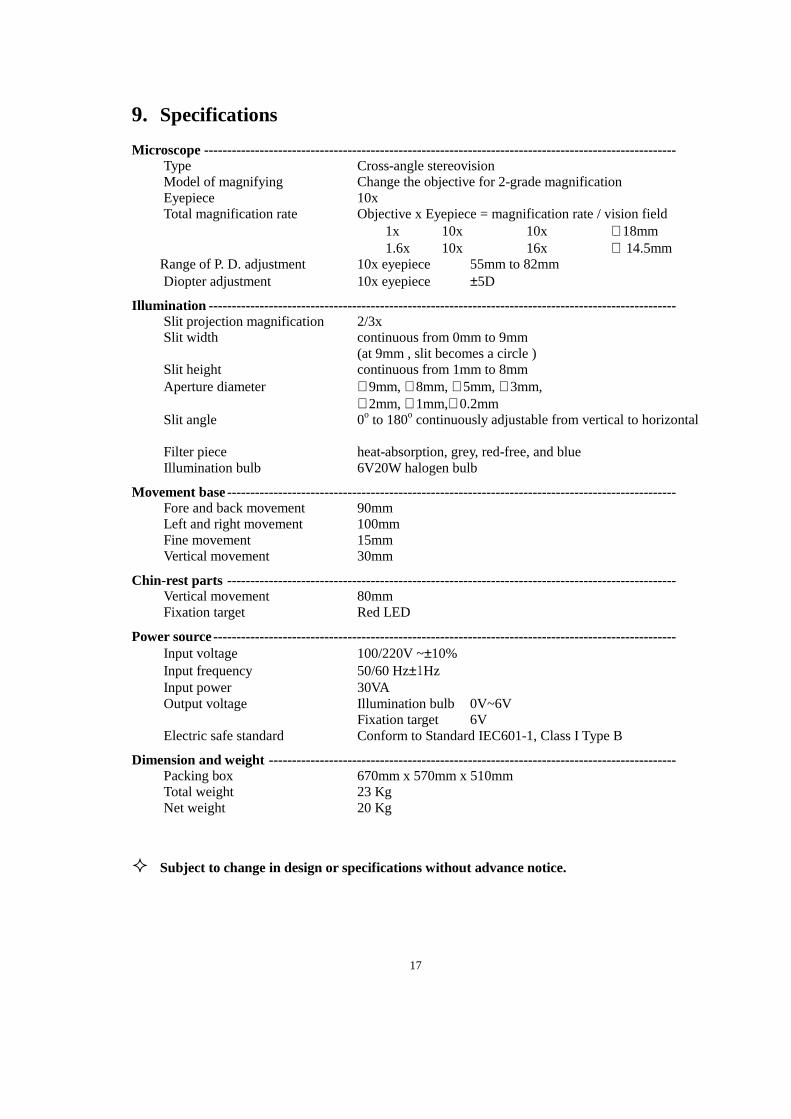
9. Specifications
Microscope ------------------------------------------------------------------------------------------------------ Type Cross-angle stereovision Model of magnifying Change the objective for 2-grade magnification Eyepiece 10x Total magnification rate Objective x Eyepiece = magnification rate / vision field
1x 10x 10x ∅18mm 1.6x 10x 16x ∅ 14.5mm Range of P. D. adjustment 10x eyepiece 55mm to 82mm
Diopter adjustment 10x eyepiece ±5D
Illumination --------------------------------------- -------------------------------------------------------------- Slit projection magnification 2/3x Slit width continuous from 0mm to 9mm (at 9mm , slit becomes a circle ) Slit height continuous from 1mm to 8mm Aperture diameter ∅9mm, ∅8mm, ∅5mm, ∅3mm, ∅2mm, ∅1mm,∅0.2mm Slit angle 0o to 180o continuously adjustable from vertical to horizontal Filter piece heat-absorption, grey, red-free, and blue Illumination bulb 6V20W halogen bulb
Movement base------------------------------------------------------------------------------------------------- Fore and back movement 90mm Left and right movement 100mm Fine movement 15mm Vertical movement 30mm
Chin-rest parts ------------------------------------------------------------------------------------------------- Vertical movement 80mm Fixation target Red LED
Power source---------------------------------------------------------------------------------------------------- Input voltage 100/220V ~±10% Input frequency 50/60 Hz±1Hz Input power 30VA Output voltage Illumination bulb 0V~6V Fixation target 6V Electric safe standard Conform to Standard IEC601-1, Class I Type B
Dimension and weight ---------------------------------------------------------------------------------------- Packing box 670mm x 570mm x 510mm Total weight 23 Kg Net weight 20 Kg
� Subject to change in design or specifications without advance notice.


![[XLS]ncseducation.comncseducation.com/Result-on-Website.xls · Web viewMordijiush J. Sangma SLIT-2247 Akash Boro SLIT-2248 Anisha Das SLIT-2249 Udit Narayan Roy SLIT-2250 Michael](https://static.fdocuments.us/doc/165x107/5ab167d47f8b9a6b468c7b61/xls-viewmordijiush-j-sangma-slit-2247-akash-boro-slit-2248-anisha-das-slit-2249.jpg)